Lower Extremity Edema Ultrasound

Brief H&P:

Ultrasound image of heart with depressed ejection fraction

Depressed ejection fraction, image from The POCUS Atlas

An 44 year-old male with no reported medical history (though limited access to medical care) presents with lower extremity swelling. He states that the symptoms have been gradually worsening over the past 3 months. He notes occasional fatigue while at work but denies chest pain, shortness of breath, leg pain or changes in urination.

A point-of-care ultrasound is performed showing decreased left ventricular ejection fraction. The patient was admitted for further evaluation and management of new-onset congestive heart failure.

Algorithm for the Evaluation of Lower Extremity Edema with Ultrasound

An algorithm for the evaluation of lower extremity edema with ultrasound

Gallery

The POCUS Atlas
The ultrasound images and videos used in this post come from The POCUS Atlas, a collaborative collection focusing on rare, exotic and perfectly captured ultrasound images.
The POCUS Atlas
Nodular liver contour, ascites

Nodular liver contour, ascites

Ascites

Ascites

Cobblestoning

Cobblestoning

Cobblestoning

Cobblestoning

Longitudinal view of a ruptured Baker cyst

Longitudinal view of a ruptured Baker cyst

References

  1. Trayes KP, Studdiford JS, Pickle S, Tully AS. Edema: diagnosis and management. Am Fam Physician. 2013;88(2):102-110.
  2. Goyal A, Cusick AS, Bhutta BS. Peripheral Edema. [Updated 2022 Nov 19]. In: StatPearls [Internet]. Treasure Island (FL): StatPearls Publishing; 2022 Jan-. Available from: https://www.ncbi.nlm.nih.gov/books/NBK554452/
  3. Smith, C. Clinical manifestations and evaluation of edema in adults. Post TW, ed. UpToDate. Waltham, MA: UpToDate Inc. http://www.uptodate.com. Accessed 2/11/2023.
This algorithm was developed by Dr. Huakang Huang. Huakang is an emergency medicine resident at UTHealth Houston.

Anticoagulant Reversal in Intracranial Hemorrhage

Brief HPI:

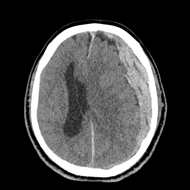
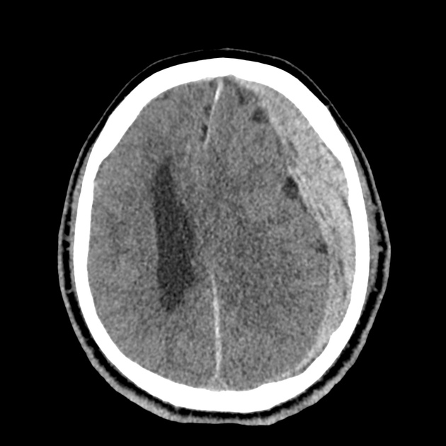
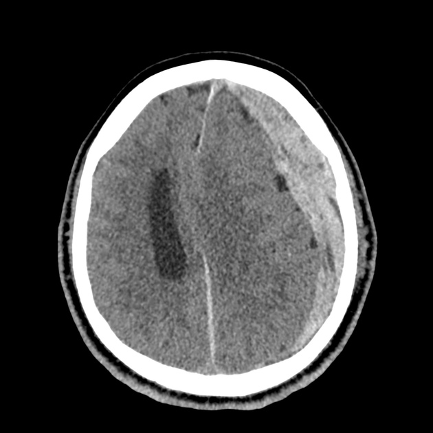
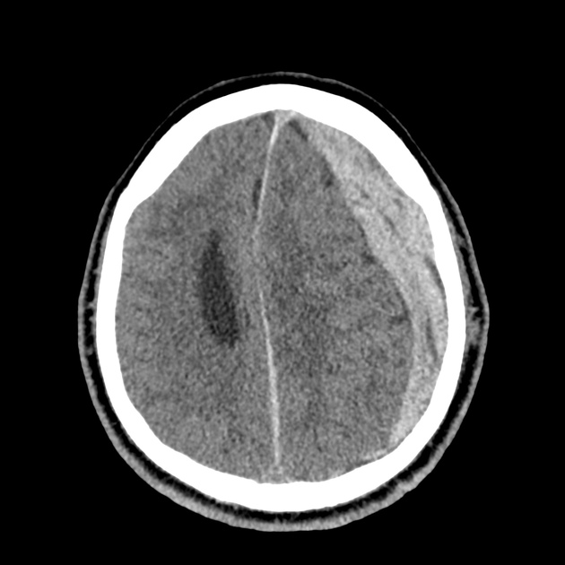
A 65-year-old male with a past medical history of hypertension, diabetes mellitus, and atrial fibrillation presents after a mechanical fall with a posterior scalp hematoma and altered mental status. The patient’s family reports that the patient is taking apixaban with his last dose 4 hours prior to arrival. Physical examination reveals a GCS of 13, blood pressure of 175/99, and asymmetric pupils. The patient is taken to CT where head imaging reveals left sided subdural hematoma with midline shift and developing uncal herniation.
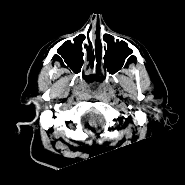
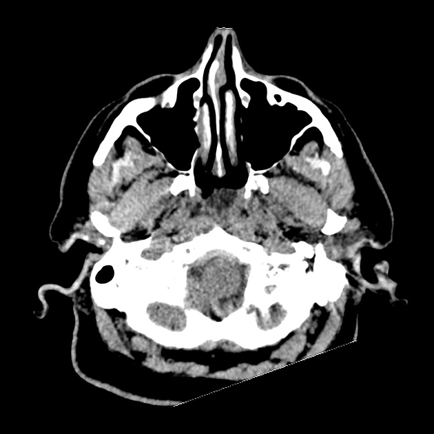
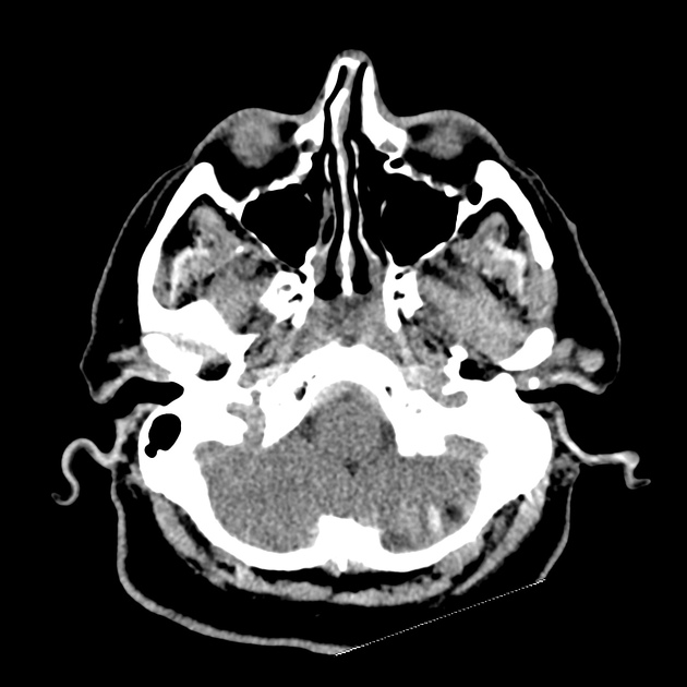
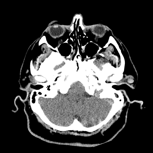
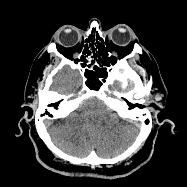
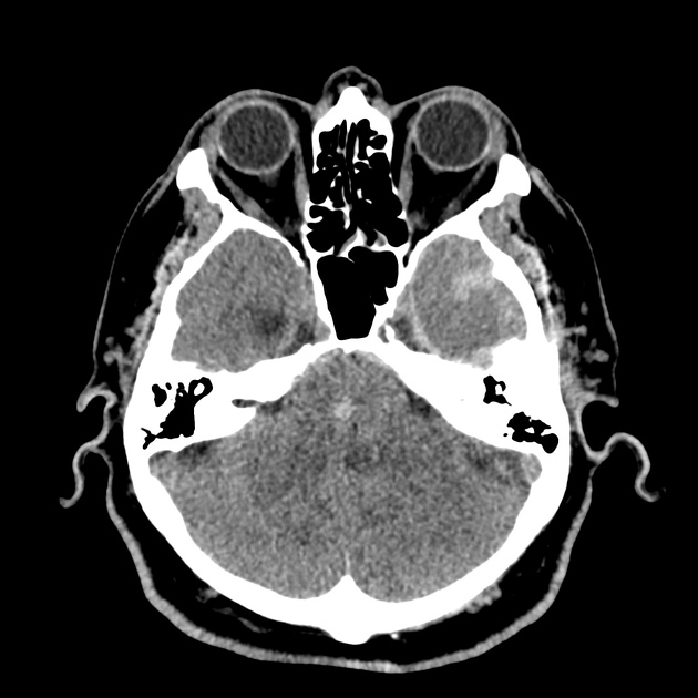
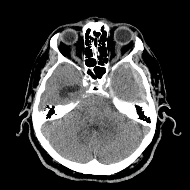
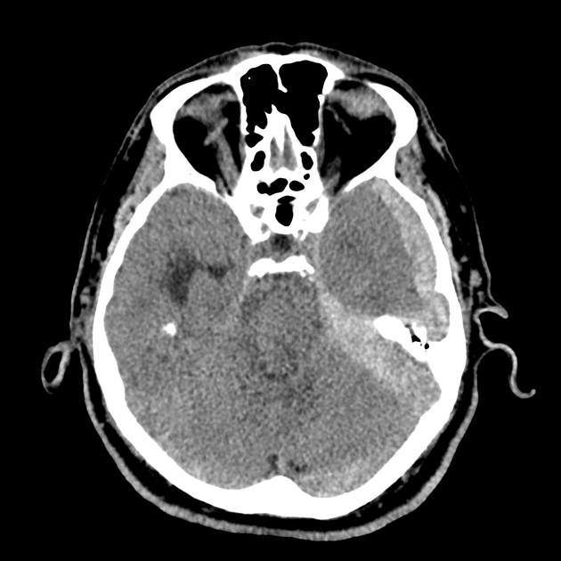
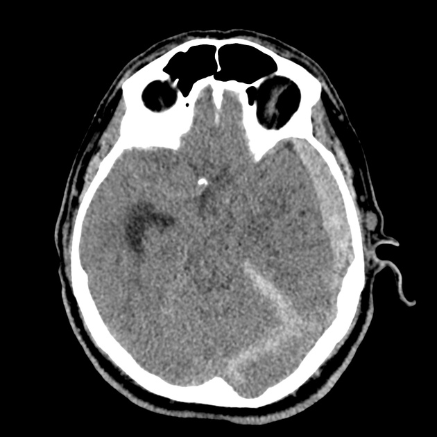
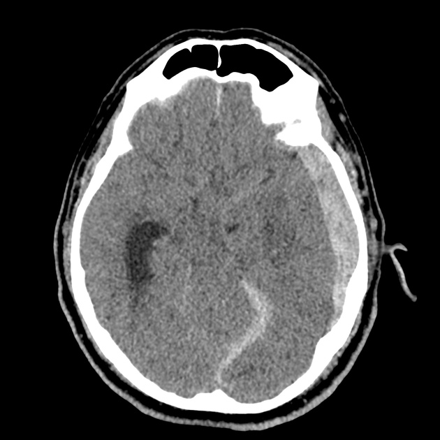
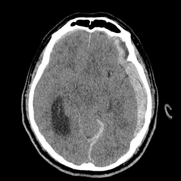
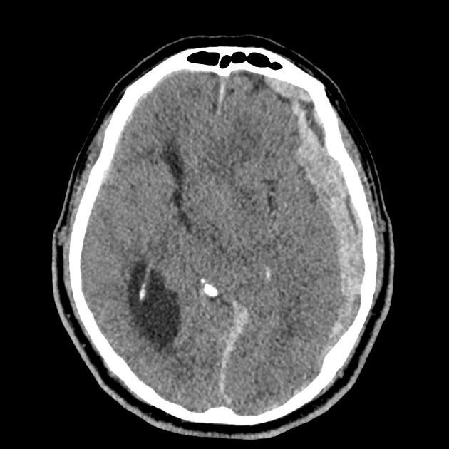
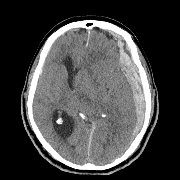
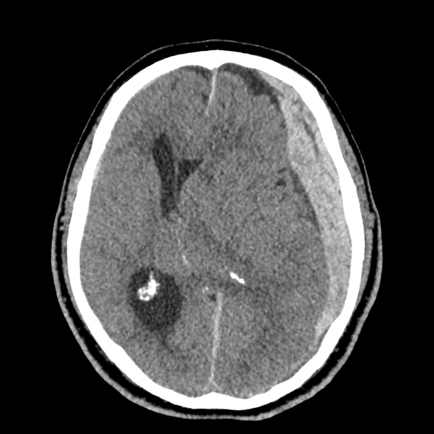
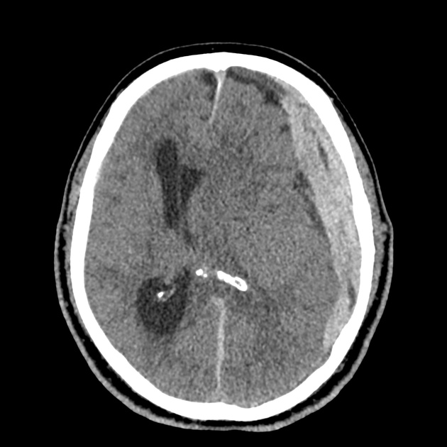
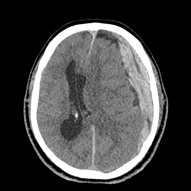
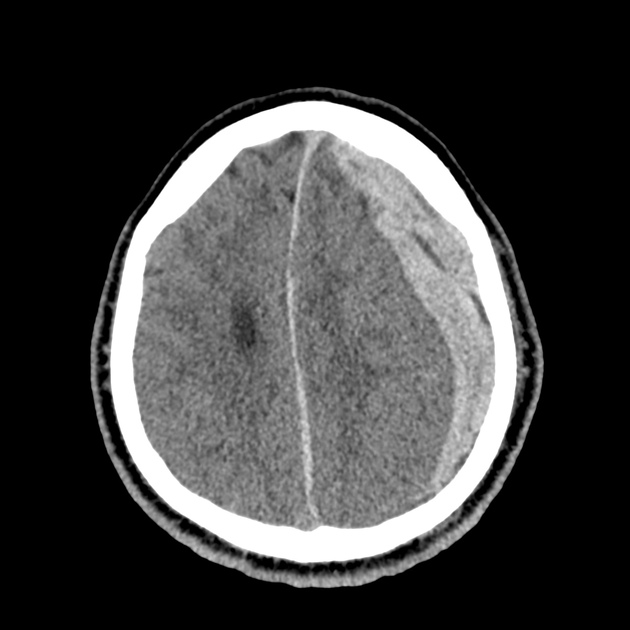
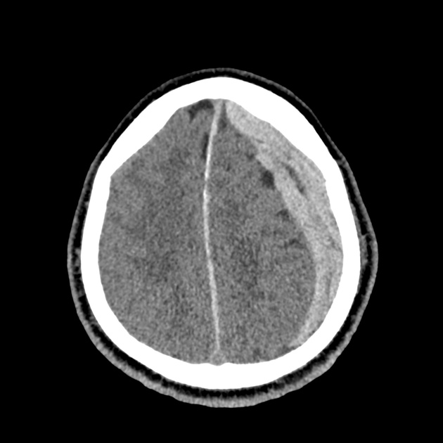
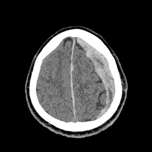
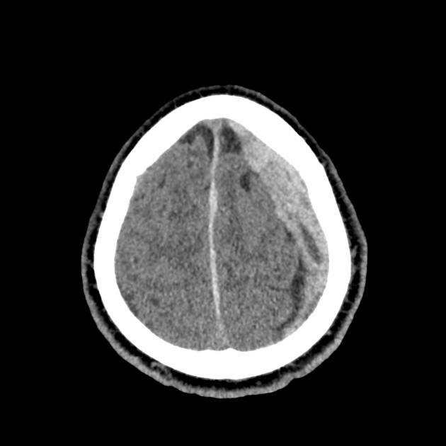
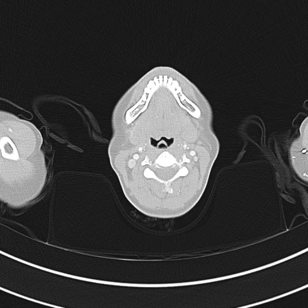
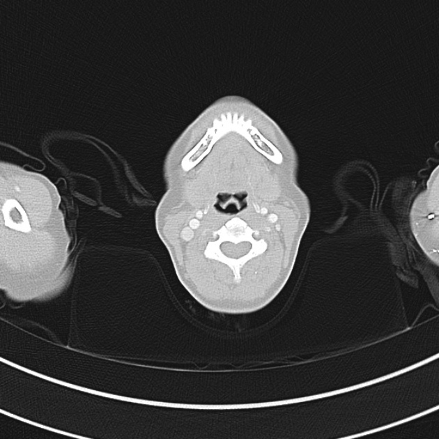
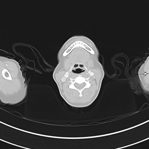
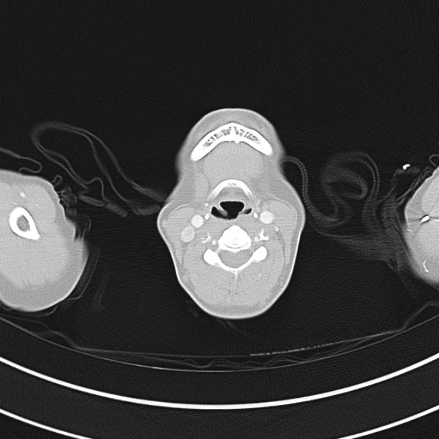
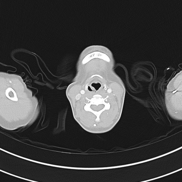
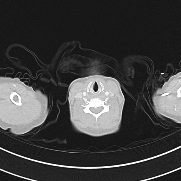
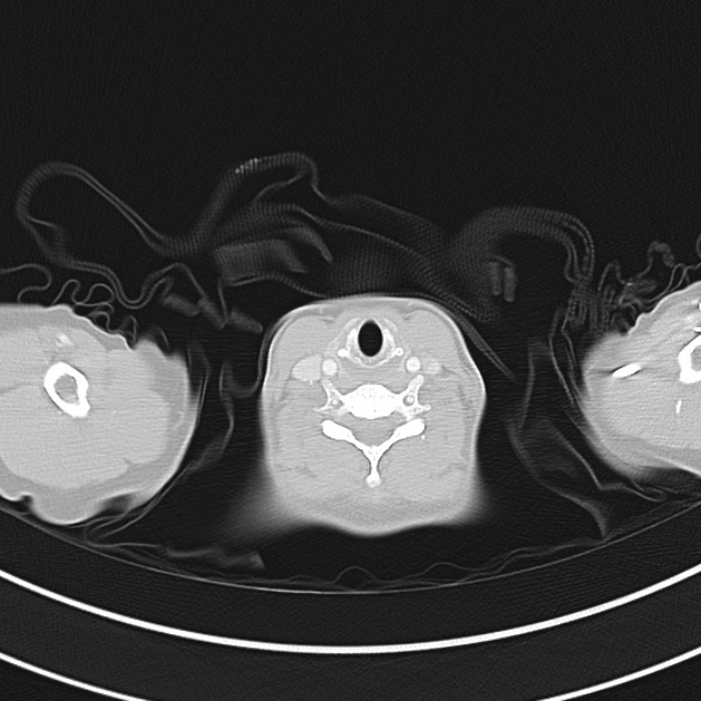
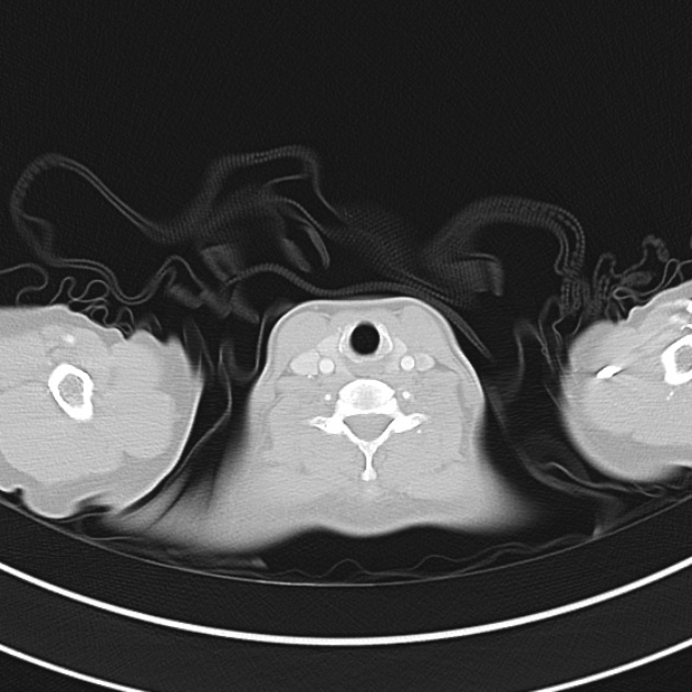
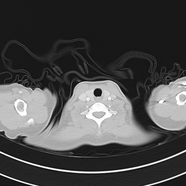
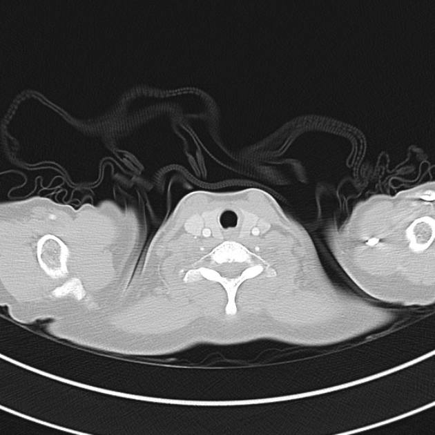
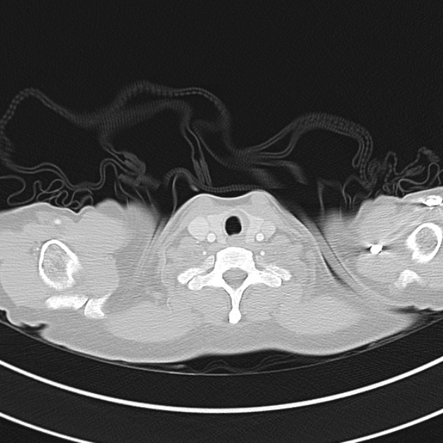
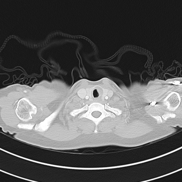
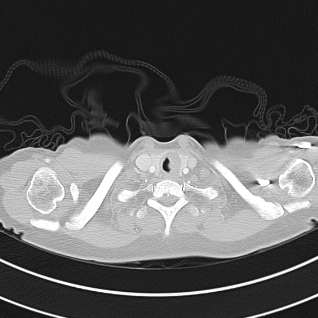
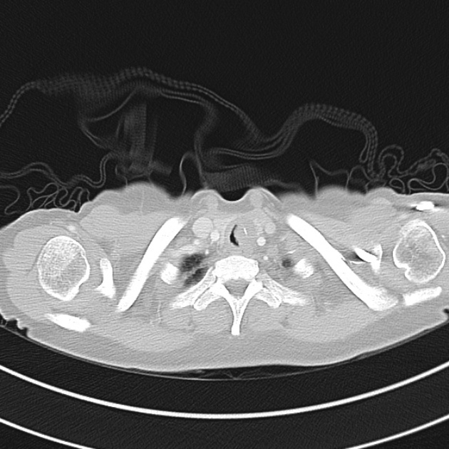
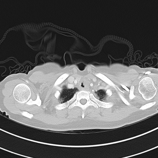
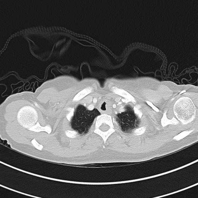
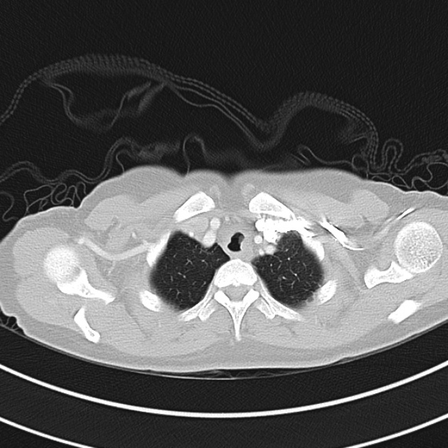
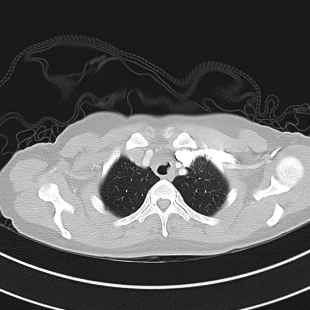
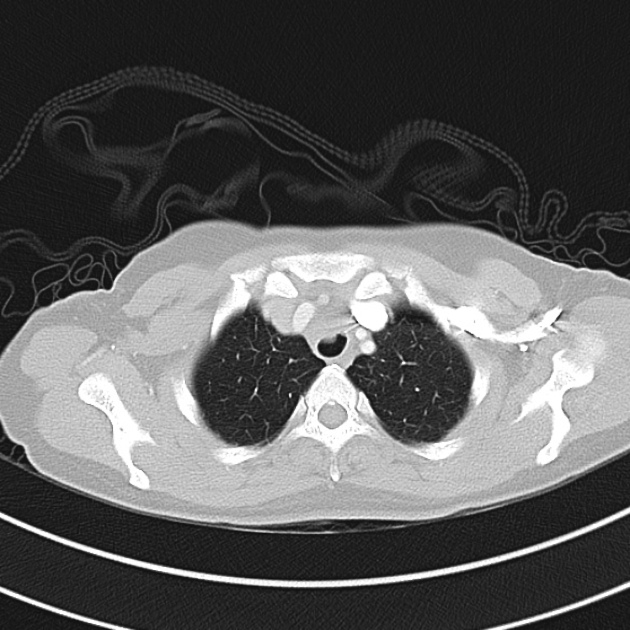
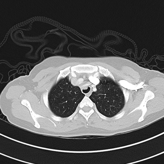
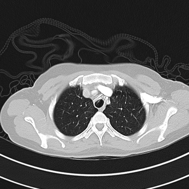
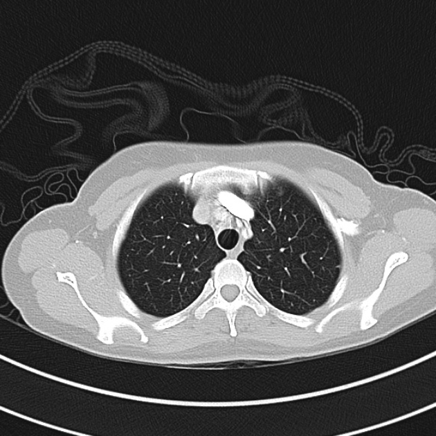
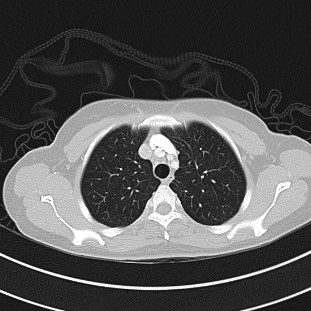
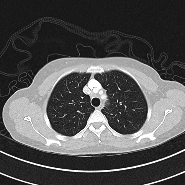
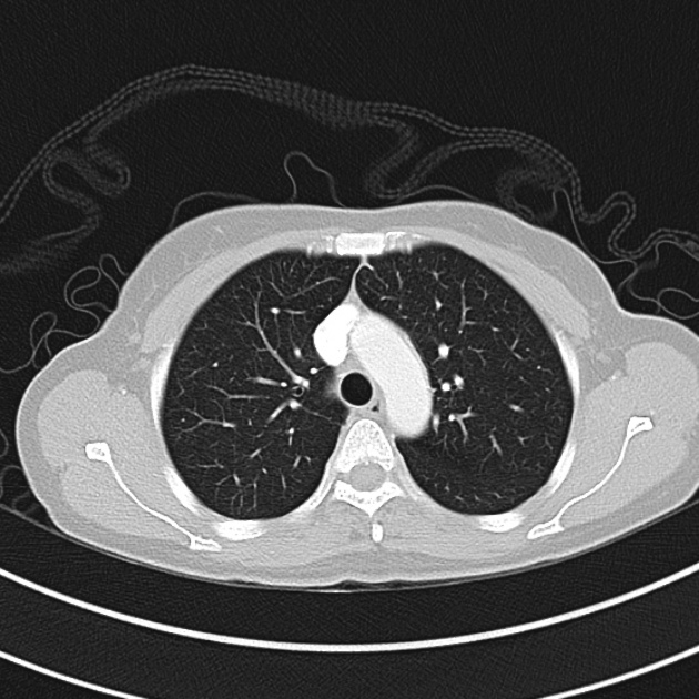
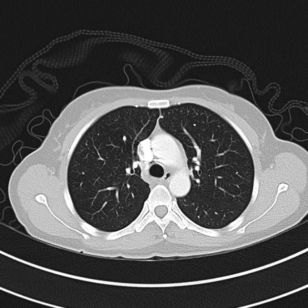
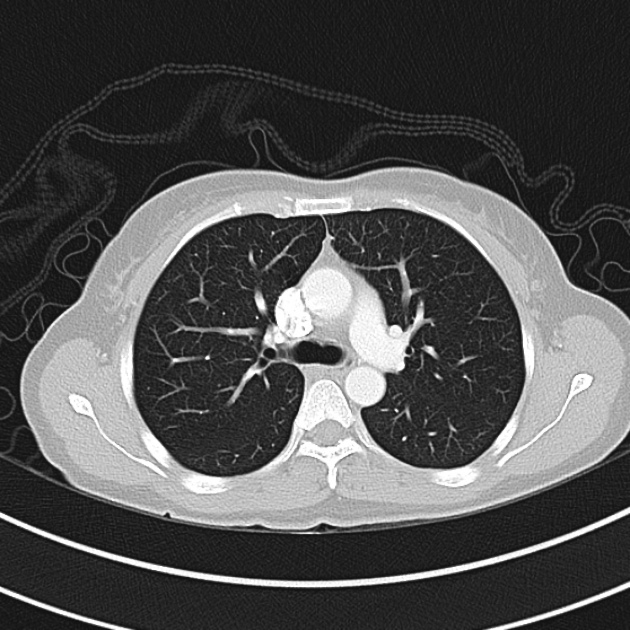
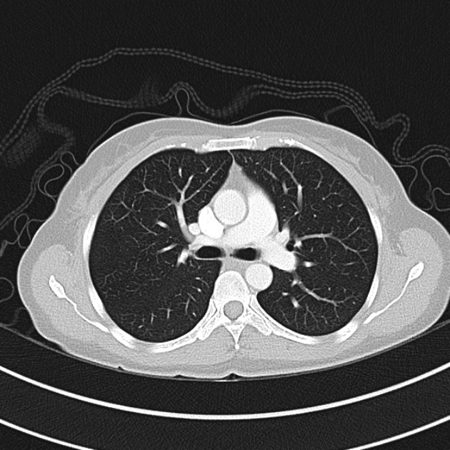
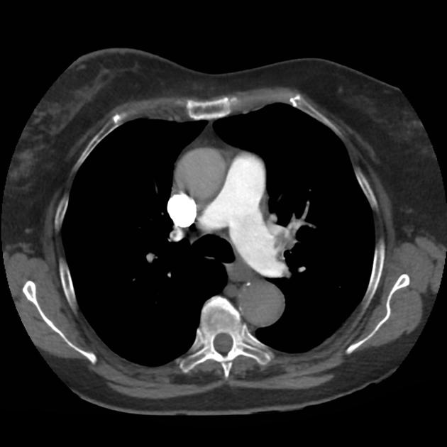
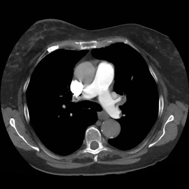
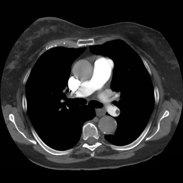
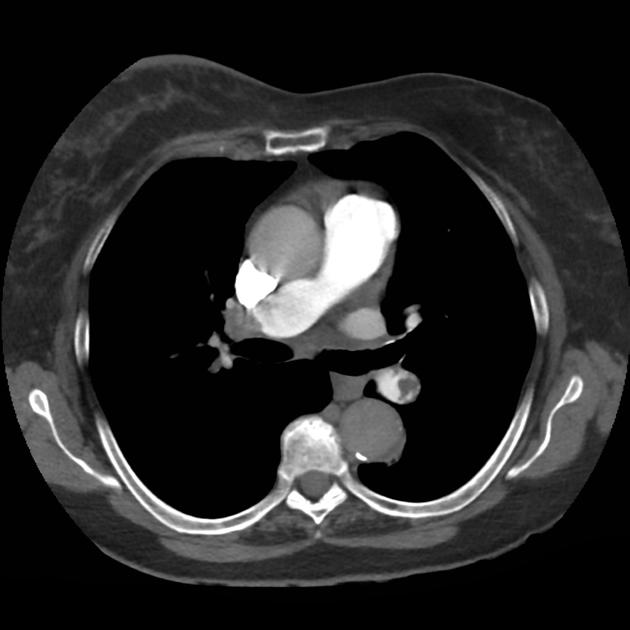
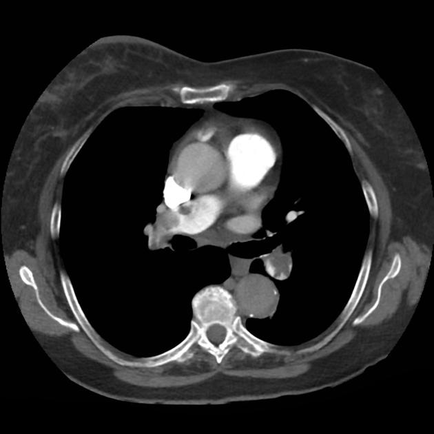
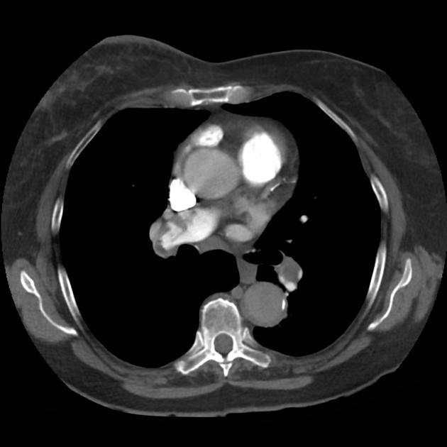
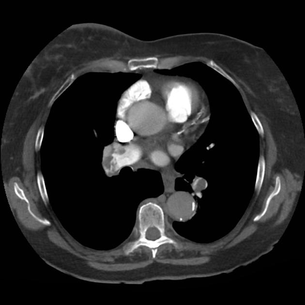
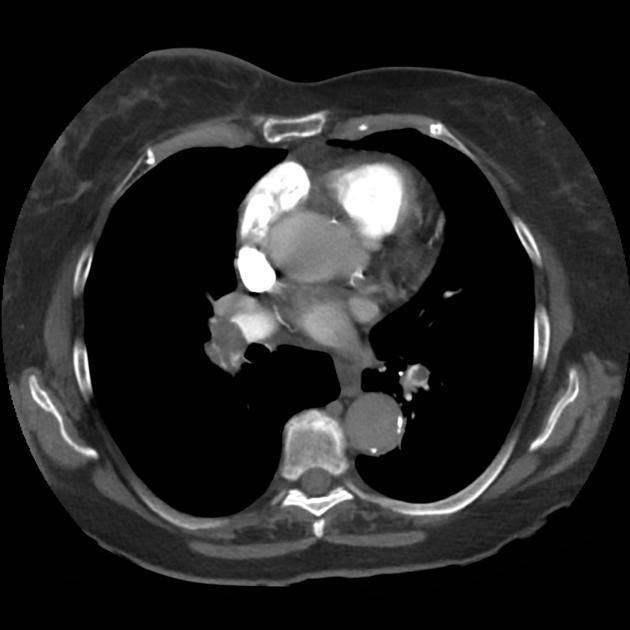
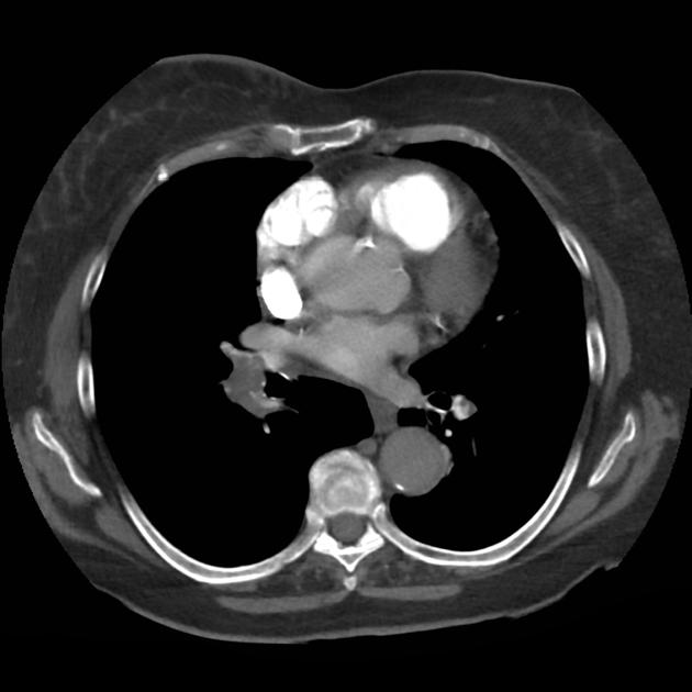
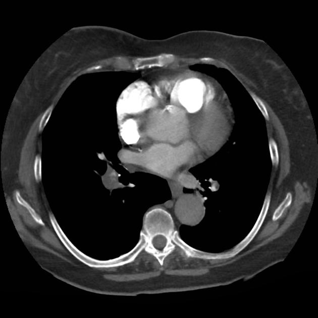
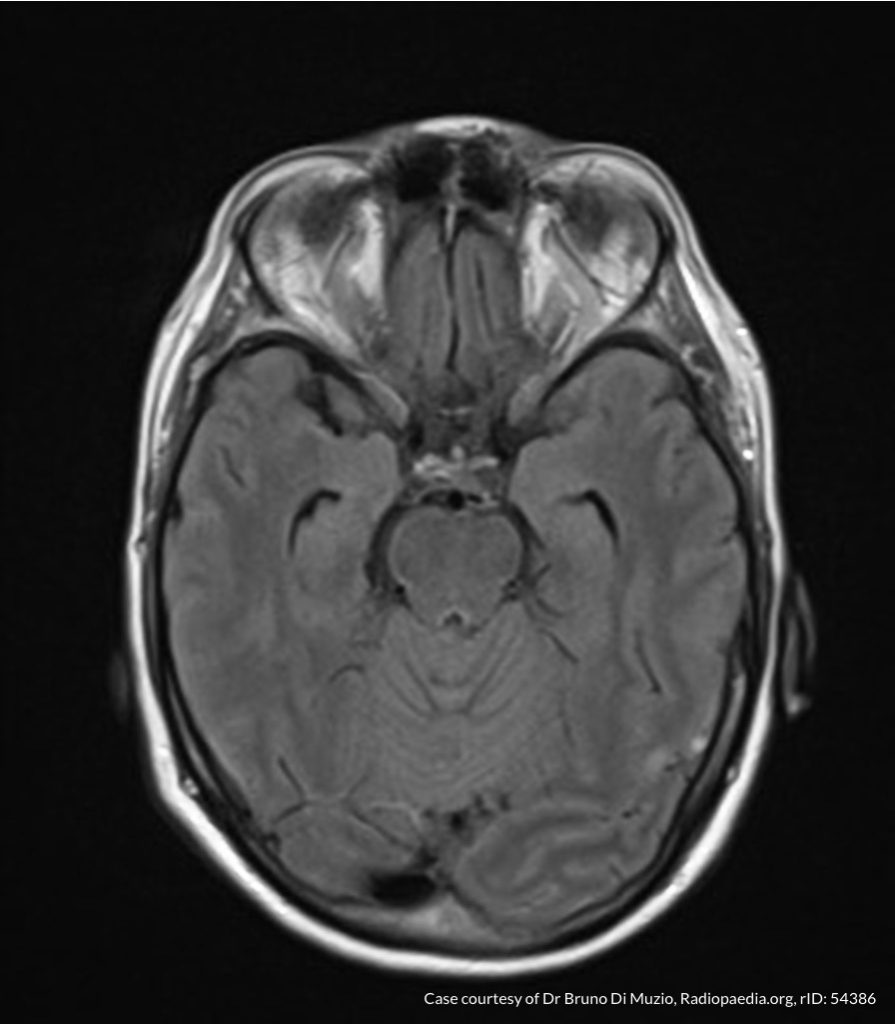
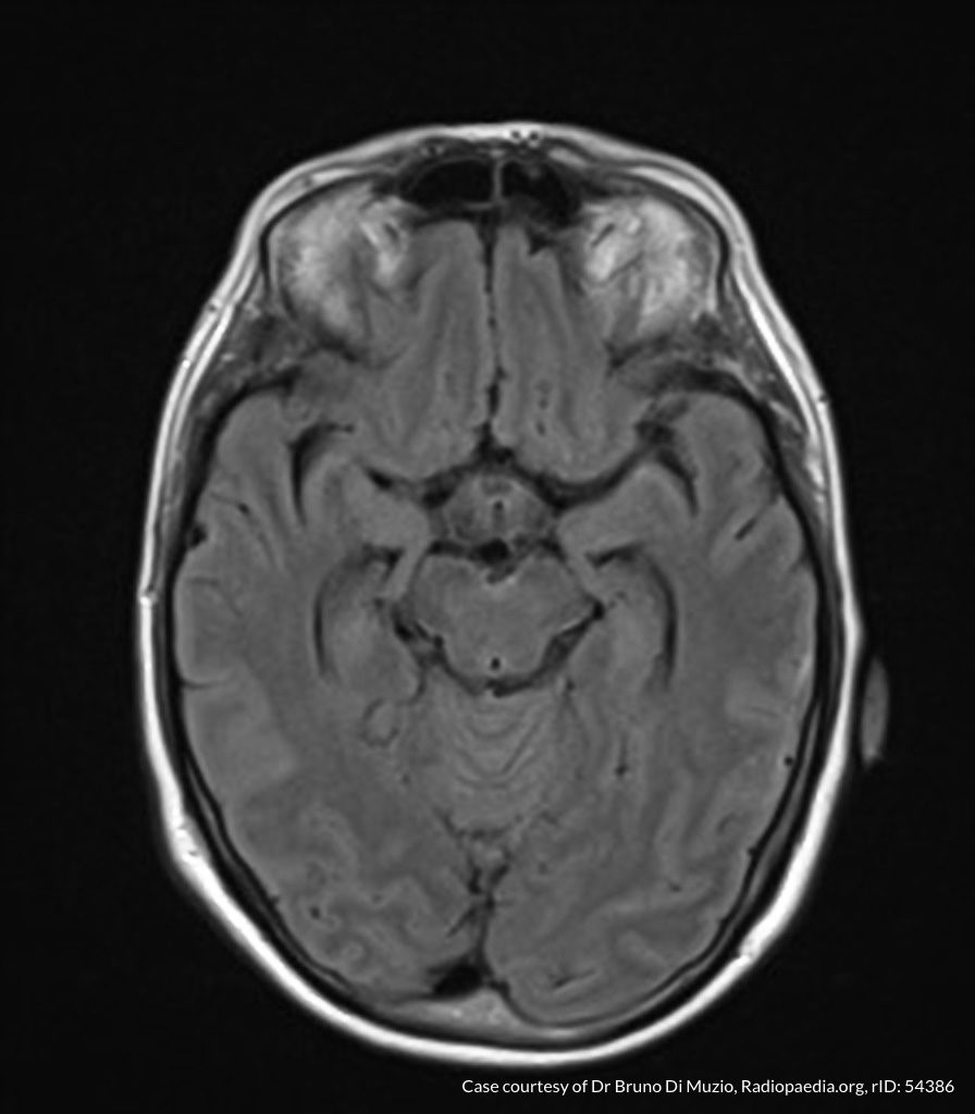
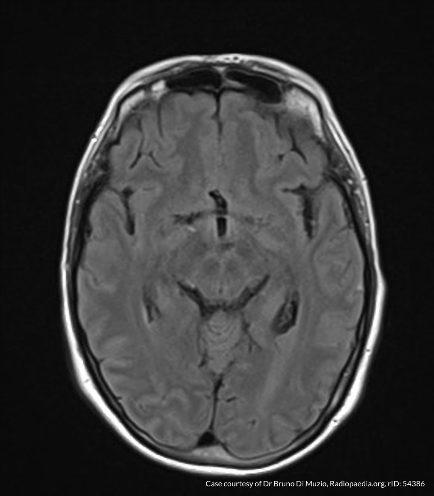
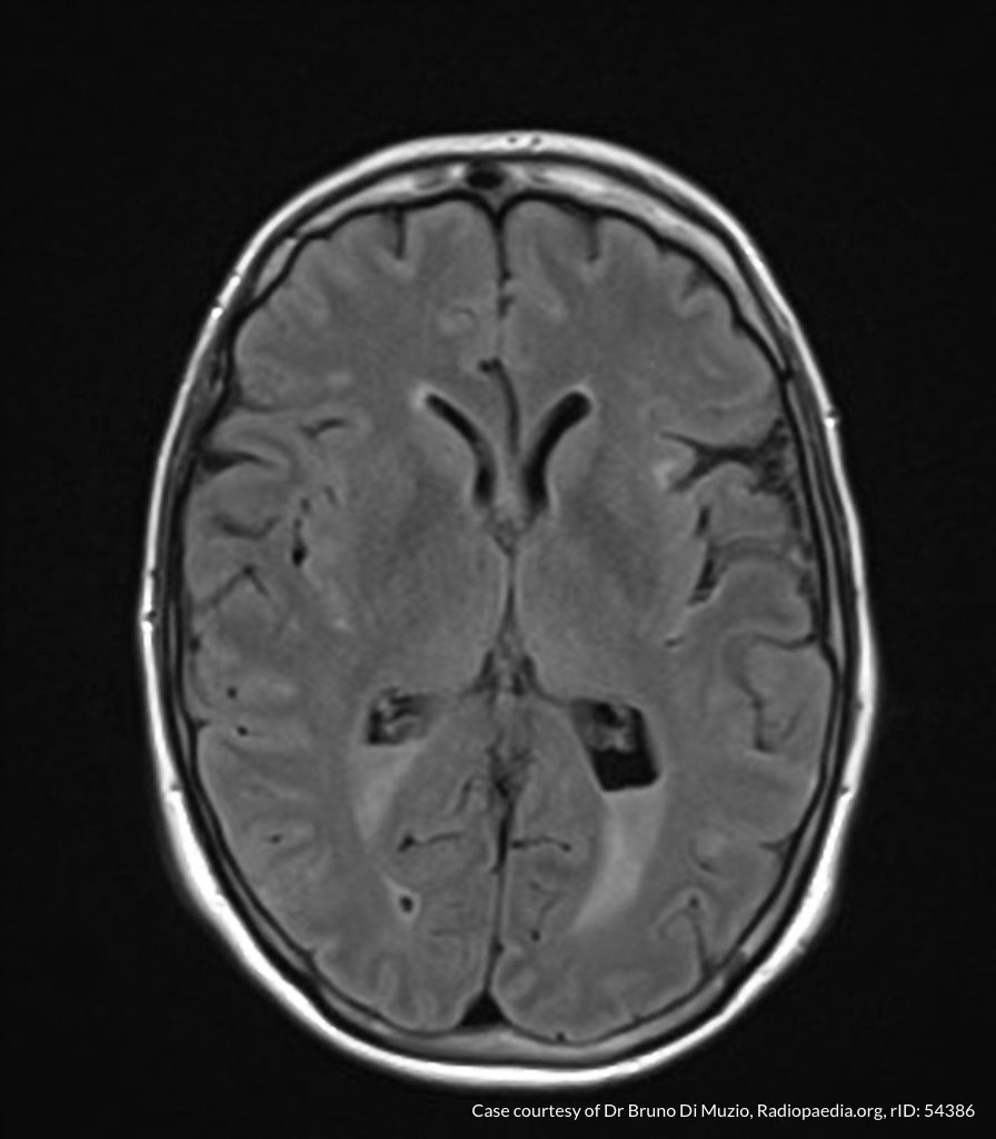
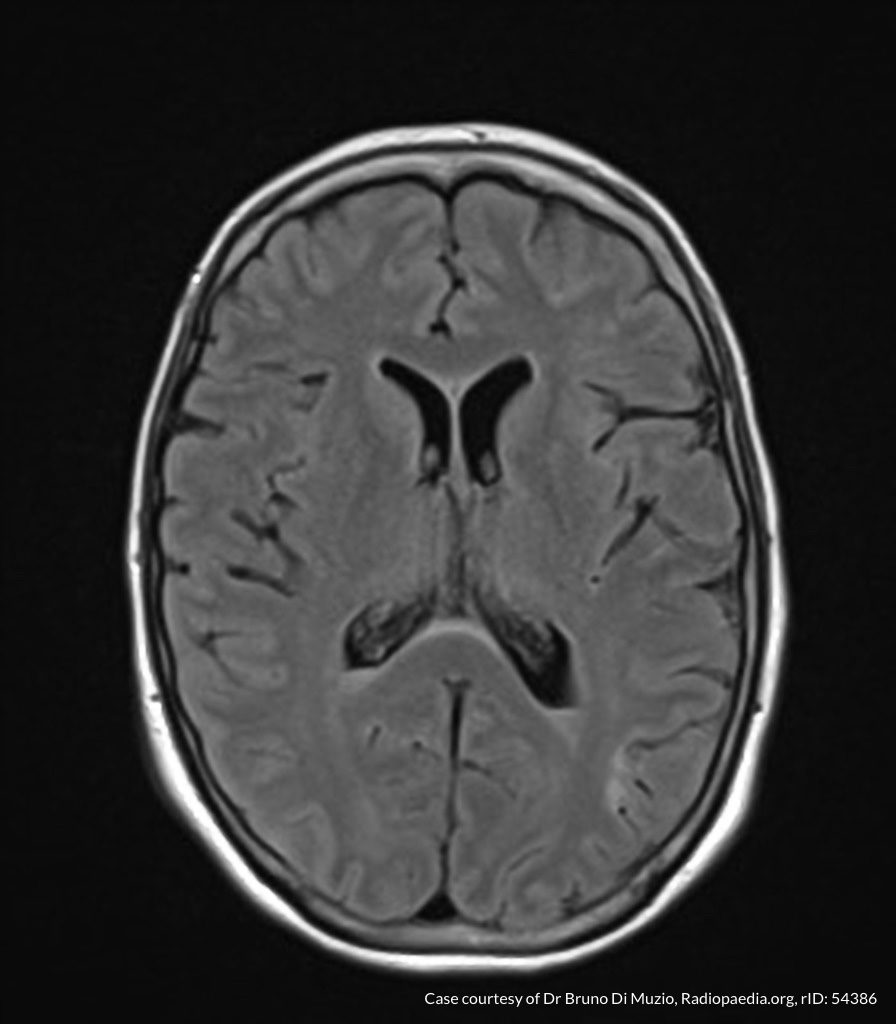
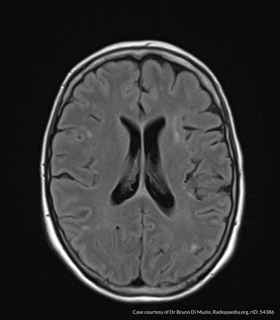
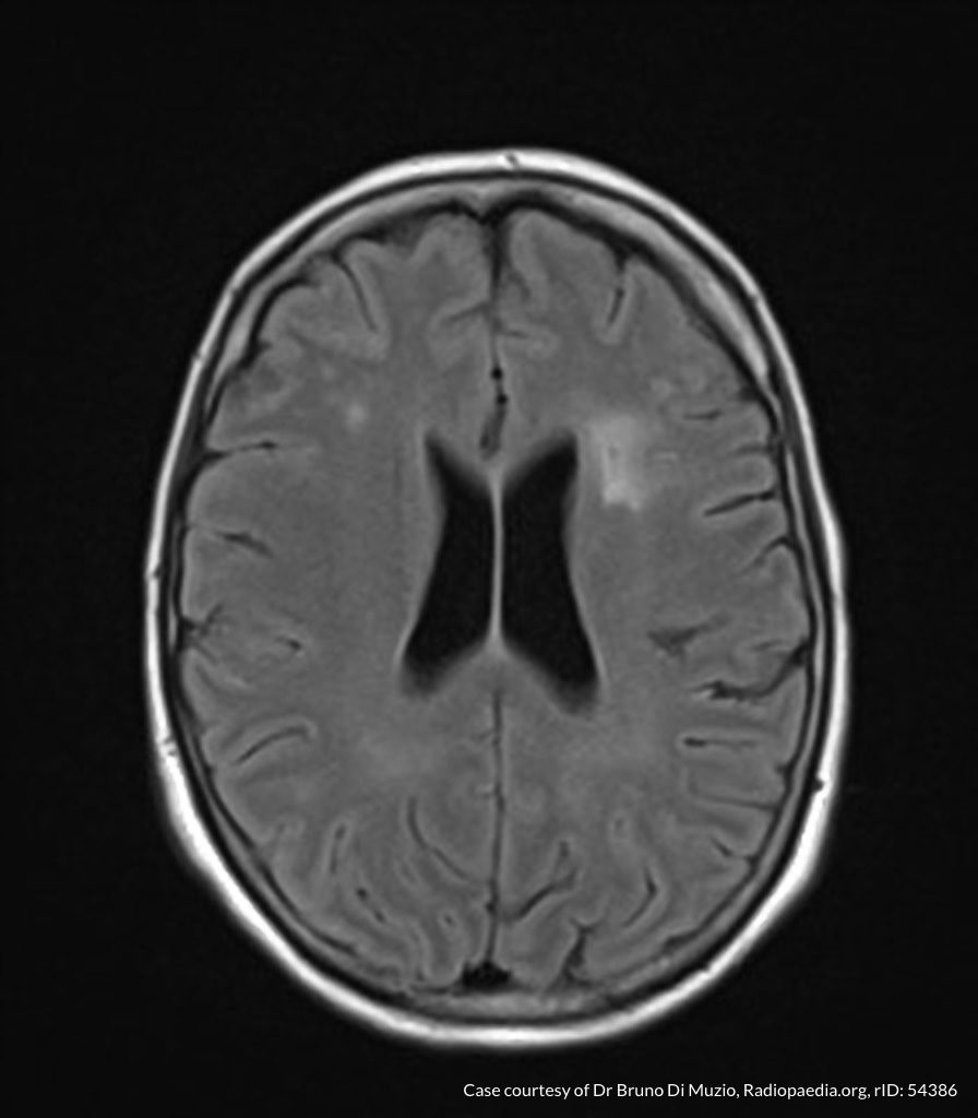
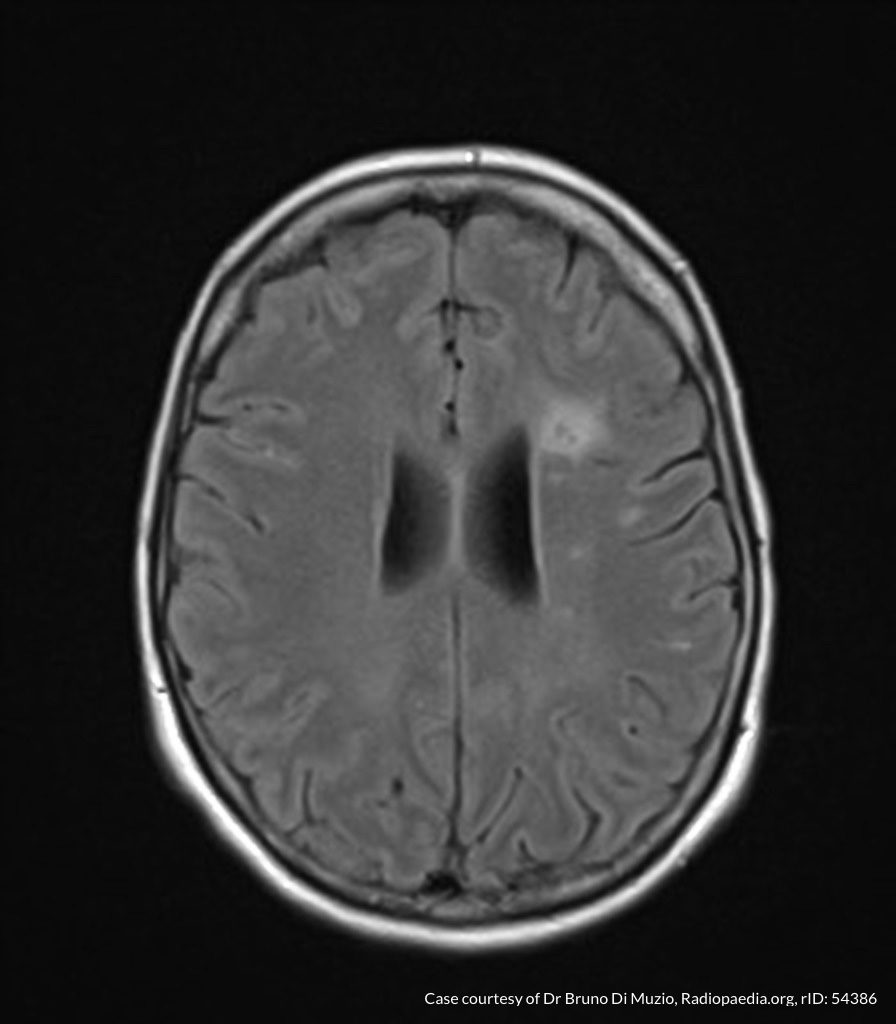
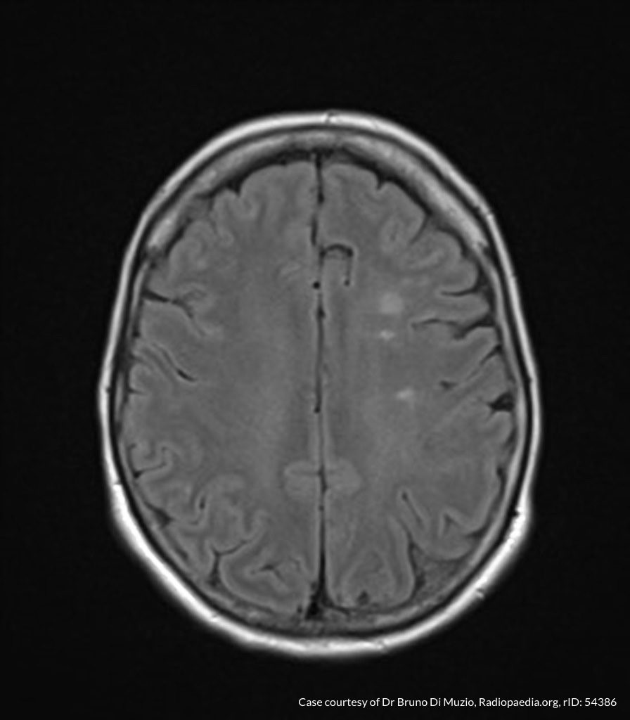
00_subdural
02_subdural
04_subdural
06_subdural
08_subdural
10_subdural
12_subdural
14_subdural
16_subdural
18_subdural
20_subdural
22_subdural
24_subdural
26_subdural
28_subdural
30_subdural
32_subdural
34_subdural
36_subdural
38_subdural
40_subdural
42_subdural
44_subdural
46_subdural

CT Head:

Left cerebral convexity acute subdural hematoma producing substantial mass effect with midline shift and left uncal herniation.
Case courtesy of Dr Andrew Dixon, Radiopaedia.org. From the case rID: 32395

A nicardipine infusion is initiated and the head of the bed is elevated. Andexanet Alfa is not available, therefore an infusion of 4-Factor PCC is initiated. The patient is taken emergently to the operating room by neurosurgery for craniotomy and hematoma evacuation.

An Algorithm for the Reversal of Anticoagulation for Intracranial Hemorrhage 1-4

An Algorithm for Anticoagulant Reversal in Intracranial Hemorrhage


All Agents

For all agents, discontinue anticoagulation. Patients may require blood pressure control including anti-hypertensive infusions (goal SBP <140). Avoid reversal for intracranial hemorrhage associated with cerebral venous thrombosis. Use cautiously in patients with concomitant life-threatening ischemia, thrombosis, or severe DIC.

Vitamin K Antagonists (ex. warfarin)

Initial Dose

A fixed dose of 4F-PCC 1500 to 2000 units can be given as an initial dose with repeat dosing based on INR measurement 15 minutes after completion of infusion. Follow local institution guidelines if available.

Monitoring and Repeat Dosing

  • Vitamin K: if INR ≥1.4 at 12 hours 5
  • 4F-PCC: May consider repeat PCC dosing based on INR, though with increased DIC and thrombotic risk, it is recommended to correct further with FFP if INR remains ≥1.4 6

Direct Factor Xa Inhibitors (ex. rivaroxaban, apixaban)

Activated charcoal may be effective for up to six hours for apixaban 7 and eight hours for rivaroxaban 8.

*Andexanet alfa Regimens 9,10

  • Low-dose: rivaroxaban <10mg, apixaban <5mg, edoxaban <30mg or 8 or more hours since last dose
  • High-dose: If greater than above thresholds, or dose/timing unknown

Pentasaccharides (ex. fondaparinux)

Use high-dose Andexanet alfa regimen 12

Direct Thrombin inhibitors (ex. dabigatran)

Monitoring and Repeat Dosing

If ongoing significant bleeding after treatment, consider redosing idarucizumab and/or hemodialysis.

Alternative Regimens

If idarucizumab is not available, aPCC (50-80 units/kg) , 4F-PCC or 3F-PCC (50 units/kg) can be used in order of preference.

Unfractionated Heparin

Dosing

Determination of units of heparin is based on estimated active agent (half-life 1-2 hours)

  • Protamine sulfate 1mg/100 units IV, maximum dose 50mg
  • Alternatively, can give fixed dose of 25-50mg

Monitoring and Repeat Dosing

If aPTT is persistently elevated, repeat 0.5 mg/100 units

Low-Molecular Weight Heparin 13

Reversal is not indicated if more than 3-5 half-lives have passed since administration:

  • Enoxaparin mean half-life: 4-5 hours
  • Dalteparin mean half-life: 2.8 hours
  • Nadroparin mean half-life: 3.7 hours

If bleeding persists, or renal insufficiency, repeat dose .5 mg/1 mg enoxaparin or .5 mg/100 anti-Xa units.

This algorithm was developed by Dr. Taylor Martin. Taylor is an emergency medicine resident at McGovern Medical School at UTHealth Houston.

References

Guidelines & Reviews

  1. Greenberg SM, Ziai WC, Cordonnier C, et al. 2022 guideline for the management of patients with spontaneous intracerebral hemorrhage: a guideline from the american heart association/american stroke association. Stroke. Published online May 17, 2022:101161STR0000000000000407.
  2. Tomaselli GF, Mahaffey KW, Cuker A, et al. 2020 acc expert consensus decision pathway on management of bleeding in patients on oral anticoagulants: a report of the american college of cardiology solution set oversight committee. J Am Coll Cardiol. 2020;76(5):594-622.
  3. Frontera JA, Lewin JJ, Rabinstein AA, et al. Guideline for reversal of antithrombotics in intracranial hemorrhage: a statement for healthcare professionals from the neurocritical care society and society of critical care medicine. Neurocrit Care. 2016;24(1):6-46.
  4.  Freeman, W. David, Weitz, Jeffrey. “Reversal of anticoagulation in intracranial hemorrhage.” UpToDate. (2022) https://www.uptodate.com/contents/reversal-of-anticoagulation-in-intracranial-hemorrhage?search=anticoagulation%20reversal (Accessed on May 26, 2022)

Vitamin K Antagonists

  1. Ansell J, Hirsh J, Hylek E, Jacobson A, Crowther M, Palareti G. Pharmacology and management of the vitamin k antagonists: american college of chest physicians evidence-based clinical practice guidelines(8th edition). Chest. 2008;133(6 Suppl):160S-198S.
  2. Pabinger I, Brenner B, Kalina U, et al. Prothrombin complex concentrate (Beriplex p/n) for emergency anticoagulation reversal: a prospective multinational clinical trial. J Thromb Haemost. 2008;6(4):622-631.

Direct Factor Xa Inhibitors

  1. http://packageinserts.bms.com/pi/pi_eliquis.pdf
  2. https://www.bayer.com/sites/default/files/2020-11/xarelto-pm-en.pdf
  3. Demchuk AM, Yue P, Zotova E, et al. Hemostatic efficacy and anti-fxa (Factor xa) reversal with andexanet alfa in intracranial hemorrhage: annexa-4 substudy. Stroke. 2021;52(6):2096-2105.
  4. Cohen AT, Lewis M, Connor A, et al. Thirty-day mortality with andexanet alfa compared with prothrombin complex concentrate therapy for life-threatening direct oral anticoagulant-related bleeding. J Am Coll Emerg Physicians Open. 2022;3(2):e12655.
  5. Scaglione F. New oral anticoagulants: comparative pharmacology with vitamin K antagonists. Clin Pharmacokinet. 2013;52(2):69-82.

Pentasaccharides (ex. fondaparinux)

  1. Lu G, DeGuzman FR, Hollenbach SJ, et al. A specific antidote for reversal of anticoagulation by direct and indirect inhibitors of coagulation factor Xa. Nat Med. 2013;19(4):446-451.

Low-Molecular Weight Heparin

  1. Fareed J, Hoppensteadt D, Walenga J, et al. Pharmacodynamic and pharmacokinetic properties of enoxaparin : implications for clinical practice. Clin Pharmacokinet. 2003;42(12):1043-1057.

Wheezing and Stridor

Brief HPI:

A 66 year-old male with a history of hypertension and COPD presents with shortness of breath. He states that his symptoms are unimproved with home nebulizer treatments and denies fever, cough or new sputum production. On examination, he has stridor appreciated during inspiratory and expiratory phases.

01_stridor
02_stridor
03_stridor
04_stridor
05_stridor
06_stridor
07_stridor
08_stridor
09_stridor
10_stridor
11_stridor
12_stridor
13_stridor
14_stridor
15_stridor
16_stridor
17_stridor
18_stridor
19_stridor
20_stridor
21_stridor
22_stridor
23_stridor
24_stridor
25_stridor
26_stridor
27_stridor
28_stridor
29_stridor
30_stridor
31_stridor
32_stridor

CT Chest:

1.9cm soft tissue thickening of the left tracheal wall at the level of the inferior thyroid gland. Luminal narrowing to 4 mm at this level.
Case courtesy of Dr Ian Bickle from Radiopaedia.org: 47677

Sound Characteristics

Stridor

An inspiratory, expiratory, or continuous monophonic sound that is loudest over the central airways.

Wheezing

A musical, high-pitched sound – more commonly expiratory. Requires sufficient airflow to induce airway oscillations.

Respiratory Phase

Inspiratory

Supraglottic: negative intratracheal pressure during inspiration causes airway collapse.

Biphasic

Glottic/Subglottic: fixed obstruction not impacted by changes in luminal/thoracic pressure.

Expiratory

Intrathoracic: increased pleural pressure compresses the narrowed airway.

An Algorithm for the Diagnosis of Wheezing and Stridor

An Algorithm for the Diagnosis of Wheezing and Stridor

Special thanks to Dr. Denna Zebda, Assistant Professor, Department of Otorhinolaryngology – McGovern Medical School for her expertise and review of this algorithm.

References

  1. Sicari V, Zabbo CP. Stridor. [Updated 2021 Jul 10]. In: StatPearls [Internet]. Treasure Island (FL): StatPearls Publishing; 2022 Jan-. Available from: https://www.ncbi.nlm.nih.gov/books/NBK525995/
  2. Patel PH, Mirabile VS, Sharma S. Wheezing. [Updated 2021 May 12]. In: StatPearls [Internet]. Treasure Island (FL): StatPearls Publishing; 2022 Jan-. Available from: https://www.ncbi.nlm.nih.gov/books/NBK482454/
  3. Bohadana A, Izbicki G, Kraman SS. Fundamentals of lung auscultation. N Engl J Med. 2014;370(21):2053.
  4. Orient JM, Sapira JD. Sapira’s Art & Science of Bedside Diagnosis. 4th ed. Wolters Kluwer Health/Lippincott Williams & Wilkins; 2010.

LVAD

Brief H&P:

A 48 year-old male with a history of congestive heart failure s/p left ventricular assist device is brought in by EMS with low-flow alarms. According to prehospital report, the patient had otherwise been in his usual state of health and had been shocked by his ICD multiple times prior to their arrival. No vital signs could be obtained en route.

On arrival in the emergency department, the patient was awake and responding appropriately to questions. His MAP was 80mmHg, an audible whir was auscultated from his device and the skin surrounding the percutaneous exit site appeared normal.

ECG

ECG with Ventricular Fibrillation

POCUS

Ultrasound showing parasternal long axis view of fibrillating heart

The patient’s device was inactivated with a magnet to prevent further ineffectual shocks. An arterial line was placed for continuous blood pressure measurement. He was sedated and externally defibrillated with return to normal sinus rhythm prior to admission to the CCU.

An Algorithm for the Evaluation of Unstable LVAD1

Algorithm for the Evaluation of Unstable LVAD

Reference

Stenberg R, Shenvi C. Targeted evaluation of patients with left ventricular assist devices and shock or hypotension. Ann Emerg Med. 2020;76(1):34-41.

Hypoglycemia

Brief HPI:

A 40 year-old male with a history of diabetes presents after accidental insulin overdose. He reports inadvertently administering 50 units of rapid-acting insulin when intending to take his long-acting insulin 25 minutes prior to presentation. He is currently asymptomatic, has a normal physical examination and his point-of-care glucose is 65mg/dL.

ED Course

The patient’s insulin regimen was clarified:

  • Insulin glargine 50 units SQ once daily
  • Insulin aspart 1 unit per 5g carbohydrates SQ before meals

The patient administered an insulin dose sufficient to dispose of 250g of carbohydrates with an impending peak action within minutes. The required dose of parenteral dextrose would require potentially-toxic1,2 solution concentrations (one liter of D25 or 10 ampules of D50) and there was insufficient time for central venous catheter placement. Moreover, the patient was awake and asymptomatic – able to tolerate oral intake. Juice boxes containing 15g of carbohydrates each were located and the patient drank 16-17 boxes while undergoing serial glucose measurements. His blood glucose ranged between 70mg/dL and 150mg/dL and he was discharged after an uneventful 6-hour observation.

Insulin Pharmacokinetics3

Graph of insulin pharmacokinetics

Insulin and Carbohydrates

The effects of both insulin and carbohydrate ingestion on blood glucose are individualized. If known, the patient’s insulin:carbohydrate ratio can be used to anticipate the effect of overdose. Alternatively, their mealtime dose and carbohydrate allowance can be used.

Very broadly, one gram of carbohydrates increases blood glucose by 3-5mg/dL and one unit of insulin disposes of 10 grams of carbohydrates (or reduces blood glucose by 30-50mg/dL).4

Symptoms

  • Autonomic: tremor, palpitations, anxiety, diaphoresis
  • Neuroglycopenic: cognitive impairment, psychomotor, seizure, coma

Diagnosis

  • Serum glucose <60mg/dL
  • Generally symptomatic at <55mg/dL though threshold is variable depending on chronicity
  • Whipple Triad:
    • Symptoms suggestive of hypoglycemia
    • Low glucose
    • Resolution of symptoms after administration of glucose

Differential Diagnosis of Hypoglycemia

Differential Diagnosis of Hypoglycemia

Common Anti-hyperglycemic Drugs and Pharmacology

Drug Pharmacology
Onset Peak Duration
Rapid-acting insulin

  • Aspart (Novolog)
  • Lispro (Humalog)
15-30min 1-2h 3-5h
Short-acting insulin

  • Regular
30-60min 2-4h 6-10h
Intermediate-acting insulin

  • NPH
1-3h 4-12h 18-24h
Long-acting insulin

  • Glargine (Lantus)
2-4h None 24h
Sulfonylurea

  • Glimepiride
  • Glipizide (Glucotrol)
  • Glyburide (Glycron, Micronase)
2-6h 12-24h

Evaluation of Hypoglycemia

Patients with known diabetes who are not systemically ill and can identify a clear precipitant, no extensive workup is required. In severely ill patients, consider:

  • BMP
  • LFT
  • EtOH
  • Infectious workup: CXR, UA, urine and blood cultures
  • ECG, troponin
  • Other studies: insulin, C-peptide, pro-insulin, glucagon, growth hormone, cortisol, B-OH, insulin antibodies

Management and Monitoring

An algorithm for the management and monitoring of hypoglycemia

Special thanks to Dr. Ryan Pedigo, Associate Residency Program Director at Harbor-UCLA Medical Center for his case submission and content review.
Twitter

References:

  1. Kuwahara T, Asanami S, Kubo S. Experimental infusion phlebitis: tolerance osmolality of peripheral venous endothelial cells. Nutrition. 1998;14(6):496-501.
  2. Wiegand R, Brown J. Hyaluronidase for the management of dextrose extravasation. Am J Emerg Med. 2010;28(2):257.e1-2.
  3. Hirsch IB. Insulin analogues. N Engl J Med. 2005;352(2):174-183.
  4. Your Insulin Therapy. (2004). Am Fam Physician, 70(3), 511-512.
  5. Self, W. H., & McNaughton, C. D. (2013). Hypoglycemia. In Emergency Medicine (2nd ed., pp. 1379-1390). Elsevier.
  6. Service, FJ. Hypoglycemia in adults: Clinical manifestations, definition, and causes. In: UpToDate, Post TW (Ed), UpToDate, Waltham, MA. (Accessed on March 18, 2016.)
  7. Service FJ. Hypoglycemic disorders. N Engl J Med. 1995;332(17):1144–1152. doi:10.1056/NEJM199504273321707.
  8. Krinsley JS, Grover A. Severe hypoglycemia in critically ill patients: risk factors and outcomes. Critical Care Medicine. 2007;35(10):2262–2267. doi:10.1097/01.CCM.0000282073.98414.4B.
  9. Lacherade J-C, Jacqueminet S, Preiser J-C. An overview of hypoglycemia in the critically ill. J Diabetes Sci Technol. 2009;3(6):1242–1249.

Hyperammonemia

Brief H&P

A 38 year-old male with unknown medical history is brought to the emergency department by EMS with agitation and bizarre behavior. According to prehospital report, the patient was acting erratically – shouting incomprehensibly in the middle of a busy street with possible associated seizure activity.

On evaluation, the patient was found to be tachycardic, hypertensive, and markedly agitated. Physical examination with a focus on toxidromes was notable for the presence of rotary nystagmus suggestive of hallucinogen including phencyclidine toxicity. The patient required pharmacologic sedation to allow for a broad evaluation of altered mental status.

ED Course

The patient’s workup including core temperature, head imaging and laboratory tests (including AST/ALT, albumin, INR) were unremarkable with the exception of an ammonia level of 142 umol/L (normal range 16-53), slightly elevated CK, and urine toxicology screen with multiple positive agents. Over the course of several hours in the emergency department, the patient’s mental status gradually improved reaching normal level of alertness and orientation with normal neurological examination. He acknowledged PCP use as well as a prior history of seizures (possibly related to ethanol withdrawal) without routine anti-epileptic drug use. He denied known history of liver disease.

The patient’s hyperammonemia was attributed to a hypercatabolic state secondary to phencyclidine-induced agitation with possible seizure. He was discharged with resources for assistance with substance cessation.

An Algorithm for the Differential Diagnosis of Hyperammonemia:

Algorithm for the Differential Diagnosis of Hyperammonemia

References:

  1. Kalra A, Norvell JP. Cause for Confusion: Noncirrhotic Hyperammonemic Encephalopathy. Clin Liver Dis. 2020;15(6):223-227. doi:10.1002/cld.929
  2. Mallet M, Weiss N, Thabut D, Rudler M. Why and when to measure ammonemia in cirrhosis? Clin Res Hepatol Gas. 2018;42(6):505-511. doi:10.1016/j.clinre.2018.01.004
  3. Hassan AAI, Ibrahim W, Subahi A, Mohamed A. ‘All that glitters is not gold’: when hyperammonaemia is not from hepatic aetiology. Bmj Case Reports. 2017;2017:bcr-2017-219441. doi:10.1136/bcr-2017-219441
  4. Odigwe CC, Khatiwada B, Holbrook C, et al. Noncirrhotic Hyperammonemia Causing Relapsing Altered Mental Status. Bayl Univ Medical Cent Proc. 2017;28(4):472-474. doi:10.1080/08998280.2015.11929312
  5. Upadhyay R, Bleck TP, Busl KM. Hyperammonemia: What Urea-lly Need to Know: Case Report of Severe Noncirrhotic Hyperammonemic Encephalopathy and Review of the Literature. Case Reports Medicine. 2016;2016:1-10. doi:10.1155/2016/8512721
  6. Walker V. Severe hyperammonaemia in adults not explained by liver disease. Ann Clin Biochem. 2011;49(3):214-228. doi:10.1258/acb.2011.011206
  7. Laish I, Ari ZB. Noncirrhotic hyperammonaemic encephalopathy. Liver Int. 2011;31(9):1259-1270. doi:10.1111/j.1478-3231.2011.02550.x
  8. LaBuzetta JN, Yao JZ, Bourque DL, Zivin J. Adult Nonhepatic Hyperammonemia: A Case Report and Differential Diagnosis. Am J Medicine. 2010;123(10):885-891. doi:10.1016/j.amjmed.2010.02.029
  9. Clay AS, Hainline BE. Hyperammonemia in the ICU. Chest. 2007;132(4):1368-1378. doi:10.1378/chest.06-2940
  10. Weng T-I, Shih FF-Y, Chen W-J. Unusual causes of hyperammonemia in the ED. Am J Emerg Medicine. 2004;22(2):105-107. doi:10.1016/j.ajem.2003.12.011
  11. Hawkes ND, Thomas GAO, Jurewicz A, et al. Non-hepatic hyperammonaemia: an important, potentially reversible cause of encephalopathy. Postgrad Med J. 2001;77(913):717. doi:10.1136/pmj.77.913.717

Adrenal Insufficiency

Brief H&P:

A 65 year-old female with a history of hypertension, hyperlipidemia and rheumatoid arthritis presents with generalized weakness and nausea/vomiting for 3 days. She denies fever, focal weakness, numbness or speech difficulty.

HPA Axis

HPA Axis

Vital signs were notable for hypotension (72/48mmHg), her examination revealed diffuse weakness but no focal deficits and her abdominal examination was unremarkable. Laboratory tests were notable for hyponatremia (117 mEq/L) and new renal dysfunction. She received 2L of intravenous fluids but remained hypotensive.

Additional history revealed that the patient had been out of her home medications (including prednisone) for the past 1 week. Samples for laboratory tests (cortisol, ACTH) were drawn and she was treated with hydrocortisone 100mg IV with subsequent improvement in blood pressure – she was admitted to the ICU for adrenal crisis.

An Algorithm for the Evaluation and Management of Adrenal Insufficiency and Crisis

Algorithm for the Evaluation and Management of Adrenal Insufficiency and Crisis

Special thanks to Dr. Katrin Takenaka, Professor, Department of Emergency Medicine and Dr. Reem Al-Dallal, Assistant Professor, Division of Endocrinology, Department of Internal Medicine – McGovern Medical School for their expertise and review of this algorithm.

References:

  1. Idrose, A., Tintinalli, J., Ma, O., Yealy, D., Meckler, G., Stapczynski, J., Cline, D., Thomas, S. (2020). Adrenal Insufficiency Tintinalli’s Emergency Medicine: A Comprehensive Study Guide, 9e
  2. Carroll, T., Aron, D., Findling, J., Tyrrell, J., Gardner, D., Shoback, D. (2017). Glucocorticoids and Adrenal Androgens Greenspan’s Basic & Clinical Endocrinology, 10e
  3. Dineen, R., Thompson, C., Sherlock, M. (2019). Adrenal crisis: prevention and management in adult patients Therapeutic Advances in Endocrinology and Metabolism 10(), 2042018819848218. https://dx.doi.org/10.1177/2042018819848218
  4. Rushworth, R., Torpy, D., Falhammar, H. (2019). Adrenal Crisis New England Journal of Medicine 381(9), 852-861. https://dx.doi.org/10.1056/nejmra1807486
  5. Amrein, K., Martucci, G., Hahner, S. (2018). Understanding adrenal crisis Intensive Care Medicine 44(5), 652-655. https://dx.doi.org/10.1007/s00134-017-4954-2
  6. Oelkers, W. (1996). Adrenal Insufficiency The New England Journal of Medicine 335(16), 1206-1212. https://dx.doi.org/10.1056/nejm199610173351607
  7. Tucci, V., Sokari, T. (2014). The Clinical Manifestations, Diagnosis, and Treatment of Adrenal Emergencies Emergency Medicine Clinics of North America 32(2), 465-484. https://dx.doi.org/10.1016/j.emc.2014.01.006
  8. Bleicken, B., Hahner, S., Ventz, M., Quinkler, M. (2010). Delayed Diagnosis of Adrenal Insufficiency Is Common: A Cross-Sectional Study in 216 Patients The American Journal of the Medical Sciences 339(6), 525-531. https://dx.doi.org/10.1097/maj.0b013e3181db6b7a
  9. Broersen, L., Pereira, A., Jørgensen, J., Dekkers, O. (2015). Adrenal Insufficiency in Corticosteroids Use: Systematic Review and Meta-Analysis The Journal of Clinical Endocrinology & Metabolism 100(6), 2171-2180. https://dx.doi.org/10.1210/jc.2015-1218
  10. Joseph, R., Hunter, A., Ray, D., Dixon, W. (2016). Systemic glucocorticoid therapy and adrenal insufficiency in adults: A systematic review Seminars in Arthritis and Rheumatism 46(1), 133-141. https://dx.doi.org/10.1016/j.semarthrit.2016.03.001

Isolation Precautions

An Algorithm for the Determination of Isolation Precautions

An Algorithm for the Determination of Isolation Precautions

References

  1. Siegel, J., Rhinehart, E., Jackson, M., Chiarello, L., Committee, H. (2007). 2007 Guideline for Isolation Precautions: Preventing Transmission of Infectious Agents in Health Care Settings American Journal of Infection Control 35(10), S65-S164. https://dx.doi.org/10.1016/j.ajic.2007.10.007
  2. Liang, S., Theodoro, D., Schuur, J., Marschall, J. (2014). Infection Prevention in the Emergency Department Annals of Emergency Medicine 64(3), 299-313. https://dx.doi.org/10.1016/j.annemergmed.2014.02.024
  3. Liang, S., Riethman, M., Fox, J. (2018). Infection Prevention for the Emergency Department: Out of Reach or Standard of Care? Emergency medicine clinics of North America 36(4), 873-887. https://dx.doi.org/10.1016/j.emc.2018.06.013
  4. Gottenborg, E., Barron, M. (2016). Isolation Precautions in the Inpatient Setting Hospital Medicine Clinics 5(1), 30-42. https://dx.doi.org/10.1016/j.ehmc.2015.08.004
  5. Harding, A., Almquist, L., Hashemi, S. (2011). The use and need for standard precautions and transmission-based precautions in the emergency department. Journal of emergency nursing: JEN : official publication of the Emergency Department Nurses Association 37(4), 367-73; quiz 424-5. https://dx.doi.org/10.1016/j.jen.2010.11.017

Hypoxia

Brief H&P:

A 67 year-old male with a history of hypertension and diabetes presents to the emergency department after a syncopal episode. He had been completing his normal morning routine when he developed a sensation of lightheadedness and awoke on the ground of his kitchen. He denies associated chest pain, palpitations, diaphoresis, or recent illness. He has no known sick contacts nor exposures to individuals undergoing evaluation for COVID-19.

On arrival in the emergency department, the patient was noted to be hypoxic with pulse oximetry measuring 74%. He was placed on supplemental oxygen via non-rebreather with improvement of oxygen saturation to 94%. Examination demonstrated diminished alertness (requiring constant stimulation for responses) and generalized motor weakness. Cardiac and pulmonary examinations were unremarkable with the exception of tachypnea and no extremity edema was appreciated.

A chest radiograph was obtained which demonstrated platelike atelectasis. An arterial blood gas was obtained with PaO2 of 72mmHg suggesting a prominent A-a gradient. CT pulmonary angiography was obtained:

01_pe
02_pe
03_pe
04_pe
05_pe
06_pe
07_pe
08_pe
09_pe
10_pe
11_pe
12_pe

CT Pulmonary Angiography:

Bilateral pulmonary emboli. Case courtesy of Associate Prof Frank Gaillard, Radiopaedia.org, rID: 19636

Upon return, the patient’s mental status worsened associated with hypotension and he was intubated for airway protection and received systemic thrombolysis. He was subsequently taken for emergent endovascular treatment of massive pulmonary embolus.

An Algorithm for the Differential Diagnosis of Hypoxemia & Hypoxia1-7

Hypoxemia is defined as low PaO2 while hypoxia is insufficient global or local tissue oxygen content.

An Algorithm for the Differential Diagnosis of Hypoxemia and Hypoxia

References

  1. Stapczynski J. Respiratory Distress. In: Tintinalli JE, Ma O, Yealy DM, Meckler GD, Stapczynski J, Cline DM, Thomas SH. eds. Tintinalli’s Emergency Medicine: A Comprehensive Study Guide, 9e New York, NY: McGraw-Hill; . http://accessmedicine.mhmedical.com/content.aspx?bookid=2353&sectionid=219642010. Accessed April 12, 2020.
  2. Gas Transport & pH. In: Barrett KE, Barman SM, Brooks HL, Yuan JJ. eds. Ganong’s Review of Medical Physiology, 26e New York, NY: McGraw-Hill; . http://accessmedicine.mhmedical.com/content.aspx?bookid=2525&sectionid=204297654. Accessed April 12, 2020.
  3. Loscalzo J. Hypoxia and Cyanosis. In: Jameson J, Fauci AS, Kasper DL, Hauser SL, Longo DL, Loscalzo J. eds. Harrison’s Principles of Internal Medicine, 20e New York, NY: McGraw-Hill; . http://accessmedicine.mhmedical.com/content.aspx?bookid=2129&sectionid=192012521. Accessed April 12, 2020.
  4. West NE, Lechtzin N. Chapter 93. Hypoxia. In: McKean SC, Ross JJ, Dressler DD, Brotman DJ, Ginsberg JS. eds. Principles and Practice of Hospital Medicine New York, NY: McGraw-Hill; 2012. http://accessmedicine.mhmedical.com/content.aspx?bookid=496&sectionid=41304065. Accessed April 12, 2020.
  5. Pulmonary Physiology. In: Kibble JD, Halsey CR. eds. Medical Physiology: The Big Picture New York, NY: McGraw-Hill; 2014. http://accessmedicine.mhmedical.com/content.aspx?bookid=1291&sectionid=75576764. Accessed April 12, 2020.
  6. Petersson, J., Glenny, R. (2014). Gas exchange and ventilation–perfusion relationships in the lung European Respiratory Journal 44(4), 1023-1041. https://dx.doi.org/10.1183/09031936.00037014
  7. Morchi, R. (2011). Diagnosis Deconstructed: The Case of the Patient with No Chief Complaint Emergency Medicine News XXXIII(3)
    Rodríguez-Roisin, R., Roca, J. (2005). Mechanisms of hypoxemia Intensive Care Medicine 31(8), 1017-1019. https://dx.doi.org/10.1007/s00134-005-2678-1

COVID-19

Brief HPI:

A 38 year-old male with a history of hypertension presents to the emergency department with fever, cough and shortness of breath. He notes 4 days of symptoms which have been gradually worsening despite over-the-counter treatments. He denies recent travel or sick contacts. While he attempted to remain isolated – his symptoms grew intolerable.

On arrival in the emergency department, vital signs were notable for tachycardia and hypoxia (SpO2 85%, improving to 92% on 4L by nasal cannula). Physical examination demonstrated tachypnea and accessory muscle use but clear lung fields, and no extremity edema nor jugular venous distension. A chest radiograph revealed patchy airspace opacities. A presumptive diagnosis of COVID-19 pneumonia was made.

While awaiting hospitalization, the patient’s hypoxia worsened though he remained otherwise alert and oriented. He was placed on 15L via non-rebreather and instructed regarding self-prone positioning. He was admitted to the intensive care unit.

An Algorithm for the Management of COVID-19 Hypoxic Respiratory Failure1-6

An algorithm for the management of COVID-19 respiratory failure

References

  1. Whittle, J., Pavlov, I., Sacchetti, A., Atwood, C., Rosenberg, M. (2020). Respiratory Support for Adult Patients with COVID‐19 Journal of the American College of Emergency Physicians Open https://dx.doi.org/10.1002/emp2.12071
  2. Hui, D., Chow, B., Chu, L., Ng, S., Lee, N., Gin, T., Chan, M. (2012). Exhaled Air Dispersion during Coughing with and without Wearing a Surgical or N95 Mask PLoS ONE  7(12), e50845. https://dx.doi.org/10.1371/journal.pone.0050845
  3. Hui, D., Chow, B., Lo, T., Ng, S., Ko, F., Gin, T., Chan, M. (2015). Exhaled Air Dispersion During Noninvasive Ventilation via Helmets and a Total Facemask Chest  147(5), 1336-1343. https://dx.doi.org/10.1378/chest.14-1934
  4. Hui, D., Chow, B., Lo, T., Tsang, O., Ko, F., Ng, S., Gin, T., Chan, M. (2019). Exhaled air dispersion during high-flow nasal cannula therapy versus CPAP via different masks European Respiratory Journal  53(4), 1802339. https://dx.doi.org/10.1183/13993003.02339-2018
  5. Sun, Q., Qiu, H., Huang, M., Yang, Y. (2020). Lower mortality of COVID-19 by early recognition and intervention: experience from Jiangsu Province Annals of Intensive Care  10(1), 33. https://dx.doi.org/10.1186/s13613-020-00650-2
  6. Roca, O., Caralt, B., Messika, J., Samper, M., Sztrymf, B., Hernández, G., García-de-Acilu, M., Frat, J., Masclans, J., Ricard, J. (2018). An Index Combining Respiratory Rate and Oxygenation to Predict Outcome of Nasal High-Flow Therapy American Journal of Respiratory and Critical Care Medicine  199(11), 1368-1376. https://dx.doi.org/10.1164/rccm.201803-0589oc

Hyponatremia

HPI:

A 62 year-old male with a history of hepatitis C cirrhosis complicated by hepatocellular carcinoma s/p radiofrequency ablation presenting after referral from hepatology clinic for hyponatremia. One week ago, the patient developed abdominal distension and shortness of breath that resolved after large-volume paracentesis and was started on furosemide 40mg p.o. daily and aldactone 100mg p.o. daily. After initiating diuretics, the patient noted worsening lower extremity edema, and increased thirst/fluid intake.

He reports two days of fatigue and intermittent confusion supported by family members who reported slowed speech. He otherwise denies abdominal pain, distension, nausea/vomiting, diarrhea/constipation, chest pain or shortness of breath. In the ED, the patient received 1L NS bolus.

PMH:

  • Hepatitis C cirrhosis c/b HCC s/p RFA
  • Rheumatoid arthritis

PSH:

  • None

Family History:

  • Non-contributory.

Social History:

  • Lives with partner, denies current or prior t/e/d abuse
  • HepC contracted from blood transfusions

Medications:

  • Furosemide 40mg p.o. daily
  • Spironolactone 100mg p.o. daily
  • Rifaximin 550mg p.o. b.i.d.

Allergies:

  • NKDA

Physical Exam:

VS: T 98.2 HR 80 RR 14 BP 95/70 O2 98% RA
Gen: Elderly male in no acute distress, alert and answering questions appropriately.
HEENT: NC/AT, PERRL, EOMI, faint scleral icterus, MMM.
CV: RRR, normal S1/S2, no murmurs. JVP 8cm.
Lungs: Faint basilar crackles on bilateral lung bases.
Abd: Normoactive bowel sounds, mildly distended but non-tender, without rebound/guarding.
Ext: 2+ pitting edema in lower extremities to knees bilaterally. 2+ peripheral pulses, warm and well perfused.
Neuro: AAOx3. CN II-XII intact. No asterixis. Normal gait. Normal FTN/RAM.

Labs/Studies:

  • BMP (admission): 112/5.6/88/22/28/1.1/97
  • BMP (+10h): 118/5.4/93/23/26/1.0/133
  • sOsm: 264
  • Urine: Na <20, K 26, Osm 453
  • BNP: 40
  • AST/ALT/AP/TB/Alb: 74/57/91/2.4/2.2

Assessment/Plan:

62M with HepC cirrhosis, with e/o decompensation (new-onset ascites) and hyponatremia.

  1. #Hyponatremia: Sodium 114, likely chronic, patient currently asymptomatic without concerning findings on neurological exam. Clinical findings suggestive of hypervolemic hyponatremia 2/2 decompensated cirrhosis resulting in decreased effective arterial blood volume and volume retention. However, the recent initiation of diuretics, mild AKI and early response to isotonic fluids in the ED suggests possible hypovolemic component.
    • 1L fluid restriction
    • q.4.h. sodium check, goal increase of 8mEq per 24h
    • hold diuretics
  2. #Hyperkalemia: Potassium 5.6, asymptomatic, AKI vs. medication-induced (aldactone). Continue monitoring.
  3. #AKI: Elevated creatinine 1.1 from baseline 0.7. Likely pre-renal given recent initiation of diuretics. Consider hepatorenal syndrome given decompensated cirrhosis. Follow-up repeat creatinine after 1L NS bolus in ED.
  4. #Hepatitis C: decompensated with new-onset ascites. No e/o encephalopathy, continue home rifaximin.

Physiology of Hyponatremia: 1,2,3,4

Physiology of Hyponatremia

Differential Diagnosis of Hyponatremia: 5

Differential Diagnosis of Hyponatremia

Evaluation of Hyponatremia: 2

  1. Identification of onset (acute vs. chronic)
  2. Presence of symptoms (HA, nausea, confusion, seizures)
  3. Assessment of volume status (edema, JVD, skin turgor, postural BP)
  4. Medical history (cardiac, liver, renal disease), drug history

References:

  1. Freda BJ, Davidson MB, Hall PM. Evaluation of hyponatremia: a little physiology goes a long way. Cleve Clin J Med. 2004;71(8):639–650.
  2. Biswas M, Davies JS. Hyponatraemia in clinical practice. Postgrad Med J. 2007;83(980):373–378. doi:10.1136/pgmj.2006.056515.
  3. Adrogué HJ, Madias NE. Hyponatremia. N. Engl. J. Med. 2000;342(21):1581–1589. doi:10.1056/NEJM200005253422107.
  4. Marx JA, Hockberger RS, Walls RM, Adams JG. Rosen’s emergency medicine: concepts and clinical practice. 2010;1.
  5. Milionis HJ, Liamis GL, Elisaf MS. The hyponatremic patient: a systematic approach to laboratory diagnosis. CMAJ. 2002;166(8):1056–1062.

Transfusion Reactions

Brief HPI:

A 28 year-old female with a history of systemic lupus erythematosus and end-stage renal disease without access to scheduled hemodialysis presents to the emergency department with 1 week of worsening dyspnea, fatigue and leg swelling. Her symptoms are reminiscent of prior episodes resolving with hemodialysis. On evaluation, vital signs are normal and laboratory tests demonstrate microcytic anemia (Hb 5.9g/dL) but no hyperkalemia. A plain chest radiograph is normal and the patient ambulates without hypoxia.

The patient was deemed to not meet any requirements for emergent hemodialysis. One unit of packed red blood cells was ordered for transfusion for symptomatic anemia. During transfusion, the patient developed worsening dyspnea and was found to be hypertensive and hypoxic. A chest radiograph was obtained and is shown below.

Chest x-ray with pulmonary edema

Pulmonary vascular congestion and bilateral pleural effusions.


The transfusion was discontinued, the patient was placed on non-invasive positive pressure ventilation, and emergent hemodialysis was initiated with subsequent resolution of presumed transfusion associated circulatory overload.

Algorithm for the Evaluation and Management of Transfusion Reactions

Algorithm for the Evaluation and Management of Transfusion Reactions

This algorithm was developed by Dr. Eric Madden, chief resident in emergency medicine at McGovern Med EM.

References

  1. Carson JL, Triulzi DJ, Ness PM. Indications for and Adverse Effects of Red-Cell Transfusion. N Engl J Med. 2017;377(13):1261-1272. doi:10.1056/NEJMra1612789.
  2. Delaney M, Wendel S, Bercovitz RS, et al. Transfusion reactions: prevention, diagnosis, and treatment. Lancet. 2016;388(10061):2825-2836. doi:10.1016/S0140-6736(15)01313-6.
  3. Goel R, Tobian AAR, Shaz BH. Noninfectious transfusion-associated adverse events and their mitigation strategies. Blood. 2019;133(17):1831-1839. doi:10.1182/blood-2018-10-833988.
  4. Osterman JL, Arora S. Blood product transfusions and reactions. Emerg Med Clin North Am. 2014;32(3):727-738. doi:10.1016/j.emc.2014.04.012.
  5. Silvergleid AJ. Approach to the patient with a suspected acute transfusion reaction. Post TW, ed. UpToDate. Waltham, MA: UpToDate Inc. https://www.uptodate.com (Accessed on September 01, 2019.)
  6. Suddock JT, Crookston KP. Transfusion Reactions. January 2019.

Hypertensive Emergency

Brief HPI:

A 62 year-old female with a history of hypertension, diabetes and coronary artery disease is brought to the emergency department with altered mental status. The patient is confused and unable to provide history. Her family note that symptoms have been gradually worsening for the past one day and she had previously been in her usual state of good health. There was no history of recent illness, medication changes, recreational substance use, sick contacts, or travel.

On evaluation, vital signs were notable for hypertension (224/120mmHg, comparable on all extremities) though otherwise normal including afebrile core temperature – capillary glucose was 114mg/dL. On examination, the patient was awake and alert, making coordinated movements symmetrically in all four extremities without hyperreflexia or increased tone. Speech was unintelligible and the patient was unable to follow simple commands.

Labs/Imaging

Laboratory tests were notable for a serum creatinine of 1.2mg/dL (baseline unknown) but otherwise normal including CBC, troponin, TSH, and UA. ECG demonstrated left ventricular hypertrophy without acute ischemic changes. Imaging including chest radiograph and CT head non-contrast and CTA brain/neck were normal. Lumbar puncture was performed and CSF was normal.

Hospital Course

The patient was initiated on a continuous infusion of nicardipine for presumed hypertensive encephalopathy and admitted to the medical intensive care unit. An MRI was performed on hospital day 1 and demonstrated chronic microvascular ischemic changes. The patient’s mental status gradually improved over the course of her hospitalization and she was discharged home on hospital day 4.

An Algorithm for the Evaluation and Management of Hypertensive Emergencies

An Algorithm for the Evaluation and Management of Hypertensive Emergencies

References

General

  1. Lloyd-Jones DM, Morris PB, Ballantyne CM, et al. 2017 Focused Update of the 2016 ACC Expert Consensus Decision Pathway on the Role of Non-Statin Therapies for LDL-Cholesterol Lowering in the Management of Atherosclerotic Cardiovascular Disease Risk: A Report of the American College of Cardiology Task Force on Expert Consensus Decision Pathways. In: Vol 70. 2017:1785-1822. doi:10.1016/j.jacc.2017.07.745.
  2. Janke AT, McNaughton CD, Brody AM, Welch RD, Levy PD. Trends in the Incidence of Hypertensive Emergencies in US Emergency Departments From 2006 to 2013. J Am Heart Assoc. 2016;5(12). doi:10.1161/JAHA.116.004511.
  3. Rodriguez MA, Kumar SK, De Caro M. Hypertensive crisis. Cardiology in Review. 2010;18(2):102-107. doi:10.1097/CRD.0b013e3181c307b7.
  4. Katz JN, Gore JM, Amin A, et al. Practice patterns, outcomes, and end-organ dysfunction for patients with acute severe hypertension: the Studying the Treatment of Acute hyperTension (STAT) registry. Am Heart J. 2009;158(4):599–606.e1. doi:10.1016/j.ahj.2009.07.020.
  5. Elliott WJ. Clinical features in the management of selected hypertensive emergencies. Prog Cardiovasc Dis. 2006;48(5):316-325. doi:10.1016/j.pcad.2006.02.004.
  6. Aggarwal M, Khan IA. Hypertensive crisis: hypertensive emergencies and urgencies. Cardiol Clin. 2006;24(1):135-146. doi:10.1016/j.ccl.2005.09.002.
  7. Varon J, Marik PE. Clinical review: the management of hypertensive crises. Crit Care. 2003;7(5):374-384. doi:10.1186/cc2351.
  8. Shayne PH, Pitts SR. Severely increased blood pressure in the emergency department. YMEM. 2003;41(4):513-529. doi:10.1067/mem.2003.114.
  9. Vaughan CJ, Delanty N. Hypertensive emergencies. The Lancet. 2000;356(9227):411-417. doi:10.1016/S0140-6736(00)02539-3.

Ischemic Stroke

  1. Powers WJ, Rabinstein AA, Ackerson T, et al. 2018 Guidelines for the Early Management of Patients With Acute Ischemic Stroke: A Guideline for Healthcare Professionals From the American Heart Association/American Stroke Association. Stroke. 2018;49(3):e46-e110. doi:10.1161/STR.0000000000000158.

Hemorrhagic Stroke

  1. Hemphill JC, Greenberg SM, Anderson CS, et al. Guidelines for the Management of Spontaneous Intracerebral Hemorrhage: A Guideline for Healthcare Professionals From the American Heart Association/American Stroke Association. Stroke. 2015;46(7):2032-2060. doi:10.1161/STR.0000000000000069.

Subarachnoid Hemorrhage

  1. Connolly ES, Rabinstein AA, Carhuapoma JR, et al. Guidelines for the management of aneurysmal subarachnoid hemorrhage: a guideline for healthcare professionals from the American Heart Association/american Stroke Association. Stroke. 2012;43(6):1711-1737. doi:10.1161/STR.0b013e3182587839.

Renal

  1. Gillies MA, Kakar V, Parker RJ, Honoré PM, Ostermann M. Fenoldopam to prevent acute kidney injury after major surgery-a systematic review and meta-analysis. Crit Care. 2015;19(1):449. doi:10.1186/s13054-015-1166-4.
  2. Tumlin JA, Dunbar LM, Oparil S, et al. Fenoldopam, a dopamine agonist, for hypertensive emergency: a multicenter randomized trial. Fenoldopam Study Group. Academic Emergency Medicine. 2000;7(6):653-662.
  3. Shusterman NH, Elliott WJ, White WB. Fenoldopam, but not nitroprusside, improves renal function in severely hypertensive patients with impaired renal function. Am J Med. 1993;95(2):161-168.

Aortic Disease

  1. Hiratzka LF, Bakris GL, Beckman JA, et al. 2010 ACCF/AHA/AATS/ACR/ASA/SCA/SCAI/SIR/STS/SVM guidelines for the diagnosis and management of patients with Thoracic Aortic Disease: a report of the American College of Cardiology Foundation/American Heart Association Task Force on Practice Guidelines, American Association for Thoracic Surgery, American College of Radiology, American Stroke Association, Society of Cardiovascular Anesthesiologists, Society for Cardiovascular Angiography and Interventions, Society of Interventional Radiology, Society of Thoracic Surgeons, and Society for Vascular Medicine. Circulation. 2010;121(13):e266-e369. doi:10.1161/CIR.0b013e3181d4739e.

Pregnancy

  1. Townsend R, O’Brien P, Khalil A. Current best practice in the management of hypertensive disorders in pregnancy. Integr Blood Press Control. 2016;9:79-94. doi:10.2147/IBPC.S77344.
  2. Al-Safi Z, Imudia AN, Filetti LC, Hobson DT, Bahado-Singh RO, Awonuga AO. Delayed Postpartum Preeclampsia and Eclampsia. Obstet Gynecol. 2011;118(5):1102-1107. doi:10.1097/AOG.0b013e318231934c.
  3. Hypertension in pregnancy: diagnosis and management. National Institute for Health and Care Excellence. https://www.nice.org.uk/guidance/cg107. Published August 1, 2010. Accessed May 20, 2019.

Cerebrospinal Fluid

Brief HPI:

An approximately 70 year-old male with unknown medical history is brought to the emergency department with altered mental status. A community member contacted police after not seeing the patient for the past three days which was unusual. Upon entering the patient’s home, EMS found the patient on the ground, unresponsive. Capillary glucose was normal and naloxone was administered without appreciable effect.

On arrival in the emergency department, the patient remained unresponsive to verbal and noxious stimulation and was intubated for airway protection. Vital signs were notable for hypotension (BP 88/45mmHg) and a core temperature of 96.5°F. Physical examination demonstrated cool extremities and ecchymosis and edema involving the right upper and lower extremities. The patient’s blood pressure improved with fluid resuscitation and empiric broad-spectrum antibiotics were administered due to concern for infection in the setting of hypothermia.

Laboratory/Imaging Results

Laboratory tests were notable for leukocytosis and creatine kinase above the threshold for detection. Radiology preliminary interpretation of non-contrast head imaging was normal. A lumbar puncture was performed with grossly purulent cerebrospinal fluid.

00_ventriculitis
01_ventriculitis
02_ventriculitis
03_ventriculitis
04_ventriculitis
05_ventriculitis
06_ventriculitis
07_ventriculitis
08_ventriculitis
09_ventriculitis

MRI Brain

Dependent material within the occipital horns of the lateral ventricles consistent with ventriculitis.

Hospital Course

The patient was admitted for the treatment of presumed meningitis. Radiology final interpretation of non-contrast head computed tomography commented on ventricular debris suggestive of ventriculitis which was later confirmed on magnetic resonance imaging1,2. Due to poor response to systemic antibiotics, neurosurgery was consulted, a ventricular drain was placed with administration of intrathecal antibiotics. The patient’s condition continued to deteriorate and family members elected to allow his natural death.

An Algorithm for the Analysis of Cerebrospinal Fluid (CSF)3-14

An Algorithm for the Analysis of Cerebrospinal Fluid (CSF)

References

  1. Lesourd A, Magne N, Soares A, et al. Primary bacterial ventriculitis in adults, an emergent diagnosis challenge: report of a meningoccal case and review of the literature. BMC Infect Dis. 2018;18(1):226. doi:10.1186/s12879-018-3119-4.
  2. Gofman N, To K, Whitman M, Garcia-Morales E. Successful treatment of ventriculitis caused by Pseudomonas aeruginosa and carbapenem-resistant Klebsiella pneumoniae with i.v. ceftazidime-avibactam and intrathecal amikacin. Am J Health Syst Pharm. 2018;75(13):953-957. doi:10.2146/ajhp170632.
  3. Dorsett M, Liang SY. Diagnosis and Treatment of Central Nervous System Infections in the Emergency Department. Emerg Med Clin North Am. 2016;34(4):917-942. doi:10.1016/j.emc.2016.06.013.
  4. Perry JJ, Alyahya B, Sivilotti MLA, et al. Differentiation between traumatic tap and aneurysmal subarachnoid hemorrhage: prospective cohort study. BMJ. 2015;350:h568. doi:10.1136/bmj.h568.
  5. Lee SCM, Lueck CJ. Cerebrospinal fluid pressure in adults. J Neuroophthalmol. 2014;34(3):278-283. doi:10.1097/WNO.0000000000000155.
  6. Brouwer MC, Thwaites GE, Tunkel AR, van de Beek D. Dilemmas in the diagnosis of acute community-acquired bacterial meningitis. Lancet. 2012;380(9854):1684-1692. doi:10.1016/S0140-6736(12)61185-4.
  7. Wright BLC, Lai JTF, Sinclair AJ. Cerebrospinal fluid and lumbar puncture: a practical review. J Neurol. 2012;259(8):1530-1545. doi:10.1007/s00415-012-6413-x.
  8. Gorchynski J, Oman J, Newton T. Interpretation of traumatic lumbar punctures in the setting of possible subarachnoid hemorrhage: who can be safely discharged? Cal J Emerg Med. 2007;8(1):3-7.
  9. Deisenhammer F, Bartos A, Egg R, et al. Guidelines on routine cerebrospinal fluid analysis. Report from an EFNS task force. Eur J Neurol. 2006;13(9):913-922. doi:10.1111/j.1468-1331.2006.01493.x.
  10. Seehusen DA, Reeves MM, Fomin DA. Cerebrospinal fluid analysis. Am Fam Physician. 2003;68(6):1103-1108.
  11. Shah KH, Edlow JA. Distinguishing traumatic lumbar puncture from true subarachnoid hemorrhage. J Emerg Med. 2002;23(1):67-74.
  12. Walker HK, Hall WD, Hurst JW. Clinical Methods: The History, Physical, and Laboratory Examinations. 1990.
  13. Mayefsky JH, Roghmann KJ. Determination of leukocytosis in traumatic spinal tap specimens. Am J Med. 1987;82(6):1175-1181.
  14. Geiseler PJ, Nelson KE, Levin S, Reddi KT, Moses VK. Community-acquired purulent meningitis: a review of 1,316 cases during the antibiotic era, 1954-1976. Rev Infect Dis. 1980;2(5):725-745.

Pleural Fluid

Brief HPI:

A 43 year-old female with no reported medical history presents with shortness of breath. She notes 2 months of gradually worsening symptoms associated with unproductive cough and intermittent subjective fevers. Symptoms are worsened with activity and when laying flat. She has no history of similar symptoms in the past.

Vital signs are notable for tachycardia, tachypnea and hypoxia. Examination demonstrates absent breath sounds in the entire right lung field. A plain chest radiograph is obtained and shown below. The patient was placed on non-invasive positive pressure with minimal improvement and an emergent therapeutic thoracentesis was performed. Pleural fluid was exudative and a large volume was submitted for cytology.

Whiteout right lung field Whiteout right lung field

An Algorithm for the Analysis of Pleural Fluid

An Algorithm for the Analysis of Pleural Fluid

References

  1. Light RW, Girard WM, Jenkinson SG, George RB. Parapneumonic effusions. Am J Med. 1980;69(4):507-512.
  2. Heffner JE, Brown LK, Barbieri CA. Diagnostic value of tests that discriminate between exudative and transudative pleural effusions. Primary Study Investigators. Chest. 1997;111(4):970-980. doi:10.1378/chest.111.4.970.
  3. Romero S, Martinez A, Hernandez L, et al. Light’s criteria revisited: consistency and comparison with new proposed alternative criteria for separating pleural transudates from exudates. Respiration. 2000;67(1):18-23. doi:10.1159/000029457.
  4. Light RW. Clinical practice. Pleural effusion. N Engl J Med. 2002;346(25):1971-1977. doi:10.1056/NEJMcp010731.
  5. Sahn SA, Huggins JT, San Jose E, Alvarez-Dobano JM, Valdes L. The Art of Pleural Fluid Analysis. Clinical Pulmonary Medicine. 2013;20(2):77-96. doi:10.1097/CPM.0b013e318285ba37.
  6. Light RW. The Light criteria: the beginning and why they are useful 40 years later. Clinics in Chest Medicine. 2013;34(1):21-26. doi:10.1016/j.ccm.2012.11.006.
  7. Aggarwal AN, Agarwal R, Sehgal IS, Dhooria S, Behera D. Meta-analysis of Indian studies evaluating adenosine deaminase for diagnosing tuberculous pleural effusion. Int J Tuberc Lung Dis. 2016;20(10):1386-1391. doi:10.5588/ijtld.16.0298.

Synovial Fluid

Brief HPI:

A 38 year-old female with a history of obesity and obstructive sleep apnea presents with right knee pain. She cannot identify a clear precipitant for her symptoms which she first noted 2 weeks ago. Her pain is worsened with ambulation and while previously tolerable, has grown more severe despite over-the-counter analgesics over the past two days. She denies fevers, intravenous drug use, recent travel or instrumentation.

On evaluation, vital signs are normal. Physical examination demonstrates a moderate-sized right knee effusion with overlying warmth though no edema. There is minimal pain with range of motion, no pain with heel percussion, and she is ambulatory independently with a mildly antalgic gait. Clinical suspicion for septic arthritis was low. A diagnostic arthrocentesis was performed without complication. Synovial fluid was less-viscous than normal with slight debris. Laboratory analysis revealed 14,230 white blood cells with 85% neutrophils and no crystals visualized. The patient was discharged with supportive care and outpatient follow-up – cultures were ultimately negative.

An Algorithm for the Analysis of Synovial Fluid

An Algorithm for the Analysis of Synovial Fluid

References

  1. Margaretten ME, Kohlwes J, Moore D, Bent S. Does this adult patient have septic arthritis? JAMA. 2007;297(13):1478-1488. doi:10.1001/jama.297.13.1478.
  2. Brannan SR, Jerrard DA. Synovial fluid analysis. J Emerg Med. 2006;30(3):331-339. doi:10.1016/j.jemermed.2005.05.029.
  3. Couderc M, Pereira B, Mathieu S, et al. Predictive value of the usual clinical signs and laboratory tests in the diagnosis of septic arthritis. CJEM. 2015;17(4):403-410. doi:10.1017/cem.2014.56.
  4. MD HJC, MD LAB, MD ML. Septic Arthritis. Hospital Medicine Clinics. 2014;3(4):494-503. doi:10.1016/j.ehmc.2014.06.009.

Ascitic Fluid

Brief HPI:

A 56 year-old male with a history of alcoholic cirrhosis complicated by esophageal varices presents to the emergency department with abdominal distension. He notes gradually worsening symptoms over the past 2 weeks – roughly correlating with the timing of his last paracentesis. He has limited access to medical care and typically presents to emergency departments for palliative paracenteses. He is otherwise in his usual state of health and denies fevers, chills, abdominal pain, vomiting blood, or dark/bloody stools.

Vital signs are notable for a heart rate of 97bpm and blood pressure of 110/65mmHg – otherwise normal. Examination demonstrates a distended abdomen which is non-tender, dull to percussion and with a palpable fluid wave. Bedside ultrasonography shows large, homogenous-appearing ascites with readily-accessible pockets for drainage in the bilateral lower quadrants. A palliative paracentesis is performed with uncomplicated extraction of 4 liters of translucent, straw-colored fluid. Ascitic fluid analysis shows 90 white blood cells of which 10% are polymorphonuclear. The patient is observed briefly in the emergency department, noted symptomatic improvement and was discharged with a plan for telephone follow-up of fluid culture results.

An Algorithm for the Analysis of Ascitic Fluid

Algorithm for the Analysis of Ascitic Fluid

References

  1. Runyon BA. Care of patients with ascites. N Engl J Med. 1994;330(5):337-342. doi:10.1056/NEJM199402033300508.
  2. Wong CL, Holroyd-Leduc J, Thorpe KE, Straus SE. Does this patient have bacterial peritonitis or portal hypertension? How do I perform a paracentesis and analyze the results? JAMA. 2008;299(10):1166-1178. doi:10.1001/jama.299.10.1166.
  3. Tarn AC, Lapworth R. Biochemical analysis of ascitic (peritoneal) fluid: what should we measure? Ann Clin Biochem. 2010;47(Pt 5):397-407. doi:10.1258/acb.2010.010048.
  4. Li PK-T, Szeto CC, Piraino B, et al. ISPD Peritonitis Recommendations: 2016 Update on Prevention and Treatment. Perit Dial Int. 2016;36(5):481-508. doi:10.3747/pdi.2016.00078.
  5. MacIntosh T. Emergency Management of Spontaneous Bacterial Peritonitis – A Clinical Review. Cureus. 2018;10(3):e2253. doi:10.7759/cureus.2253.

Pericardial Effusion

HPI:

43F with a history of HTN and diastolic heart failure presenting with two days of shortness of breath. Reports that symptoms are worse at night when lying down to sleep and associated with a cough productive of white sputum. She also reports intermittent left-sided chest pain, described as sharp and exacerbated by cough or deep inspiration. She denies fevers/chills, nausea/vomiting, sick contacts or recent travel.
m

PMH:

  • Hypertension
  • Diabetes Mellitus (Type II)
  • Hyperlipidemia
  • Diastolic heart failure

PSH:

  • Cesarean section

FH:

  • Father with MI at 76 years-old

SHx:

  • Lives at home.
  • Denies tobacco, alcohol or drug abuse.

Meds:

  • Lasix 40mg p.o. daily
  • Lisinopril 20mg p.o. daily
  • Atenolol 50mg p.o. daily
  • Omeprazole 20mg p.o. daily
  • Lantus 14 units daily
  • Novolin 6 units t.i.d

Allergies:

NKDA

Physical Exam:

VS: T 98.2 HR 81 RR 19 BP 219/91 O2 95% RA
Gen: Adult female in no acute distress, alert and responding appropriately to questions.
HEENT: PERRL, EOMI, mucous membranes moist.
CV: RRR, no murmurs appreciated, no JVD.
Lungs: Crackles at right lung base.
Abd: Soft, non-tender, non-distended, without rebound/guarding.
Ext: 1+ pitting edema in bilateral lower extremities to knee.
Neuro: AAOx4, grossly normal peripheral sensation and motor strength.

Labs/Studies:

  • Troponin: 0.15
  • Procalcitonin: 0.15
  • CBC: 10.9/9.1/26.4/296
  • BMP: 134/4.6/104/22/56/2.87/214

Imaging:

Pericardial Effusion

Pericardial Effusion

Measured in the largest dimension, suggestive of a moderate to large pericardial effusion.

E-Point Septal Separation

E-Point Septal Separation

E-Point Septal Separation (EPSS), estimated here is the smallest distance between the anterior leaflet of the mitral valve and intraventricular septum. Values > 12mm are suggestive of depressed ejection fraction.

Left Ventricular Hypertrophy

Left Ventricular Hypertrophy

Thickened left ventricular wall.

Pericardial Effusion - Subxiphoid

Pericardial Effusion - Subxiphoid

Pericardial Effusion - Parasternal Long

Pericardial Effusion - Parasternal Long

Pericardial Effusion - Parasternal Short

Pericardial Effusion - Parasternal Short

  • CXR: Consolidation involving the majority of the right lung, cardiomegaly.
  • Bedside Echo: LVEF 55%, concentric LVH, no wall motion abnormality, moderate pericardial effusion noted, RV not collapsed.

Assessment/Plan:

43F with a history of HTN, diastolic heart failure presenting with SOB.

#SOB: CXR finding of right-sided consolidation with history of productive cough, evidence of leukocytosis with neutrophil predominance, and relative hypoxemia suggestive of community-acquired pneumonia. No evidence of systemic inflammatory response. PE unlikely, patient is not bed-bound and alternative diagnosis more likely.
– Start empiric antimicrobial therapy ceftriaxone 1g IV q24h, azithromycin 500mg IV q24h.

#Pericardial Effusion: Noted on bedside echo, no evidence of RV collapse to suggest cardiac tamponade. Also, no JVD and pulsus paradoxus measured at 8mmHg.
– Obtain formal transthoracic echocardiogram to evaluate effusion.
– Consult cardiology if worsening hemodynamics

#Elevated Troponin: No ECG changes suggestive of acute ST-elevation MI. May represent NSTEMI though historical features not consistent with ACS.
– Trend troponin/EKG q.8.h. x3
– Give aspirin 325mg, consider anti-coagulation.
– Consider stress echo prior to discharge

#Elevated Creatinine: Baseline unknown, likely acute component with or without chronic kidney disease.
– Volume resuscitation as tolerated, follow repeat chemistry.

#Hypertension: Asymptomatic, resume home medications.

Physiology of Cardiac Tamponade 1

  • Intrapericardial pressure (IPP) normally reflects intrathoracic pressure (ITP).
  • Inspiration: low ITP → low RAP → increased RA filling.
  • Expiration: high ITP → low LAP → increased LA filling.
  • Increased pericardial fluid → increased IPP → increased LA/RA filling pressures (diastolic dysfunction) → increased variation with respiration.
  • Earliest hemodynamic change in cardiac tamponade is JVD or IVC dilation.

IVC variation as marker for RAP 1

IVC Diameter (cm) Change with Respiration (%) RAP (mmHg)
<2.1 >50% 0-5
<2.1 <50% 5-10
>2.1 >50% 5-10
>2.1 <50% >15

Grading Pericardial Effusions 1

Grade Echo-free space (mm) Size (mL)
Small <10 100
Moderate 10-20 100-500
Large >20 >500

History and Physical Exam in Patients with Acute Pericarditis 2,3

Symptom/Sign ACS Pericarditis PE
Quality Pressure Sharp Sharp
Pleuritic No Yes Yes
Positional No Yes (worse when supine) No
Duration Minutes to hours Hours to days Hours to days
Improves with NG Yes No No
Friction Rub No Yes No
S3 Maybe No No

Differential Diagnosis of Pericardial Effusion 2-8

Differential Diagnosis of Pericardial Effusion

References:

  1. Schairer, J. R., Biswas, S., Keteyian, S. J., & Ananthasubramaniam, K. (2011). A Systematic Approach to Evaluation of Pericardial Effusion and Cardiac Tamponade. Cardiology in Review, 19(5), 233–238. doi:10.1097/CRD.0b013e31821e202c
  2. Khandaker MH, Espinosa RE, Nishimura RA, et al. Pericardial Disease: Diagnosis and Management. Mayo Clinic Proceedings. 2010;85(6):572-593. doi:10.4065/mcp.2010.0046.
  3. Lange, RA, Hillis, LD. Clinical practice. Acute pericarditis. The New England journal of medicine. 2004;351(21), 2195–2202. doi:10.1056/NEJMcp041997
  4. Imazio M, Adler Y. Management of pericardial effusion. Eur Heart J. 2013;34(16):1186-1197. doi:10.1093/eurheartj/ehs372.
  5. LeWinter MM. Clinical practice. Acute pericarditis. N Engl J Med. 2014;371(25):2410-2416. doi:10.1056/NEJMcp1404070.
  6. Vakamudi S, Ho N, Cremer PC. Pericardial Effusions: Causes, Diagnosis, and Management. Prog Cardiovasc Dis. 2017;59(4):380-388. doi:10.1016/j.pcad.2016.12.009.
  7. Imazio M, Mayosi BM, Brucato A, et al. Triage and management of pericardial effusion. J Cardiovasc Med (Hagerstown). 2010;11(12):928-935. doi:10.2459/JCM.0b013e32833e5788.
  8. Maisch B, Seferović PM, Ristić AD, et al. Guidelines on the diagnosis and management of pericardial diseases executive summary; The Task force on the diagnosis and management of pericardial diseases of the European society of cardiology. Eur Heart J. 2004;25(7):587-610. doi:10.1016/j.ehj.2004.02.002.

Low Voltage ECG

Definition

  • QRS in limb leads <5mm
  • QRS in precordial leads <10mm

General Causes

  • Fluid, fat or air attenuating signal
  • Myocardial infiltration
  • Loss of viable myocardium

Example

Low Voltage ECG
Low Voltage ECG

Low Voltage ECG

ECG of patient with pericardial effusion

Baseline ECG
Baseline ECG

Baseline ECG

Old ECG from same patient

Differential Diagnosis of Low Voltage ECG

Differential Diagnosis of Low-Voltage ECG

References

  1. Madias JE. Low QRS voltage and its causes. J Electrocardiol. 2008;41(6):498–500. doi:10.1016/j.jelectrocard.2008.06.021.
  2. WikEM: Low ECG voltage

Palpitations

Brief H&P

48F with a history of Grave disease (off medications for 4 months), presenting with palpitations. Noted gradual onset of palpitations while at rest, describing a pounding sensation lasting 3-4 hours and persistent (though improved) on presentation. Symptoms not associated with chest pain, shortness of breath, loss of consciousness, nor triggered by exertion. She reported a history of 8-10 episodes in the past for which she did not seek medical attention. Review of systems notable only for heat intolerance.

On physical examination, vital signs were notable for tachycardia (HR 138bpm). No alteration in mental status, murmur, tremor or hyperreflexia appreciated.

Labs

  • Hb: 14.7
  • Urine hCG: negative
  • TSH: <0.01
  • Total T3: 311ng/dL
  • Free T4: 2.64ng/dL

ECG

Palpitations - Sinus Tachycardia

Sinus Tachycardia

Impression/Plan

Palpitations due to sinus tachycardia from symptomatic hyperthyroidism secondary to medication non-adherence. Improved with propranolol, discharged with methimazole and PMD follow-up.

Algorithm for the Evaluation and Management of Palpitations1, 2

Algorithm for the Evaluation and Management of Palpitations

Evaluation of Palpitations

History and Physical

Subjective description of symptom quality
Rapid and regular beating suggests paroxysmal SVT or VT. Rapid and irregular beating suggests atrial fibrillation, atrial flutter, or variable conduction block.
Stop/start sensation: PAC or PVC
Rapid fluttering: Sustained supraventricular or ventricular tachycardia
Pounding in neck: Produced by canon A waves from AV dissociation (VT, complete heart block, SVT)
Onset and offset
Random, episodic, lasting instants: Suggests PAC or PVC
Gradual onset and offset: Sinus tachycardia
Abrupt onset and offset: SVT or VT
Syncope
Suggests hemodynamically significant arrhythmia, often VT
Examination
Identify evidence of structural, valvular heart disease

ECG1

ECG Finding Presumed etiology
Short PR, Delta waves WPW, AVRT
LAA, LVH Atrial fibrillation
PVC, BBB Idiopathic VT
Q-waves Prior MI, VT
QT-prolongation VT (polymorphic)
LVH, septal Q-waves HCM
Blocks  

References

  1. Zimetbaum P, Josephson ME. Evaluation of patients with palpitations. N Engl J Med. 1998;338(19):1369-1373. doi:10.1056/NEJM199805073381907.
  2. Probst MA, Mower WR, Kanzaria HK, Hoffman JR, Buch EF, Sun BC. Analysis of emergency department visits for palpitations (from the National Hospital Ambulatory Medical Care Survey). The American Journal of Cardiology. 2014;113(10):1685-1690. doi:10.1016/j.amjcard.2014.02.020.
  3. Abbott AV. Diagnostic approach to palpitations. Am Fam Physician. 2005;71(4):743-750.