Wheezing and Stridor

Brief HPI:

A 66 year-old male with a history of hypertension and COPD presents with shortness of breath. He states that his symptoms are unimproved with home nebulizer treatments and denies fever, cough or new sputum production. On examination, he has stridor appreciated during inspiratory and expiratory phases.

01_stridor
02_stridor
03_stridor
04_stridor
05_stridor
06_stridor
07_stridor
08_stridor
09_stridor
10_stridor
11_stridor
12_stridor
13_stridor
14_stridor
15_stridor
16_stridor
17_stridor
18_stridor
19_stridor
20_stridor
21_stridor
22_stridor
23_stridor
24_stridor
25_stridor
26_stridor
27_stridor
28_stridor
29_stridor
30_stridor
31_stridor
32_stridor

CT Chest:

1.9cm soft tissue thickening of the left tracheal wall at the level of the inferior thyroid gland. Luminal narrowing to 4 mm at this level.
Case courtesy of Dr Ian Bickle from Radiopaedia.org: 47677

Sound Characteristics

Stridor

An inspiratory, expiratory, or continuous monophonic sound that is loudest over the central airways.

Wheezing

A musical, high-pitched sound – more commonly expiratory. Requires sufficient airflow to induce airway oscillations.

Respiratory Phase

Inspiratory

Supraglottic: negative intratracheal pressure during inspiration causes airway collapse.

Biphasic

Glottic/Subglottic: fixed obstruction not impacted by changes in luminal/thoracic pressure.

Expiratory

Intrathoracic: increased pleural pressure compresses the narrowed airway.

An Algorithm for the Diagnosis of Wheezing and Stridor

An Algorithm for the Diagnosis of Wheezing and Stridor

Special thanks to Dr. Denna Zebda, Assistant Professor, Department of Otorhinolaryngology – McGovern Medical School for her expertise and review of this algorithm.

References

  1. Sicari V, Zabbo CP. Stridor. [Updated 2021 Jul 10]. In: StatPearls [Internet]. Treasure Island (FL): StatPearls Publishing; 2022 Jan-. Available from: https://www.ncbi.nlm.nih.gov/books/NBK525995/
  2. Patel PH, Mirabile VS, Sharma S. Wheezing. [Updated 2021 May 12]. In: StatPearls [Internet]. Treasure Island (FL): StatPearls Publishing; 2022 Jan-. Available from: https://www.ncbi.nlm.nih.gov/books/NBK482454/
  3. Bohadana A, Izbicki G, Kraman SS. Fundamentals of lung auscultation. N Engl J Med. 2014;370(21):2053.
  4. Orient JM, Sapira JD. Sapira’s Art & Science of Bedside Diagnosis. 4th ed. Wolters Kluwer Health/Lippincott Williams & Wilkins; 2010.

Ear Pain

Brief HPI:

A 48 year-old female with a history of hypertension, diabetes, and hyperlipidemia presents with ear pain and discharge. She notes an associated headache and fevers. Examination demonstrates external auditory canal edema with granulation tissue along the floor. No cranial nerve abnormalities identified.

CT suggestive of malignant otitis externa

Image courtesy of Dr. Charlie Chia-Tsong Hsu, Radiopaedia.org case rID: 19938.

ED Course:

A CT was obtained which demonstrated edema of the external auditory meatus, pinna and periauricular soft tissue with fluid in the left mastoid sinuses without evidence of bone erosion. The patient was diagnosed with malignant otitis externa, started on intravenous ciprofloxacin and admitted with otolaryngology consultation.

An Algorithm for the Differential Diagnosis of Ear Pain1-5

An Algorithm for the Differential Diagnosis of Ear Pain

Otalgia Gallery

References

  1. Earwood JS, Rogers TS, Rathjen NA. Ear pain: diagnosing common and uncommon causes. Am Fam Physician. 2018;97(1):20-27.
  2. Coulter J, Kwon E. Otalgia. [Updated 2020 Aug 15]. In: StatPearls [Internet]. Treasure Island (FL): StatPearls Publishing; 2021 Jan-. Available from: https://www.ncbi.nlm.nih.gov/books/NBK549830/
  3. Pfaff, J. A., & Moore, G. P. (2018). Rosen’s Emergency Medicine: Concepts and Clinical Practice. In 1325336653 972907711 R. M. Walls (Author), Rosen’s Emergency Medicine: Concepts and Clinical Practice (9th ed., Vol. 1, pp. 820-831). Philadelphia, PA: Elsevier.
  4. Safavi Naini A, Ghorbani J, Montazer Lotfe Elahi S, Beigomi M. Otologic manifestations and progression in patients with wegener’s granulomatosis: a survey in 55 patients. Iran J Otorhinolaryngol. 2017;29(95):327-331.
  5. Conover K. Earache. Emerg Med Clin North Am. 2013;31(2):413-442.

Epistaxis

Brief HPI:

A 63 year-old female with a history of hypertension, diabetes, and deep venous thrombosis on warfarin presents with epistaxis. She noted the spontaneous onset of nose bleeding 15 minutes prior to presentation. She had attempted compression but symptoms persisted so she was brought to the emergency department. On initial evaluation, she was in no acute distress and vital signs were normal. She was compressing her distal nares and was spitting up blood.

Oxymetolazone was administered and the patient was instructed regarding the appropriate position for compression, however bleeding continued when reassessed at 10- and then 30-minutes of compression. A bleeding focus could not be visualized on rhinoscopy so a nasal tampon was inserted with resolution of bleeding. Bleeding did not recur after two hours of observation in the emergency department. The patient’s INR was therapeutic two days prior to presentation and she was instructed to continue her usual regimen. At primary care follow-up two days later, the compression device was successfully removed.

Algorithm for the Management of Epistaxis1,2
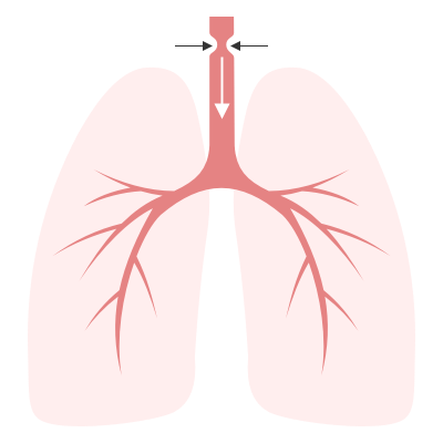
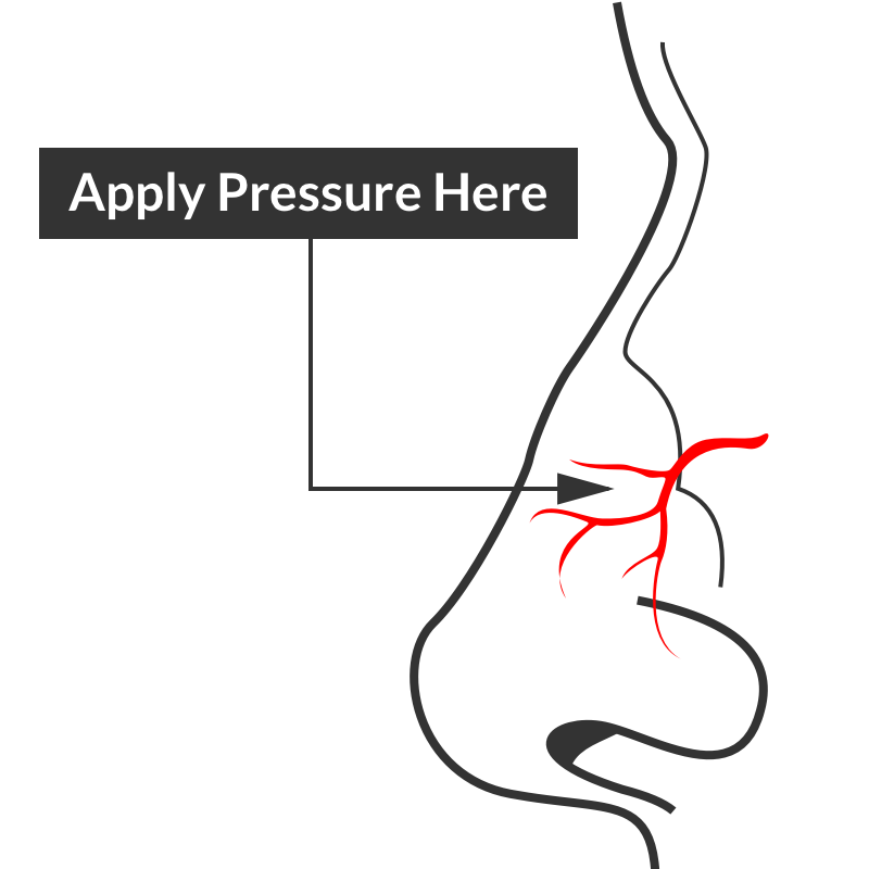
Algorithm for the Management of Epistaxis

Epistaxis site of compression

Site of compression

External Compression

Begin with simple measures while preparing the necessary equipment and medications. Request that the patient gently blow their nose to clear clots, administer oxymetolazone 0.05% two sprays into the affected side. Apply firm pressure below the nasal bridge continuously for at least 10 minutes before reassessment. Commercial compression devices are available, or can be fashioned with tongue depressors3. Alternatively, the patient can apply pressure themselves.
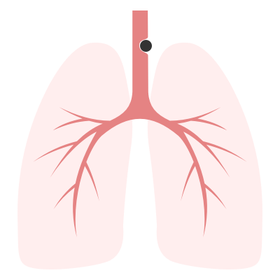
Cautery

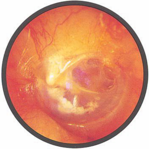
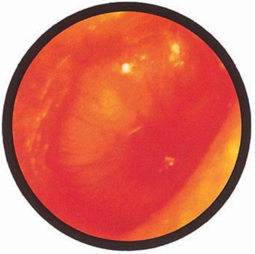
Again ask the patient to blow their nose to remove clots. Apply topical anesthetic for patient comfort prior to inspection with a nasal speculum. Additional suction (small tip, Frazier) may be required to improve visualization. If the bleeding site is identified, apply silver nitrate circumferentially around the source, then directly over the site. Avoid prolonged exposure or exposure to opposing sides of the nasal septum. If hemorrhage control is successful, patients may be discharged with a topical antimicrobial ointment such as polymixin-bacitracin-neomycin.
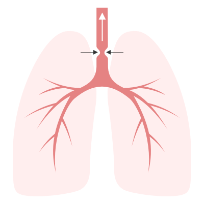
Packing 4,5

Multiple commercial anterior packing devices are available. Placement technique is similar for most, generally involving lubrication of the device with antimicrobial ointment or sterile water, sliding the device along the floor of the nasal cavity, followed by injection or inflation of the device to support tamponade. The incorporation of tranexamic acid (500mg in 5mL) into any phase of anterior packing may be beneficial 6,7. Packing the contralateral side to further support tamponade may be required.

Commonly used commercial devices are:

  • Merocel: lubricate with antimicrobial ointment, once deployed can rehydrate with saline or topical vasoconstrictor
  • Rapid Rhino
  • Rhino Rocket

Packing material should remain for 48-72 hours, during which patients should be re-evaluated. Prophylactic systemic antibiotics for the prevention of sinusitis or toxic shock are likely not required8.

Thrombogenic materials such as Floseal or Surgicel can also be used and may be better tolerated than packing materials9.
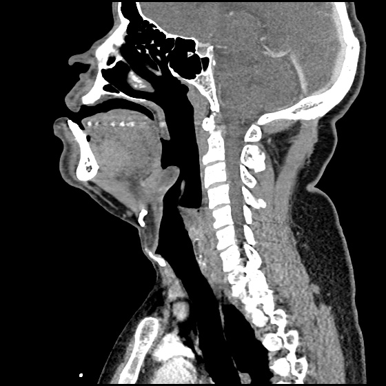
Posterior Control

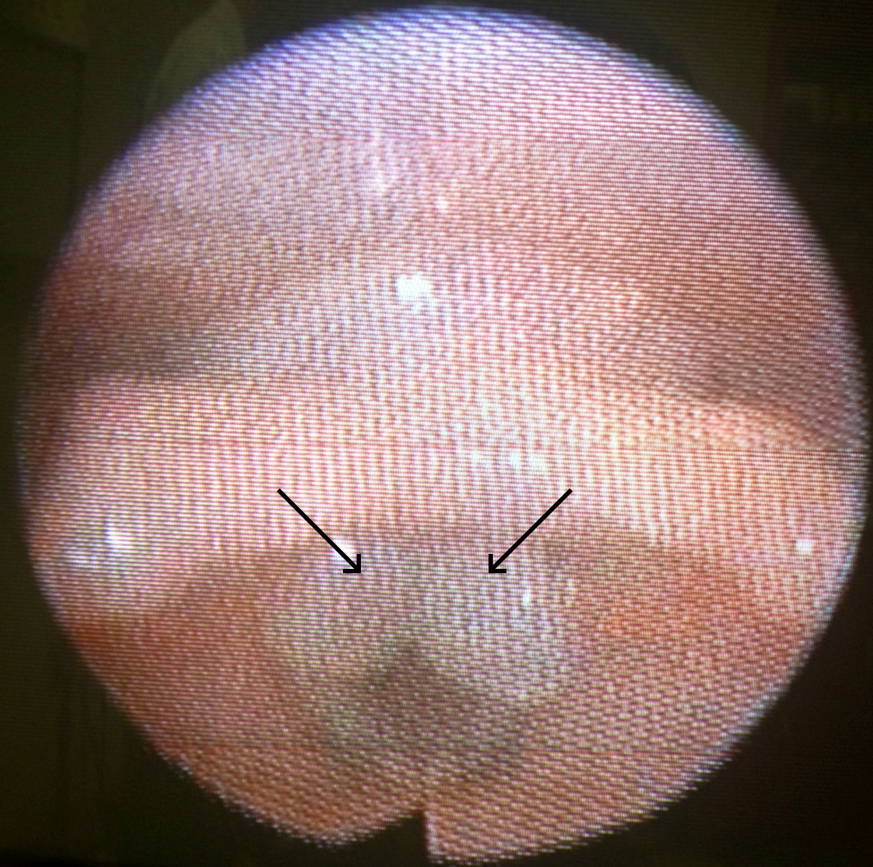
If bleeding persists despite the above measures, a posterior site should be considered. Dual-balloon commercial devices are available for the control of posterior epistaxis and are deployed in a similar fashion to anterior devices. Once inserted, the posterior balloon should be inflated with air – with the volume guided by tension of the pilot cuff. The anterior balloon can then be inflated in a similar fashion. The posterior balloon cuff should be reinspected after 5 minutes as additional inflation may be required.

Commonly used commercial devices are:

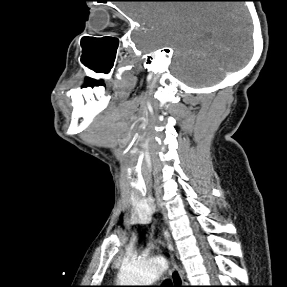
If a commercial device is unavailable, a Foley catheter may be used. The catheter is introduced into the affected side. Once the tip is visualized in the posterior oropharynx, the balloon is inflated with approximately 10mL of sterile water. The catheter is then withdrawn gently to seat the balloon posteriorly. The catheter is secured in position against the nares with a clamp (taking care to pad the nares with gauze to prevent trauma) 10,11.

Patients with posterior epistaxis should be admitted with otolaryngology consultation. If bleeding continues despite these measures, emergent otolaryngology consultation for operative management is warranted.

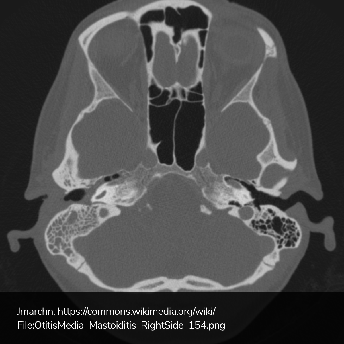
Causes of Epistaxis12

Causes of Epistaxis

References

  1. Leong SCL, Roe RJ, Karkanevatos A. No frills management of epistaxis. Emerg Med J. 2005;22(7):470-472. doi:10.1136/emj.2004.020602.
  2. Barnes ML, Spielmann PM, White PS. Epistaxis: a contemporary evidence based approach. Otolaryngol Clin North Am. 2012;45(5):1005-1017. doi:10.1016/j.otc.2012.06.018.
  3. Moxham V, Reid C. Controlling epistaxis with an improvised device. Emergency Medicine Journal. 2001;18(6):518. doi:10.1136/emj.18.6.518.
  4. Singer AJ, Blanda M, Cronin K, et al. Comparison of nasal tampons for the treatment of epistaxis in the emergency department: A randomized controlled trial. Ann Emerg Med. 2005;45(2):134-139. doi:10.1016/j.annemergmed.2004.10.002.
  5. Iqbal IZ, Jones GH, Dawe N, et al. Intranasal packs and haemostatic agents for the management of adult epistaxis: systematic review. J Laryngol Otol. 2017;131(12):1065-1092. doi:10.1017/S0022215117002055.
  6. MD RZ, MD PM, MD SA, PhD AG, MD MS. A new and rapid method for epistaxis treatment using injectable form of tranexamic acid topically: a randomized controlled trial. American Journal of Emergency Medicine. 2013;31(9):1389-1392. doi:10.1016/j.ajem.2013.06.043.
  7. Kamhieh Y, Fox H. Tranexamic acid in epistaxis: a systematic review. Clin Otolaryngol. 2016;41(6):771-776. doi:10.1111/coa.12645.
  8. MD BC. Are Prophylactic Antibiotics Necessary for Anterior Nasal Packing in Epistaxis? YMEM. 2015;65(1):109-111. doi:10.1016/j.annemergmed.2014.08.011.
  9. Mathiasen RA, Cruz RM. Prospective, Randomized, Controlled Clinical Trial of a Novel Matrix Hemostatic Sealant in Patients with Acute Anterior Epistaxis. The Laryngoscope. 2005;115(5):899-902. doi:10.1097/01.MLG.0000160528.50017.3C.
  10. Holland NJ, Sandhu GS, Ghufoor K, Frosh A. The Foley catheter in the management of epistaxis. Int J Clin Pract. 2001;55(1):14-15.
  11. Hartley C, Axon PR. The Foley catheter in epistaxis management–a scientific appraisal. J Laryngol Otol. 1994;108(5):399-402.
  12. Kucik CJ, Clenney T. Management of epistaxis. Am Fam Physician. 2005;71(2):305-311.

Epiglottitis

Brief H&P:

30 year-old male with no significant medical history presenting with 24 hours of progressively worsening throat pain, difficulty swallowing and voice hoarseness. He reports subjective fevers and chills.
Vital signs notable for Tmax 38.4°C. On physical examination, the patient was sitting upright, unable to swallow secretions with faint inspiratory stridor and dysphonia (though he was able to speak in full sentences and without apparent respiratory distress). Oropharyngeal examination showed minimal right parapharyngeal edema without uvular or palatal deviation and there was exquisite right lateral neck tenderness to palpation.

Labs

  • CBC: 24.2/14.4/43.4/202
  • Wound culture: MSSA
IM-0001-0060
IM-0001-0062
IM-0001-0064
IM-0001-0066
IM-0001-0068
IM-0001-0070
IM-0001-0072
IM-0001-0074
IM-0001-0076
IM-0001-0078
IM-0001-0080

CT Neck/Soft Tissue with Contrast

Edema of the oropharynx/hypopharynx, consistent with epiglottitis and early abscess formation.

ED/Hospital Course

The patient acutely decompensated prior to fiberoptic laryngoscopy and proceeded emergently to the operating room for controlled intubation. The operative report described the following findings: “The patient had diffuse edema of the posterior oropharyngeal wall. The epiglottis was severely thickened, Omega shaped, soft to palpation and with moderate pressure, it appeared to come to a head and pus was expressed from the lingual side of the epiglottis.” The patient was extubated on hospital day three and discharged soon thereafter, he was doing well on follow-up.

Evaluation of Sore Throat – Applied

Evaluation of Sore Throat - Applied

Sore Throat

Evaluation of Sore Throat

Evaluation of Sore Throat

Physical Examination:

Neck
Stiffness, limitation of extension suggestive of retropharyngeal abscess.
Jaw
Trismus associated with peritonsillar cellulitis or abscess.
Oral Cavity
Dry mucous membranes suggest dehydration (from odynophagia) and indicates severity of symptoms.
Tongue elevation, sublingual/submental induration, poor dentition (particularly of mandibular molars) associated with Ludwig Angina.
Unilateral tonsillar enlargement with contralateral uvular deviation suggests peritonisllar abscess. Fluctuance may be palpated.
Tonsilar exudates suggest infectious pharyngitis (non-specific).
Palatal petechiae suggest bacterial pharyngitis.
Ulcerations of the anterior oral cavity are associated with herpes infection, lesions on the soft palate are suggestive of coxsackievirus infection.
Rarely, a grey membrane in the posterior pharynx will suggest diphtheria.
Lymphadenopathy
Tender anterior cervical lymphadenopathy may suggest bacterial pharyngitis.
Posterior cervical lymphadenopathy is associated with infectious mononucleosis.
Large, firm, non-mobile lymph nodes may suggest malignancy.
Eyes
Presence of conjunctivitis (also rhinorrhea, exanthema) associated with viral pharyngitis.
Skin
Ulcers involving the hands, feet, in addition to pharyngeal lesions suggest coxsackievirus infection.
Scarlatiniform rash associated with pharyngitis (particularly in school-age children) suggests streptococcal pharyngitis.
Abdomen
Splenomegaly is associated with infectious mononucleosis.

Centor Criteria (Modified)

  • +1: Fever
  • +1: Tonsillar Exudate
  • +1: Tender anterior cervical lymphadenopathy
  • +1: Absence of cough
  • -1: Age >45yo

Incidence of GABHS by Centor Criteria

  • 0, -1: 1%
  • 1: 10%
  • 2: 17%
  • 3: 35%
  • 4: 51%

References:

  1. Newman, D., & Shreves, A. (2013). Sore Throat. In Rosen’s Emergency Medicine – Concepts and Clinical Practice (8th ed., Vol. 1, pp. 198-202). Elsevier Health Sciences.
  2. King, B. R., & Charles, R. A. (2004). Pharyngitis In The ED Diagnostic Challenges And Management Dilemmas. Emergency medicine practice, 6(5), 1–24.

Dizziness and Vertigo

Types of Dizziness

Types of Dizziness

Distinguishing Central vs. Peripheral Vertigo

Characteristic Peripheral Central
Onset Sudden Gradual
Intensity Severe Mild
Duration Minutes Weeks
Timing Intermittent Continuous
Nystagmus Horizontal Vertical, bidirectional
Exacerbation with head movement +
Auditory symptoms +
Neurological findings +

Causes of Vertigo

Causes of Vertigo

Characteristics of common causes of vertigo

Cause Mechanism Onset Symptoms Findings
Peripheral
BPPV Otolith Brief, positional episodes Nausea, vomiting, absent auditory symptoms. Dix-Hallpike positive
Vestibular neuronitis Viral, post-viral inflammation of vestibular portion of CNVIII Acute and severe, subsiding over days. Nausea, vomiting, absent auditory symptoms. Head thrust abnormal
Meniere Endolymphatic hydrops Recurrent, lasting hours Tinnitus, hearing loss. SNHL
Central
Vertebrobasilar insufficiency Atherosclerosis (vascular risk factors) Acute onset, recurrent episodes if TIA Headache, gait impairment, diplopia, absent auditory symptoms. Neurologic deficits
Cerebellar stroke Atherosclerosis (vascular risk factors) Acute and severe Headache, dysphagia, gait impairment Dysmetria, dysdiadochokinesia, ataxia, CN palsy
Brainstem stroke Atherosclerosis (vascular risk factors), dissection Acute and severe Dysphagia, dysphonia, gait impairment, sensory disturbances Loss of pain/temperature on ipsilateral face, contralateral body, palatal/pharyngeal paralysis
MS Demyelination Subacute onset History of other, variable symptoms INO

History

  • Onset, duration, timing, severity, exacerbating factors
  • Vascular risk factors: age, male, HTN, CAD, DM, atrial fibrillation
  • Vestibulotoxic medications: aminoglycosides, AED

Key Physical Examination Findings

  • VS: Presence of hypotension suggests presyncope
  • Head: Examine for evidence of trauma
  • Neck: Auscultate for carotid bruit
  • Ear: Effusion or perforation suggests peripheral process (possible perilymphatic fistula)
  • Eye: Examine for pupillary defects (CNIII), papilledema, extraoccular muscles
  • Neuro: Cerebellar testing

Positional Testing

Dix-Hallpike
Turn head 45°
Upright sitting → supine (head overhanging bed)
Positive: nystagmus + symptoms on one side
Roll
Supine
Turn head 90°
Positive: nystagmus + symptoms on both sides, more severe on affected

HINTS1

Normal head impulse, direction-changing nystagmus, or skew deviation suggests stroke.

Head impulse
Focus on examiner’s nose
Rapidly turn head 10° in horizontal plan
Presence of corrective saccade suggests defect of peripheral vestibular nerve
Nystagmus
Peripheral: Horizontal, unidirectional. Increases on gaze in direction of fast phase (decreases or resolves opposite)
Central: Direction changing
Skew deviation
Cross cover
Presence of vertical disconjugate gaze suggests brainstem dysfunction

HINTS Gallery

Positive Head Impulse Test
Central Changing Nystagmus
Skew Deviation

Labs

  • Glucose
  • CBC/Chemistry
  • ECG

Imaging

  • Warranted if findings concerning for central process
  • MRI preferred

Management

Specific etiologies
Vestibular neuronitis: steroids
Meniere: dietary changes
BPPV: canalith repositioning
Symptomatic relief
Promethazine (Phenergan) 12.5-25mg PO
Ondansetron (Zofran) 4mg IV
Lorazepam (Ativan) 1-2mg PO/IV
Meclizine (Antivert) 25mg PO q6-8h PRN

References

  1. Kattah, J. C., Talkad, A. V., Wang, D. Z., Hsieh, Y.-H., & Newman-Toker, D. E. (2009). HINTS to diagnose stroke in the acute vestibular syndrome: three-step bedside oculomotor examination more sensitive than early MRI diffusion-weighted imaging. Stroke; a journal of cerebral circulation, 40(11), 3504–3510. doi:10.1161/STROKEAHA.109.551234
  2. Chang, A., & Olshaker, J. (2013). Dizziness and Vertigo. In Rosen’s Emergency Medicine – Concepts and Clinical Practice (8th ed., Vol. 1, pp. 162-169). Elsevier Health Sciences.

Angioedema

AngioedemaHPI:

63-year old African American male with a history of HTN presenting with lip swelling x1 day. The patient states he was well until this morning when he noticed progressive swelling of his lips. The swelling is not associated with any difficulty speaking, swallowing or breathing and is not painful.

He denies new rashes or itching, and has no history of such swelling. He also denies any exposure to known allergens, recent insect bites or travel. He has been taking lisinopril for his blood pressure regularly for the past several months and denies any prior adverse effects (cough, rash).

PMH:

  • Parkinson Disease
  • HTN

PSH:

None

FH:

No family history of angioedema

SHx:

  • No t/e/d use
  • Lives at home with caretaker

Meds:

  • Lisinopril 20mg p.o. daily
  • Carbidopa/levodopa 50mg p.o. t.i.d.

Allergies:

NKDA

Physical Exam:

VS: T 37.8 HR 84 RR 14 BP 146/98 O2 99% RA
Gen: Well-appearing, no respiratory distress, speaking comfortably
HEENT: PERRL, significant external upper/lower lips swelling extending to lateral cheeks, non-tender, no fluctuance or overlying skin changes. No visible tongue swelling, floor of mouth swelling/tenderness, uvular/palatal deviation.
CV: RRR, no M/R/G
Lungs: CTAB, no crackles/wheezing, good air movement b/l
Abd: +BS, soft, NT/ND, no rebound/guarding
Ext: Warm, well-perfused, 2+ peripheral pulses
Skin: No visible skin lesions/rashes
Neuro: AAOx4, CN II-XII intact

Assessment/Plan:

63M with acute onset, progressive facial swelling. Currently restricted to external lips, with no evidence of airway compromise. Likely ACE inhibitor-induced angioedema given patient is on lisinopril and has no history of hereditary angioedema. Doubt anaphylaxis given no allergies, suspicious exposures or history of pruritus. Doubt infection given afebrile and painless swelling without e/o erythema.

Pathophysiology of ACE inhibitor-induced angioedema1

Pathophysiology of ACE-inhibitor induced angioedema

Angioedema is a vascular reaction associated with tissue (subcutaneous, submucosal) edema resulting from increased activity of vasoactive substances. The vasoactive substances in ACE inhibitor-induced angioedema are bradykinin and substance P. In the presence of ACE inhibition, these enzymes are inactivated through alternative pathways which, if disturbed, lead to angioedema.

Epidemiology of ACE inhibitor-induced angioedema

Angioedema occurs in 0.1-0.7% of patients taking ACE inhibitors, and 60% of cases occur within the first week of starting an ACE inhibitor (though it can occur as much as years later).2,3 ACE inhibitors are implicated as the cause of 20-40% of all ED visits for angioedema.4

Risk Factors2,5,6

  • Female
  • Age > 65yo
  • African American
  • Prior angioedema
  • Smoking
  • ACE inhibitor-associated cough

Clinical Features of ACE inhibitor-induced angioedema

Affected Sites:

  • Mucous membranes of the head and neck
    • Face
    • Tongue
    • Lips
    • Pharynx
    • Larynx
  • GI tract
    • Diffuse abdominal pain
    • Nausea/vomiting/diarrhea

Signs/Symptoms at initial presentation:4

  • SOB (89%)
  • Lip swelling (70%)
  • Tongue swelling (52%)
  • Voice change/hoarseness (29%)
  • Stridor (11%)

Key Clinical Features:

  • Onset in minutes with resolution in 24-72 hours
  • Absence of itching/urticaria7

Staging and Disposition:8

Stage Affected Site Outpatient (%) Floor (%) ICU (%) Intervention (%)
I Face, lip 48 52 0 0
II Soft palate 60 40 0 0
III Tongue 26 7 67 7
IV Larynx 0 0 100 24

Management of ACE inhibitor-induced angioedema

  • Proven benefit
    • Airway management
    • Withdrawal of ACE inhibitor
  • Unclear benefit
    • Epinephrine 0.3mg IM q15min
    • Diphenhydramine 50mg IV
    • Famotidine 20mg IV
    • Solumedrol 125mg IV
  • Future treatment options
    • FFP: contains ACE9
    • Icatibant: bradykinin B2 receptor antagonist10,11

References:

  1. Vleeming, W., van Amsterdam, J. G., Stricker, B. H. C., & de Wildt, D. J. (1998). ACE inhibitor-induced angioedema. Drug Safety, 18(3), 171–188. doi:10.2165/00002018-199818030-00003
  2. Grant, N. N., Deeb, Z. E., & Chia, S. H. (2007). Clinical experience with angiotensin-converting enzyme inhibitor-induced angioedema. Otolaryngology – head and neck surgery, 137(6), 931–935. doi:10.1016/j.otohns.2007.08.012
  3. Slater, E. E., Merrill, D. D., Guess, H. A., Roylance, P. J., Cooper, W. D., Inman, W. H., & Ewan, P. W. (1988). Clinical profile of angioedema associated with angiotensin converting-enzyme inhibition. JAMA : the journal of the American Medical Association, 260(7), 967–970.
  4. Banerji, A., Clark, S., Blanda, M., LoVecchio, F., Snyder, B., & Camargo, C. A. (2008). Multicenter study of patients with angiotensin-converting enzyme inhibitor-induced angioedema who present to the emergency department. Annals of allergy, asthma & immunology, 100(4), 327–332. doi:10.1016/S1081-1206(10)60594-7
  5. Gibbs, C. R., Lip, G. Y., & Beevers, D. G. (1999). Angioedema due to ACE inhibitors: increased risk in patients of African origin. British journal of clinical pharmacology, 48(6), 861–865.
  6. Morimoto, T., Gandhi, T. K., Fiskio, J. M., Seger, A. C., So, J. W., Cook, E. F., Fukui, T., et al. (2004). An evaluation of risk factors for adverse drug events associated with angiotensin-converting enzyme inhibitors. Journal of evaluation in clinical practice, 10(4), 499–509. doi:10.1111/j.1365-2753.2003.00484.x
  7. Kanani, A., Schellenberg, R., & Warrington, R. (2011). Urticaria and angioedema. Allergy, Asthma & Clinical Immunology, 7(Suppl 1), S9. doi:10.1186/1710-1492-7-S1-S9
  8. Ishoo, E., Shah, U. K., Grillone, G. A., Stram, J. R., & Fuleihan, N. S. (1999). Predicting airway risk in angioedema: staging system based on presentation. Otolaryngology – head and neck surgery, 121(3), 263–268.
  9. Hassen, G. W., Kalantari, H., Parraga, M., Chirurgi, R., Meletiche, C., Chan, C., Ciarlo, J., et al. (2013). Fresh frozen plasma for progressive and refractory angiotensin-converting enzyme inhibitor-induced angioedema. The Journal of emergency medicine, 44(4), 764–772. doi:10.1016/j.jemermed.2012.07.055
  10. Bas, M., Greve, J., Stelter, K., Bier, H., Stark, T., Hoffmann, T. K., & Kojda, G. (2010). Therapeutic Efficacy of Icatibant in Angioedema Induced by Angiotensin-Converting Enzyme Inhibitors: A Case Series. Annals of emergency medicine, 56(3), 278–282. doi:10.1016/j.annemergmed.2010.03.032
  11. MD, M. G., & MD, M. A. (2012). Icatibant: a novel approach to the treatment of angioedema related to the use of angiotensin-converting enzyme inhibitors. American Journal of Emergency Medicine, 30(8), 1664.e1–1664.e2. doi:10.1016/j.ajem.2011.09.014

Hearing loss and Tinnitus

HPI:

42 year-old male with no significant medical history presenting to ENT clinic after referral from PMD for perforated TM. The patient last had normal hearing approximately 1yr ago when he noted acute onset of right ear pain, discharge, hearing loss and ringing in the setting of fever and a productive cough. He does not recall an inciting event (trauma, swimming) to this initial episode, and had no previous history of ear infections. He saw his PMD several days later, was told he had a perforated ear drum and was treated with antibiotics.

Since then, the patient has not had any further ear pain or discharge but is left with persistent and constant hearing loss and ringing (high-pitched, non-pulsatile).

PMH:

None

PSH:

None

FH:

Non-contributory

SHx:

Worked in construction for 20yrs

Meds:

None

Allergies:

NKDA

Physical Exam:

Gen: Well-appearing, no acute distress
Head: NC/AT
Eyes: PERRL (4-2mm), disc margins sharp
Ears: Weber lateralizes to left, AC > BC b/l
AD: Decreased acuity to finger rub, EAC with some cerumen, cleared to reveal central perforation in posterior-superior quadrant of tympanic membrane.
AS: EAC clear, TMI
Nose: Nasal mucosa pink, septum midline
Mouth: MMM, no lesions, good dentition, no pharyngeal erythema/exudates
Neck: Trachea midline, supple, no cervical lymphadenopathy, no thyroid enlargement

Studies

Audiogram

Audiogram: Severe low-mid frequency progressing to profound high frequency mixed hearing loss

Assessment/Plan:

42M, no significant PMH, with perforated TM and audiogram showing mixed hearing deficit. The patient describes a history suggestive of acute otitis media complicated by TM perforation. Persistent perforation seen on examination today can result in the tinnitus and hearing loss the patient complains of. However the marked sensorineural component remains unexplained, particularly given the patient reported previously normal hearing. While there is some evidence that acute otitis media can lead to sensorineural hearing loss, it is typically only mild and only in high-frequency ranges.1,2  Plan for further evaluation with repeat audiogram and MRI IAC, RTC when studies completed.


Examination of the Ear3

  1. External Auditory Canal: in acute otitis externa, the canal will be narrowed, swollen and erythematous
  2. Tympanic Membrane: use cone of light for orientation, identify malleus and move speculum to visualize all four quadrants
Tympanic Membrane
Ear Anatomy
Normal
Perforation
Tympanosclerosis
Acute Otitis Media

Differential Diagnosis of Hearing Loss4

Differential Diagnosis of Hearing Loss

Differential Diagnosis of Tinnitus5

Differential Diagnosis of Tinnitus

References:

  1. Tarlow, M. (1998). Otitis media: pathogenesis and medical sequelae. Ear, nose, & throat journal, 77(6 Suppl), 3–6.
  2. Tsuprun, V., Cureoglu, S., Schachern, P. A., Ferrieri, P., Briles, D. E., Paparella, M. M., & Juhn, S. K. (2008). Role of pneumococcal proteins in sensorineural hearing loss due to otitis media. Otology & neurotology, 29(8), 1056–1060.
  3. Bickley, Lynn S., Peter G. Szilagyi, and Barbara Bates. Bates’ guide to physical examination and history taking. Philadelphia: Wolters Kluwer Health/Lippincott Williams & Wilkins, 2009. Print.
  4. Isaacson, J. E., & Vora, N. M. (2003). Differential diagnosis and treatment of hearing loss. American family physician, 68(6), 1125–1132.
  5. Crummer, R. W., & Hassan, G. A. (2004). Diagnostic approach to tinnitus. American family physician, 69(1), 120–126.

Dysphonia (Hoarseness)

Case 1

HPI:

36 year-old female with no significant medical history who presents after referral for voice hoarseness. According to the patient, she underwent a C-section 3 months ago (at an outside hospital) complicated by bleeding requiring a second operation (L salpingoophorectomy); however, neither procedure required emergent intubation. She reports that she had some vomiting associated with anesthesia which ultimately required intubation and admission to the MICU for 5-6d. She was discharged 10 days after the initial operation, and both she and her baby were in good health. Two weeks after discharge, she began experiencing throat irritation and 1 month after discharge she noticed voice hoarseness which has been persistent. Today, she denies difficulty swallowing or breathing, F/C, N/V, abdominal pain.

PMH:

None

PSH:

Cesarean x2, L salpingoophorectomy

FH:

Non-contributory

SHx:

Lives at home taking care of 3 children, denies t/e/d

Meds:

None

Allergies:

NKDA

Physical Exam:

Gen: WA, NAD
Head: NC/AT
OC: MMM, no lesions, no pharyngeal erythema/exudates, hoarse voice
Ears: EAC clear, TMI b/l
Flex: Posterior commissure edema, cobblestoning, b/l TVC with shiny white masses
Flexible nasolaryngoscopy image showing trauma granulomata.

Flexible nasolaryngoscopy image showing trauma granulomata.

Assessment/Plan:

36F, no significant PMH, recent Cesarean and L salpingoophorectomy c/b likely aspiration requiring intubation and mechanical ventilation for several days with onset of progressive voice hoarseness 1mo later. History and flexible nasolaryngoscopy consistent with trauma granulomata, as well as laryngopharyngeal reflux.

  • Discussed treatment options with patient, recommend voice rest x2mo and continued monitoring
  • Laryngopharyngeal reflux, given dietary/lifestyle education, start omeprazole 20mg p.o. b.i.d.
  • RTC 1mo

Case 2

HPI:

51 year-old female with a history of Hepatitis C, COPD and an 80 pack-year smoking history presents with concern about progressive voice hoarseness x2mo. She reports quitting smoking two months ago and is not sure if the hoarseness preceded or followed quitting. She has occasional throat discomfort which is mild. She otherwise denies difficulty or pain with swallowing, worsening shortness of breath, unintentional weight loss.

She also reports a new mass on her neck which she first noticed yesterday. Denies associated pain, or surrounding skin changes.

PMH:

  • Hepatitis C
  • COPD

PSH:

  • Hysterectomy

FH:

Non-contributory

SHx:

80 pack-year smoking history, no current EtOH, drug use (previously used heroin and opiates)

Meds:

  • Methadone
  • Elavil
  • Multiple unknown inhaled medications

Allergies:

  • Naproxen (swelling)

Physical Exam:

Gen: WA, NAD
Head: NC/AT
Eyes: PERRL, EOMI
Ears: b/l EAC erythema, TMI, no lesions/exudates
OC: MMM, no lesions
Neck: Supple, no thyroid enlargement, no cervical lymphadenopathy, 5x6cm soft, round, mobile, non-tender mass on left lateral neck w/o overlying skin changes
Flex: Diffuse laryngeal damage, thickened posterior commissure, right TVC with area of leukoplakia, left TVC appears irregular

Assessment/Plan:

51F hx HepC, COPD, 80py smoking, presenting with voice hoarseness x2mo. History concerning for malignancy, exam today shows significant laryngeal damage and vocal cord irregularities warranting further evaluation. Possible component of fungal infection 2/2 inhaled steroid use for COPD, plan to reduce potentially aggravating factors (treat fungal infection, voice rest) and repeat evaluation. Neck mass possibly lipoma however will evaluate further given concern for malignancy.

  • Start fluconazole 100mg two tables p.o. on day1, 100mg p.o. daily x7d
  • Start nystatin 100,000 units/mL 10mL gargle and swallow t.i.d. x2wks
  • Advised voice rest
  • RTC in 3wks
  • CT neck/soft tissue w/wo IV contrast to evaluate neck mass

Anatomy of the Pharynx/Larynx:

Vocal cord anatomy
Pharyngeal wall anatomy
Structure of the pharynx

Physiology of Voice Production: 1

Voice is produced through the passive vibration of vocal folds in an air stream and requires:

  1. Adequate Air Stream
  2. Smooth vocal fold edges
  3. Vocal folds with normal vibratory properties
  4. Appropriate vocal fold positioning

Differential diagnosis of dysphonia (hoarseness): 1,2,3

 Differential Diagnosis of Dysphonia (hoarseness)

Characteristics of Hoarse Voice: 5

Characteristic Likely cause
Breathy Vocal cord paralysis
Hoarse Vocal cord lesion, LPR
Low-pitched Reinke’s edema, vocal abuse, LPR

References:

  1. Mau, T. (2010). Diagnostic Evaluation and Management of Hoarseness. Medical Clinics of North America, 94(5), 945–960. doi:10.1016/j.mcna.2010.05.010
  2. Feierabend, R. H., & Shahram, M. N. (2009). Hoarseness in adults. American family physician, 80(4), 363–370.
  3. Schwartz, S. R., Cohen, S. M., Dailey, S. H., Rosenfeld, R. M., Deutsch, E. S., Gillespie, M. B., Granieri, E., et al. (2009, September). Clinical practice guideline: hoarseness (dysphonia). Otolaryngology. doi:10.1016/j.otohns.2009.06.744
  4. Bruch, J.W., Kamani D.V. Diaphragmatic pacing. In: UpToDate, Basow, DS (Ed), UpToDate, Waltham, MA, 2013.
  5. Rosen, C. A., Anderson, D., & Murry, T. (1998). Evaluating hoarseness: keeping your patient’s voice healthy. American family physician, 57(11), 2775–2782.

Sore Throat

Oropharynx AnatomyID:

17 year-old female presenting to the pediatric ED with sore throat for 2 days.

HPI:

The patient reports steadily worsening sore throat over the past 2 days, associated with a sensation of swelling. The pain is described as sharp, 4/10 in severity, located on the left side of her throat, and worsened with swallowing. She denies inability to swallow or difficulty breathing, she also denies fever, cough, new skin rashes or genital lesions.

She has no PMH/PSH, takes no medications, denies t/e/d use and is not currently sexually active.

PE:

  • VS: 111/65mmHg, 80bpm, 97.8°, 16/min, 100% RA
  • Gen: Well-appearing, NAD
  • HEENT: PERRL, no conjunctival injection, TM clear b/l, minimal pharyngeal erythema on left with 6mm white circular lesion on left tonsil, no tonsillar enlargement, no uvular deviation, no cervical LAD, neck supple no masses, normal neck ROM
  • CV: RRR, no M/R/G, Lungs: CTAB
  • Abdomen: +BS, soft, NT/ND
  • Ext: Warm, well-perfused, normal peripheral pulses

Assessment & Plan:

17yo female with no significant PMH with acute pharyngitis for 2 days. The most likely cause of the patient’s symptoms is viral pharyngitis, potentially herpangina (given the appearance of the tonsillar lesion). A more serious viral/bacterial pharyngitis is less likely given the absence of fever or significant erythema/exudate. There was no uvular deviation to suggest peritonsillar abscess and no evidence of airway obstruction to suggest other acute processes (epiglottitis, retropharyngeal abscess). The plan is to recommend supportive care and ibuprofen for symptomatic relief. The patient will be discharged home in good condition with precautions to return if symptoms worsen or she begins to have difficulty swallowing/breathing.

Differential Diagnosis of Acute Pharyngitis:

Acute Pharyngitis


Evaluation (history):

  • Respiratory distress: epiglottitis, retropharyngeal abscess, peritonsillar abscess, EBV (obstruction in or near pharynx)
  • Fatigue: infectious mononucleuosis
  • Abrupt onset: epiglottitis

Evaluation (physical examination):

  • Vesicles anterior: herpetic stomatitis, SJS, Behcet
  • Vesicles posterior: herpangina (± involvement of extremities)
  • Asymmetry: peritonsillar abscess
  • Stridor, drooling, respiratory distress: airway obstruction
  • Generalized inflammation: Kawasaki