Headache

Brief HPI:

A 48 year-old male with hypertension and hyperlipidemia presents with headache. Notes onset of symptoms 8 hours prior to presentation, reaching maximal severity within seconds. Headache improved with over-the-counter analgesics. On examination, there are no neurological deficits, neck is supple. A CT head non-contrast is obtained:
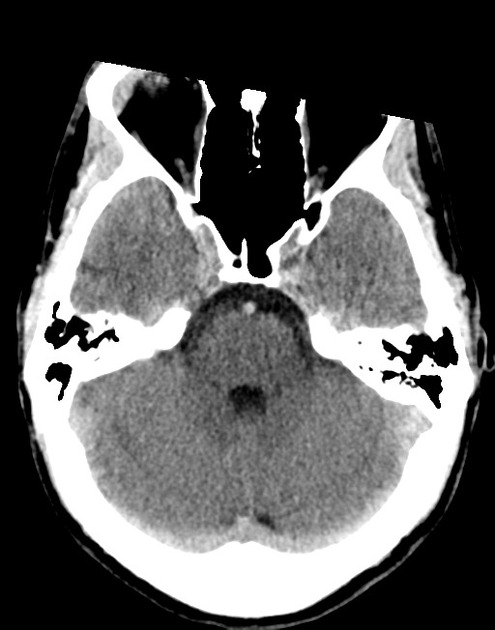
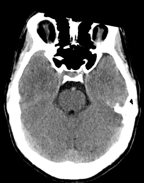
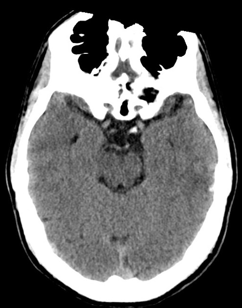
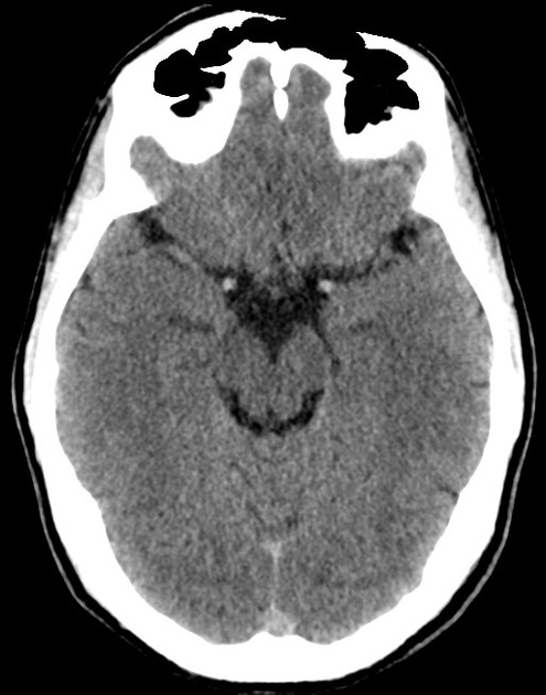
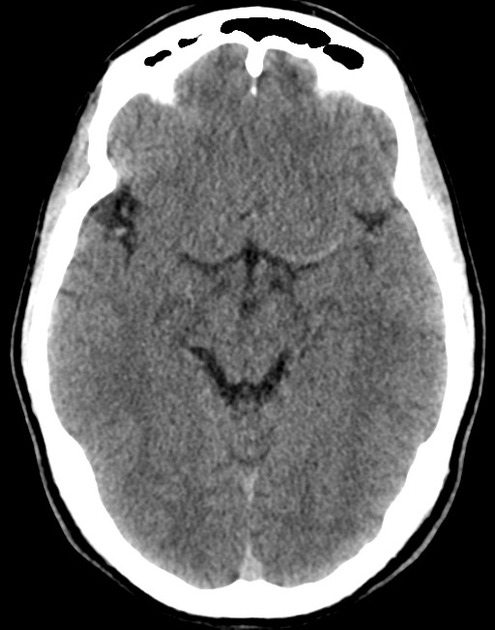
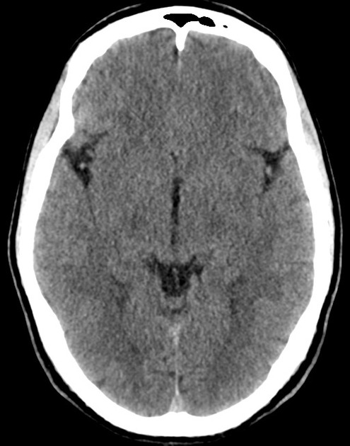
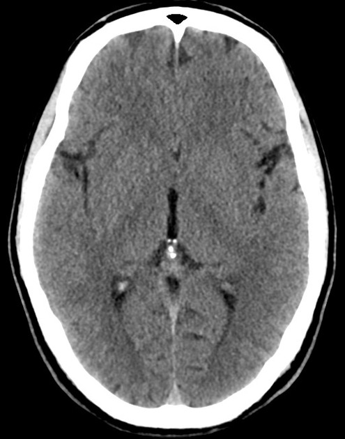
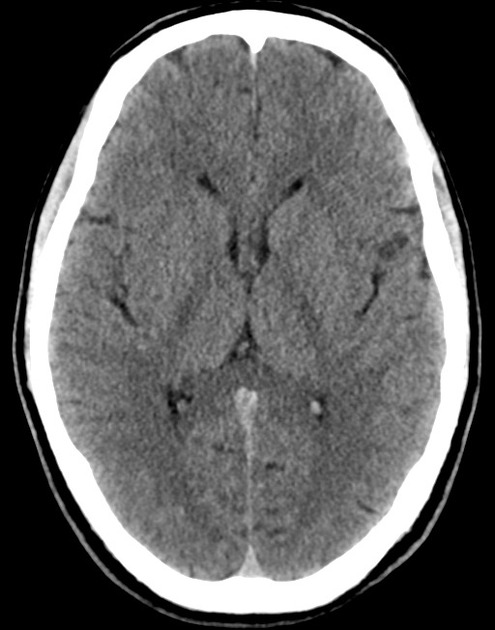
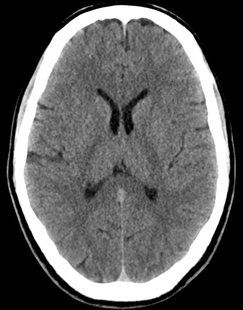
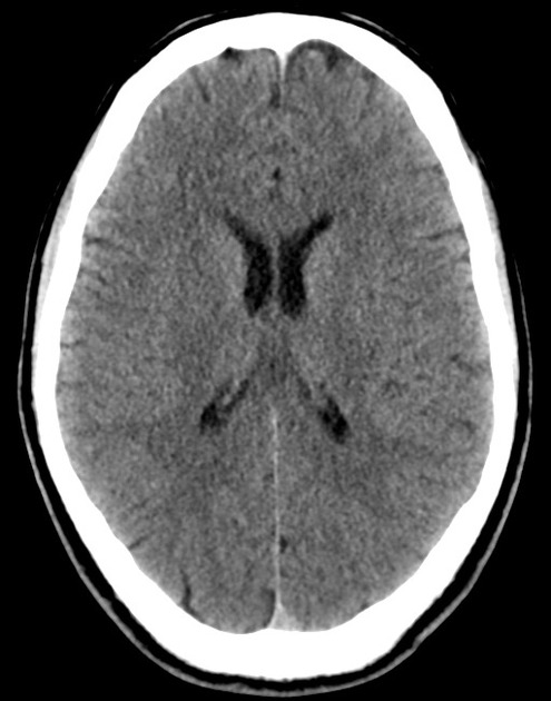
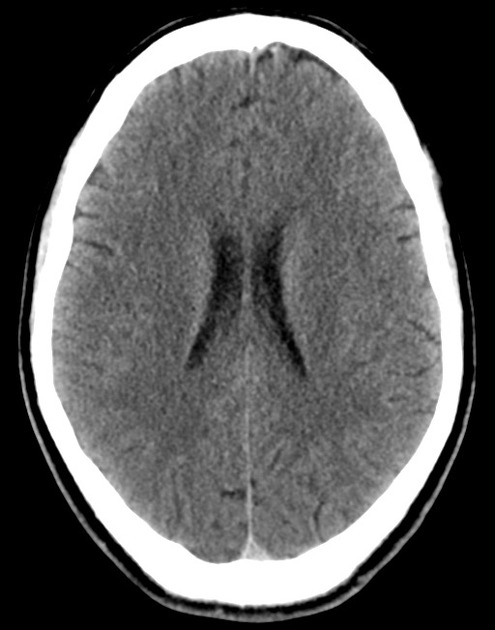
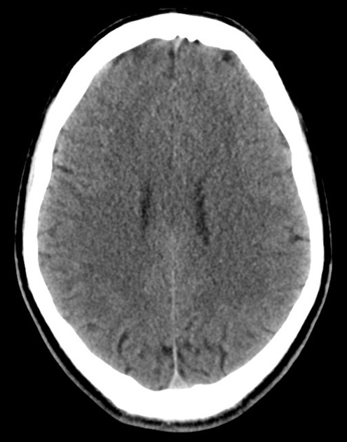
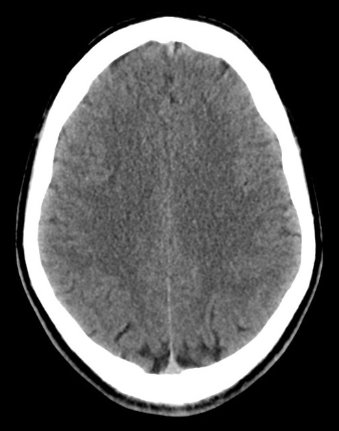
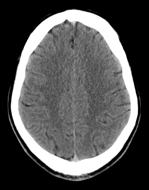
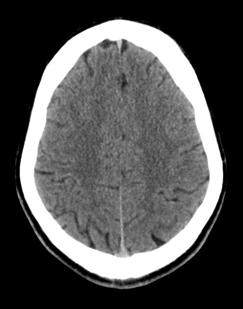
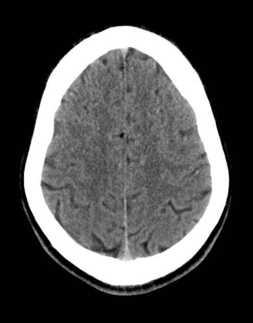
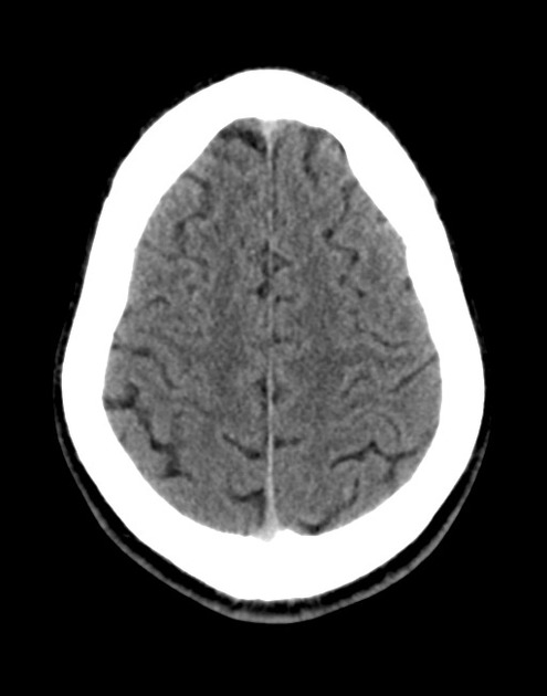
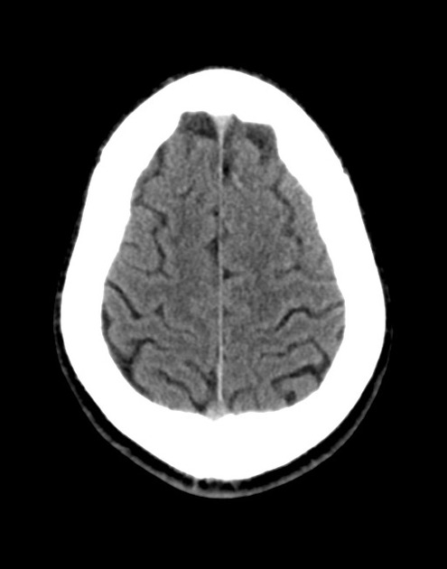
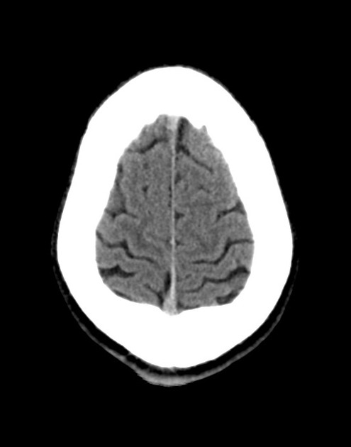
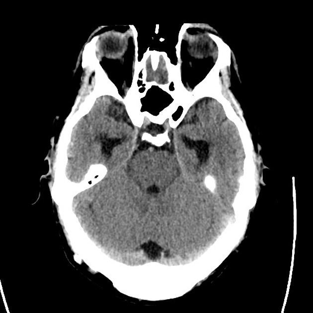
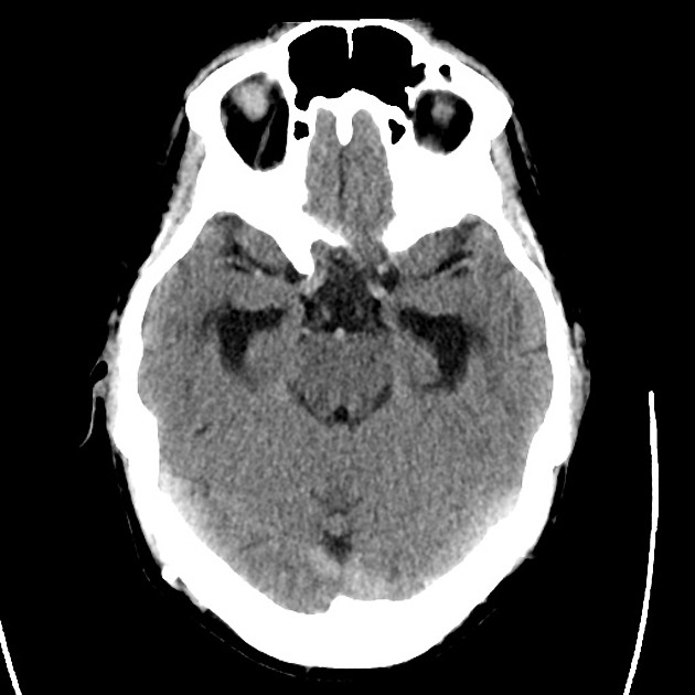
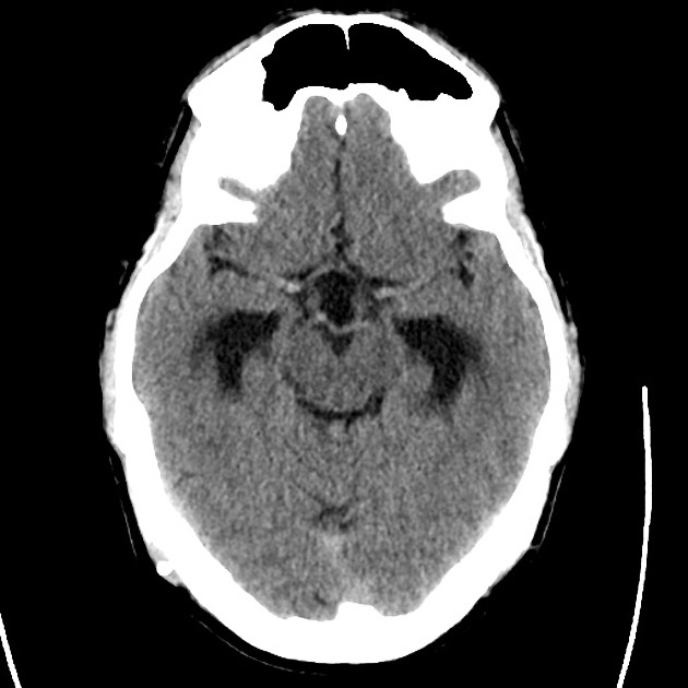
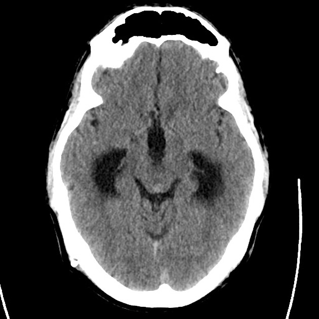
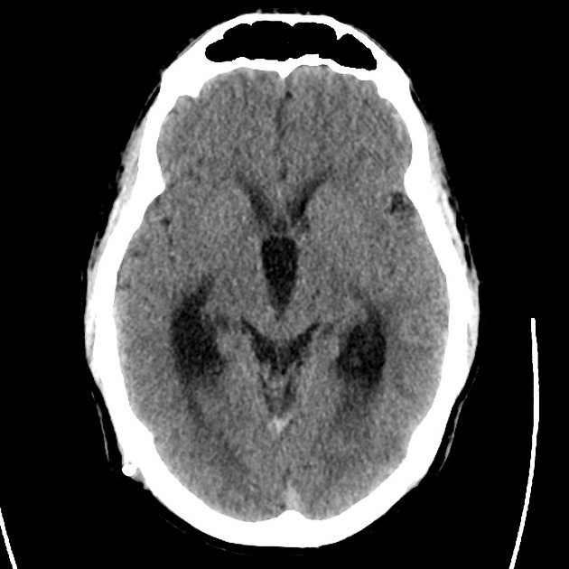
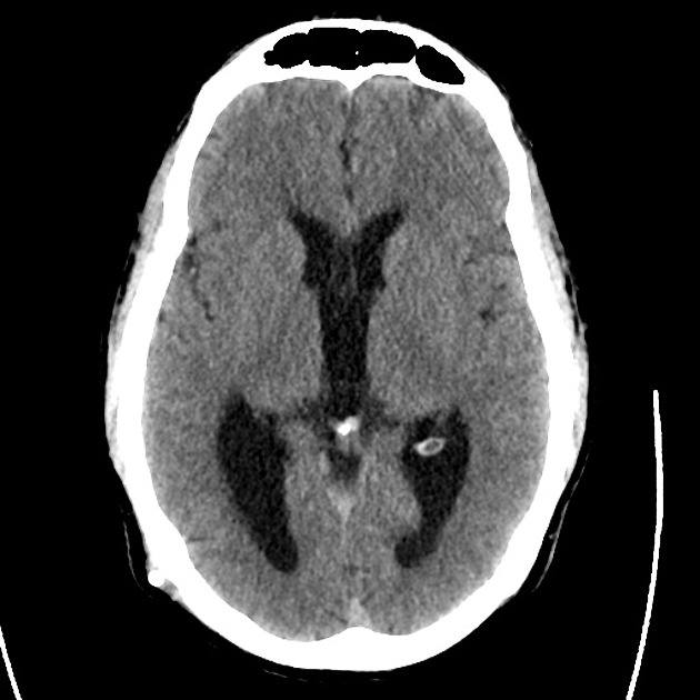
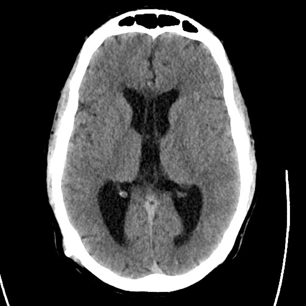
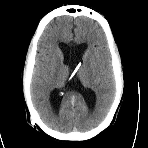
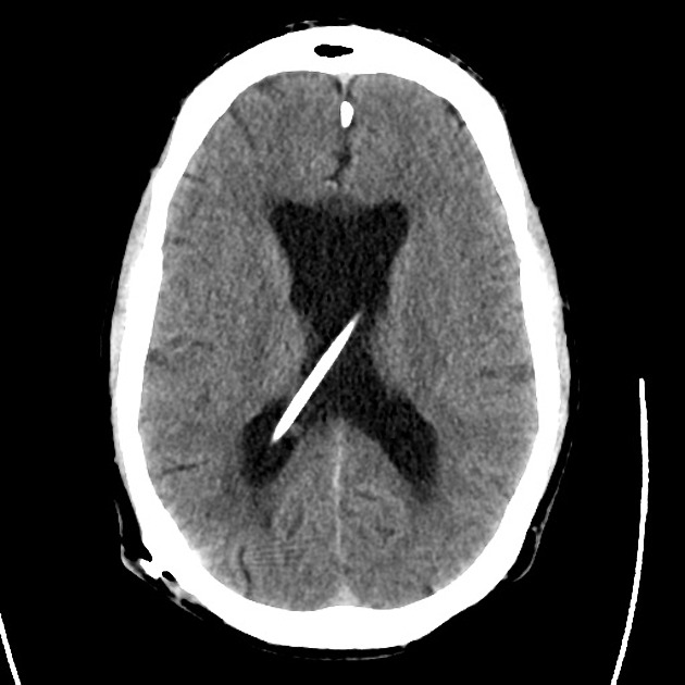
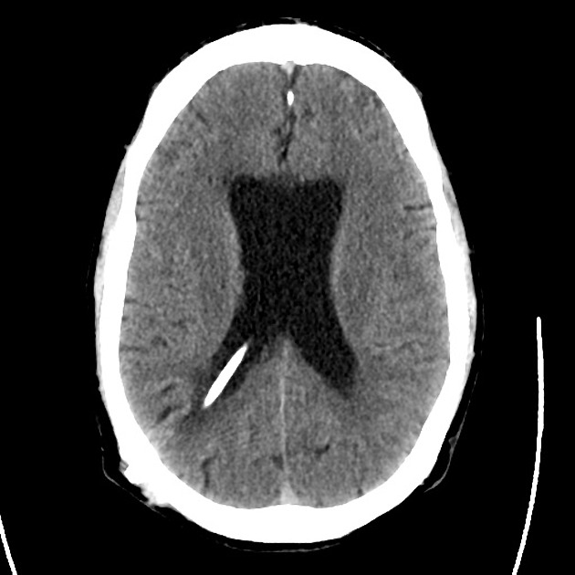
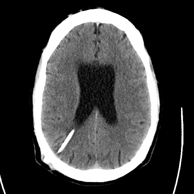
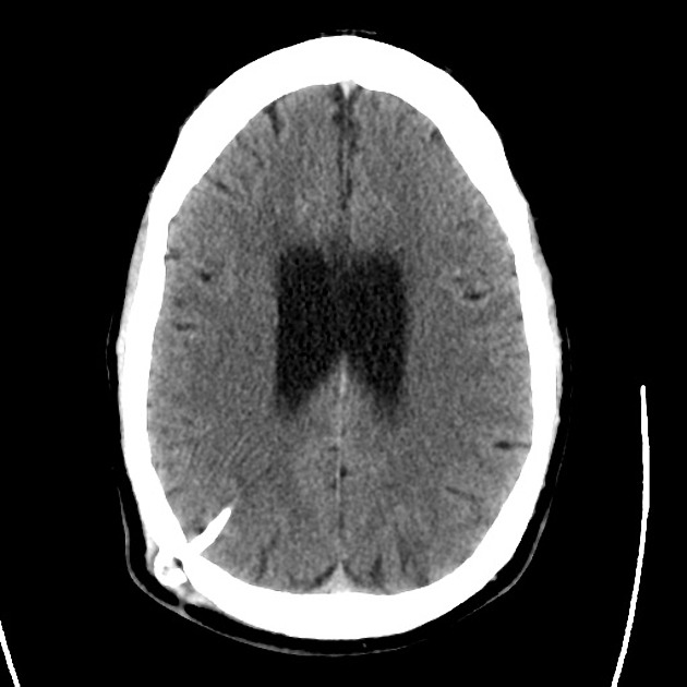
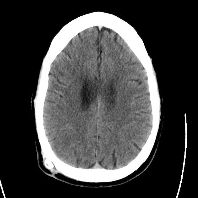
01_normal
02_normal
03_normal
04_normal
05_normal
06_normal
07_normal
08_normal
09_normal
10_normal
11_normal
12_normal
13_normal
14_normal
15_normal
16_normal
18_normal
19_normal
20_normal

CT Head:

No acute intracranial process. Case courtesy of Assoc Prof Craig Hacking, Radiopaedia.org, rID: 37118

ED Course:

A lumbar puncture is performed, CSF sampling reveals xanthochromia – neurosurgery is consulted and the patient is admitted for angiography and possible intervention.


An Algorithm for the Evaluation of Headache

An Algorithm for the Evaluation of Headache

High-Risk Historical Features

  • Sudden onset (seconds/minutes), patient recalls activity at onset
  • Worst in life or change in character from established headache
  • Fever, neck pain/stiffness
  • Altered mental status
  • Malignancy
  • Coagulopathy: iatrogenic, hepatopathy, dialysis
  • Immunocompromised
  • Rare: CO exposure, jaw claudication, PCKD

Location of Pain

Headache Location

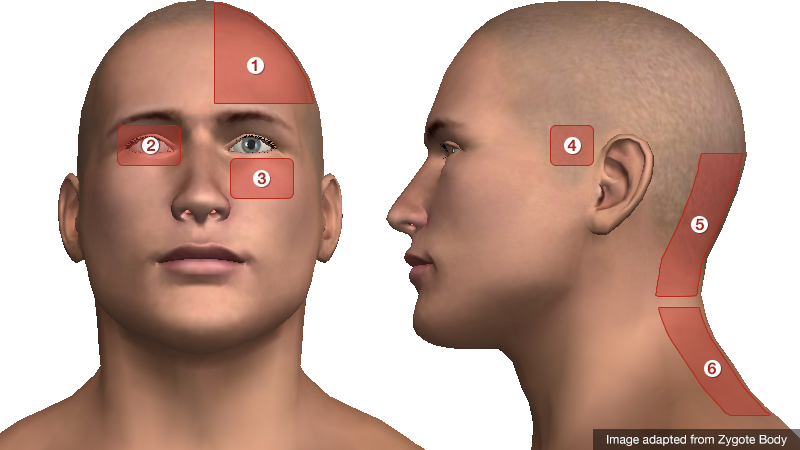
  1. Unilateral: migraine
  2. Periorbital: glaucoma, CVT, optic neuritis, cluster
  3. Facial/maxillary: trigeminal neuralgia, sinusitis
  4. Temporal: GCA
  5. Occipital: cerebellar stroke
  6. Nuchal: meningitis

Characteristics of Primary Headaches

Type Location Duration Quality Associated symptoms Comment
Migraine Unilateral Hours to days Throbbing Photophobia, phonophobia Atypical migraines with neurological findings (basilar, ophthalmoplegic, ophthalmic, hemiplegic)
Tension Bilateral Minutes to days Constricting None
Cluster Unilateral, periorbital Minutes to hours Throbbing Conjunctival injection, lacrimation, rhinorrhea, miosis, eyelid edema Males 90%, triggered by EtOH.

Physical Examination Findings

Vital Signs
Fever: present in 95% of patients with meningitis
Head
Trauma: signs of basilar skull fracture
Temporal artery tenderness/induration: GCA
Pericranial muscle tenderness: tension headache
Trigger point, Tinnel sign: occipital neuralgia
Eyes
Pupillary defects: aneurysm with CN III compression
Papilledema, absence of spontaneous venous pulsations: elevated intracranial pressure
EOM abnormalities: ICH, mass lesion, neuropathy (DM, Lyme)
Horner syndrome (ptosis, miosis, anhidrosis): carotid dissection
Visual field defect: stroke, atypical migraine
Conjunctival injection: glaucoma (fixed, mid-size pupil, elevated intraocular pressure), cluster headache
Mouth
Thrush: immunocompromise
Sinuses
Tenderness to palpation, abnormal transillumination: sinusitis
Neck
Resistance to supine neck flexion: meningitis
Kernig: supine position, hip flexed, knee flexed, resistance to knee extension
Brudzinski: supine position, neck flexion results in knee flexion
Jolt accentuation: patient rotates head side-to-side, 2-3 times/sec exacerbates headache

References:

  1. Russi, C. (2013). Headache. In Rosen’s Emergency Medicine – Concepts and Clinical Practice (8th ed., Vol. 1, pp. 170-175). Elsevier Health Sciences.
  2. Godwin SA, Villa J. “Acute headache in the ED: Evidence-Based Evaluation and Treatment Options.” Emerg Med Pract 2001; 3(6): 1-32.
  3. Edlow, J. A., Panagos, P. D., Godwin, S. A., Thomas, T. L., & Decker, W. W. (2008). Clinical Policy: Critical Issues in the Evaluation and Management of Adult Patients Presenting to the Emergency Department With Acute Headache. Annals of emergency medicine, 52(4), 407–436. doi:10.1016/j.annemergmed.2008.07.001
  4. WikEM: Headache

CSF Shunt Complications

Brief HPI:

A 33 year-old female with a history of idiopathic intracranial hypertension and ventriculoperitoneal shunt placement presents with headache and confusion. She denies fever, trauma, neck pain or stiffness. She has not had symptoms like this since her shunt was placed 2 years ago. Imaging was obtained which showed ventriculomegaly and a fracture of the shunt at the level of the cervical spine. Neurosurgery was consulted and the patient was admitted for shunt repair.

00_ct_csf_shunt
01_ct_csf_shunt
02_ct_csf_shunt
03_ct_csf_shunt
04_ct_csf_shunt
05_ct_csf_shunt
06_ct_csf_shunt
07_ct_csf_shunt
08_ct_csf_shunt
09_ct_csf_shunt
10_ct_csf_shunt
11_ct_csf_shunt
12_ct_csf_shunt

CT Head

Ventriculomegaly with dilatation of the temporal horns in particular. Right parietal approach ventricular drain. Case courtesy of Dr. Henry Knipe, Radiopaedia.org, rID: 39615

XR Shunt Series

XR Shunt Series

Shunt tubing fractured at the level of the upper cervical spine

An Algorithm for CSF Shunt Complications

An Algorithm for CSF Shunt Complications

References:

  1. Madsen MA. Emergency department management of ventriculoperitoneal cerebrospinal fluid shunts. Ann Emerg Med. 1986;15(11):1330-1343.
  2. Ferras M, McCauley N, Stead T, Ganti L, Desai B. Ventriculoperitoneal shunts in the emergency department: a review. Cureus. 2020;12(2):e6857.
  3. Paff M, Alexandru-Abrams D, Muhonen M, Loudon W. Ventriculoperitoneal shunt complications: A review. Interdisciplinary Neurosurgery. 2018;13:66-70.
  4. Pitetti R. Emergency department evaluation of ventricular shunt malfunction: is the shunt series really necessary? Pediatr Emerg Care. 2007;23(3):137-141.
  5. Fowler JB, De Jesus O, Mesfin FB. Ventriculoperitoneal Shunt. [Updated 2021 Feb 7]. In: StatPearls [Internet]. Treasure Island (FL): StatPearls Publishing; 2021 Jan-. Available from: https://www.ncbi.nlm.nih.gov/books/NBK459351/
  6. Broggi M, Zattra CM, Schiariti M, et al. Diagnosis of ventriculoperitoneal shunt malfunction: a practical algorithm. World Neurosurg. 2020;137:e479-e486.

Headache, vertigo and weakness

ID:

30 year-old male with no significant PMH presenting with right-sided “weakness”, vertigo and headache for three days.

HPI:

The patient was in his normal state of good health until three days prior to admission when he was out shopping and felt a headache and right-sided weakness. The headache came on suddenly, felt “sharp”, 7/10 in severity and radiated from the posterior occiput to his forehead on the right side. The headache was associated with vertigo and nausea/vomiting. At the same time, the patient noticed that he was no longer able to walk, describing “weakness” on the right side. He also began feeling dizzy, describing the sensation as “room spinning”. He denied changes in vision, hearing, difficulty speaking or swallowing (though he has had persistent hiccups for the past few days). He also denied CP, palpitations, SOB. He did seek immediate medical attention at an OSH but is unable to recall what was done and he was discharged from the ER on the same day. He presents 3 days later with persistent symptoms.

PE:

  • Mental status: normal
  • CN II-XII: intact with the exception of decreased sensation to sharp touch on right face and decreased gag reflex
  • Motor: normal bulk/tone, strength 5/5 in UE/LE bilaterally
  • Sensory: decreased sensation to pain and temperature on left body
  • Gait: wide-based, unable to tandem, heel, toe walk. Walking in place, he turns to the right.

Assessment:

30 year-old male with no PMH presenting with 3 days of HA, vertigo, hiccups, right-sided ataxia, and alternating decreased pain and temperature sensation on ipsilateral face and contralateral hemibody. These symptoms are suggestive of a brainstem lesion, localizing to the medulla. Hiccups suggest involvement of nucleus ambiguus (CN IX, X, XII). Alternating decreased pain/temperature sensation suggests involvement of the spinal tract of CN V, and interruption of fibers of the descending spinothalamic tract. These findings point further to a lesion in the right lateral medulla, likely vascular given the rapid onset of symptoms. Associated findings of right-sided ataxia suggests involvement of the superior cerebellar peduncle (restiform body) in the posterior lateral medulla. An MRI brain showed the lesion shown in Fig-1, and a CTA head/neck the following day showed dissection of the right vertebral artery.

A System for Cerebrovascular Disease:

Types of strokes

Clinical characteristics of strokes

Strokes are characterized by the sudden onset of focal neurological deficits. These are typically unilateral and consciousness is generally maintained.

  • dysphasia
  • dysarthria
  • weakness
  • ataxia
  • sensory loss
  • neglect
  • hemianopsia

If some of these typical features are not present (ex. gradual onset, significantly impaired consciousness, seizures early), consider alternative diagnoses (ex. hypoglycemia, subdural hematoma, mass, postictal paresis, migraine).

Common causes of stroke

The most common causes are atherosclerosis (leading to thromboembolism or local occlusion) and cardioembolism. If the patient does not have risk factors, consider alternatives:

  • contralateral ptosis/miosis: carotid artery dissection affecting sympathetic fibers
  • fever + murmur: infective endocarditis
  • HA + ↑ESR: giant-cell arteritis

References:

  1. Agabegi, S. (2013). Step-up to medicine. Philadelphia: Wolters Kluwer/Lippincott Williams & Wilkins.
  2. van der Worp, H. B., & van Gijn, J. (2007). Acute Ischemic Stroke. The New England journal of medicine, 357(6), 572–579. doi:10.1056/NEJMcp072057