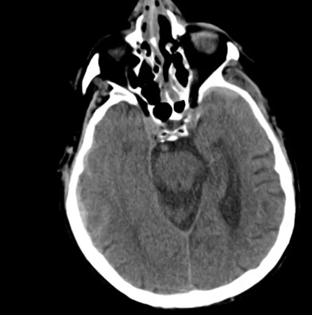
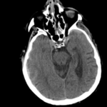
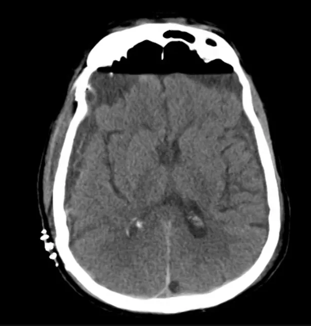
CT Head (Initial)
- Noncontrast axial images through the head demonstrate no evidence of skull fracture.
- Large lentiform-shaped mixed density extra-axial acute epidural hematoma in the right parietal occipital
- Associated subdural hematoma tracking along right convexity toward the right temporal lobe.
- There is no evidence of midline shift.
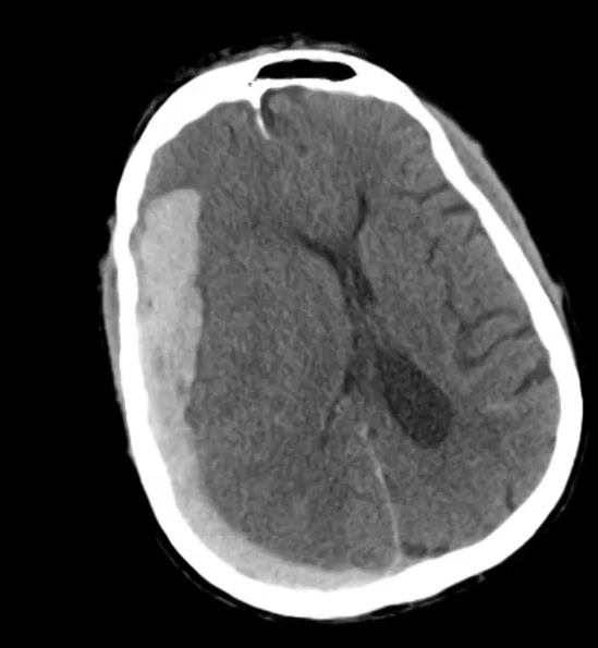
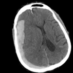
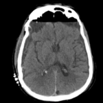
CT Head (+8h)
- Significant interval increase in the size of the right hemispheric subdural hematoma
- There is now midline shift from right to left at the level of the septum pellucidum measuring 10 mm, partial effacement of the right lateral ventricle and subfalcial herniation.
- Scattered subarachnoid blood is redemonstrated.
- Comminuted fractures of the nasal bone are present and there is overlying and associated periorbital soft tissue swelling.
CT Head (+16h, s/p SDH evacuation)
- Interval gross total evacuation of right hemispheric subdural hematoma.
- Moderate anterior bifrontal subdural and right epidural air is present.
- Small scattered subarachnoid and intraventricular blood is redemonstrated.