Monocular Painless Vision Loss

Brief H&P:

A 29 year-old female with no known medical history presents with vision changes for two weeks. She describes blurring and darkening of her vision in the left eye without associated pain after which her vision returns to normal. She notes occasional headaches which she describes as her usual migraines.

Physical examination including a detailed neurological examination, visual acuity, and visual fields is normal with the exception of bilateral optic disc edema. A non-contrast head CT is performed and is similarly normal. The patient underwent uncomplicated lumbar puncture with identification of elevated opening pressure with otherwise unremarkable cerebrospinal fluid analyses.

The patient’s evaluation was consistent with idiopathic intracranial hypertension, she was started on acetazolamide 500mg p.o. b.i.d and was discharged with close neurology follow-up.

An Algorithm for the Evaluation of Painless Monocular Vision Loss

An algorithm for the evaluation of painless monocular vision loss

This algorithm was developed by Dr. Lara Samarneh. Lara is an emergency medicine resident at UTHealth Houston. She attended medical school at UTHealth Houston McGovern Medical School and graduated from The University of Texas at Austin with a Bachelor’s degree in Biomedical Engineering. She is passionate about education and is currently serving as Vice Chief of Education.
Special thanks to Dr. Ore-ofe O. Adesina, MD , Associate Professor, Department of Ophthalmology and Visual Science – McGovern Medical School, Medical Director, Cizik Eye Clinic for his expertise and review of this algorithm.

References

  1. Fundoscopy images in the algorithm are sourced from Ophthalmic Atlas Images by EyeRounds.org, The University of Iowa which are licensed under a Creative Commons Attribution-NonCommercial-NoDerivs 3.0 Unported License.
  2. Guluma, K, Lee, J. Ophthalmology. In Rosen’s Emergency Medicine: Concepts and Clinical Practice (10th ed., pp. 750-780). Philadelphia, PA: Elsevier.
  3. Sharma P, Sridhar J, Mehta S. Flashes and floaters. Prim Care. 2015;42(3):425-435.
  4. Bagheri N, Mehta S. Acute vision loss. Prim Care. 2015;42(3):347-361.
  5. Pula JH, Kwan K, Yuen CA, Kattah JC. Update on the evaluation of transient vision loss. Clin Ophthalmol. 2016;10:297-303.
  6. Dehghani A, Giti M, Akhlaghi MR, Karami M, Salehi F. Ultrasonography in distinguishing optic neuritis from nonarteritic anterior ischemic optic neuropathy. Adv Biomed Res. 2012;1:3.
  7. Prasad S, Galetta SL. Approach to the patient with acute monocular visual loss. Neurol Clin Pract. 2012;2(1):14-23.
  8. Abbatemarco JR, Patell R, Buccola J, Willis MA. Acute monocular vision loss: Don’t lose sight of the differential. Rutecki GW, ed. CCJM. 2017;84(10):779-787.
  9. Current management of amaurosis fugax. The amaurosis fugax study group. Stroke. 1990;21(2):201-208.

Wheezing and Stridor

Brief HPI:

A 66 year-old male with a history of hypertension and COPD presents with shortness of breath. He states that his symptoms are unimproved with home nebulizer treatments and denies fever, cough or new sputum production. On examination, he has stridor appreciated during inspiratory and expiratory phases.

01_stridor
02_stridor
03_stridor
04_stridor
05_stridor
06_stridor
07_stridor
08_stridor
09_stridor
10_stridor
11_stridor
12_stridor
13_stridor
14_stridor
15_stridor
16_stridor
17_stridor
18_stridor
19_stridor
20_stridor
21_stridor
22_stridor
23_stridor
24_stridor
25_stridor
26_stridor
27_stridor
28_stridor
29_stridor
30_stridor
31_stridor
32_stridor

CT Chest:

1.9cm soft tissue thickening of the left tracheal wall at the level of the inferior thyroid gland. Luminal narrowing to 4 mm at this level.
Case courtesy of Dr Ian Bickle from Radiopaedia.org: 47677

Sound Characteristics

Stridor

An inspiratory, expiratory, or continuous monophonic sound that is loudest over the central airways.

Wheezing

A musical, high-pitched sound – more commonly expiratory. Requires sufficient airflow to induce airway oscillations.

Respiratory Phase

Inspiratory

Supraglottic: negative intratracheal pressure during inspiration causes airway collapse.

Biphasic

Glottic/Subglottic: fixed obstruction not impacted by changes in luminal/thoracic pressure.

Expiratory

Intrathoracic: increased pleural pressure compresses the narrowed airway.

An Algorithm for the Diagnosis of Wheezing and Stridor

An Algorithm for the Diagnosis of Wheezing and Stridor

Special thanks to Dr. Denna Zebda, Assistant Professor, Department of Otorhinolaryngology – McGovern Medical School for her expertise and review of this algorithm.

References

  1. Sicari V, Zabbo CP. Stridor. [Updated 2021 Jul 10]. In: StatPearls [Internet]. Treasure Island (FL): StatPearls Publishing; 2022 Jan-. Available from: https://www.ncbi.nlm.nih.gov/books/NBK525995/
  2. Patel PH, Mirabile VS, Sharma S. Wheezing. [Updated 2021 May 12]. In: StatPearls [Internet]. Treasure Island (FL): StatPearls Publishing; 2022 Jan-. Available from: https://www.ncbi.nlm.nih.gov/books/NBK482454/
  3. Bohadana A, Izbicki G, Kraman SS. Fundamentals of lung auscultation. N Engl J Med. 2014;370(21):2053.
  4. Orient JM, Sapira JD. Sapira’s Art & Science of Bedside Diagnosis. 4th ed. Wolters Kluwer Health/Lippincott Williams & Wilkins; 2010.

LVAD

Brief H&P:

A 48 year-old male with a history of congestive heart failure s/p left ventricular assist device is brought in by EMS with low-flow alarms. According to prehospital report, the patient had otherwise been in his usual state of health and had been shocked by his ICD multiple times prior to their arrival. No vital signs could be obtained en route.

On arrival in the emergency department, the patient was awake and responding appropriately to questions. His MAP was 80mmHg, an audible whir was auscultated from his device and the skin surrounding the percutaneous exit site appeared normal.

ECG

ECG with Ventricular Fibrillation

POCUS

Ultrasound showing parasternal long axis view of fibrillating heart

The patient’s device was inactivated with a magnet to prevent further ineffectual shocks. An arterial line was placed for continuous blood pressure measurement. He was sedated and externally defibrillated with return to normal sinus rhythm prior to admission to the CCU.

An Algorithm for the Evaluation of Unstable LVAD1

Algorithm for the Evaluation of Unstable LVAD

Reference

Stenberg R, Shenvi C. Targeted evaluation of patients with left ventricular assist devices and shock or hypotension. Ann Emerg Med. 2020;76(1):34-41.

Ear Pain

Brief HPI:

A 48 year-old female with a history of hypertension, diabetes, and hyperlipidemia presents with ear pain and discharge. She notes an associated headache and fevers. Examination demonstrates external auditory canal edema with granulation tissue along the floor. No cranial nerve abnormalities identified.

CT suggestive of malignant otitis externa

Image courtesy of Dr. Charlie Chia-Tsong Hsu, Radiopaedia.org case rID: 19938.

ED Course:

A CT was obtained which demonstrated edema of the external auditory meatus, pinna and periauricular soft tissue with fluid in the left mastoid sinuses without evidence of bone erosion. The patient was diagnosed with malignant otitis externa, started on intravenous ciprofloxacin and admitted with otolaryngology consultation.

An Algorithm for the Differential Diagnosis of Ear Pain1-5

An Algorithm for the Differential Diagnosis of Ear Pain

Otalgia Gallery

References

  1. Earwood JS, Rogers TS, Rathjen NA. Ear pain: diagnosing common and uncommon causes. Am Fam Physician. 2018;97(1):20-27.
  2. Coulter J, Kwon E. Otalgia. [Updated 2020 Aug 15]. In: StatPearls [Internet]. Treasure Island (FL): StatPearls Publishing; 2021 Jan-. Available from: https://www.ncbi.nlm.nih.gov/books/NBK549830/
  3. Pfaff, J. A., & Moore, G. P. (2018). Rosen’s Emergency Medicine: Concepts and Clinical Practice. In 1325336653 972907711 R. M. Walls (Author), Rosen’s Emergency Medicine: Concepts and Clinical Practice (9th ed., Vol. 1, pp. 820-831). Philadelphia, PA: Elsevier.
  4. Safavi Naini A, Ghorbani J, Montazer Lotfe Elahi S, Beigomi M. Otologic manifestations and progression in patients with wegener’s granulomatosis: a survey in 55 patients. Iran J Otorhinolaryngol. 2017;29(95):327-331.
  5. Conover K. Earache. Emerg Med Clin North Am. 2013;31(2):413-442.

CSF Shunt Complications

Brief HPI:

A 33 year-old female with a history of idiopathic intracranial hypertension and ventriculoperitoneal shunt placement presents with headache and confusion. She denies fever, trauma, neck pain or stiffness. She has not had symptoms like this since her shunt was placed 2 years ago. Imaging was obtained which showed ventriculomegaly and a fracture of the shunt at the level of the cervical spine. Neurosurgery was consulted and the patient was admitted for shunt repair.

00_ct_csf_shunt
01_ct_csf_shunt
02_ct_csf_shunt
03_ct_csf_shunt
04_ct_csf_shunt
05_ct_csf_shunt
06_ct_csf_shunt
07_ct_csf_shunt
08_ct_csf_shunt
09_ct_csf_shunt
10_ct_csf_shunt
11_ct_csf_shunt
12_ct_csf_shunt

CT Head

Ventriculomegaly with dilatation of the temporal horns in particular. Right parietal approach ventricular drain. Case courtesy of Dr. Henry Knipe, Radiopaedia.org, rID: 39615

XR Shunt Series

XR Shunt Series

Shunt tubing fractured at the level of the upper cervical spine

An Algorithm for CSF Shunt Complications

An Algorithm for CSF Shunt Complications

References:

  1. Madsen MA. Emergency department management of ventriculoperitoneal cerebrospinal fluid shunts. Ann Emerg Med. 1986;15(11):1330-1343.
  2. Ferras M, McCauley N, Stead T, Ganti L, Desai B. Ventriculoperitoneal shunts in the emergency department: a review. Cureus. 2020;12(2):e6857.
  3. Paff M, Alexandru-Abrams D, Muhonen M, Loudon W. Ventriculoperitoneal shunt complications: A review. Interdisciplinary Neurosurgery. 2018;13:66-70.
  4. Pitetti R. Emergency department evaluation of ventricular shunt malfunction: is the shunt series really necessary? Pediatr Emerg Care. 2007;23(3):137-141.
  5. Fowler JB, De Jesus O, Mesfin FB. Ventriculoperitoneal Shunt. [Updated 2021 Feb 7]. In: StatPearls [Internet]. Treasure Island (FL): StatPearls Publishing; 2021 Jan-. Available from: https://www.ncbi.nlm.nih.gov/books/NBK459351/
  6. Broggi M, Zattra CM, Schiariti M, et al. Diagnosis of ventriculoperitoneal shunt malfunction: a practical algorithm. World Neurosurg. 2020;137:e479-e486.

Pneumomediastinum

Brief HPI:

A 34-year-old male with a history of rheumatoid arthritis and interstitial lung disease presents to the emergency department with joint pain unimproved with home medications. He suspects the precipitant is a recent illness, describing cough and nasal congestion. He also noted a “crunching” sensation when turning his neck not otherwise associated with fevers, recurrent vomiting, chest pain, abdominal pain or difficulty breathing.

A chest radiograph was obtained which demonstrated pneumomediastinum.

Chest x-ray showing pneumomediastinum

Imaging from several months prior to presentation is shown below:

ct1_1
ct1_2
ct1_3
ct1_4
ct1_5
ct1_6
ct1_7
ct1_8
ct1_9
ct1_10

Prior CT Chest:

Extensive peripheral reticular and ground glass opacities and traction bronchiectasis predominates in the lower lobes. Imaging findings are most suggestive of usual interstitial pneumonia. Small focus of pneumomediastinum at carina.

The patient was placed on supplemental oxygen, a repeat chest CT was obtained.

ct2_1
ct2_2
ct2_3
ct2_4
ct2_5
ct2_6
ct2_7
ct2_8
ct2_9
ct2_10
ct2_11
ct2_12
ct2_13
ct2_14
ct2_15
ct2_16

Current CT Chest:

Large pneumomediastinum extends superiorly into the bilateral lower neck and bilateral anterior and posterior chest walls. It extends inferiorly to the anterior diaphragmatic space. This most likely represents spontaneous pneumomediastinum in the clinical setting of interstitial lung disease. Pneumorrhachis is seen, related to pneumomediastinum.

The etiology of the patient’s spontaneous pneumomediastinum was deemed to be related to his underlying interstitial lung disease provoked by viral respiratory tract infection related coughing. He was observed for two days without decompensation and was discharged with outpatient follow-up.

Pathophysiology of Pneumomediastinum

Spontaneous pneumomediastinum results from the rupture of terminal alveoli with subsequent tracking of gas along the bronchovascular tree through interstitial lung tissue to the mediastinum and adjacent structures (pleural, pericardial, retropharyngeal, retroperitoneal, intraperitoneal and subcutaneous spaces)1.

Secondary pneumomediastinum arises from non-alveolar sources including the gastrointestinal tract (most gravely, esophageal rupture though also from other intraperitoneal sources2), and upper respiratory tract (including facial fractures3).

Management of Pneumomediastinum5-7

The management of spontaneous pneumomediastinum focuses on treatment of the underlying precipitant, supportive care, administration of supplemental oxygen (to promote gas reabsorption) and observation for complications including rare progression to tension pneumomediastinum4.

Secondary pneumomediastinum is of significantly more concern and should be suspected in patients with any of the following features:

Symptoms

  • History of forceful vomiting
  • Dysphagia

Signs

  • Fever
  • Hemodynamic instability
  • Left-sided pleural effusion
  • Abdominal tenderness
  • Leukocytosis

Management is aggressive including resuscitation, maintenance of NPO status, broad-spectrum antibiotics, and emergent surgical consultation.

Differential Diagnosis of Pneumomediastinum5-11

An Algorithm for the Evaluation of Pneumomediastinum

References:

  1. Macklin, M., Macklin, C. (1944). Malignant Interstitial Emphysema of the Lungs and Mediastinum as an Important Occult Complication in Many Respiratory Diseases and Other Conditions: an Interpretation of the Clinical Literature in the Light of Laboratory Experiment Medicine 23(4)
  2. Fosi, S., Giuricin, V., Girardi, V., Caprera, E., Costanzo, E., Trapano, R., Simonetti, G. (2014). Subcutaneous Emphysema, Pneumomediastinum, Pneumoretroperitoneum, and Pneumoscrotum: Unusual Complications of Acute Perforated Diverticulitis Case Reports in Radiology 2014(), 1-5. https://dx.doi.org/10.1155/2014/431563
  3. Luca, G., Petteruti, F., Tanga, M., Luciano, A., Lerro, A. (2011). Pneumomediastinum and Subcutaneous Emphysema Unusual Complications of Blunt Facial Trauma Indian Journal of Surgery 73(5), 380-381. https://dx.doi.org/10.1007/s12262-011-0310-x
  4. Shennib, H., Barkun, A., Matouk, E., Blundell, P. (1988). Surgical Decompression of a Tension Pneumomediastinum Chest 93(6), 1301-1302. https://dx.doi.org/10.1378/chest.93.6.1301
  5. Bakhos, C., Pupovac, S., Ata, A., Fantauzzi, J., Fabian, T. (2014). Spontaneous pneumomediastinum: an extensive workup is not required. Journal of the American College of Surgeons 219(4), 713-7. https://dx.doi.org/10.1016/j.jamcollsurg.2014.06.001
  6. Iyer, V., Joshi, A., Ryu, J. (2009). Spontaneous Pneumomediastinum: Analysis of 62 Consecutive Adult Patients Mayo Clinic Proceedings 84(5), 417-421. https://dx.doi.org/10.4065/84.5.417
  7. Takada, K., Matsumoto, S., Hiramatsu, T., Kojima, E., Shizu, M., Okachi, S., Ninomiya, K., Morioka, H. (2009). Spontaneous pneumomediastinum: an algorithm for diagnosis and management. Therapeutic advances in respiratory disease 3(6), 301-7. https://dx.doi.org/10.1177/1753465809350888
  8. Al-Mufarrej, F., Badar, J., Gharagozloo, F., Tempesta, B., Strother, E., Margolis, M. (2008). Spontaneous pneumomediastinum: diagnostic and therapeutic interventions. Journal of cardiothoracic surgery 3(1), 59. https://dx.doi.org/10.1186/1749-8090-3-59
  9. Takada, K., Matsumoto, S., Hiramatsu, T., Kojima, E., Watanabe, H., Sizu, M., Okachi, S., Ninomiya, K. (2008). Management of spontaneous pneumomediastinum based on clinical experience of 25 cases Respiratory Medicine 102(9), 1329-1334. https://dx.doi.org/10.1016/j.rmed.2008.03.023
  10. Bejvan, S., Godwin, J. (1996). Pneumomediastinum: old signs and new signs. American Journal of Roentgenology 166(5), 1041-1048. https://dx.doi.org/10.2214/ajr.166.5.8615238
  11. Langwieler, T., Steffani, K., Bogoevski, D., Mann, O., Izbicki, J. (2004). Spontaneous pneumomediastinum The Annals of Thoracic Surgery 78(2), 711-713. https://dx.doi.org/10.1016/j.athoracsur.2003.09.021

Emphysematous Urinary Tract Infections

Brief HPI:

A 45 year-old female with a history of ureterolithiasis s/p bilateral percutaneous nephrostomies, hypertension and diabetes presents to the emergency department with flank pain and dysuria for two days. She noted that output from her right nephrostomy had diminished. On evaluation, her vital signs are notable for fever and tachycardia but are otherwise normal. Examination demonstrates right costovertebral angle tenderness to percussion. Drain sites appeared normal, without overlying erythema. Urinalyses from both nephrostomy collection bags were submitted. Computed tomography of the abdomen and pelvis was obtained to evaluate for nephrostomy malposition.

01_pyelo
03_pyelo
04_pyelo
05_pyelo
06_pyelo
07_pyelo
08_pyelo
09_pyelo
10_pyelo
11_pyelo
12_pyelo
13_pyelo
14_pyelo
15_pyelo
16_pyelo
17_pyelo

CT Abdomen/Pelvis Interpretation

Complex perirenal fluid collection with gas suggestive of emphysematous pyelonephritis with abscess.

Hospital Course

The patient was treated with parenteral antibiotics based on prior culture data and was admitted to the intensive care unit with urology consultation and plan for interventional radiology percutaneous drainage. The patient underwent uncomplicated perinephric drain placement and nephrostomy exchange and was discharged on hospital day five to complete a course of oral antibiotics.

An Algorithm for the Evaluation and Management of Emphysematous Urinary Tract Infections

An Algorithm for the Evaluation and Management of Emphysematous Urinary Tract Infections

References

  1. Evanoff GV, Thompson CS, Foley R, Weinman EJ. Spectrum of gas within the kidney. Emphysematous pyelonephritis and emphysematous pyelitis. Am J Med. 1987;83(1):149-154.
  2. Wan YL, Lee TY, Bullard MJ, Tsai CC. Acute gas-producing bacterial renal infection: correlation between imaging findings and clinical outcome. Radiology. 1996;198(2):433-438. doi:10.1148/radiology.198.2.8596845.
  3. Shokeir AA, El-Azab M, Mohsen T, El-Diasty T. Emphysematous pyelonephritis: a 15-year experience with 20 cases. Urology. 1997;49(3):343-346. doi:10.1016/S0090-4295(96)00501-8.
  4. Chen MT, Huang CN, Chou YH, Huang CH, Chiang CP, Liu GC. Percutaneous drainage in the treatment of emphysematous pyelonephritis: 10-year experience. JURO. 1997;157(5):1569-1573.
  5. Huang JJ, Tseng CC. Emphysematous pyelonephritis: clinicoradiological classification, management, prognosis, and pathogenesis. Arch Intern Med. 2000;160(6):797-805.
  6. Roy C, Pfleger DD, Tuchmann CM, Lang HH, Saussine CC, Jacqmin D. Emphysematous pyelitis: findings in five patients. Radiology. 2001;218(3):647-650. doi:10.1148/radiology.218.3.r01fe14647.
  7. Park BS, Lee S-J, Kim YW, Huh JS, Kim JI, Chang S-G. Outcome of nephrectomy and kidney-preserving procedures for the treatment of emphysematous pyelonephritis. Scand J Urol Nephrol. 2006;40(4):332-338. doi:10.1080/00365590600794902.
  8. Grupper M, Kravtsov A, Potasman I. Emphysematous cystitis: illustrative case report and review of the literature. Medicine (Baltimore). 2007;86(1):47-53. doi:10.1097/MD.0b013e3180307c3a.
  9. Mokabberi R, Ravakhah K. Emphysematous urinary tract infections: diagnosis, treatment and survival (case review series). Am J Med Sci. 2007;333(2):111-116.
  10. Yao J, Gutierrez OM, Reiser J. Emphysematous pyelonephritis. Kidney Int. 2007;71(5):462-465. doi:10.1038/sj.ki.5002001.
  11. Thomas AA, Lane BR, Thomas AZ, Remer EM, Campbell SC, Shoskes DA. Emphysematous cystitis: a review of 135 cases. BJU Int. 2007;100(1):17-20. doi:10.1111/j.1464-410X.2007.06930.x.
  12. Falagas ME, Alexiou VG, Giannopoulou KP, Siempos II. Risk factors for mortality in patients with emphysematous pyelonephritis: a meta-analysis. JURO. 2007;178(3 Pt 1):880–5–quiz1129. doi:10.1016/j.juro.2007.05.017.
  13. Somani BK, Nabi G, Thorpe P, et al. Is percutaneous drainage the new gold standard in the management of emphysematous pyelonephritis? Evidence from a systematic review. J Urol. 2008;179(5):1844-1849. doi:10.1016/j.juro.2008.01.019.
  14. Aswathaman K, Gopalakrishnan G, Gnanaraj L, Chacko NK, Kekre NS, Devasia A. Emphysematous pyelonephritis: outcome of conservative management. Urology. 2008;71(6):1007-1009. doi:10.1016/j.urology.2007.12.095.
  15. Kapoor R, Muruganandham K, Gulia AK, et al. Predictive factors for mortality and need for nephrectomy in patients with emphysematous pyelonephritis. BJU Int. 2010;105(7):986-989. doi:10.1111/j.1464-410X.2009.08930.x.
  16. Ubee SS, McGlynn L, Fordham M. Emphysematous pyelonephritis. BJU Int. 2011;107(9):1474-1478. doi:10.1111/j.1464-410X.2010.09660.x.
  17. Lu Y-C, Chiang B-J, Pong Y-H, et al. Predictors of failure of conservative treatment among patients with emphysematous pyelonephritis. BMC Infect Dis. 2014;14(1):418. doi:10.1186/1471-2334-14-418.

Synovial Fluid

Brief HPI:

A 38 year-old female with a history of obesity and obstructive sleep apnea presents with right knee pain. She cannot identify a clear precipitant for her symptoms which she first noted 2 weeks ago. Her pain is worsened with ambulation and while previously tolerable, has grown more severe despite over-the-counter analgesics over the past two days. She denies fevers, intravenous drug use, recent travel or instrumentation.

On evaluation, vital signs are normal. Physical examination demonstrates a moderate-sized right knee effusion with overlying warmth though no edema. There is minimal pain with range of motion, no pain with heel percussion, and she is ambulatory independently with a mildly antalgic gait. Clinical suspicion for septic arthritis was low. A diagnostic arthrocentesis was performed without complication. Synovial fluid was less-viscous than normal with slight debris. Laboratory analysis revealed 14,230 white blood cells with 85% neutrophils and no crystals visualized. The patient was discharged with supportive care and outpatient follow-up – cultures were ultimately negative.

An Algorithm for the Analysis of Synovial Fluid

An Algorithm for the Analysis of Synovial Fluid

References

  1. Margaretten ME, Kohlwes J, Moore D, Bent S. Does this adult patient have septic arthritis? JAMA. 2007;297(13):1478-1488. doi:10.1001/jama.297.13.1478.
  2. Brannan SR, Jerrard DA. Synovial fluid analysis. J Emerg Med. 2006;30(3):331-339. doi:10.1016/j.jemermed.2005.05.029.
  3. Couderc M, Pereira B, Mathieu S, et al. Predictive value of the usual clinical signs and laboratory tests in the diagnosis of septic arthritis. CJEM. 2015;17(4):403-410. doi:10.1017/cem.2014.56.
  4. MD HJC, MD LAB, MD ML. Septic Arthritis. Hospital Medicine Clinics. 2014;3(4):494-503. doi:10.1016/j.ehmc.2014.06.009.

Resuscitative Thoracotomy

Brief HPI:

A call is received from pre-hospital providers regarding an inbound trauma patient. An estimated 30 year-old male with unknown history sustained a penetrating wound to the right flank. On EMS arrival the patient was unresponsive but had a weakly-palpable radial pulse which was lost en-route. Their estimated time of arrival is 5 minutes.

Algorithm for the Selection of Patients for Resuscitative Thoracotomy

Algorithm for the Selection of Patients for Resuscitative Thoracotomy

References:

  1. Seamon MJ, Haut ER, Van Arendonk K, Barbosa RR, Chiu WC, Dente CJ, et al. An evidence-based approach to patient selection for emergency department thoracotomy: A practice management guideline from the Eastern Association for the Surgery of Trauma. J Trauma Acute Care Surg. 2015 Jul;79(1):159–73.
  2. Burlew CC, Moore EE, Moore FA, Coimbra R, McIntyre RC, Davis JW, et al. Western Trauma Association critical decisions in trauma: resuscitative thoracotomy. J Trauma Acute Care Surg. 2012 Dec;73(6):1359–63.
  3. Sherren PB, Reid C, Habig K, Burns BJ. Algorithm for the resuscitation of traumatic cardiac arrest patients in a physician-staffed helicopter emergency medical service. Crit Care. 2013 Mar 12;17(2):308.
  4. Cothren CC, Moore EE. Emergency department thoracotomy for the critically injured patient: Objectives, indications, and outcomes. World J Emerg Surg. 2006 Mar 24;1:4.

Red and Painful Eye

Brief HPI

A 60-year-old female with rheumatoid arthritis presents with unilateral eye pain and redness without reported vision changes. Physical examination demonstrates radially-oriented engorged episcleral vessels and normal visual acuity – she was diagnosed with episcleritis and discharged with follow-up.

Algorithm for the Evaluation of the Red and Painful Eye

Algorithm for the Evaluation of the Red and Painful Eye

Overview

The red or painful eye is a common presentation in the emergency department and the rapid identification and management of potentially sight-threatening causes is critical.

The diagnostic approach to the red or painful eye begins with identifying a history of caustic exposure where immediate and copious irrigation (even before detailed examination) may limit further injury. Alkaline agents induce more severe liquefactive necrosis leading to keratoconjunctivitis, while acidic agents are generally less destructive. Management is identical for both: irrigation with lactated Ringer solution through a Morgan lens applied to a topically anesthetized eye for 5-10 minutes repeated until the pH of the eye is neutral1.

Ocular or facial trauma presents a spectrum of differential diagnoses. Suspicion for globe rupture is increased by a suggestive mechanism such as a high-velocity projectile or high-impact blunt facial trauma. Characteristic examination findings include obvious globe deformity, an irregularly-shaped pupil, extrusion of vitreous, markedly decreased visual acuity, or parting of fluorescein (Seidel sign). If globe rupture is suspected, further manipulation is unadvisable and the affected eye should be shielded. Measures should be taken to avoid increases in intra-ocular pressure including elevation of the patient’s head-of-bed, anti-emetics (to prevent intra-ocular hypertension with vomiting), and the avoidance of medications potentially implicated in intra-ocular hypertension (ketamine, succinylcholine). Tetanus and antimicrobial prophylaxis should be provided while awaiting emergent ophthalmology consultation2,3.

Another traumatic diagnosis warranting rapid identification and possible intervention is retrobulbar hematoma – identified by proptosis, eye pain, decreased visual acuity, and elevated intra-ocular pressure. Pressures exceeding 40mmHg warrant lateral cantholysis in conjunction with medical management to prevent optic nerve ischemia and preserve vision4,5.

Diagnostic Approach

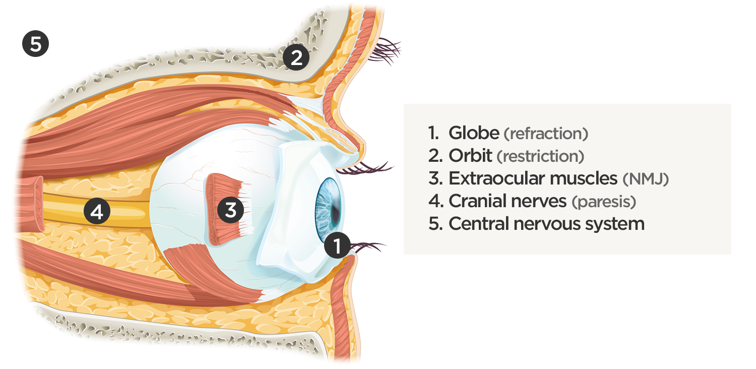
The evaluation of the non-traumatic red or painful eye follows a systematic and anatomically-based approach, starting with external components and moving inward:

1. External

The examination begins externally with an assessment of function (visual acuity) and inspection and palpation of the periorbital region. Periorbital edema, erythema, and tenderness to palpation in the setting of systemic illness (fever) is concerning for orbital cellulitis. When associated with elevated intra-ocular pressure or proptosis, a retrobulbar abscess may be present. Both warrant admission and parenteral antibiotics and the latter may require operative management such as aspiration or cantholysis. Less severe features without impact on visual acuity is suggestive of a periorbital cellulitis which may be treated as an outpatient with close follow-up2,6.

2. Lids and Lashes

Several non-emergent processes may affect the lids and lashes including blepharitis (inflammation of the eyelid margin), chalazion (inflammation of the Meibomian glands), hordeolum (eyelash follicle abscess), or dacrocystitis (infection of the lacrimal sac)2,7,8.

3. Conjunctiva and Sclera

Again, proceeding from superficial to deeper structures we encounter the epithelial layer (including palpebral and bulbar components) covering the sclera which is subject to allergic or infectious inflammation. Conjunctivitis is characterized by engorgement of superficial conjunctival blood vessels, potentially associated with conjunctival edema (chemosis), or discharge. Most conjunctivitis is self-limited and not sight-threatening, treatment is aimed at symptomatic relief though topical antibiotics have few adverse effects and may be prescribed if the diagnosis of bacterial conjunctivitis is unclear9.

When associated with pain, a deeper inflammatory process is implicated. Scleritis is a frequently immune-mediated inflammatory process (though infection, malignancy and medications have been implicated) associated with pain, photophobia, and examination findings of globe tenderness and engorged scleral blood vessels. Management in the emergency department is trivial (systemic NSAID’s), however ophthalmology consultation should be secured due to the risk of vision-compromising complications, as well as the intimation of an underlying systemic disorder9,10. Episcleritis is similarly immune-mediated, though generally self-limiting. The diagnosis is made by identification of characteristic, radially-oriented engorged episcleral vessels. When the diagnosis of scleritis versus episcleritis or conjunctivitis is in question the application of a topical vasoconstrictor (phenylephrine 2.5%) will blanch vessels in the conjunctival or superficial episcleral plexuses – sparing scleral vessels10,11.

4. Cornea

Keratitis can be caused by infection, ultraviolet light exposure, or contact lens use. Patients may have photophobia and a foreign-body sensation. Gross inspection or slit-lamp examination will show epithelial erosions that stain with fluorescein or the characteristic dendritic pattern accompanying herpes simplex virus infection. Management includes ophthalmology consultation, topical antibiotics if a bacterial process is suggested, and close follow-up7-9,12.

5. Anterior Chamber

A critical process occurring in the anterior chamber is angle-closure glaucoma. The patient commonly presents with severe pain, circumcorneal injection, and a pupil fixed at mid-dilation. Diagnosis is confirmed by the measurement of elevated intra-ocular pressure (greater than 20mmHg). Reduction of intra-ocular pressure with topical and systemic agents should begin immediately while awaiting emergent ophthalmology consultation13.

The slit-lamp microscope facilitates examination of the anterior chamber. The presence of cells (floating white and red blood cells, or layering hypopyon or hyphema) and flare (protein) suggest inflammation in the anterior segment caused by a systemic inflammatory process, infection, or trauma and warrants close ophthalmologic follow-up2,7,9.

6. Vitreous

An ocular examination mimicking orbital cellulitis with evidence of anterior chamber involvement, particularly in a patient with a history of recent ocular surgery or trauma suggests endophthalmitis. Management requires admission for parenteral antibiotics with ophthalmology consultation2.

Additional Diagnostic Modalities

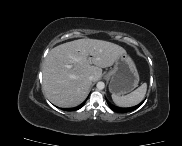
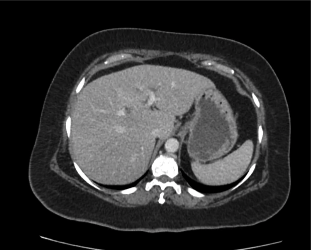
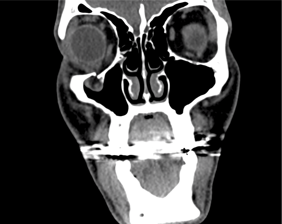
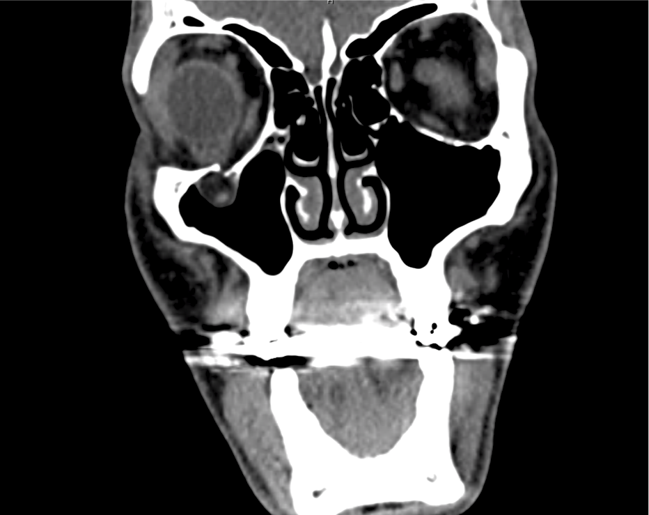
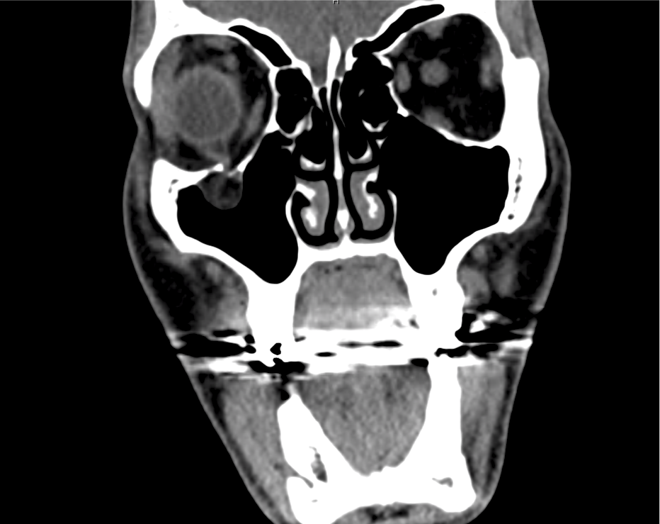
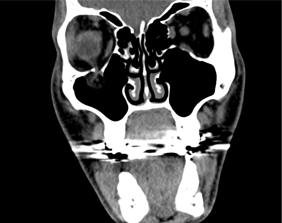
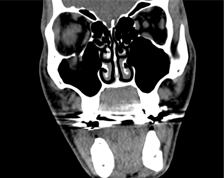
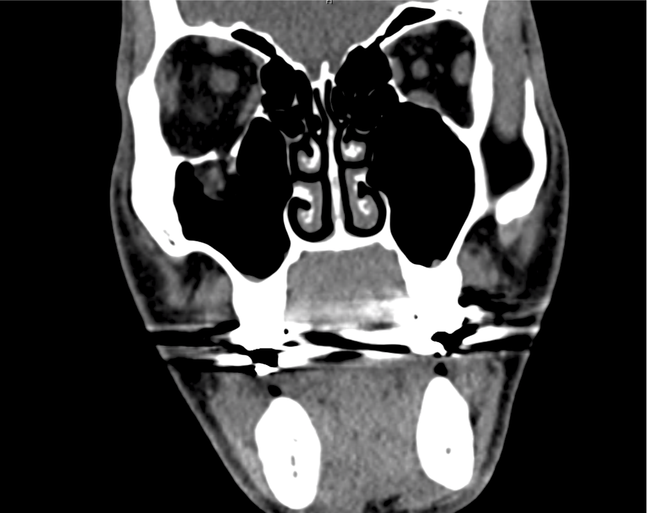
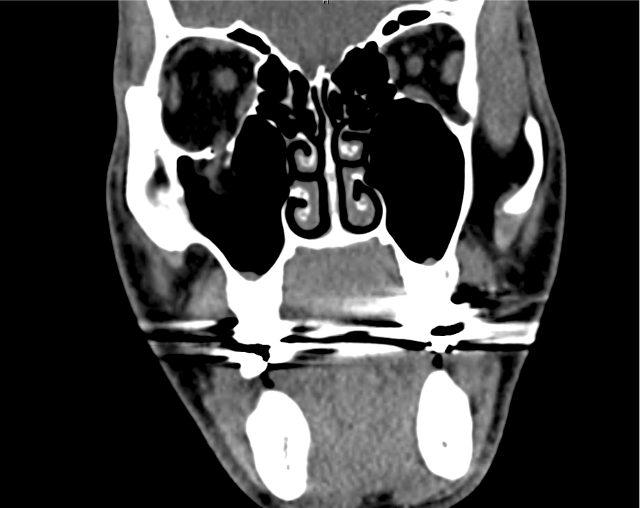
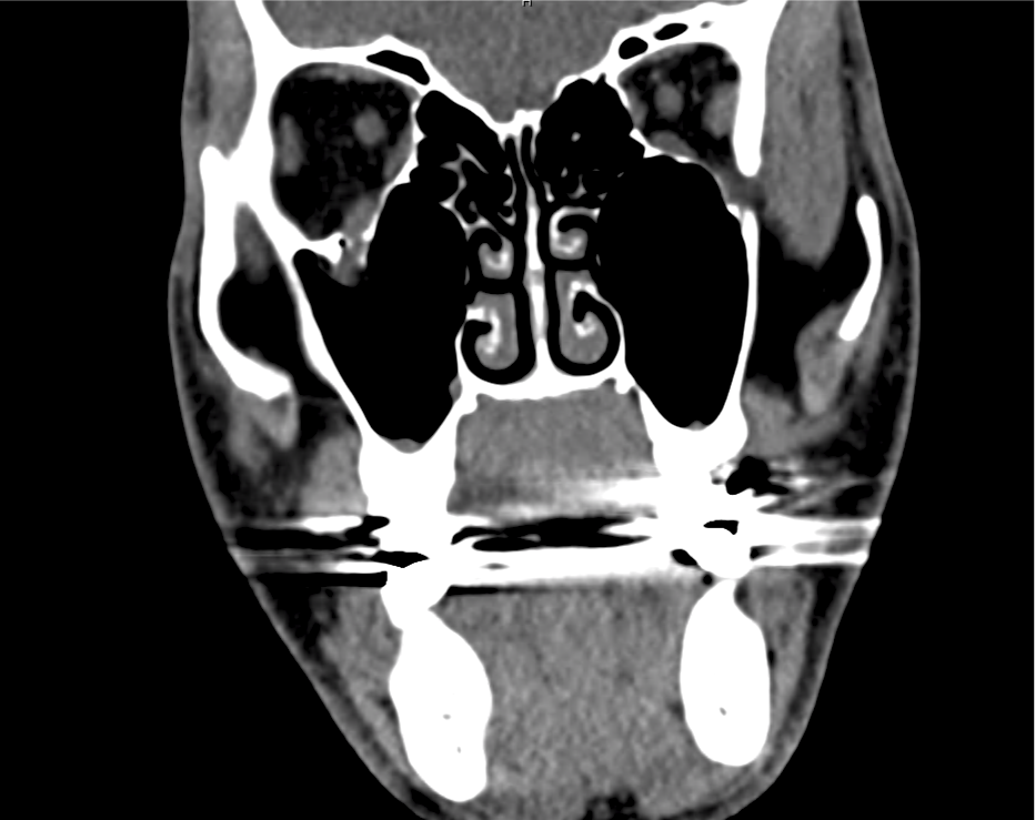
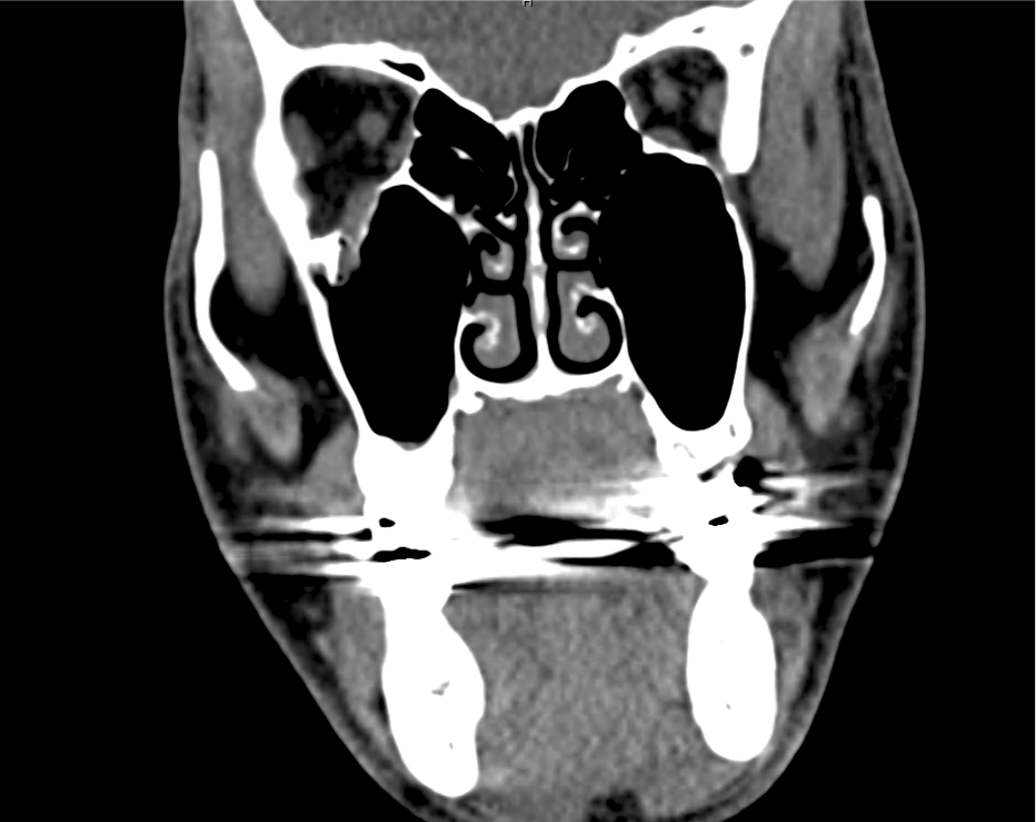
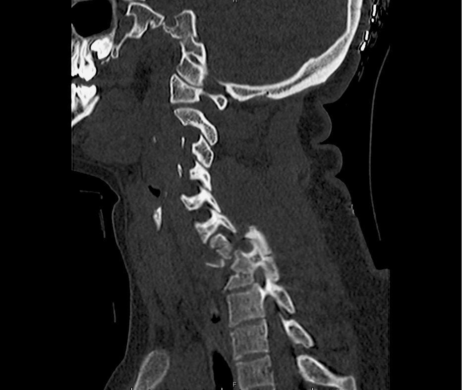
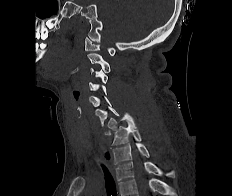
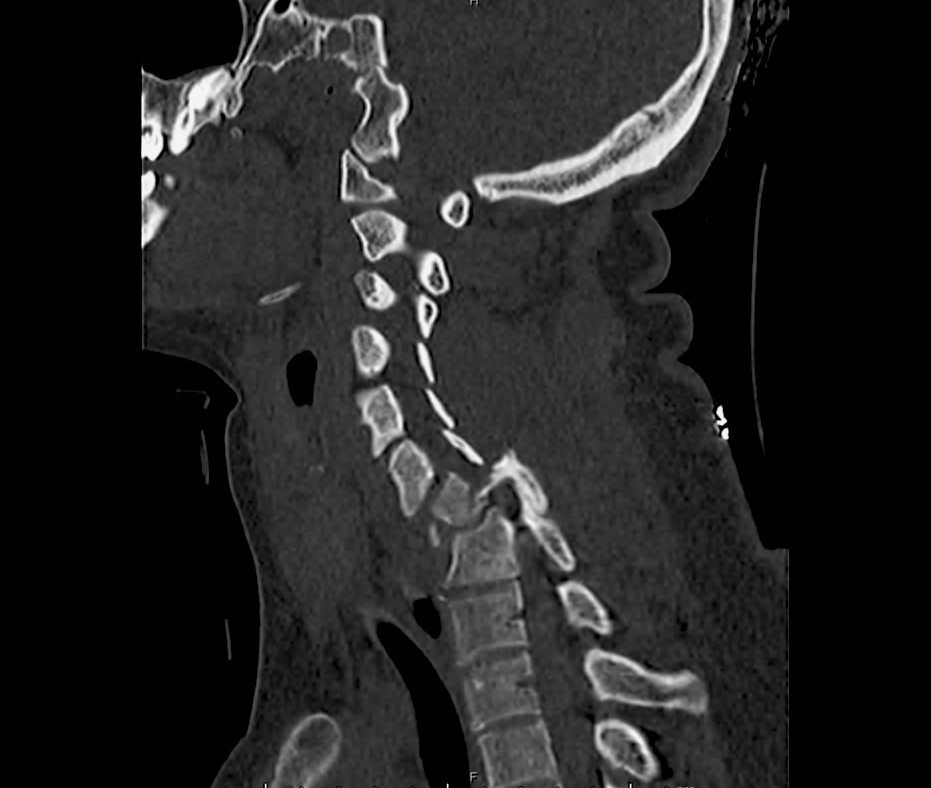
Advanced imaging may be useful in the diagnosis of traumatic and non-traumatic orbital pathology. Multi-detector computed tomography (MD-CT) is readily available and rapidly performed in the emergency department and can aid in the diagnosis of critical infectious processes, including extension beyond the orbital septum in orbital cellulitis, scleral thickening in endophthalmitis, and characterization of hematoma or abscess in the retrobulbar space. The addition of intravenous contrast media can identify critical vascular processes such as cavernous sinus thrombosis14,15. For traumatic pathology, CT can assist with the evaluation of globe integrity, lens position, vitreous/retinal detachment, and foreign bodies16. Imaging cannot be relied upon exclusively to exclude pathology, and the patient’s presentation and clinician’s examination should determine the need for consultation and evaluation. For globe rupture, for example, in one study of 59 patients with severe ocular trauma and diagnostic uncertainty regarding the presence of globe rupture, CT failed to diagnosed open globe injury in 1/3 of patients (with surgical scleral inspection as a reference standard)17. Another retrospective analysis of 48 eyes sustaining trauma revealed sensitivity ranging from 56-68% for CT identification of open globe injury18.

In addition to potential diagnostic inaccuracy, computed tomography exposes patients to risks including ionizing radiation, and the possibility of contrast-induced nephropathy19. Ultrasound is becoming increasingly accessible and comfortable for the emergency physician, and has the benefit of being relatively non-invasive – including facilitating ocular examination in patients with significant periorbital swelling limiting eye-opening. Ocular ultrasound may aid with the diagnosis of a wide variety of ocular pathology including vitreous hemorrhage, retinal detachment, central retinal arterial/venous occlusions, foreign body identification, lens dislocation and retrobulbar hematoma. In one study of 61 patients presenting with trauma or acute vision changes, ultrasound interpretation agreed with criterion standard (orbital computed tomography or ophthalmology evaluation) for 98% of cases20.

View Ocular Ultrasound Algorithm

References

  1. Messman AM. Ocular Injuries: New Strategies In Emergency Department Management. Emergency Medicine Practice. 2015;17(11):1–21–quiz21–2.
  2. Wright JL, Wightman JM. Red and painful eye. … Concepts and Clinical Practice 8th ed …. 2014.
  3. Romaniuk VM. Ocular trauma and other catastrophes. Emerg Med Clin North Am. 2013;31(2):399-411. doi:10.1016/j.emc.2013.02.003.
  4. Babineau MR, Sanchez LD. Ophthalmologic Procedures in the Emergency Department. Emerg Med Clin North Am. 2008;26(1):17-34. doi:10.1016/j.emc.2007.11.003.
  5. Rowh AD, Ufberg JW, Chan TC, Vilke GM, Harrigan RA. Lateral canthotomy and cantholysis: emergency management of orbital compartment syndrome. J Emerg Med. 2015;48(3):325-330. doi:10.1016/j.jemermed.2014.11.002.
  6. Henderson M, Tierney L, Smetana G. The Patient History: Evidence-Based Approach. McGraw Hill Professional; 2012.
  7. Alteveer JG, Mccans KM, Hemphill RR. The Red Eye, The Swollen Eye, And Acute Vision Loss. … Practice+ Em Practice …. 2002.
  8. Leibowitz HM. The red eye. N Engl J Med. 2000;343(5):345-351. doi:10.1056/NEJM200008033430507.
  9. Mahmood AR, Narang AT. Diagnosis and management of the acute red eye. Emerg Med Clin North Am. 2008. doi:10.1016/j.emc.2007.10.002.
  10. Albini TA, Rao NA, Smith RE. The Diagnosis and Management of Anterior Scleritis. International Ophthalmology Clinics. 2005;45(2):191.
  11. Roscoe M, Landis T. How to diagnose the acute red eye with confidence. JAAPA. 2006;19(3):24–30–quiz45–6.
  12. Deborah S Jacobs MD. Evaluation of the red eye. UpToDate. https://www.uptodate.com/contents/evaluation-of-the-red-eye. Published February 24, 2016. Accessed April 18, 2017.
  13. Prum BE, Herndon LW, Moroi SE, et al. Primary Angle Closure Preferred Practice Pattern(®) Guidelines. Ophthalmology. 2016;123(1):P1-P40. doi:10.1016/j.ophtha.2015.10.049.
  14. LeBedis CA, Sakai O. Nontraumatic orbital conditions: diagnosis with CT and MR imaging in the emergent setting. Radiographics. 2008;28(6):1741-1753. doi:10.1148/rg.286085515.
  15. Platnick J, Crum AV, Soohoo S, Cedeño PA, Johnson MH. The globe: infection, inflammation, and systemic disease. YSULT. 2011;32(1):38-50. doi:10.1053/j.sult.2010.12.003.
  16. Dunkin JM, Crum AV, Swanger RS, Bokhari SAJ. Globe trauma. YSULT. 2011;32(1):51-56. doi:10.1053/j.sult.2010.09.003.
  17. Hoffstetter P, Schreyer AG, Schreyer CI, et al. Multidetector CT (MD-CT) in the diagnosis of uncertain open globe injuries. Rofo. 2010;182(2):151-154. doi:10.1055/s-0028-1109659.
  18. Arey ML, Mootha VV, Whittemore AR, Chason DP, Blomquist PH. Computed tomography in the diagnosis of occult open-globe injuries. Ophthalmology. 2007;114(8):1448-1452. doi:10.1016/j.ophtha.2006.10.051.
  19. Custer PL, Kent TL. Pitfalls of ophthalmic radiographic imaging. Curr Opin Ophthalmol. 2014;25(5):432-435. doi:10.1097/ICU.0000000000000064.
  20. Blaivas M, Theodoro D, Sierzenski PR. A study of bedside ocular ultrasonography in the emergency department. Academic Emergency Medicine. 2002;9(8):791-799.

Blunt Cardiac Injury

Case Presentation

A 35-year-old female with no past medical history is brought in by ambulance to the emergency department. She was struck by a firework (“Roman Candle”) which lodged in her mid-chest until the propellant was consumed. She transiently lost consciousness but was awake upon EMS arrival. She complains of pleuritic chest pain. Examination reveals a circular 4x4cm full-thickness burn to the mid-chest with surrounding deep and superficial partial-thickness burns. Her ECG shows normal sinus rhythm, the initial serum troponin I is 32.9 (normal <0.012). CT angiography of the thorax is obtained.

bci_01
bci_02
bci_03
bci_04
bci_05
bci_06
bci_07
bci_08
bci_09
bci_10
bci_11
bci_12
bci_13

Mechanisms

Blunt cardiac injury (BCI) may be induced by multiple forces including direct thoracic trauma, crush injury of mediastinal contents between the sternum and thoracic spine, rapid deceleration causing tears at venous-atrial confluences, abrupt pressure changes from rapid compression of abdominal contents, blast injury, or laceration from bone fracture fragments1. The most common mechanisms of injury are motor vehicle collisions (50%), auto versus pedestrian (35%), motorcycle accidents (9%) and falls from significant height (>6m)2.

BCI represents a spectrum of conditions. Diagnosis is both challenging and critical as clinical manifestations can be absent or rapidly fatal.

At one end of the spectrum is myocardial contusion. The lack of a gold-standard for the diagnosis of this clinical entity has led to a preference for describing associated abnormalities if present3,4, including cardiac dysfunction (identified on echocardiography) or the next entity along the spectrum – arrhythmia.

The most common arrhythmia identified in blunt cardiac injury is sinus tachycardia, followed by premature atrial or ventricular contractions, T-wave changes, and atrial fibrillation or flutter5. Commotio cordis is a unique arrhythmia induced by untimely precordial impact (often in sports) during a vulnerable phase of ventricular excitability, resulting in ventricular fibrillation2.

ST-segment elevations after blunt cardiac injury should raise concern for myocardial infarction due to coronary artery dissection, laceration or thrombosis (often in already-diseased vessels) 5,6.

The remaining disease entities are increasingly rare, require careful examination or imaging for diagnosis, and are more likely to be non-survivable. Septal injury can range from small tears to rupture. Valvular injury most commonly affects the aortic valve (followed by mitral and tricuspid valves) and involves damage to leaflets, or rupture of papillary muscles or chordae tendineae. The clinical presentation is of acute valvular insufficiency, including acute heart failure and murmur2,7,8. A widened pulse pressure may be noted with aortic valve injury, and the manifestations of valvular injury may be delayed9. Finally, myocardial wall rupture is unlikely to be survivable, though patients may present with cardiac tamponade if rupture is small or contained2.

Evaluation

The primary diagnostic modalities for the assessment of BCI in the emergency department include assessment for pericardial fluid during the Focused Assessment with Sonography for Trauma (FAST), electrocardiography and cardiac enzymes.

While specific for identifying patients at risk of complications of BCI, electrocardiography alone is not sufficient to exclude BCI. In one study, only 59% of patient with echocardiographic evidence of BCI (wall-motion abnormalities, other chamber abnormalities) had initially abnormal ECG’s10. In another study, 41% of patients with initially normal ECG’s developed clinically significant abnormalities11. The use of specialized electrocardiography including right-sided ECG (proposed to better detect right-ventricular abnormalities which are more commonly associated with BCI) and signal-averaged ECG is not supported11,12.

Several studies have supported the use of serum troponin for the detection of clinically significant BCI – particularly in combination with electrocardiography. A prospective study in 2001 evaluating patients with blunt thoracic trauma using ECG at admission and 8-hours, as well as troponin I at admission, 4- and 8-hours had a negative predictive value of 100% for significant BCI (arrhythmia requiring treatment, shock, or structural cardiac abnormalities) in patients with initially normal ECG and troponin13.

Another prospective study adding to the population evaluated by Salim et al. included 41 patients with normal ECG’s and troponin levels at admission and 8-hours who were admitted for significant mechanisms, none developed significant BCI (again described as arrhythmia requiring treatment, shock, or structural cardiac abnormalities) after 1 to 3 days of observation14. The precise timing of serum troponin analysis remains unclear.

While FAST may detect hemopericardium warranting immediate intervention, formal echocardiography is indicated for patients with unexplained hypotension (to evaluate for valvular injury or regional wall-motion abnormalities) or persistent arrhythmias (to evaluate for arrhythmogenic intramural hematomas)15. The presence of sternal fractures was previously thought to increase risk of BCI and mandate echocardiography, however this notion is no longer supported16-18. The role of advanced imaging including helical CT (cardiac-gated), and MRI remains unclear19.

Algorithm for the Evaluation of Blunt Thoracic Trauma

Notes:
† Arrest in ED, immediate chest tube output >20ml/kg (>1.5L) or >200mL/hr for 2-4hr.

Management

Management of BCI depends on the pathologic process localized along the spectrum defined above. Persistent hypotension after appropriate evaluation for alternative etiologies may represent myocardial contusion with cardiac dysfunction and should be evaluated with echocardiography. Similarly, echocardiography and observation with continuous telemetry monitoring is indicated for any new arrhythmia or persistent and unexplained tachycardia. Patients with only elevation of the serum troponin without electrocardiographic abnormalities or obvious cardiac dysfunction should also be admitted for observation and serial cardiac enzymes. Traumatic myocardial infarction, valvular injury, or post-traumatic structural myocardial defects should be managed in consultation with cardiothoracic surgery5,19-21.

Case Conclusion

The CT interpretation noted the soft-tissue defect identified on examination as well as associated pulmonary contusions and a non-displaced sternal fracture. The patient went to the operating room for washout and debridement. A transthoracic echocardiogram demonstrated trace mitral regurgitation and a small pericardial effusion. She remained hemodynamically stable and serial troponin measures downtrended – no dysrhythmias were noted on telemetry monitoring. She was discharged on hospital day four with a negative-pressure wound dressing.

References

  1. Schultz JM, Trunkey DD. Blunt cardiac injury. Crit Care Clin. 2004;20(1):57-70.
  2. Yousef R, Carr JA. Blunt cardiac trauma: a review of the current knowledge and management. Ann Thorac Surg. 2014;98(3):1134-1140. doi:10.1016/j.athoracsur.2014.04.043.
  3. Mattox KL, Flint LM, Carrico CJ, et al. Blunt cardiac injury. The Journal of Trauma: Injury, Infection, and Critical Care. 1992;33(5):649-650.
  4. Sybrandy KC, Cramer MJM, Burgersdijk C. Diagnosing cardiac contusion: old wisdom and new insights. Heart. 2003;89(5):485-489.
  5. Elie M-C. Blunt cardiac injury. Mt Sinai J Med. 2006;73(2):542-552.
  6. Edouard AR, Felten M-L, Hebert J-L, Cosson C, Martin L, Benhamou D. Incidence and significance of cardiac troponin I release in severe trauma patients. Anesthesiology. 2004;101(6):1262-1268.
  7. Cordovil A, Fischer CH, Rodrigues ACT, et al. Papillary Muscle Rupture After Blunt Chest Trauma. Journal of the American Society of Echocardiography. 2006;19(4):469.e1-469.e3. doi:10.1016/j.echo.2005.12.005.
  8. Pasquier M, Sierro C, Yersin B, Delay D, Carron P-N. Traumatic Mitral Valve Injury After Blunt Chest Trauma: A Case Report and Review of the Literature. The Journal of Trauma: Injury, Infection, and Critical Care. 2010;68(1):243-246. doi:10.1097/TA.0b013e3181bb881e.
  9. Ismailov RM, Weiss HB, Ness RB, Lawrence BA, Miller TR. Blunt cardiac injury associated with cardiac valve insufficiency: trauma links to chronic disease? Injury. 2005;36(9):1022-1028. doi:10.1016/j.injury.2005.05.028.
  10. García-Fernández MA, López-Pérez JM, Pérez-Castellano N, et al. Role of transesophageal echocardiography in the assessment of patients with blunt chest trauma: correlation of echocardiographic findings with the electrocardiogram and creatine kinase monoclonal antibody measurements. Am Heart J. 1998;135(3):476-481.
  11. Fulda GJ, Giberson F, Hailstone D, Law A, Stillabower M. An evaluation of serum troponin T and signal-averaged electrocardiography in predicting electrocardiographic abnormalities after blunt chest trauma. The Journal of Trauma: Injury, Infection, and Critical Care. 1997;43(2):304–10–discussion310–2.
  12. Walsh P, Marks G, Aranguri C, et al. Use of V4R in patients who sustain blunt chest trauma. The Journal of Trauma: Injury, Infection, and Critical Care. 2001;51(1):60-63.
  13. Salim A, Velmahos GC, Jindal A, et al. Clinically significant blunt cardiac trauma: role of serum troponin levels combined with electrocardiographic findings. The Journal of Trauma: Injury, Infection, and Critical Care. 2001;50(2):237-243.
  14. Velmahos GC, Karaiskakis M, Salim A, et al. Normal electrocardiography and serum troponin I levels preclude the presence of clinically significant blunt cardiac injury. The Journal of Trauma: Injury, Infection, and Critical Care. 2003;54(1):45–50–discussion50–1. doi:10.1097/01.TA.0000046315.73441.D8.
  15. Nagy KK, Krosner SM, Roberts RR, Joseph KT, Smith RF, Barrett J. Determining which patients require evaluation for blunt cardiac injury following blunt chest trauma. World J Surg. 2001;25(1):108-111.
  16. Roy-Shapira A, Levi I, Khoda J. Sternal fractures: a red flag or a red herring? The Journal of Trauma: Injury, Infection, and Critical Care. 1994;37(1):59-61.
  17. Hills MW, Delprado AM, Deane SA. Sternal fractures: associated injuries and management. The Journal of Trauma: Injury, Infection, and Critical Care. 1993;35(1):55-60.
  18. Rashid MA, Ortenwall P, Wikström T. Cardiovascular injuries associated with sternal fractures. Eur J Surg. 2001;167(4):243-248. doi:10.1080/110241501300091345.
  19. Clancy K, Velopulos C, Bilaniuk JW, et al. Screening for blunt cardiac injury: an Eastern Association for the Surgery of Trauma practice management guideline. J Trauma Acute Care Surg. 2012;73(5 Suppl 4):S301-S306. doi:10.1097/TA.0b013e318270193a.
  20. El-Menyar A, Thani Al H, Zarour A, Latifi R. Understanding traumatic blunt cardiac injury. Ann Card Anaesth. 2012;15(4):287-295. doi:10.4103/0971-9784.101875.
  21. Hockberger RS, Walls RM. Rosen’s Emergency Medicine. Mosby Incorporated; 2002.

Arterial Pressure Indices

Indications

  • Assess for peripheral arterial disease (PAD)
  • Assess for risk of arterial injury in trauma

Test characteristics

  • PAD: ABI <0.90 sensitivity 95%, specificity 100% for >50% stenosis on arteriography1
  • Trauma: API <0.90 sensitivity 95%, specificity 97% for major arterial injury2

Technique for obtaining arterial pressure indices3

  1. Patient lies supine with extremities at level of the heart for 10 minutes
  2. Ankle: cuff positioned just above malleolus
  3. Brachial: cuff positioned just above antecubital fossa
  4. Doppler SBP
  5. Sequence: first arm, first PT, first DP, other PT, other DP, other arm. If SBP of first arm >10mmHg compared to second arm, repeat first arm and disregard first measure
  6. Calculation: ABI = higher of DP or PT / higher arm

Interpretation of ABI for PAD3

Value Interpretation
0-0.40 Severe PAD, rest pain, gangrene
0.41-0.90 PAD, claudication
0.91-1.30 Normal
>1.30 Non-compressible, severely calcified

Algorithm for the Evaluation of Arterial Injury4, 5

Algorithm for the Evaluation of Arterial Injury

Notes:

  • † If unable to palpate pulses due to patient habitus or shock/hypothermia, reattempt with oversized cuff and after appropriate rewarming/resuscitation. If remains challenging, proceed with imaging.
  • ‡ Proximal LE arterial injuries refers to the major named arteries of the thigh (excluding the profunda femoris) and proximal to the anterior tibial artery and tibioperoneal bifurcation.

Arteries of the Lower Leg

References:

  1. Mohler ER. Peripheral arterial disease: identification and implications. Arch Intern Med. 2003;163(19):2306-2314. doi:10.1001/archinte.163.19.2306.
  2. Johansen K, Lynch K, Paun M, Copass M. Non-invasive vascular tests reliably exclude occult arterial trauma in injured extremities. The Journal of Trauma: Injury, Infection, and Critical Care. 1991;31(4):515–9–discussion519–22.
  3. Aboyans V, Criqui MH, Abraham P, et al. Measurement and interpretation of the ankle-brachial index: a scientific statement from the American Heart Association. Circulation. 2012;126(24):2890-2909. doi:10.1161/CIR.0b013e318276fbcb.
  4. Feliciano DV, Moore FA, Moore EE, et al. Evaluation and management of peripheral vascular injury. Part 1. Western Trauma Association/critical decisions in trauma. J Trauma. 2011;70(6):1551-1556. doi:10.1097/TA.0b013e31821b5bdd.
  5. Fox N, Rajani RR, Bokhari F, et al. Evaluation and management of penetrating lower extremity arterial trauma: an Eastern Association for the Surgery of Trauma practice management guideline. J Trauma Acute Care Surg. 2012;73(5 Suppl 4):S315-S320. doi:10.1097/TA.0b013e31827018e4.
  6. Inaba K, Branco BC, Reddy S, et al. Prospective evaluation of multidetector computed tomography for extremity vascular trauma. J Trauma. 2011;70(4):808-815. doi:10.1097/TA.0b013e3182118384.

Epistaxis

Brief HPI:

A 63 year-old female with a history of hypertension, diabetes, and deep venous thrombosis on warfarin presents with epistaxis. She noted the spontaneous onset of nose bleeding 15 minutes prior to presentation. She had attempted compression but symptoms persisted so she was brought to the emergency department. On initial evaluation, she was in no acute distress and vital signs were normal. She was compressing her distal nares and was spitting up blood.

Oxymetolazone was administered and the patient was instructed regarding the appropriate position for compression, however bleeding continued when reassessed at 10- and then 30-minutes of compression. A bleeding focus could not be visualized on rhinoscopy so a nasal tampon was inserted with resolution of bleeding. Bleeding did not recur after two hours of observation in the emergency department. The patient’s INR was therapeutic two days prior to presentation and she was instructed to continue her usual regimen. At primary care follow-up two days later, the compression device was successfully removed.

Algorithm for the Management of Epistaxis1,2

Algorithm for the Management of Epistaxis

Epistaxis site of compression

Site of compression

External Compression

Begin with simple measures while preparing the necessary equipment and medications. Request that the patient gently blow their nose to clear clots, administer oxymetolazone 0.05% two sprays into the affected side. Apply firm pressure below the nasal bridge continuously for at least 10 minutes before reassessment. Commercial compression devices are available, or can be fashioned with tongue depressors3. Alternatively, the patient can apply pressure themselves.

Cautery

Again ask the patient to blow their nose to remove clots. Apply topical anesthetic for patient comfort prior to inspection with a nasal speculum. Additional suction (small tip, Frazier) may be required to improve visualization. If the bleeding site is identified, apply silver nitrate circumferentially around the source, then directly over the site. Avoid prolonged exposure or exposure to opposing sides of the nasal septum. If hemorrhage control is successful, patients may be discharged with a topical antimicrobial ointment such as polymixin-bacitracin-neomycin.

Packing 4,5

Multiple commercial anterior packing devices are available. Placement technique is similar for most, generally involving lubrication of the device with antimicrobial ointment or sterile water, sliding the device along the floor of the nasal cavity, followed by injection or inflation of the device to support tamponade. The incorporation of tranexamic acid (500mg in 5mL) into any phase of anterior packing may be beneficial 6,7. Packing the contralateral side to further support tamponade may be required.

Commonly used commercial devices are:

  • Merocel: lubricate with antimicrobial ointment, once deployed can rehydrate with saline or topical vasoconstrictor
  • Rapid Rhino
  • Rhino Rocket

Packing material should remain for 48-72 hours, during which patients should be re-evaluated. Prophylactic systemic antibiotics for the prevention of sinusitis or toxic shock are likely not required8.

Thrombogenic materials such as Floseal or Surgicel can also be used and may be better tolerated than packing materials9.

Posterior Control

If bleeding persists despite the above measures, a posterior site should be considered. Dual-balloon commercial devices are available for the control of posterior epistaxis and are deployed in a similar fashion to anterior devices. Once inserted, the posterior balloon should be inflated with air – with the volume guided by tension of the pilot cuff. The anterior balloon can then be inflated in a similar fashion. The posterior balloon cuff should be reinspected after 5 minutes as additional inflation may be required.

Commonly used commercial devices are:

If a commercial device is unavailable, a Foley catheter may be used. The catheter is introduced into the affected side. Once the tip is visualized in the posterior oropharynx, the balloon is inflated with approximately 10mL of sterile water. The catheter is then withdrawn gently to seat the balloon posteriorly. The catheter is secured in position against the nares with a clamp (taking care to pad the nares with gauze to prevent trauma) 10,11.

Patients with posterior epistaxis should be admitted with otolaryngology consultation. If bleeding continues despite these measures, emergent otolaryngology consultation for operative management is warranted.

Causes of Epistaxis12

Causes of Epistaxis

References

  1. Leong SCL, Roe RJ, Karkanevatos A. No frills management of epistaxis. Emerg Med J. 2005;22(7):470-472. doi:10.1136/emj.2004.020602.
  2. Barnes ML, Spielmann PM, White PS. Epistaxis: a contemporary evidence based approach. Otolaryngol Clin North Am. 2012;45(5):1005-1017. doi:10.1016/j.otc.2012.06.018.
  3. Moxham V, Reid C. Controlling epistaxis with an improvised device. Emergency Medicine Journal. 2001;18(6):518. doi:10.1136/emj.18.6.518.
  4. Singer AJ, Blanda M, Cronin K, et al. Comparison of nasal tampons for the treatment of epistaxis in the emergency department: A randomized controlled trial. Ann Emerg Med. 2005;45(2):134-139. doi:10.1016/j.annemergmed.2004.10.002.
  5. Iqbal IZ, Jones GH, Dawe N, et al. Intranasal packs and haemostatic agents for the management of adult epistaxis: systematic review. J Laryngol Otol. 2017;131(12):1065-1092. doi:10.1017/S0022215117002055.
  6. MD RZ, MD PM, MD SA, PhD AG, MD MS. A new and rapid method for epistaxis treatment using injectable form of tranexamic acid topically: a randomized controlled trial. American Journal of Emergency Medicine. 2013;31(9):1389-1392. doi:10.1016/j.ajem.2013.06.043.
  7. Kamhieh Y, Fox H. Tranexamic acid in epistaxis: a systematic review. Clin Otolaryngol. 2016;41(6):771-776. doi:10.1111/coa.12645.
  8. MD BC. Are Prophylactic Antibiotics Necessary for Anterior Nasal Packing in Epistaxis? YMEM. 2015;65(1):109-111. doi:10.1016/j.annemergmed.2014.08.011.
  9. Mathiasen RA, Cruz RM. Prospective, Randomized, Controlled Clinical Trial of a Novel Matrix Hemostatic Sealant in Patients with Acute Anterior Epistaxis. The Laryngoscope. 2005;115(5):899-902. doi:10.1097/01.MLG.0000160528.50017.3C.
  10. Holland NJ, Sandhu GS, Ghufoor K, Frosh A. The Foley catheter in the management of epistaxis. Int J Clin Pract. 2001;55(1):14-15.
  11. Hartley C, Axon PR. The Foley catheter in epistaxis management–a scientific appraisal. J Laryngol Otol. 1994;108(5):399-402.
  12. Kucik CJ, Clenney T. Management of epistaxis. Am Fam Physician. 2005;71(2):305-311.

Thromboelastography

Thromboelastography (TEG) is an assessment of hemostatic function intended to evaluate in vivo coagulation parameters, guiding the targeted correction of coagulopathy1. TEG has predominantly been studied in cardiac surgery, though research has extended to other peri-operative and peri-procedural transfusion management2-5.

Recently, a randomized trial explored the use of TEG to guide transfusion in trauma patients requiring massive transfusion6. 111 patients meeting requirements for massive transfusion protocol activation were randomized to a conventional coagulation assay (CCA) or TEG-guided transfusion algorithm. Patients in the TEG group demonstrated significantly decreased mortality at 28 days and reductions in plasma and platelet transfusion requirements.

More research is needed before TEG can be recommended for use in trauma resuscitation or other common emergency department applications7,8, however it may be useful to prepare by becoming familiar with the most basic aspects of thromboelastography.

Thromboelastography Summary

Thromboelastography Summary

Examples

Normal

Normal

Anti-coagulants

Anti-coagulants

R,K: Increased
Angle: Decreased

Anti-Platelet

Anti-Platelet

R: Normal
K: Increased
MA: Decreased

Hypercoagulable

Hypercoagulable

R,K: Decreased
MA: Increased

FIbrinolysis

FIbrinolysis

MA: Decreasing
LY30: Increased

DIC (Phase 1)

DIC (Phase 1)

R,K: Decreased
MA: Increased
LY30: Increased

DIC (Phase 2)

DIC (Phase 2)

R,K: Increased
MA: Decreased

References

  1. Bolliger D, Seeberger MD, Tanaka KA. Principles and Practice of Thromboelastography in Clinical Coagulation Management and Transfusion Practice. Transfusion Medicine Reviews. 2012;26(1):1-13. doi:10.1016/j.tmrv.2011.07.005.
  2. Porte RJ, Bontempo FA, Knot EA, Lewis JH, Kang YG, Starzl TE. Systemic effects of tissue plasminogen activator-associated fibrinolysis and its relation to thrombin generation in orthotopic liver transplantation. Transplantation. 1989;47(6):978-984.
  3. Rahe-Meyer N, Solomon C, Hanke A, et al. Effects of fibrinogen concentrate as first-line therapy during major aortic replacement surgery: a randomized, placebo-controlled trial. Anesthesiology. 2013;118(1):40-50. doi:10.1097/ALN.0b013e3182715d4d.
  4. Weber CF, Klages M, Zacharowski K. Perioperative coagulation management during cardiac surgery. Current Opinion in Anaesthesiology. 2013;26(1):60-64. doi:10.1097/ACO.0b013e32835afd28.
  5. De Pietri L, Bianchini M, Montalti R, et al. Thrombelastography-guided blood product use before invasive procedures in cirrhosis with severe coagulopathy: A randomized, controlled trial. Hepatology. 2016;63(2):566-573. doi:10.1002/hep.28148.
  6. Gonzalez E, Moore EE, Moore HB, et al. Goal-directed Hemostatic Resuscitation of Trauma-induced Coagulopathy. Ann Surg. 2016;263(6):1051-1059. doi:10.1097/SLA.0000000000001608.
  7. Afshari A, Wikkelsø A, Brok J, Møller AM, Wetterslev J. Thrombelastography (TEG) or Thromboelastometry (ROTEM) to Monitor Haemotherapy Versus Usual Care in Patients with Massive Transfusion. Vol 24. (Afshari A, ed.). Chichester, UK: John Wiley & Sons, Ltd; 1996:404–3. doi:10.1002/14651858.CD007871.pub2.
  8. da Luz LT, Nascimento B, Rizoli S. Thrombelastography (TEG®): practical considerations on its clinical use in trauma resuscitation. Scand J Trauma Resusc Emerg Med. 2013;21(1):29. doi:10.1186/1757-7241-21-29.

Pneumobilia: Hepatic Gas Applied

Brief HPI

A 45 year-old female with a history of pre-diabetes and gastroesophageal reflux disease presents with 3 days of epigastric abdominal pain. She describes constant, burning abdominal pain which worsened on the day of presentation associated with two episodes of non-bloody and non-bilious emesis. The patient was tender to palpation in the epigastrium and right upper quadrant.

Right upper quadrant ultrasound

Laboratory studies were largely normal. A complete blood count demonstrated minimal leukocytosis (11.6 with normal differential), and liver function tests were normal.

A right-upper quadrant ultrasound was obtained which demonstrated “strongly shadowing structures in the gallbladder fossa which might represent a wall-echo-shadow, calcified gallbladder wall, or air within the gallbladder”.

The patient underwent contrast-enhanced computed tomography of the abdomen and pelvis which is shown below.

Imaging

cyst-gastric_01
cyst-gastric_02
cyst-gastric_03
cyst-gastric_04
cyst-gastric_05
cyst-gastric_06
cyst-gastric_07
cyst-gastric_08
cyst-gastric_09
cyst-gastric_10
cyst-gastric_11
cyst-gastric_12
cyst-gastric_13
cyst-gastric_14
cyst-gastric_15

CT Abdomen/Pelvis with Contrast

Pneumobilia, intra- and extra-hepatic biliary duct dilation, pericholecystic fat stranding, and an air-fluid level within a contracted gallbladder. Mildly dilated loops of ileal bowel with a possible transition point in the right lower quadrant. Findings suggestive of possible gallstone ileus.

The patient was taken to the operating room for exploratory laparotomy, possible cholecystectomy and possible small bowel resection for presumed gallstone ileus. Intra-operative findings were notable for a cholecystogastric fistula which was repaired.

Differentiation between Portal Venous Gas and Pneumobilia

The patient’s CT demonstrated mostly central hepatic gas. This finding combined with the presence of an air-fluid level in the gallbladder was most consistent with pneumobilia. This case demonstrates an application of the previously-developed algorithm for the evaluation of hepatic gas in a relatively unique pathologic process.
Hepatic Gas: Pneumobilia vs. Portal Venous Gas

Penetrating Neck Trauma

Brief H&P

A young male presents to the emergency department after a self-inflicted stab wound to the neck. Examination revealed a knife handle protruding from the left lateral neck. A plain radiograph is shown below.

CXR: Radiopaque foreign body in left neck.

The patient was initially stable but developed shortness of breath upon attempting to lie flat for advanced imaging and was taken emergently to the operating room. Neck exploration showed no obvious neurovascular injuries, and the course of the 6cm blade was posterior to the trachea and esophagus. The knife was removed with “considerable force” as it was likely lodged within a portion of vertebral bone. The patient underwent esophagoscopy and bronchoscopy without identified tracheoesophageal injuries. The patient did well post-operatively and was discharged home.

Zones of Injury1-3

Previously, the evaluation and management of hemodynamically stable patients with penetrating neck injury was guided by the anatomic “zone” of injury. The affected zone guided the performance of additional diagnostic procedures including potentially morbid neck explorations.

Neck Zones of Injury

Understanding zone definitions remains important for the emergency physician to appreciate potentially implicated underlying structures. However, the advent of modern imaging modalities, specifically computed tomography with angiography, provides appropriate sensitivity for vascular and tracheoesophageal injuries when combined with detailed physical examination and maintenance of an appropriate threshold for the performance of additional studies if warranted by the clinical presentation (suboptimal imaging, concerning projectile trajectory, etc).

Zone Definition
I Clavicles/sternum to cricoid cartilage
II Cricoid cartilage to the angle of mandible
III Superior to the angle of mandible to the skull base


Algorithm for the Evaluation of Penetrating Neck Trauma


References

  1. Sperry JL, Moore EE, Coimbra R, et al. Western Trauma Association critical decisions in trauma: penetrating neck trauma. J Trauma Acute Care Surg. 2013;75(6):936-940. doi:10.1097/TA.0b013e31829e20e3.
  2. Brywczynski JJ, Barrett TW, Lyon JA, Cotton BA. Management of penetrating neck injury in the emergency department: a structured literature review. Emerg Med J. 2008;25(11):711-715. doi:10.1136/emj.2008.058792.
  3. Shiroff AM, Gale SC, Martin ND, et al. Penetrating neck trauma: a review of management strategies and discussion of the “No Zone” approach. Am Surg. 2013;79(1):23-29. doi:10.1007/978-3-662-49859-0_29.


Diplopia Applied

Brief H&P:

A young male with no past medical history presents to the emergency department after assault. He was punched multiple times in the face and has since noted double vision, worse with upward gaze. Examination revealed right peri-orbital edema with associated limitation to upward gaze.

Imaging:

entrapment_1
entrapment_2
entrapment_3
entrapment_4
entrapment_5
entrapment_6
entrapment_7
entrapment_8
entrapment_9

CT Maxillofacial Non-contrast

Inferior orbital wall fracture with herniation of the inferior rectus muscle.

Extraocular Muscle Actions:

Extra-ocular movement actions.

Affected Anatomic Sites in Diplopia:

Coordinated eye positioning is affected by voluntary movements (requiring cranial nerve control for conjugate eye movements), vergence (for depth adjustments), as well as reflexive adjustments for head movement (requiring vestibular input). As with any motor activity, neuromuscular control must be normal with unrestricted movement of the globe within the orbit.

Sites causing diplopia

Algorithm for the Evaluation of Diplopia:

Diplopia has been explored previously on ddxof. The earlier algorithm was focused on identifying the paretic nerve. This algorithm uses features of the history and physical examination to identify potential etiologic causes of diplopia.

Algorithm for the Evaluation of Diplopia

References:

  1. Rucker JC, Tomsak RL. Binocular diplopia. A practical approach. Neurologist. 2005;11(2):98-110. doi:10.1097/01.nrl.0000156318.80903.b1.
  2. Friedman DI. Pearls: diplopia. Semin Neurol. 2010;30(1):54-65. doi:10.1055/s-0029-1244995.
  3. Alves M, Miranda A, Narciso MR, Mieiro L, Fonseca T. Diplopia: a diagnostic challenge with common and rare etiologies. Am J Case Rep. 2015;16:220-223. doi:10.12659/AJCR.893134.
  4. Dinkin M. Diagnostic approach to diplopia. Continuum (Minneap Minn). 2014;20(4 Neuro-ophthalmology):942-965. doi:10.1212/01.CON.0000453310.52390.58.
  5. Marx J, Walls R, Hockberger R. Rosen’s Emergency Medicine – Concepts and Clinical Practice. 8 ed. Elsevier Health Sciences; 2013:176-183.
  6. Nazerian P, Vanni S, Tarocchi C, et al. Causes of diplopia in the emergency department: diagnostic accuracy of clinical assessment and of head computed tomography. Eur J Emerg Med. 2014;21(2):118-124. doi:10.1097/MEJ.0b013e3283636120.
  7. Low L, Shah W, MacEwen CJ. Double vision. BMJ. 2015;351:h5385. doi:10.1136/bmj.h5385.
  8. Danchaivijitr C, Kennard C. Diplopia and eye movement disorders. J Neurol Neurosurg Psychiatry. 2004;75 Suppl 4:iv24-iv31. doi:10.1136/jnnp.2004.053413.
  9. Huff JS, Austin EW. Neuro-Ophthalmology in Emergency Medicine. Emerg Med Clin North Am. 2016;34(4):967-986. doi:10.1016/j.emc.2016.06.016.

Cervical Spine Injuries

Brief H&P

A young patient with no past medical history is brought in by ambulance after a high-speed motor vehicle accident. Trauma survey demonstrates absent motor/sensation in bilateral lower extremities with sensory level at T3-T4. Computed tomography of the cervical spine was obtained and is shown below.

Imaging

c-spine_01
c-spine_02
c-spine_03
c-spine_04
c-spine_05
c-spine_06
c-spine_07
c-spine_08
c-spine_09
c-spine_10
c-spine_11
c-spine_12
c-spine_13
c-spine_14
c-spine_15

CT C-Spine

Fracture-dislocation at C6-C7 and C7-T1 with comminuted burst fracture to C7 and locked facet joint with resultant anterior migration of C6 over C7, unstable cervical spine fracture.

Anatomy

Atlas and Axis
Axis (C2 vertebra)
C-spine Lateral View
C-spine Radiographs
Skull base and C1/C2
Vertebral Columns

Flexion

C1/C2

Wedge fracture

  • Stretch on strong nuchal ligament transmits force to vertebral body.
  • Stability: Generally stable unless >50% compression or multiple contiguous.

Flexion-teardrop fracture

  • Severe flexion force, avulsion of fragment of anterior/inferior portion of vertebral body.
  • Stability: Unstable, involves anterior/posterior ligamentous disruptions.

Clay shoveler’s fracture

  • Oblique fracture of spinous process of lower cervical spine.
  • Stability: Stable

Subluxation

  • Pure ligamentous injury without associated fracture.
  • Imaging: Widening of interspinous and intervertebral spaces on lateral.
  • Stability: Potentially unstable.

Bilateral facet dislocation

  • Anterior displacement of spine above level of injury caused by dislocation of upper inferior facet from lower superior facet.
  • Imaging: Anterior displacement greater than ½ AP diameter of vertebral body.
  • Stability: Unstable

Odontoid process fracture

  • Head trauma with shear force directed at odontoid.
  • Sub-classification: Type I (above transverse ligament), type II (odontoid base), type III (extension to body of C2)
  • Stability: Types II, III unstable.

Flexion/Rotation

Rotary atlantoaxial dislocation

  • Imaging: Open-mouth odontoid, asymmetric lateral masses of C1.
  • Stability: Unstable

Unilateral facet dislocation

  • Flexion and rotation centered around single facet results in contralateral facet dislocation.
  • Imaging: AP radiograph shows spinous processes above dislocation displaced from midline, lateral radiograph shows anterior displacement of lower vertebra (less than ½ AP diameter of vertebral body).

Extension

Posterior neural arch fracture (C1)

  • Forced extension causes compressive force on posterior elements of C1 between occiput and C2.
  • Stability: Unstable

Hangman’s fracture (spondylolysis C2)

  • Abrupt deceleration causes fracture of bilateral pedicles of C2, potentially with associated subluxation. Rarely associated with SCI due to large diameter of neural canal at C2.
  • Imaging: May be associated with retropharyngeal space edema.
  • Stability: Unstable

Extension-teardrop fracture

  • Abrupt extension (ex. diving) results in stretch along anterior longitudinal ligament with avulsion of anterior/inferior fragment of vertebral body (usually C5-C7).
  • Imaging: May be radiographically similar to flexion-teardrop fracture.
  • Complications: Central cord syndrome
  • Stability: Unstable in extension

Vertical compression

Burst fracture

  • Force applied from above or below causes transmission of force to intervertebral disc and vertebral body.
  • Imaging: Comminuted vertebral body, >40% compression of anterior vertebral body.
  • Complications: Fracture fragments may impinge on spinal cord.
  • Stability: Stable

Jefferson fracture (C1)

  • Vertical force transmitted from occipital condyles to superior articular facets of atlas, resulting in fractures of anterior and posterior arches.
  • Imaging: Widening of predental space. Open-mouth odontoid view may reveal bilateral offset distance of >7mm between lateral masses of C1/C2.
  • Stability: Unstable

Cervical Spine Imaging Decision Rule (Canadian)

Algorithm for the Evaluation of Cervical Spine Trauma (Canadian)

References:

  1. MD RK, MD BED, CAQ-SM KHM, MD WF. Emergency Department Evaluation and Treatment of Cervical Spine Injuries. Emergency Medicine Clinics of NA. 2015;33(2):241-282. doi:10.1016/j.emc.2014.12.002.
  2. Denis F. Spinal instability as defined by the three-column spine concept in acute spinal trauma. Clin Orthop Relat Res. 1984;(189):65-76.
  3. Munera F, Rivas LA, Nunez DB, Quencer RM. Imaging evaluation of adult spinal injuries: emphasis on multidetector CT in cervical spine trauma. Radiology. 2012;263(3):645-660. doi:10.1148/radiol.12110526.

Pediatric Head Trauma

Brief H&P:

A young child, otherwise healthy, is brought to the pediatric emergency department after a fall. The parents report a fall from approximately 2 feet after which the patient cried immediately and without apparent loss of consciousness. Over the course of the day, the patient developed an enlarging area of swelling over the left head. The parents were concerned about a progressive decrease in activity and interest in oral intake by the child, and they were brought to the emergency department for evaluation. Examination demonstrated a well-appearing and interactive child – appropriate for age. Head examination was notable for a 5x5cm hematoma over the left temporoparietal skull with an underlying palpable skull irregularity not present on the contralateral side. Non-contrast head computed tomography was obtained.

Imaging

ct_head_0007_Screen-Shot-2017-06-05-at-4.18.59-PM
ct_head_0006_Screen-Shot-2017-06-05-at-4.19.02-PM
ct_head_0005_Screen-Shot-2017-06-05-at-4.19.04-PM
ct_head_0004_Screen-Shot-2017-06-05-at-4.19.08-PM
ct_head_0003_Screen-Shot-2017-06-05-at-4.19.10-PM
ct_head_0002_Screen-Shot-2017-06-05-at-4.19.15-PM
ct_head_0001_Screen-Shot-2017-06-05-at-4.19.18-PM
ct_head_0000_Screen-Shot-2017-06-05-at-4.19.22-PM

CT Head

Fracture of the left temporal and parietal bone with overlying scalp hematoma.

Algorithm for the Evaluation of Pediatric Head Trauma (PECARN)1,2,3

Algorithm for the evaluation of pediatric head trauma

References

  1. Kuppermann N, Holmes JF, Dayan PS, et al. Identification of children at very low risk of clinically-important brain injuries after head trauma: a prospective cohort study. Lancet. 2009;374(9696):1160-1170. doi:10.1016/S0140-6736(09)61558-0.
  2. Brenner D, Elliston C, Hall E, Berdon W. Estimated risks of radiation-induced fatal cancer from pediatric CT. American Journal of Roentgenology. 2001;176(2):289-296. doi:10.2214/ajr.176.2.1760289.
  3. Schonfeld D, Bressan S, Da Dalt L, Henien MN, Winnett JA, Nigrovic LE. Pediatric Emergency Care Applied Research Network head injury clinical prediction rules are reliable in practice. Archives of Disease in Childhood. 2014;99(5):427-431. doi:10.1136/archdischild-2013-305004.

Acute Urinary Retention

Brief H&P:

A 62 year-old male with no significant medical history, presented to the emergency department with several days of vomiting. Examination showed suprapubic fullness with tenderness to palpation and a bedside ultrasound was performed:

RUQ
RUQ

RUQ

Right upper quadrant ultrasound with moderate hydronephrosis.

LUQ
LUQ

LUQ

Left upper quadrant ultrasound with moderate hydronephrosis.

Bladder
Bladder

Bladder

Relatively non-distended bladder.

Bladder Volume
Bladder Volume

Bladder Volume

Post-void bladder volume.

Ultrasound revealed moderate bilateral hydronephrosis with a relatively non-distended bladder. Labs were notable for new renal failure and the patient was admitted for continued evaluation. He was ultimately diagnosed with idiopathic retroperitoneal fibrosis with bilateral distal ureteral obstruction requiring stenting.

Anatomy of Acute Urinary Retention:

Differential Diagnosis of Acute Urinary Retention:1,2,3

Algorithm for the Evaluation of Acute Urinary Retention