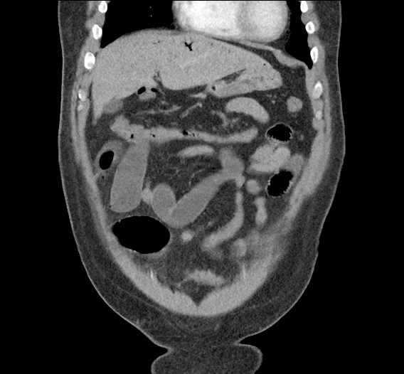
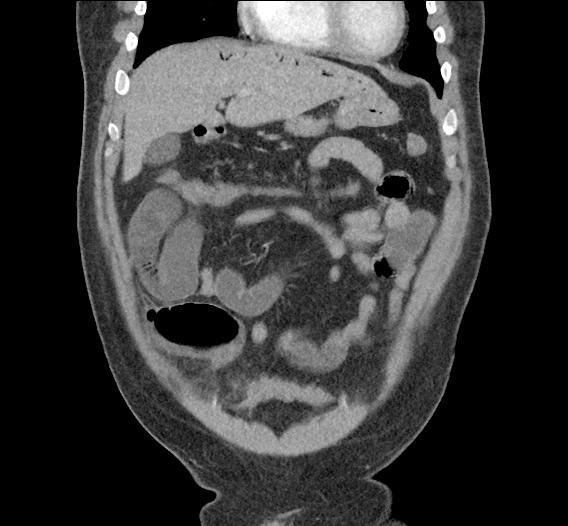
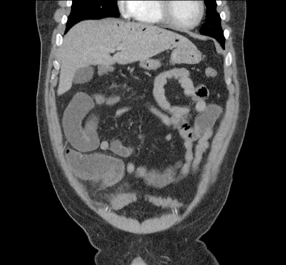
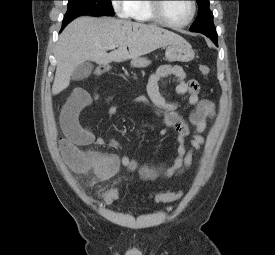
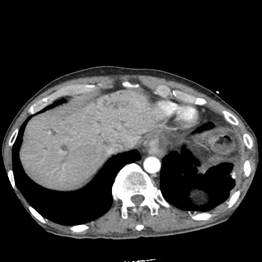
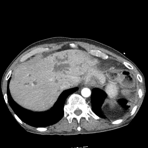
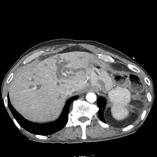
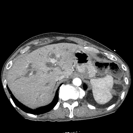
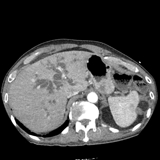
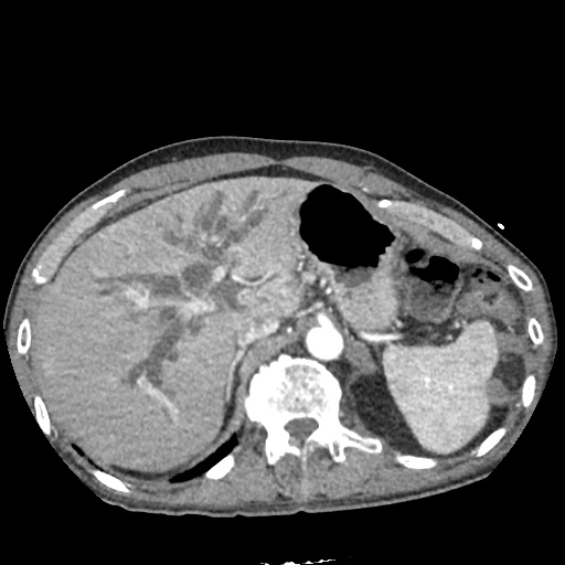
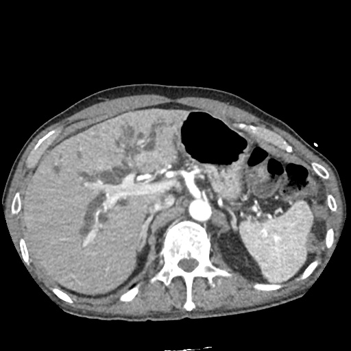
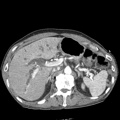
Hyperammonemia

Brief H&P

A 38 year-old male with unknown medical history is brought to the emergency department by EMS with agitation and bizarre behavior. According to prehospital report, the patient was acting erratically – shouting incomprehensibly in the middle of a busy street with possible associated seizure activity.

On evaluation, the patient was found to be tachycardic, hypertensive, and markedly agitated. Physical examination with a focus on toxidromes was notable for the presence of rotary nystagmus suggestive of hallucinogen including phencyclidine toxicity. The patient required pharmacologic sedation to allow for a broad evaluation of altered mental status.

ED Course

The patient’s workup including core temperature, head imaging and laboratory tests (including AST/ALT, albumin, INR) were unremarkable with the exception of an ammonia level of 142 umol/L (normal range 16-53), slightly elevated CK, and urine toxicology screen with multiple positive agents. Over the course of several hours in the emergency department, the patient’s mental status gradually improved reaching normal level of alertness and orientation with normal neurological examination. He acknowledged PCP use as well as a prior history of seizures (possibly related to ethanol withdrawal) without routine anti-epileptic drug use. He denied known history of liver disease.

The patient’s hyperammonemia was attributed to a hypercatabolic state secondary to phencyclidine-induced agitation with possible seizure. He was discharged with resources for assistance with substance cessation.

An Algorithm for the Differential Diagnosis of Hyperammonemia:

Algorithm for the Differential Diagnosis of Hyperammonemia

References:

  1. Kalra A, Norvell JP. Cause for Confusion: Noncirrhotic Hyperammonemic Encephalopathy. Clin Liver Dis. 2020;15(6):223-227. doi:10.1002/cld.929
  2. Mallet M, Weiss N, Thabut D, Rudler M. Why and when to measure ammonemia in cirrhosis? Clin Res Hepatol Gas. 2018;42(6):505-511. doi:10.1016/j.clinre.2018.01.004
  3. Hassan AAI, Ibrahim W, Subahi A, Mohamed A. ‘All that glitters is not gold’: when hyperammonaemia is not from hepatic aetiology. Bmj Case Reports. 2017;2017:bcr-2017-219441. doi:10.1136/bcr-2017-219441
  4. Odigwe CC, Khatiwada B, Holbrook C, et al. Noncirrhotic Hyperammonemia Causing Relapsing Altered Mental Status. Bayl Univ Medical Cent Proc. 2017;28(4):472-474. doi:10.1080/08998280.2015.11929312
  5. Upadhyay R, Bleck TP, Busl KM. Hyperammonemia: What Urea-lly Need to Know: Case Report of Severe Noncirrhotic Hyperammonemic Encephalopathy and Review of the Literature. Case Reports Medicine. 2016;2016:1-10. doi:10.1155/2016/8512721
  6. Walker V. Severe hyperammonaemia in adults not explained by liver disease. Ann Clin Biochem. 2011;49(3):214-228. doi:10.1258/acb.2011.011206
  7. Laish I, Ari ZB. Noncirrhotic hyperammonaemic encephalopathy. Liver Int. 2011;31(9):1259-1270. doi:10.1111/j.1478-3231.2011.02550.x
  8. LaBuzetta JN, Yao JZ, Bourque DL, Zivin J. Adult Nonhepatic Hyperammonemia: A Case Report and Differential Diagnosis. Am J Medicine. 2010;123(10):885-891. doi:10.1016/j.amjmed.2010.02.029
  9. Clay AS, Hainline BE. Hyperammonemia in the ICU. Chest. 2007;132(4):1368-1378. doi:10.1378/chest.06-2940
  10. Weng T-I, Shih FF-Y, Chen W-J. Unusual causes of hyperammonemia in the ED. Am J Emerg Medicine. 2004;22(2):105-107. doi:10.1016/j.ajem.2003.12.011
  11. Hawkes ND, Thomas GAO, Jurewicz A, et al. Non-hepatic hyperammonaemia: an important, potentially reversible cause of encephalopathy. Postgrad Med J. 2001;77(913):717. doi:10.1136/pmj.77.913.717

Ascitic Fluid

Brief HPI:

A 56 year-old male with a history of alcoholic cirrhosis complicated by esophageal varices presents to the emergency department with abdominal distension. He notes gradually worsening symptoms over the past 2 weeks – roughly correlating with the timing of his last paracentesis. He has limited access to medical care and typically presents to emergency departments for palliative paracenteses. He is otherwise in his usual state of health and denies fevers, chills, abdominal pain, vomiting blood, or dark/bloody stools.

Vital signs are notable for a heart rate of 97bpm and blood pressure of 110/65mmHg – otherwise normal. Examination demonstrates a distended abdomen which is non-tender, dull to percussion and with a palpable fluid wave. Bedside ultrasonography shows large, homogenous-appearing ascites with readily-accessible pockets for drainage in the bilateral lower quadrants. A palliative paracentesis is performed with uncomplicated extraction of 4 liters of translucent, straw-colored fluid. Ascitic fluid analysis shows 90 white blood cells of which 10% are polymorphonuclear. The patient is observed briefly in the emergency department, noted symptomatic improvement and was discharged with a plan for telephone follow-up of fluid culture results.

An Algorithm for the Analysis of Ascitic Fluid

Algorithm for the Analysis of Ascitic Fluid

References

  1. Runyon BA. Care of patients with ascites. N Engl J Med. 1994;330(5):337-342. doi:10.1056/NEJM199402033300508.
  2. Wong CL, Holroyd-Leduc J, Thorpe KE, Straus SE. Does this patient have bacterial peritonitis or portal hypertension? How do I perform a paracentesis and analyze the results? JAMA. 2008;299(10):1166-1178. doi:10.1001/jama.299.10.1166.
  3. Tarn AC, Lapworth R. Biochemical analysis of ascitic (peritoneal) fluid: what should we measure? Ann Clin Biochem. 2010;47(Pt 5):397-407. doi:10.1258/acb.2010.010048.
  4. Li PK-T, Szeto CC, Piraino B, et al. ISPD Peritonitis Recommendations: 2016 Update on Prevention and Treatment. Perit Dial Int. 2016;36(5):481-508. doi:10.3747/pdi.2016.00078.
  5. MacIntosh T. Emergency Management of Spontaneous Bacterial Peritonitis – A Clinical Review. Cureus. 2018;10(3):e2253. doi:10.7759/cureus.2253.

Hepatobiliary Ultrasound

Brief H&P:

A 43-year-old female with a history of hypertension, diabetes and obesity presents with right-upper quadrant abdominal pain for the past 1 week. The pain is characterized as burning, non-radiating, intermittent (with episodes lasting 10-30 minutes), resolving spontaneously and without apparent provoking features. She notes nausea but no vomiting, no changes in bowel or urinary habits. She similarly denies fevers, chest pain or shortness of breath. Vital signs were normal, and physical examination was notable only for right upper quadrant tenderness to palpation without rigidity or guarding.

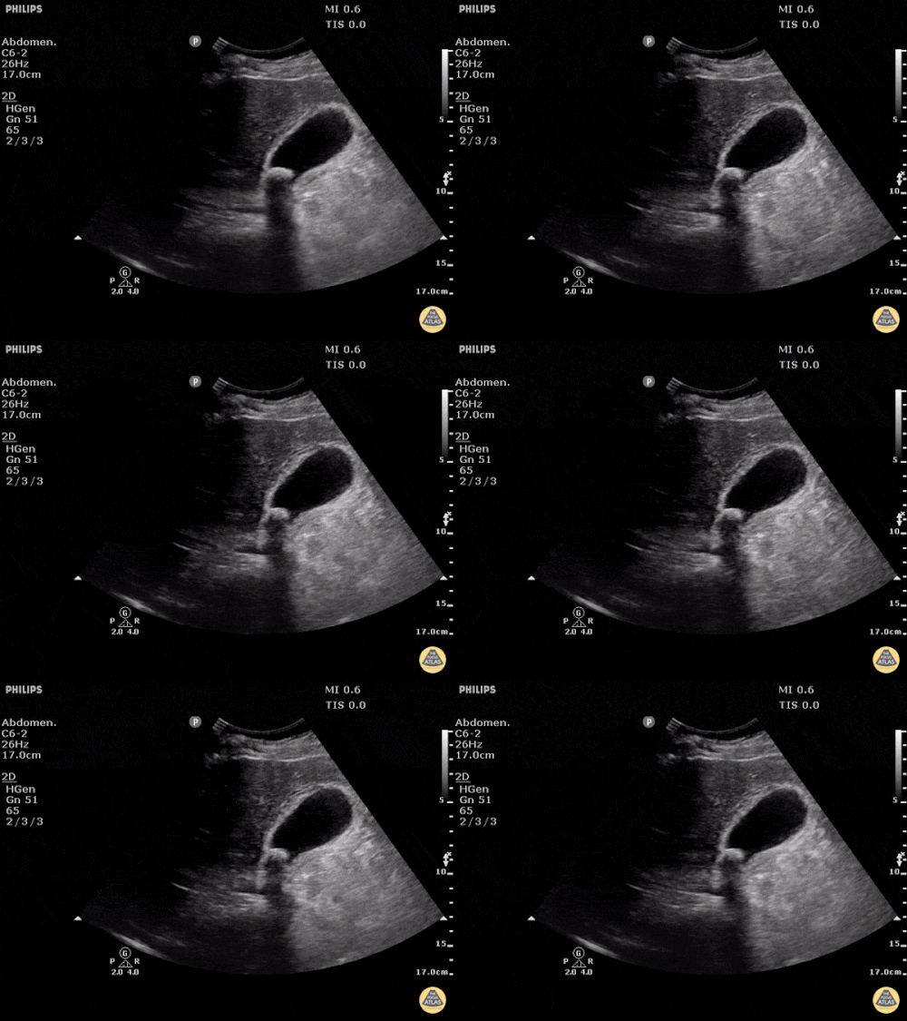
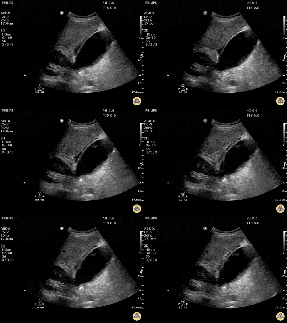
An ECG demonstrates normal sinus rhythm, laboratory tests including liver function tests and lipase were normal and a bedside ultrasound of the right upper quadrant was performed demonstrating gallstones and a positive sonographic Murphy sign. The patient was diagnosed with acute cholecystitis, antibiotics were initiated, the patient was maintained NPO while general surgery was consulted.

Evaluation of Right-Upper Quadrant Abdominal Pain

The initial evaluation of a patient presenting with right-upper quadrant (or adjacent) abdominal pain typically includes laboratory tests such as a complete blood count, chemistry panel, liver function tests and serum lipase. In patients at risk for atypical presentations for an acute coronary syndrome or with other concerning symptoms, electrocardiography and cardiac enzymes may be indicated.

The differential diagnosis is broad. A systematic approach proceeds anatomically from superficial to deeper structures centered around the site of maximal pain.

Skin

Skin

Herpes zoster, erysipelas, or cellulitis

Connective Tissue

Connective Tissue

Intercostal muscle strain, myositis, fasciitis

Bone

Bone

Rib contusion or fracture

Hepatobiliary

Hepatobiliary

Hepatitis (infectious, toxin-mediated), perihepatitis (Fitz-Hugh-Curtis), hepatic abscess, symptomatic cholelithiasis, acute cholecystitis, ascending cholangitis, pancreatitis

Gastric

Gastric

Peptic ulcer disease, gastroesophageal reflux, gastritis, gastroparesis

Small Bowel

Small Bowel

Duodenal ulcer, small bowel obstruction

Large Bowel

Large Bowel

Retrocecal appendicitis, inflammatory bowel disease

Genitourinary

Genitourinary

Pyelonephritis, ureterolithiasis

Referred

Referred

Acute coronary syndrome, lower-lobe pneumonia, pulmonary embolus

Ultrasound in the Evaluation of Right Upper Quadrant Abdominal Pain

The diagnosis is unlikely to be made based on laboratory tests alone 1. However, the addition of bedside ultrasound, particularly for the evaluation of gallbladder pathology, is both rapid and reliable 2-8. The algorithm below provides a pathway for the incorporation of bedside ultrasound of the right upper quadrant in the evaluation of suspected gallbladder disease.

Algorithm for the Use of Ultrasound in the Evaluation of Right Upper Quadrant Abdominal Pain

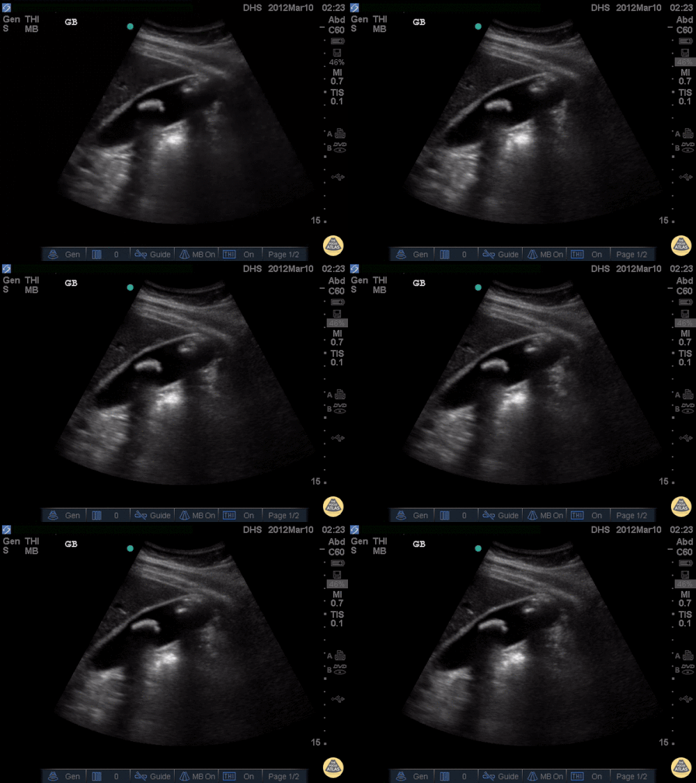
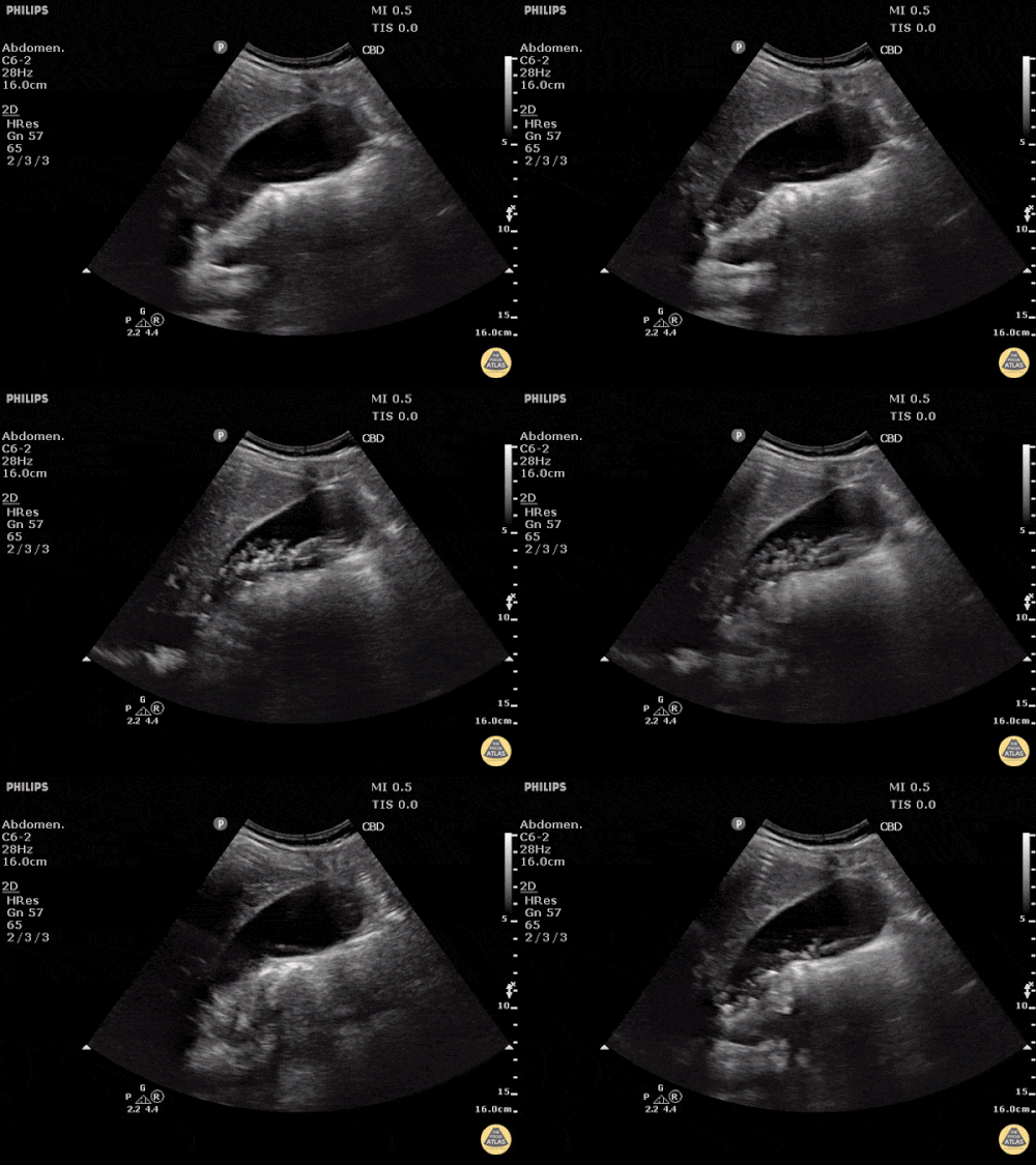
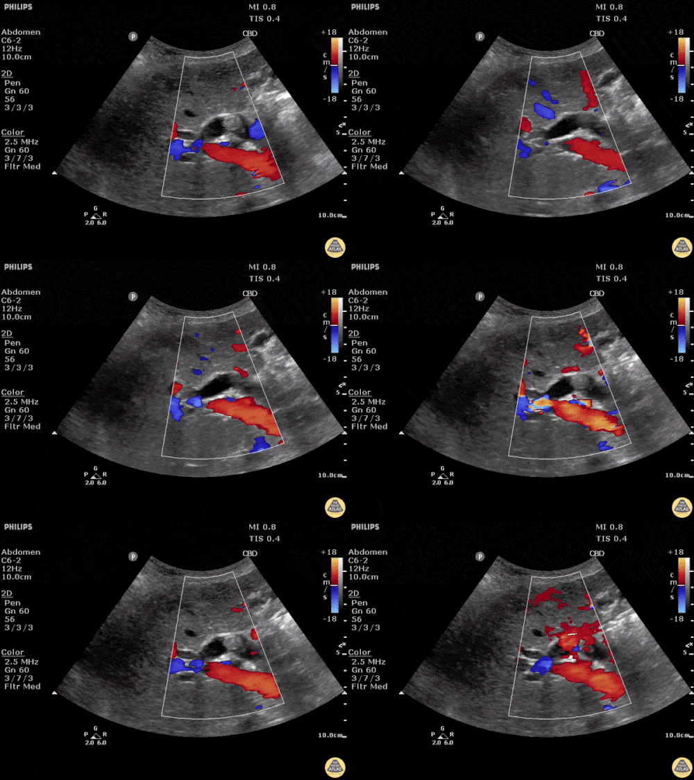
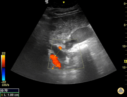
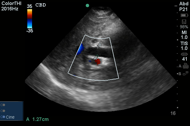
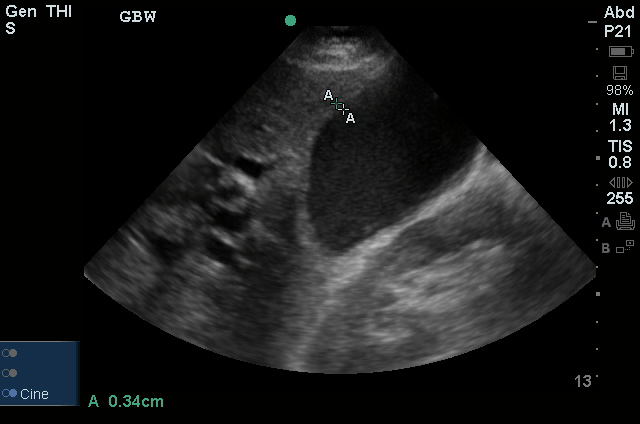
A normal-appearing gallbladder absent gallstones should prompt a traversal of the anatomic approach to the differential diagnosis detailed above. If gallstones are identified, the association with a positive sonographic Murphy sign is highly predictive of acute cholecystitis 2,5,6,9. Acute cholecystitis may be associated with inflammatory gallbladder changes such as wall-thickening (>3mm) or pericholecystic fluid 3,5,6,10-13. However, in the absence of cholelithiasis or a positive sonographic Murphy sign, these features are non-specific and may be the result of generalized edematous states such as congestive heart failure, renal failure, or hepatic failure and critically-ill patients may develop acalculous cholecystitis 7,11,14. Finally, common bile duct dilation may be due to intra-luminal obstruction as in choledocholithiasis, luminal abnormalities such as strictures, or extra-luminal compression from masses or malignancy.  Dilation is generally described as a diameter >6mm – allowing an additional 1mm for every decade over 60 years-old as well as more vague accommodations for patients with prior cholecystectomy 3,5,7,15.

Gallery

The POCUS Atlas
The ultrasound images and videos used in this post come from The POCUS Atlas, a collaborative collection focusing on rare, exotic and perfectly captured ultrasound images.
The POCUS Atlas

Gallstones

Many gallstones

Gallbladder wall thickening

Pericholecystic fluid

Choledocholithiasis

Common bile duct dilation

All illustrations are available for free, licensed (along with all content on this site) under Creative Commons Attribution-ShareAlike 4.0 International Public License.

Downloads Page License

References

  1. Trowbridge RL, Rutkowski NK, Shojania KG. Does this patient have acute cholecystitis? JAMA. 2003;289(1):80-86.
  2. Scruggs W, Fox JC, Potts B, et al. Accuracy of ED Bedside Ultrasound for Identification of gallstones: retrospective analysis of 575 studies. West J Emerg Med. 2008;9(1):1-5.
  3. Ross M, Brown M, McLaughlin K, et al. Emergency physician-performed ultrasound to diagnose cholelithiasis: a systematic review. Acad Emerg Med. 2011;18(3):227-235. doi:10.1111/j.1553-2712.2011.01012.x.
  4. Jang T, Chauhan V, Cundiff C, Kaji AH. Assessment of emergency physician-performed ultrasound in evaluating nonspecific abdominal pain. Am J Emerg Med. 2014;32(5):457-460. doi:10.1016/j.ajem.2014.01.004.
  5. Kendall JL, Shimp RJ. Performance and interpretation of focused right upper quadrant ultrasound by emergency physicians. J Emerg Med. 2001;21(1):7-13.
  6. Summers SM, Scruggs W, Menchine MD, et al. A prospective evaluation of emergency department bedside ultrasonography for the detection of acute cholecystitis. Ann Emerg Med. 2010;56(2):114-122. doi:10.1016/j.annemergmed.2010.01.014.
  7. Rubens DJ. Ultrasound Imaging of the Biliary Tract. Ultrasound Clinics. 2007;2(3):391-413. doi:10.1016/j.cult.2007.08.007.
  8. Rosen CL, Brown DF, Chang Y, et al. Ultrasonography by emergency physicians in patients with suspected cholecystitis. American Journal of Emergency Medicine. 2001;19(1):32-36. doi:10.1053/ajem.2001.20028.
  9. Shea JA. Revised Estimates of Diagnostic Test Sensitivity and Specificity in Suspected Biliary Tract Disease. Arch Intern Med. 1994;154(22):2573-2581. doi:10.1001/archinte.1994.00420220069008.
  10. Miller AH, Pepe PE, Brockman CR, Delaney KA. ED ultrasound in hepatobiliary disease. J Emerg Med. 2006;30(1):69-74. doi:10.1016/j.jemermed.2005.03.017.
  11. Shah K, Wolfe RE. Hepatobiliary ultrasound. Emergency Medicine Clinics of NA. 2004;22(3):661–73–viii. doi:10.1016/j.emc.2004.04.015.
  12. Matcuk GR, Grant EG, Ralls PW. Ultrasound measurements of the bile ducts and gallbladder: normal ranges and effects of age, sex, cholecystectomy, and pathologic states. Ultrasound Q. 2014;30(1):41-48. doi:10.1097/RUQ.0b013e3182a80c98.
  13. Engel JM, Deitch EA, Sikkema W. Gallbladder wall thickness: sonographic accuracy and relation to disease. American Journal of Roentgenology. 1980;134(5):907-909. doi:10.2214/ajr.134.5.907.
  14. Gerstenmaier JF, Hoang KN, Gibson RN. Contrast-enhanced ultrasound in gallbladder disease: a pictorial review. Abdom Radiol (NY). 2016;41(8):1640-1652. doi:10.1007/s00261-016-0729-4.
  15. Becker BA, Chin E, Mervis E, Anderson CL, Oshita MH, Fox JC. Emergency biliary sonography: utility of common bile duct measurement in the diagnosis of cholecystitis and choledocholithiasis. J Emerg Med. 2014;46(1):54-60. doi:10.1016/j.jemermed.2013.03.024.

Dysphagia

Brief H&P

A 47-year-old male with no known medical history presents with dysphagia. He reports 3 weeks of symptoms, describing difficulty predominantly with swallowing solid foods which is aided by the concomitant ingestion of liquids. He points to his throat as the area of discomfort, but has not noted any choking or coughing after attempts at swallowing. He occasionally suffers from “heartburn”, describing a burning sensation in his chest provoked by certain foods and was previously prescribed omeprazole which he has not taken for several years. He denies any prior surgeries, tobacco or alcohol use, relevant family history or similar symptoms in the past.

Physical examination was unrevealing, demonstrating a normal neurological examination, normal phonation, normal oropharynx and no appreciable neck masses. The patient was observed to comfortably swallow water.

He was discharged with gastroenterology follow-up and ultimately underwent esophagogastroduodenoscopy which demonstrated narrowing of the distal esophagus suggestive of a peptic stricture. Dilation was deferred in favor of resumption of proton pump inhibitor therapy.


Types of Dysphagia1,2

Oropharyngeal3
Characterized by difficulty initiating swallowing and accompanied by choking/coughing, nasopharyngeal regurgitation or aspiration.
Involved anatomy: Tongue, muscles of mastication, soft palate (elevation to close nasopharynx), suprahyoid muscles (elevate larynx), epiglottis (occlude airway), cricopharyngeus muscle (release upper esophageal sphincter). Neurological control predominantly coordinated by cranial nerves (V, VII, IX, X, XII)
Esophageal4
Delayed after initiating swallowing and characterized by a sensation of food bolus arresting in transit.
Involved anatomy: Skeletal and smooth muscle along the esophagus and lower esophageal sphincter. Neurological control predominantly coordinated by medulla

Important Historical Features5,6

  • Difficulty with liquids suggests motility problem
  • Difficulty with solids only or solids progressing to liquids suggests mechanical obstruction
  • Identify a history of head and neck surgery or radiation therapy
  • Identify a personal or family history of connective tissue disorder (scleroderma, RA, SLE) which may be associated with esophageal dysmotility
  • Review home medications (NSAID, bisphosphonate, potassium chloride, ferrous sulfate)
  • Immunocompromised patients are at risk for infectious esophagitis (Candida, CMV, HSV) which are generally associated with odynophagia
  • A history of heartburn may be associated with reflux-mediated complications such as erosive esophagitis, peptic stricture, and adenocarcinoma of the esophagus
  • Young patients are more likely to be affected by eosinophilic esophagitis
  • Patient localization of site of obstruction is generally accurate, patients are more accurate at localizing proximal than distal obstructions7

Algorithm for the Evaluation of Dysphagia8

Algorithm for the Evaluation of Dysphagia

Management9-11

Patients who are safely tolerating oral intake can be referred for outpatient gastroenterology evaluation. Admission should be considered for patients at high-risk for aspiration.

References

  1. Spieker MR. Evaluating dysphagia. Am Fam Physician. 2000;61(12):3639-3648.
  2. Abdel Jalil AA, Katzka DA, Castell DO. Approach to the patient with dysphagia. Am J Med. 2015;128(10):1138.e17-.e23. doi:10.1016/j.amjmed.2015.04.026.
  3. Shaker R. Oropharyngeal Dysphagia. Gastroenterol Hepatol (N Y). 2006;2(9):633-634.
  4. Galmiche JP, Clouse RE, Bálint A, et al. Functional esophageal disorders. Gastroenterology. 2006;130(5):1459-1465. doi:10.1053/j.gastro.2005.08.060.
  5. McCullough GH, Martino R. Clinical Evaluation of Patients with Dysphagia: Importance of History Taking and Physical Exam. In: Manual of Diagnostic and Therapeutic Techniques for Disorders of Deglutition. New York, NY: Springer New York; 2012:11-30. doi:10.1007/978-1-4614-3779-6_2.
  6. Cook IJ. Diagnostic evaluation of dysphagia. Nat Clin Pract Gastroenterol Hepatol. 2008;5(7):393-403. doi:10.1038/ncpgasthep1153.
  7. Wilcox CM, Alexander LN, Clark WS. Localization of an obstructing esophageal lesion. Is the patient accurate? Dig Dis Sci. 1995;40(10):2192-2196.
  8. Trate DM, Parkman HP, Fisher RS. Dysphagia. Evaluation, diagnosis, and treatment. Prim Care. 1996;23(3):417-432.
  9. American Gastroenterological Association medical position statement on management of oropharyngeal dysphagia. Gastroenterology. 1999;116(2):452-454. doi:10.1016/S0016-5085(99)70143-5.
  10. Spechler SJ. American Gastroenterological Association medical position statement on treatment of patients with dysphagia caused by benign disorders of the distal esophagus. Gastroenterology. 1999;117(1):229-232. doi:10.1016/S0016-5085(99)70572-X.
  11. Varadarajulu S, Eloubeidi MA, Patel RS, et al. The yield and the predictors of esophageal pathology when upper endoscopy is used for the initial evaluation of dysphagia. Gastrointest Endosc. 2005;61(7):804-808.


Portal Venous Gas

Brief HPI

Young male with no significant medical history presenting with progressively worsening right lower quadrant abdominal pain with marked tenderness to palpation and involuntary guarding.

Imaging

portal_venous_gas_0000_Layer-Comp-1
portal_venous_gas_0001_Layer-Comp-2
portal_venous_gas_0002_Layer-Comp-3
portal_venous_gas_0003_Layer-Comp-4
portal_venous_gas_0004_Layer-Comp-5
portal_venous_gas_0005_Layer-Comp-6
portal_venous_gas_0006_Layer-Comp-7
portal_venous_gas_0007_Layer-Comp-8
portal_venous_gas_0008_Layer-Comp-9
portal_venous_gas_0009_Layer-Comp-10
portal_venous_gas_0010_Layer-Comp-11

CT Abdomen/Pelvis with Contrast

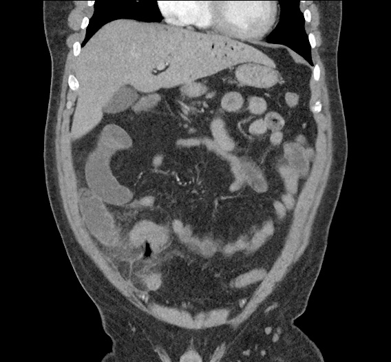
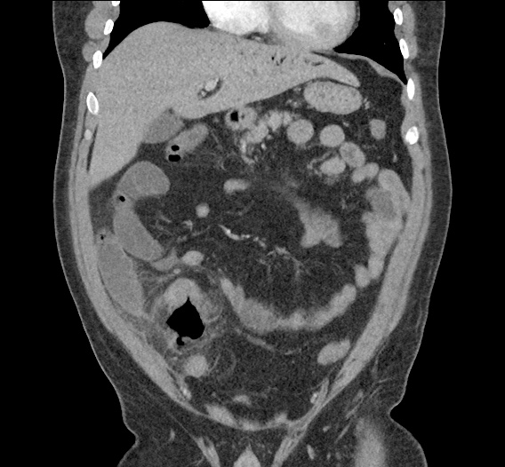
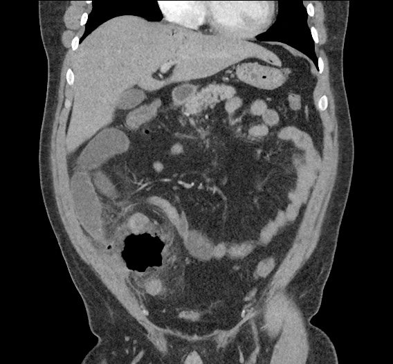
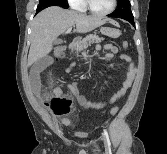
Inflammatory changes in the right lower quadrant concerning for ruptured appendicitis with approximately 9 cm abscess.
Gas in the liver likely representing portal venous gas which can be seen in the setting of appendicitis vs less likely secondary to bowel ischemia.

Differentiation between Portal Venous Gas and Pneumobilia

Portal venous gas vs. Pneumobilia

References

  1. Rabou Ahmed A and Frank Gaillard. “Pneumobilia.” Radiopaedia. http://radiopaedia.org/articles/pneumobilia.
  2. Morgan Matt A and Donna D’Souza. “Portal venous gas.” Radiopaedia. http://radiopaedia.org/articles/portal-venous-gas
  3. Sebastià C, Quiroga S, Espin E, Boyé R, Alvarez-Castells A, Armengol M. Portomesenteric vein gas: pathologic mechanisms, CT findings, and prognosis. Radiographics. 2000;20(5):1213–24–discussion1224–6. doi:10.1148/radiographics.20.5.g00se011213.
  4. Sherman SC, Tran H. Pneumobilia: benign or life-threatening. J Emerg Med. 2006;30(2):147-153. doi:10.1016/j.jemermed.2005.05.016.

Gastrointestinal Bleeding

Evaluation and Management of Gastrointestinal Bleeding

Evaluation and Management of Gastrointestinal Bleeding

Key Historical Features

Quantity
Patient’s estimate
Symptoms suggestive of anemia/volume depletion: (pre)syncope, dyspnea
Appearance/Location
Distinguish upper from lower GI bleding
PMH
Prior episodes and source
History of aortic aneurysm graft
Comorbidities: presence of CAD, CHF, liver disease or diabetes increases mortality
Medications/substance use
Gastrotoxic, anti-coagulants, anti-platelet agents
Alcohol abuse

Key Physical Findings

Vital signs
Tachycardia or hypotension
Eyes
Conjuntival pallor suggests anemia
Scleral icterus suggests liver disease
Abdomen
Hyperactive bowel sounds may be present in UGIB (blood is cathartic)
Epigastric tenderness to palpation suggests PUD
Diffuse tenderness suggests bowel ischemia, obstruction/ileus, or perforation
Rectal (digital, anoscopy)
May reveal fissures, hemorrhoids or polyps

Labs/Diagnostic Tests

  • CBC: consider transfusion for Hb <8-10g/dL particularly in elderly or those with CAD
  • BMP: BUN:creatinine > 36 in the absence of renal failure suggests UGIB
  • PT/PTT/INR: coagulopathy
  • Lactate: elevated in bowel ischemia or systemic hypoperfusion
  • T&S or T&C
  • ECG: screen for myocardial ischemia

Blatchford Scoring System

Item Value Points
BUN 18-22 2
22-28 3
28-70 4
>70 6
Hb (male) 12-13 1
10-12 3
<10 6
Hb (female) 10-12 1
<10 6
SBP 100-109 1
90-99 2
<90 3
Other HR > 100 1
Melena 1
Syncope 2
Liver disease 2
Heart failure 2

References:

  1. Goralnick, E., & Meguerdichian, D. (2013). Gastrointestinal Bleeding. In Rosen's Emergency Medicine – Concepts and Clinical Practice (8th ed., Vol. 1, pp. 248-253). Elsevier Health Sciences.

Nausea and Vomiting

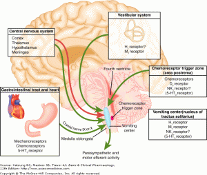
Pathophysiology of Nausea and Vomiting

Pathophysiology of Nausea and Vomiting

Complications of Nausea and Vomiting

  • Hypovolemia: activates RAAS
  • Metabolic alkalosis: loss of hydrogen ions in vomitus
  • Hypokalemia: RAAS promotes sodium retention and potassium excretion
  • Esophageal injury: Mallory-Weiss tear, Boerhaave syndrome
  • Aspiration

Key Historical Findings

Duration of vomiting
Acute: Episodic and occurring for <1 week. Suggestive of obstructive, toxic/metabolic, infectious, ischemic or neurologic process.
Chronic: Episodic and occurring for >1 month. Suggestive of partial obstruction, motility disorder or neurologic process.
Onset
Acute onset: suggests pancreatitis, gastroenteritis, or cholecystitis.
Timing
Post prandial: delayed >1 hour suggests gastric outlet obstruction or gastroparesis.
Contents
Bile: presence of bile suggests patent connection between duodenum and stomach (no GOO)
Undigested food: suggests upper GI tract process (achalasia, esophageal stricture, Zenker)
Feculent: suggests distal bowel obstruction
Associated symptoms
Headache: suggests elevated ICP

Causes of Nausea and Vomiting

Causes of Nausea and Vomiting

References

  1. Zun, L. (2013). Nausea and Vomiting. In Rosen’s Emergency Medicine – Concepts and Clinical Practice (8th ed., Vol. 1, pp. 238-247). Elsevier Health Sciences.

Abdominal Pain

Pathophysiology of Abdominal Pain

  1. Visceral: distension of hollow organs or capsular stretch of solid organs.
  2. Somatic: parietal peritoneal irritation
  3. Referred

    • Extra-abdominopelvic

      • Epigastric: inferior MI
      • Pelvic: hip
      • Abdominal: lower lobe pneumonia/infarction
    • Abdominopelvic

      • Shoulder: diaphragmatic irritation (ex. perforated duodenal ulcer, splenic pathology)
      • Mid-back: aortopathy, pancreatitis
      • Flank: renal pathology
      • Low back: uterus, rectum

Concerning Historical Features

  • Elderly: increased probability for severe disease with poor clinical diagnostic accuracy
  • Immunocompromised: HIV/AIDS, uncontrolled diabetes, chronic liver disease, chemotherapy, other immunosuppression
  • Pain preceding nausea/vomiting: increased likelihood of surgical process
  • Abrupt onset, duration <48h, constant timing
  • Prior abdominal surgical history: consider bowel obstruction
  • No prior episodes of similar pain
  • Recent antibiotic or steroid use: may mask signs of infection
  • Cardiac risk factors (HTN, vascular disease, atrial fibrillation: increased risk for mesenteric ischemia or aortic aneurysm
  • Heavy NSAID use or anticoagulation: increase concern for gastrointestinal bleeding

Imaging

  • Plain film reserved for those who would otherwise not undergo CT. XR abdomen for bowel obstruction or radiopaque foreign body.
  • CT abdomen/pelvis with IV contrast, particularly if elderly or immunocompromised.
  • Ultrasound preferred for hepatobiliary pathology
  • Bedside ultrasound for identification of IUP, free intraperitoneal fluid, cholecystitis, CBD dilation, ascites, hydronephrosis, aortopathy, volume status.

Causes of Abdominal Pain

Causes of Abdominal Pain

References

  1. Budhram, G., & Bengiamin, R. (2013). Abdominal Pain. In Rosen’s Emergency Medicine – Concepts and Clinical Practice (8th ed., Vol. 1, pp. 223-231). Elsevier Health Sciences.

Lactic Acidosis

HPI:

59F with a reported history of congestive heart failure, presenting with intermittent chest discomfort for three days.

She characterized this discomfort as “heartburn”, describing a mid-epigastric burning sensation radiating up her neck, not associated with exertion, lasting 1-2 hours and resolving with antacids. The patient has poor exercise tolerance at baseline and for the past several years has been able to ambulate only short distances around her home, and states that these symptoms have been worsening in the past week. She denies chest pain on exertion, orthopnea or paroxysmal nocturnal dyspnea. She states that she was diagnosed with congestive heart failure five years ago, but was never prescribed medications.

On further questioning, the patient reports several weeks of mouth and lip pain which has limited oral intake, though no dysphagia to solids or liquids. She otherwise denies fevers/chills, abdominal pain, nausea/vomiting, cough, changes in urinary or bowel habits.

In the emergency department, the patient was noted to have an elevated serum troponin, though ECG showed no changes of acute ischemia/infarction.

PMH:

  • Congestive heart failure

PSH:

  • None

FH:

  • Mother with diabetes
  • Father with MI at age 65

SHx:

  • 4-5 drinks of alcohol/day
  • No tobacco or drug use

Meds:

  • None

Allergies:

NKDA

Physical Exam:

VS: T 37.4 HR 106 RR 18 BP 145/82 O2 100% RA
Gen: Morbidly obese female, lying in bed, in no acute respiratory distress, speaking in complete sentences.
HEENT: Dry, cracked lips, slightly erythematous, otherwise moist mucous membranes, poor dentition. Mild scleral icterus. No cervical lymphadenopathy.
CV: Rapid rate, regular rhythm, normal S1/S2, II/VI systolic ejection murmur at LUSB, no radiation appreciated. No jugular venous distension.
Lungs: Clear to auscultation in posterior lung fields bilaterally, no crackles appreciated.
Chest: Well-circumscribed erythematous patch in folds beneath left breast, no underlying fluctuance, no significant tenderness to palpation. On contralateral breast, some hyperpigmentation but no erythema.
Abdomen: Obese, non-tender, non-distended. Patch of erythema below pannus, mildly tender to palpation.
Ext: Bilateral lower extremities with marked edema and overlying scaly plaques, some slightly ulcerated weeping serous fluid. Peripheral pulses are difficult to palpate, capillary refill difficult to assess.

Labs/Studies:

  • CBC: 11.1/11.1/34.5/212 (MCV 114.2)
  • BMP: 140/4.5/97/20/10/1.14/64
  • Anion Gap: 23
  • LFT: AST: 73, ALT: 26, AP: 300, TB: 4.6, DB: 2.1, Alb: 3.0, INR 1.3
  • BNP: 158
  • Troponin: 1.284
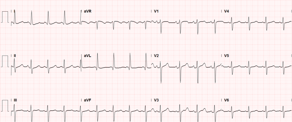
Sinus tachycardia, LVH, secondary repolarization abnormalities

Sinus tachycardia, LVH, secondary repolarization abnormalities

Imaging:

CT Pulmonary Angiography:
No evidence of central pulmonary embolism, thoracic aortic dissection, or thoracic aortic aneurysm. Evaluation of the peripheral vessels is limited due to motion artifact. No focal consolidation or pneumothorax.

CT Abdomen/Pelvis non-contrast:
No evidence of intra-abdominal abscess or definite source of infection. Marked hepatic steatosis.

CT Lower Extremity non-contrast:
Diffuse circumferential subcutaneous edema involving both lower extremities from the level of the mid thighs distally through the feet. There are bilateral subcutaneous calcifications which are likely venous calcifications in the setting of chronic venous stasis disease. There is some overlying skin thickening.

TTE:
There is moderate concentric left ventricular hypertrophy with hyperdynamic LV wall motion. The Ejection Fraction estimate is >70%. Grade I/IV (mild) LV diastolic dysfunction. No hemodynamically significant valve abnormalities.

US Abdomen:
Hepatomegaly, echogenic liver suggesting fatty infiltration. Moderately blunted hepatic vein waveforms suggesting decreased hepatic parenchymal compliance.

Assessment/Plan:

The patient was admitted to the cardiology service for management of NSTEMI and evaluation of undiagnosed CHF. She was started on a heparin continuous infusion. In addition, a CT pulmonary angiogram was obtained to evaluate for pulmonary embolism as an explanation of her progressive dyspnea on exertion. No PE, consolidation or effusion was identified.

Despite the patient’s reported history of congestive heart failure, there was no evidence that her symptoms were a result of an acute exacerbation with only a mildly elevated BNP but no jugular venous distension or evidence of pulmonary edema. The patient’s significant lower extremity edema was more suggestive of chronic venous stasis.

One notable laboratory abnormality that was explored was her elevated anion gap metabolic acidosis. Studies submitted included serum lactate, salicylates, osmolarity, CK, and urinalysis for ketonuria. This evaluation was notable for an elevated serum lactate of 13.2mmol/L and an arterial blood gas that showed adequate respiratory compensation (and no A-a gradient). Given the patient’s modest leukocytosis (with neutrophil predominance), and tachycardia, the concern for sepsis was increased though the source remained unclear. Prominent possibilities included a skin and soft-tissue infection vs. less likely intra-abdominal source though the patient’s physical examination was not suggestive of a process that would produce such a substantial lactic acidosis. Blood cultures were drawn and the patient was started on empiric antibiotics for the suspected sources. In addition, the patient was cautiously volume resuscitated given her reported history of CHF while pending a transthoracic echocardiogram to evaluate cardiac function. Additional imaging including CT abdomen/pelvis and lower extremities was obtained (though without contrast due to the patient’s recent exposure), and no obvious source was identified.

Over the next two days, the patient’s serum lactate downtrended to normal range, as did the serum troponin. A transthoracic echocardiogram showed an LVEF >70% with mild concentric hypertrophy and diastolic dysfunction. Blood and urine cultures were without growth.

Additional issues managed during the hospitalization included elevated serum transaminases (AST > ALT), conjugated hyperbilirubinemia and evidence of decreased hepatic synthetic function with hypoalbuminemia and elevated INR. Given the patient’s history of EtOH use, as well as other corroborating findings including macrocytic anemia, hypomagnesemia, folate and B12 deficiency, this was attributed to alcoholic hepatitis (discriminant function <32). Infectious hepatitis serologies were negative. The patient was started on nutritional supplements. Finally, the patient persistently complained of lip and oral mucosal pain. Examination was without discrete lesions but some mucosal redness was identified. Despite poor dentition, there was no evidence of abscess and HSV/HIV testing was negative. This was thought to be stomatitis caused by her identified nutritional deficiencies.

Differential Diagnosis of Elevated Serum Lactate 1,2

Differential Diagnosis of Elevated Serum Lactate

Algorithm for Evaluation of Acidemia 3,4

Algorithm for Evaluation of Acidemia

Algorithm for Evaluation of Alkalemia 3,4

Algorithm for Evaluation of Alkalemia

References:

  1. Fall, P. J., & Szerlip, H. M. (2005). Lactic acidosis: from sour milk to septic shock. Journal of intensive care medicine, 20(5), 255–271. doi:10.1177/0885066605278644
  2. Luft, F. C. (2001). Lactic acidosis update for critical care clinicians. Journal of the American Society of Nephrology : JASN, 12 Suppl 17, S15–9.
  3. Ingelfinger, J. R., Berend, K., de Vries, A. P. J., & Gans, R. O. B. (2014). Physiological Approach to Assessment of Acid–Base Disturbances. The New England journal of medicine, 371(15), 1434–1445. doi:10.1056/NEJMra1003327
  4. Ingelfinger, J. R., & Seifter, J. L. (2014). Integration of Acid–Base and Electrolyte Disorders. The New England journal of medicine, 371(19), 1821–1831. doi:10.1056/NEJMra1215672

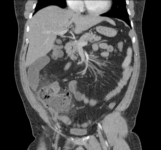
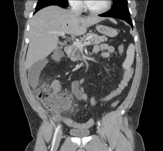
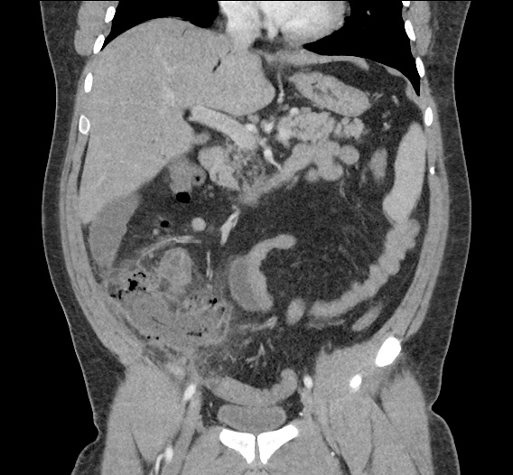
Biliary Duct Dilation

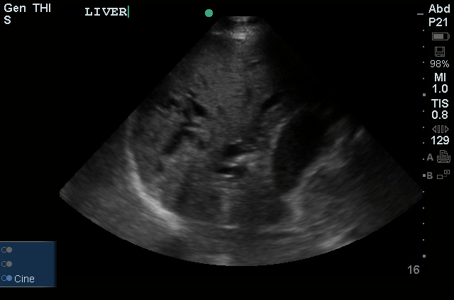
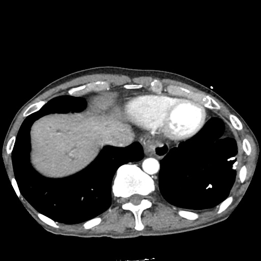
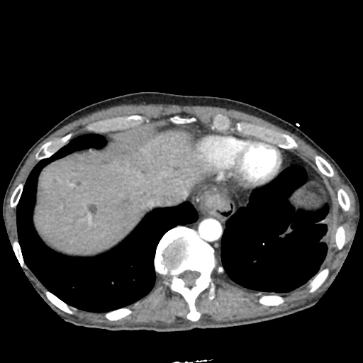
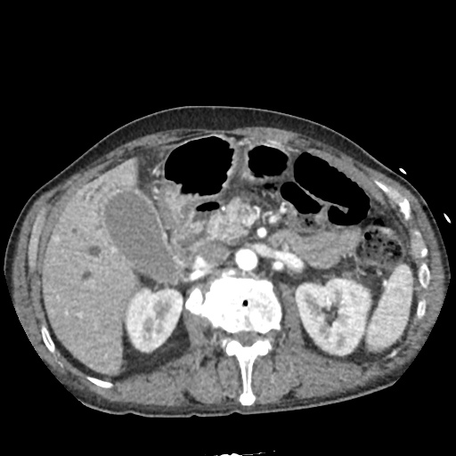
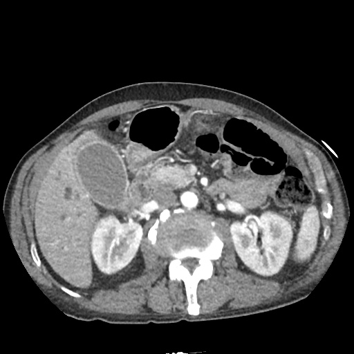
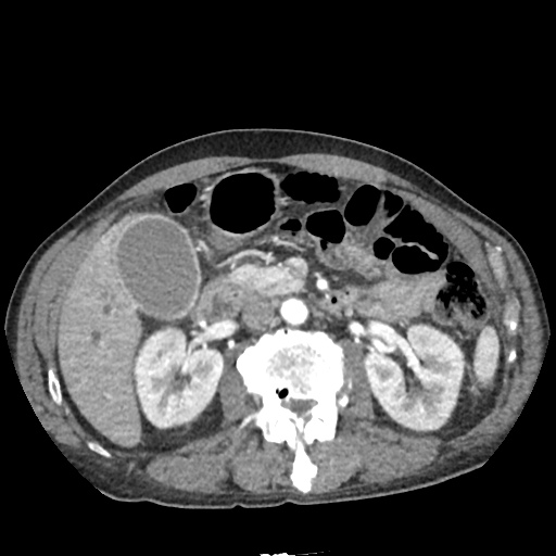
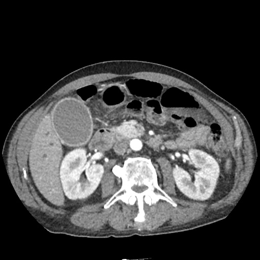
A 45 year-old male presents with progressive jaundice over 1 month, he denies abdominal pain.

Ultrasound

Liver
Common Bile Duct
Gallbladder Wall

CT Abdomen/Pelvis

IM-0001-0011
IM-0001-0013
IM-0001-0015
IM-0001-0017
IM-0001-0019
IM-0001-0021
IM-0001-0023
IM-0001-0025
IM-0001-0027
IM-0001-0029
IM-0001-0031
IM-0001-0033
IM-0001-0035
IM-0001-0037
IM-0001-0039
IM-0001-0041
IM-0001-0043
IM-0001-0045
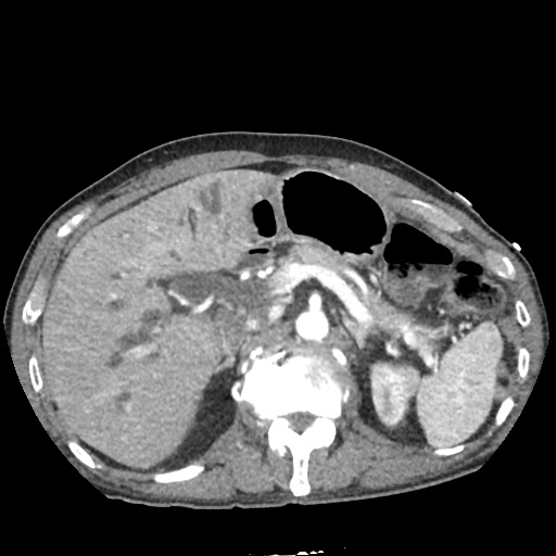
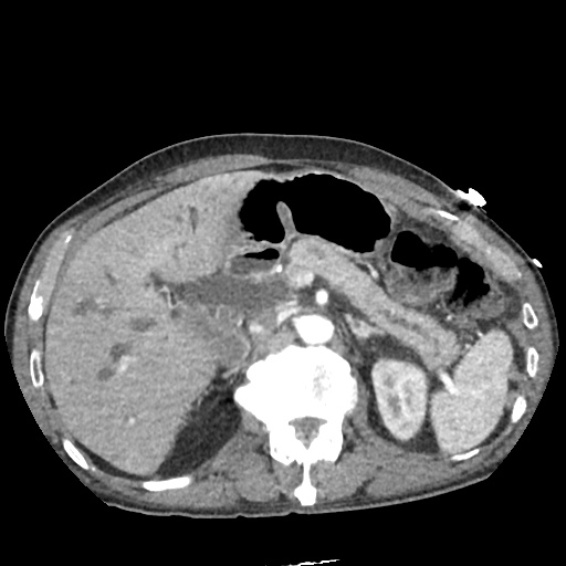
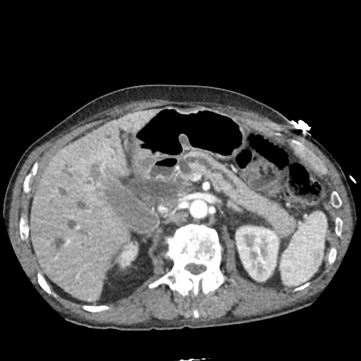
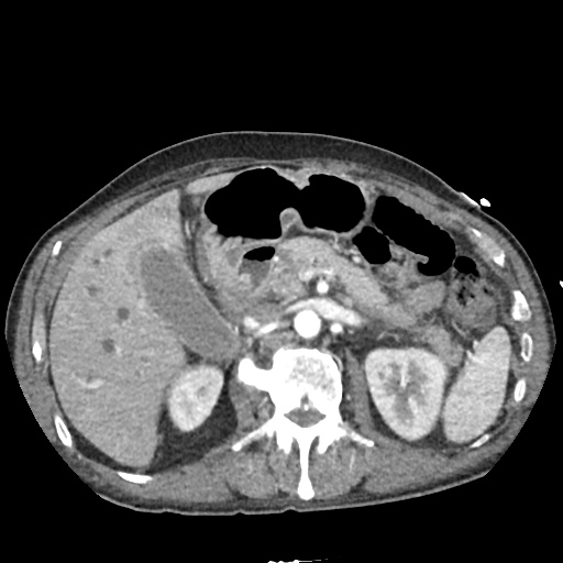
  • Markedly dilated intrahepatic biliary ducts, common bile duct and pancreatic duct.
  • Ill-defined fullness in the pancreatic head consistent with pancreatic adenocarcinoma vs. noncalcified obstructing biliary stone.

Differential Diagnosis of Biliary Duct Dilation: 1,2,3

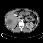
Differential Diagnosis of Biliary Duct Dilation

References:

  1. Kim, H. J., Lee, K. T., Kim, S. H., Lee, J. K., Lim, J. H., Paik, S. W., & Rhee, J. C. (2003). Differential diagnosis of intrahepatic bile duct dilatation without demonstrable mass on ultrasonography or CT: benign versus malignancy. Journal of gastroenterology and hepatology, 18(11), 1287–1292.
  2. Levy, A. D. (2009). Biliary Ducts – Pathology. The Radiology Assistant, 1–4. Retrieved from http://www.radiologyassistant.nl/en/p49e17de25294d/biliary-ducts-pathology.html
  3. Teefey, S. A., Baron, R. L., Schulte, S. J., Patten, R. M., & Molloy, M. H. (1992). Patterns of intrahepatic bile duct dilatation at CT: correlation with obstructive disease processes. Radiology, 182(1), 139–142. doi:10.1148/radiology.182.1.1727277

Hepatic Abscess

HPI:

42M with 1.5 weeks of fevers. Initially presented to ER 1wk ago and treated for possible otitis media, however follow-up ENT appointment showed no evidence of OM on exam. Fevers persisted and he developed headaches and went to urgent care where a CT head and LP were negative. A mild elevation of serum transaminases was noted and the following CT abdomen/pelvis was obtained. He denied GI symptoms.

Imaging:

Hepatic Abscess - Axial

Hepatic Abscess - Axial

- 7.4 cm cystic lesion in the inferior right lobe of the liver most consistent in appearance with hepatic abscess.
- Multiple calcified gallstones with a 10 mm gallstone in the neck of the gallbladder or possibly in the cystic duct.

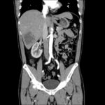
Hepatic Abscess - Coronal

Hepatic Abscess - Coronal

- 7.4 cm cystic lesion in the inferior right lobe of the liver most consistent in appearance with hepatic abscess.
- Multiple calcified gallstones with a 10 mm gallstone in the neck of the gallbladder or possibly in the cystic duct.

Assessment & Plan:

# Liver abscess: likely pyogenic s/p CT-guided drainage with 60cc purulent fluid removed. Gram stain showed GNR and WBC’s, culture grew Klebsiella pneumonia. Treated with ceftriaxone 2g IV q24h, metronidazole 500mg PO TID.

Differential Diagnosis of Hepatic Abscess:1

Differential Diagnosis of Hepatic Abscess

References:

  1. Krige J, Beckingham IJ. Liver abscesses and hydatid disease. BMJ. 2001;322(7285):537–540.

Alcoholic Hepatitis

HPI:

43 year-old female with a history of alcohol abuse and alcoholic hepatitis, presenting after referral from breast clinic for abnormal labs (notable for total bilirubin 18.1). The patient was well until two weeks ago when she noted increasing fatigue associated with morning nausea/vomiting (non-bloody) as well as yellowing of skin and eyes. She also reports darkening of urine, but no dysuria, change in volume of urine, or visible blood. She also denies fevers/chills, increased abdominal girth, abdominal pain, changes in bowel habits or bloody/dark stools.

She reports drinking 1 pint of vodka daily for the past 15 years, and perhaps more in the past 3 weeks. Her last drink was in the morning on the day of admission, she denies any history of seizures and reports withdrawal symptoms (tremor, nausea) relieved with more alcohol. She currently denies anxiety/agitation, tactile/visual/auditory hallucinations.

The patient was in breast clinic for evaluation of a painful breast mass which developed after biopsy of a lesion which was ultimately found to be benign. The patient noted the mass was growing in size and becoming more painful over the past month.

PMH:

  • EtOH abuse
  • Alcoholic hepatitis

PSH:

  • None

FH:

  • No family history of breast/gynecologic malignancy.
  • Mother with history of stroke. Father with diabetes.

SHx:

  • Lives alone.
  • Denies current or previous tobacco/drug use. Drinks 1 pint of whiskey daily for the past 15 years.
  • Not currently sexually active, no history of STI.

Meds:

  • None

Allergies:

NKDA

Physical Exam:

VS: T 98.9 HR 104 RR 19 BP 117/67 O2 99% RA
Gen: Well-appearing obese female in no acute distress
HEENT: PERRL, marked scleral icterus, sublingual icterus, MMM, no lesions
CV: Tachycardia, regular rhythm, normal S1/S2, no M/R/G
Lungs: CTAB, no crackles/wheezing
Abd: +BS, soft, non-distended, liver edge palpated 6cm below costal margin, irregular texture slightly tender to palpation, spleen not palpated, no fluid wave or shifting dullness, no rebound/guarding.
Ext: Warm, well-perfused, 2+ pulses (DP/PT), slight yellowing.
Skin: Vascular spiders on anterior chest, left breast with 5x5cm ecchymosis and tender underlying mass, no erythema, warmth, skin dimpling, nipple discharge.
Neuro: AAOx4, CN II-XII intact, no tremor noted, gait normal.

Labs/Studies:

1mo prior to admission:

  • AST/ALT/AP/TB: 444/77/234/2.5

Day 1:

  • AST/ALT/AP/TB: 185/61/184/18.1
  • PT/PTT/INR: 14.7/37.0/1.2

Day 4:

  • AST/ALT/AP/TB: 142/50/153/25.5
  • PT/PTT/INR: 20.1/38.9/1.7

Imaging:

Abdominal US

  1. Markedly echogenic and enlarged liver with a nodular surface of cirrhosis.
  2. Markedly blunted hepatic vein waveforms commonly seen due to decreased hepatic parenchymal compliance although other etiologies causes of obstruction to hepatic venous outflow.
  3. Splenomegaly.

Assessment/Plan:

44F hx EtOH abuse, alcoholic hepatitis, presenting with acute alcoholic hepatitis.
# Alcoholic hepatitis: Rapid onset of jaundice, tender hepatomegaly, and elevation of transaminases (AST > ALTx2) in the setting of chronic alcohol use suggestive of alcoholic hepatitis. Initial Maddrey discriminant hepatic function (mDH) score 37 suggestive of severe disease with high short-term mortality. Initiated trental 400mg p.o. t.i.d.
# EtOH withdrawal: Last drink <24h ago, monitor for signs of withdrawal, treat with Ativan per withdrawal protocol. # Cirrhosis: Newly diagnosed on abdominal ultrasound. Complicated by coagulopathy, and likely portal hypertension given splenomegaly/thrombocytopenia. Plan for outpatient screening EGD and continued GI follow-up. # Breast mass: Likely hematoma 2/2 biopsy associated given increased size associated with progression of coagulopathy/thrombocytopenia. Outpatient ultrasound and follow-up scheduled. # Anemia: Macrocytic, potentially related to vitamin deficiency vs. bone-marrow suppression associated with chronic alcohol use. Start thiamine/folate/multivitamin. # FEN/GI/PPx: Encourage p.o. intake (2g sodium restriction), continue ondansetron p.r.n. nausea/vomiting, obtain nutrition consult.

Hospital Course

Patient’s liver function continued to decline as evidenced by worsening coagulopathy and increased serum bilirubin. mDH had increased to 58 by day four of hospitalization and steroids were added.

Pathophysiology of Alcoholic Hepatitis: 1

Ethanol promotes translocation of bacterial components (lipopolysaccharide) across the intestinal wall, into the portal venous system and liver. These trigger a local and systemic inflammatory response which leads to hepatocellular injury and systemic effects such as fever, anorexia and weight loss.

Evaluation of Alcoholic Hepatitis: 1,2

Clinical features:

  • Rapid onset jaundice
  • Tender hepatomegaly
  • Fever
  • Ascites
  • Proximal muscle loss
  • Encephalopathy

Labs:

  • AST > ALT (x2), generally < 300IU/mL
  • Leukocytosis
  • ↑Total serum bilirubin
  • ↑INR
  • ↑Creatinine associated with poor prognosis

Other studies:

  • Screening for infection: PNA, UTI, SBP
  • Abdominal US to evaluate hepatic abscess, HCC, extrahepatic biliary obstruction

Management of Alcoholic Hepatitis: 1,2

Grading Severity:

  • Maddrey’s discriminant function
  • Glasgow score
  • Lille score (assess response to corticosteroids after 1wk)

Treatment:

  • Immediate and lifetime abstinence from alcohol
  • Trental 400mg p.o. t.i.d.
  • Prednisolone 40mg p.o. daily (controversial, some benefit in subgroup with Maddrey > 32)
  • Ascites: Sodium restriction, diuretics
  • Encephalopathy: Lactulose, rifaximin
  • Hepatorenal syndrome: albumin, vasopressors
  • Nutritional support

Interpretation of Liver Function Tests: 3

Disorder Bilirubin AST/ALT Albumin PT
Hemolysis
Gilbert
↑(indirect)
No bilirubinuria
Acute hepatocellular necrosis ↑ALT > AST
> 500IU

(poor prognosis if elevated)
Chronic liver disease

< 300IU

Alcoholic hepatitis

AST:ALT > 2

Intra- extra-hepatic cholestasis

< 500IU

↑↑

(>4x normal)

Features of Components of Liver Function Tests: 3,4

Features of Components of Liver Function Tests

References:

  1. Lucey, M. R., Mathurin, P., & Morgan, T. R. (2009). Alcoholic hepatitis. The New England journal of medicine, 360(26), 2758–2769. doi:10.1056/NEJMra0805786
  2. Sohail, U., & Satapathy, S. K. (2012). Diagnosis and management of alcoholic hepatitis. Clinics in liver disease, 16(4), 717–736. doi:10.1016/j.cld.2012.08.005
  3. Kaplan MM. Chapter 302. Evaluation of Liver Function. In: Longo DL, Fauci AS, Kasper DL, Hauser SL, Jameson JL, Loscalzo J, eds. Harrison’s Principles of Internal Medicine. 18th ed. New York: McGraw-Hill; 2012.
  4. Johnston, D. E. (1999). Special considerations in interpreting liver function tests. American family physician, 59(8), 2223–2230.

Renal Failure in Cirrhosis

CC:

Consult for acute kidney injury

HPI:

63M with a history of liver cirrhosis of cryptogenic etiology, portal vein thrombosis, and esophageal varices s/p banding (2011) who was admitted to an OSH for altered mental status and hypotension requiring dopamine and was transferred to this facility for a higher level of care.

The nephrology service was consulted for elevated serum creatinine concerning for AKI. The patient has a baseline creatinine of 1.1 (3/2013), 1.9 on transfer and continued worsening to peak of 2.6 today.

PMH:

  • Asthma
  • COPD
  • Cirrhosis (PVT, encephalopathy)
  • Inguinal hernia (recurrent)

PSH:

  • Appendectomy
  • Bilateral inguinal hernia repair

FH:

  • Non-contributory

SHx:

  • Married
  • Denies t/e/d use

Meds:

  • albumin 25g i.v. q.6.h.
  • erythromycin 1,000mg p.o. q.1.h.
  • fluticasone-salmeterol 1 puff b.i.d.
  • lactulose 45g p.o. q.6.h.
  • neomycin 1,000mg p.o. q.1.h.
  • pantoprazole 40mg i.v. daily
  • rifaximin 550mg p.o. b.i.d.
  • sodium benzoate 5g p.o. b.i.d.

Allergies:

  • Sulfa

Physical Exam:

VS: T 37.4 HR 90 RR 15 BP 86/48 O2 97% RA
Gen: Chronically ill-appearing.
HEENT: PERRL, scleral icterus, MMM
CV: RRR
Lungs: CTAB
Abd: +BS, soft, non-tender, non-distended
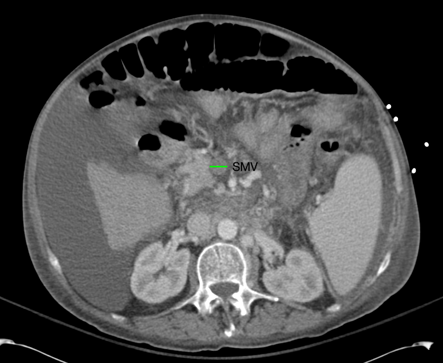
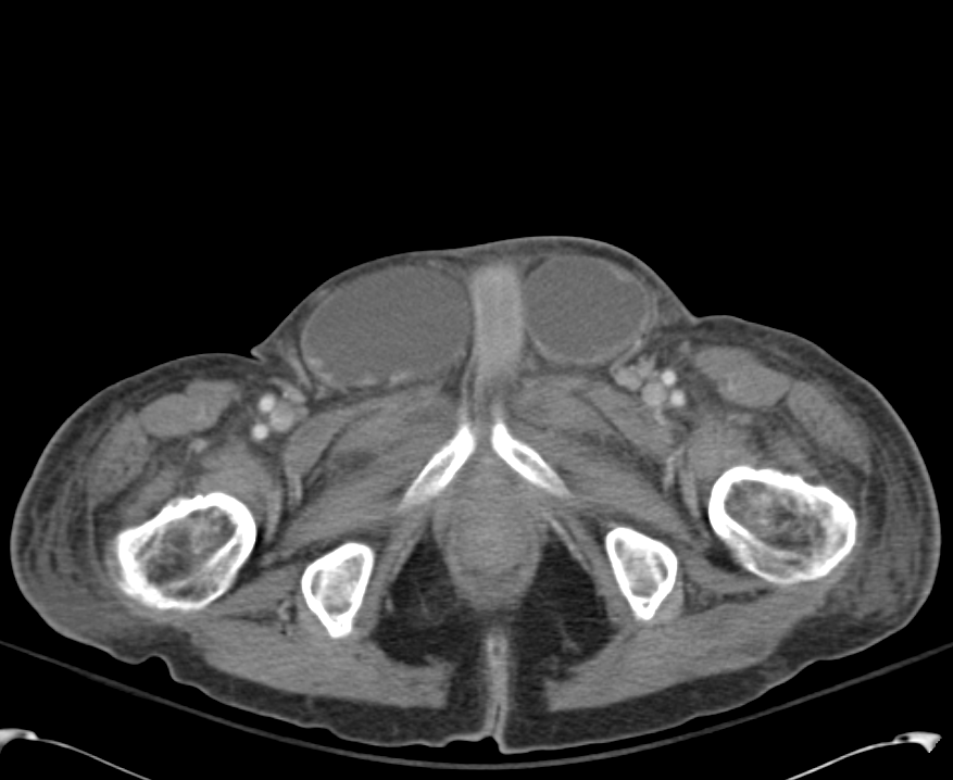
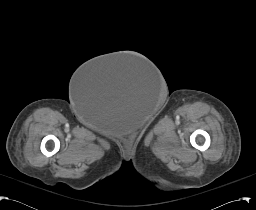
GU: Large ascites filled scrotum, testicles/inguinal canal not easily palpated
Ext: Warm, well-perfused
Skin: No palmar erythema, no vascular spiders
Neuro: AAOx4, CN II-XII grossly intact

Labs:

  • BMP: 134/4.5/103/20/41/3.0/106 (Ca 9.3, Mg 3.7, PO4 2.4)
  • LFT: AST 89, ALT 33, TB 26.6, CB 16.1, Alb 2.7
  • NH4 167

Imaging:

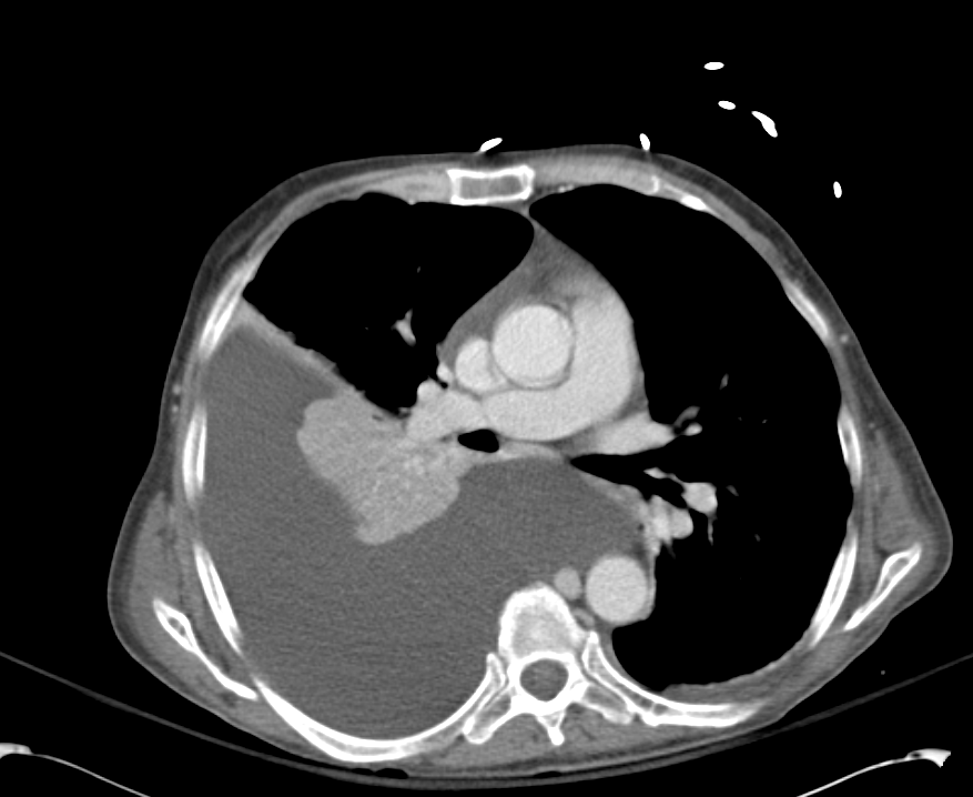
Pleural Effusion

Pleural Effusion

Large right pleural effusion with underlying compressive atelectasis.

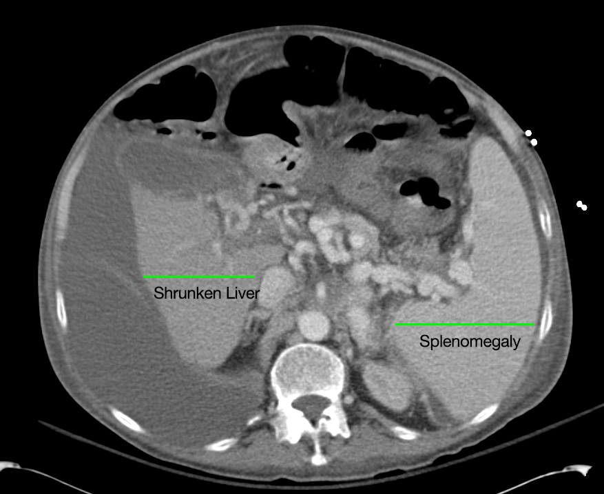
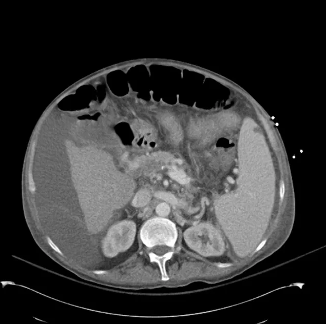
Cirrhosis and Portal Hypertension

Cirrhosis and Portal Hypertension

Shrunken/nodular liver with sequelae of portal hypertension including perisplenic collaterals, and splenomegaly.

SMV Thrombosis

SMV Thrombosis

Near-total thrombosis of the portal vein extending down to superior mesenteric vein.

B/L Inguinal Hernias

B/L Inguinal Hernias

Large volume abdominal ascites with a large amount of fluid extending into the bilateral inguinal canals.

Large Right Inguinal Hernia

Large Right Inguinal Hernia

Large volume abdominal ascites with a large amount of fluid extending into the bilateral inguinal canals.

CT Abdomen/Pelvis (PVT)

CT Abdomen/Pelvis (PVT)

Assessment/Plan:

63M with a history of liver cirrhosis of cryptogenic etiology, recently with hypotension prior to transfer to this facility and increase in creatinine from 1.9-3.0 on current admission (from baseline 1.1).

These findings indicate acute kidney injury, likely hepatorenal syndrome vs. acute tubular necrosis 2/2 prolonged hypotension. Plan to discontinue diuretics and start albumin challenge (1g/kg/day divided q6h x2d). Will also check UA, urine Na/cr/urea/eos, renal US (evaluate obstruction, kidney size). Start midodrine/octreotide for underlying HRS.

  1. Neuro: Intermittent confusion. Lactulose, rifaximin, benzoate.
  2. Resp: 2L NC. ABG 7.36/51/87/27.7/+2. CXR: Large R effusion.
  3. CV: Levo 0.075. Midodrine 15 TID. MAPs 60, HR 80s.
  4. GI: NPO/NGT. TPN.
  5. Renal: See above.
  6. Heme: Coagulopathy, keep INR <2.5
  7. ID: Afebrile. No abx.
  8. Endo: Euglycemic

Renal Failure in Cirrhosis:

Renal failure in cirrhosis is associated with higher mortality both before and after transplant. The main causes of renal failure in cirrhosis are detailed below, with particular attention to an entity unique to cirrhosis: the hepatorenal syndrome.1

Disorder Pathogenesis Diagnosis Management
HRS Dilation of splanchnic arteries initially compensated by increased CO eventually decompensates with activation of mechanisms to preserve ECBV (RAAS, SNS, ADH) leading to fluid retention (ascites, edema) and renal failure due to intrarenal vasoconstriction.Bacterial translocation and the resulting inflammatory response may contribute to splanchnic vasodilation (through production of vasoactive factors like NO).
  • Serum creatinine > 1.5mg/dl-  Not reduced with 1g/kg albumin
  • No confounding factors (2d off diuretics, no nephrotoxic agents, no shock, no e/o intrinsic renal disease)
  • Type 1: doubling creatinine > 2.5mg/dL in <2wk
  • Type 2: stable, slower progression
  • Vasoconstrictor therapy-  Albumin
  • Portasystemic shunting
  • Renal replacement therapy
  • Prevention
    • Norfloxacin
    • Albumin
Intrinsic renal Some causes of liver disease are also associated with intrinsic renal pathology (ex. GN associated with HBV, HCV).
  • Proteinuria, hematuria
  • Renal bx
  • Active urinary sediment
  • Antiviral therapy if appropriate
Pre-renal AKI Hemorrhage (GIB), fluid losses (excess diuresis, diarrhea from lactulose).
  • Suspected from patient history
  • Low FENa, bland urine sediment
  • Hemorrhage: replace volume with fluids, blood products. Control bleeding.
  • Discontinue diuretics, administer fluids if tolerated
ATN Severe ischemic or toxic (NSAID’s, nephrotoxic medications)
  • Renal tubular epithelial cells favor ATN (granular casts common in ATN, HRS)
  • Withdraw therapy
  • Avoid nephrotoxic agents

Pathophysiology of Hepatorenal Syndrome:

Pathophysiology of Hepatorenal Syndrome

Evaluation:

The evaluation of suspected renal failure in patients with cirrhosis involves assessment of renal function for evidence of acute impairment, as well as analaysis of urine for protein or active sediment to suggest intrinsic renal disease (possibly warranting renal ultrasonography or biopsy). Additionally, patients should be evaluated for evidence of bacterial infection including assessment of ascites if present as SBP produces a more severe form of the inflammatory vasodilation mechanism suspected to play a role in HRS.

Treatment:

For renal failure not caused by the hepatorenal syndrome, identification and management of the underlying cause is critical (intrinsic renal disease, hypovolemia/hemorrhage, nephrotoxicity, infection). For suspected HRS, management is dependent on the acuity and setting. In the intensive care unit, vasoconstrictor therapy (norepinephrine, vasopressin) in association with albumin is effective in the treatment of HRS.2,3  In less acute settings, a combination of midodrine, octreotide and albumin improves renal function and is associated with lower short-term mortality.4 Alternatives for patients who do not respond to medical therapy include TIPS, dialysis and transplant.

Summary:

Renal failure in ESLD is due to the causes, complications or management of cirrhosis and has important implications, with HRS in particular offering the worst prognosis.5 Early recognition and management is critical to improving outcomes.

References:

  1. Ginès, P., & Schrier, R. W. (2009). Renal failure in cirrhosis. The New England journal of medicine, 361(13), 1279–1290. doi:10.1056/NEJMra0809139
  2. Singh, V., Ghosh, S., Singh, B., Kumar, P., Sharma, N., Bhalla, A., Sharma, A. K., et al. (2012). Noradrenaline vs. terlipressin in the treatment of hepatorenal syndrome: a randomized study. Journal of hepatology, 56(6), 1293–1298. doi:10.1016/j.jhep.2012.01.012
  3. Kiser, T. H., Fish, D. N., Obritsch, M. D., Jung, R., MacLaren, R., & Parikh, C. R. (2005). Vasopressin, not octreotide, may be beneficial in the treatment of hepatorenal syndrome: a retrospective study. Nephrology, dialysis, transplantation, 20(9), 1813–1820. doi:10.1093/ndt/gfh930
  4. Esrailian, E., Pantangco, E. R., Kyulo, N. L., Hu, K.-Q., & Runyon, B. A. (2007). Octreotide/Midodrine therapy significantly improves renal function and 30-day survival in patients with type 1 hepatorenal syndrome. Digestive diseases and sciences, 52(3), 742–748. doi:10.1007/s10620-006-9312-0
  5. Alessandria, C., Ozdogan, O., Guevara, M., Restuccia, T., Jiménez, W., Arroyo, V., Rodés, J., et al. (2005). MELD score and clinical type predict prognosis in hepatorenal syndrome: relevance to liver transplantation. Hepatology (Baltimore, Md.), 41(6), 1282–1289. doi:10.1002/hep.20687

Hyperbilirubinemia

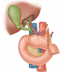
Gray's: Pancreas Anatomy

CC:

Yellow eyes

HPI:

51yo AA male with hx DM, HTN, sarcoidosis presents with yellowing of eyes and full-body itching x3wks. This was associated with dark urine and loose, light-brown stools. He denies N/V, abdominal pain, PO intolerance, F/C, recent travel, weight loss. He states that this has not occurred in the past, and he does not have any prior history of post-prandial abdominal pain.

PMH:

  • DM
  • HTN
  • Sarcoidosis

 PSH:

  • None

FH:

  • No GI malignancy

 SHx:

  • No tobacco or drug use, 10 years of 10 drinks/wk stopped 1yr ago

Meds:

  • lisinopril 20mg p.o. daily
  • pioglitazone 15mg p.o. daily
  • sitagliptin 100mg p.o. daily
  • lansoprazole 15mg p.o. daily

Allergies:

  • Vicodin (rash)

Physical Exam:

VS: T 97.9 HR 102 RR 12 BP 128/68 O2 99% RA
Gen: WA, NAD
HEENT: Marked scleral icterus, PERRL, yellowing of posterior oropharynx and floor of mouth, MMM, no cervical lymphadenopathy
CV: RRR, S1/S2 normal, no murmurs
Lungs: CTAB with good air movement
Abd: Obese, +BS, soft, NT/ND, no rebound/guarding, no palpable organomegaly, negative Murphy
GU: No inguinal lymphadenopathy
Ext: Warm, well-perfused, no LE edema, peripheral pulses 2+
Skin: No visible skin lesions
Neuro: AAOx3

Labs:

  • CBC: 16/12.4/35.1/281
  • LFT: AST 281, ALT 302, AP 264, T.bili 22.1, D.bili 16.8

Studies:

  • RUQ US: Biliary sludge, no stones, no GBW thickening, no pericholycystic fluid
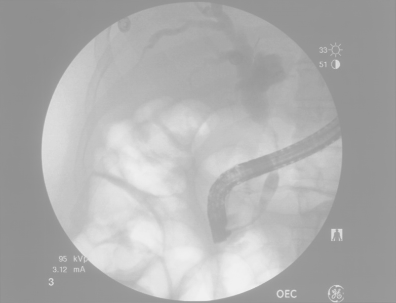
  • ERCP: 3cm stricture of distal CBD, biopsies taken

Assessment/Plan:

51AAM w/DM, HTN, sarcoidosis with 3wks painless jaundice. Obstructive pattern along with only modest elevation of liver enzymes suggests the obstruction is likely extrahepatic which was supported by ERCP finding of a distal CBD stricture. Patient has no history of prior instrumentation to cause iatrogenic stricture, and while sarcoidosis is associated with cholestatic complications (portal granulomas), pathology from biopsy showed papillary adenocarcinoma. The patient was scheduled for surgery with a plan for initial laparoscopic survey of the abdomen followed by Whipple if no evidence of widespread disease.

Imaging:

ERCP

ERCP

3cm stricture of distal CBD

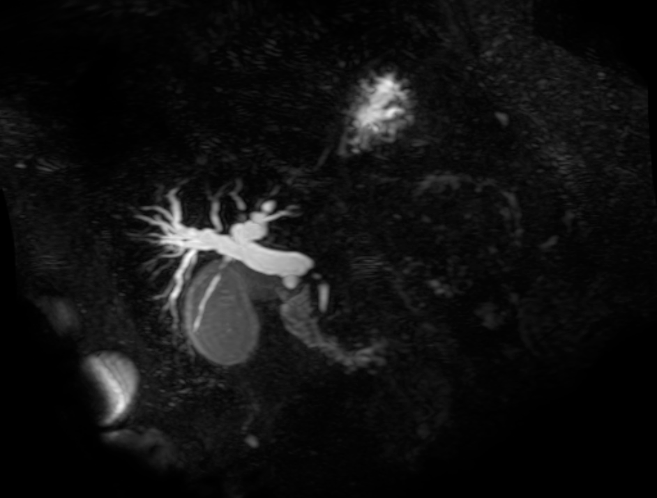
MRCP

MRCP

Filling defect in the common bile duct with marked dilatation of the common duct and intrahepatic ducts.
Findings may reflect an intraluminal mass or stone.

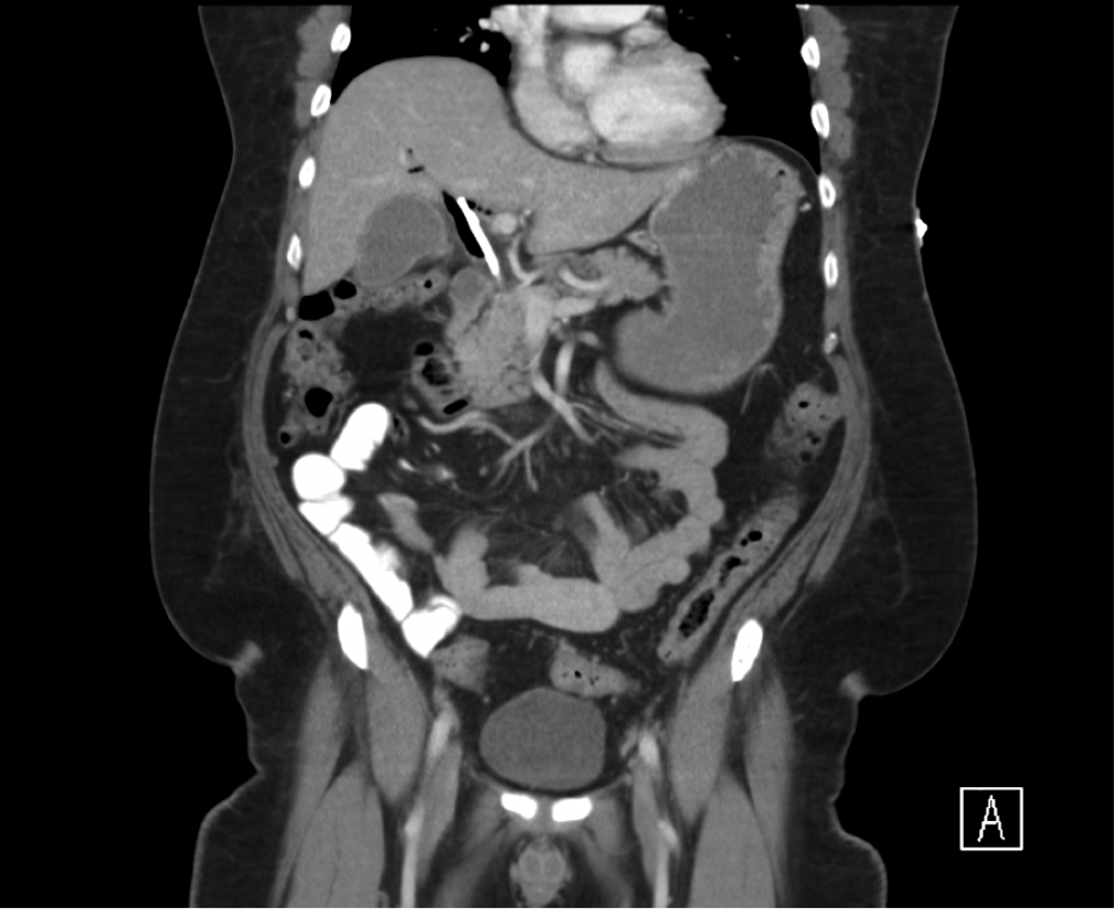
CT Abdomen/Pelvis

CT Abdomen/Pelvis

Common bile duct stent present
Expected air in the intrahepatic biliary tree and mild biliary ductal dilatation

Differential Diagnosis of Hyperbilirubinemia: 1, 2

A System for Hyperbilirubinemia

Evaluation of Hyperbilirubinemia: 3

Evaluation of Hyperbilirubinemia

References:

  1. Heathcote, E. J. (2007). Diagnosis and Management of Cholestatic Liver Disease. Clinical Gastroenterology and Hepatology, 5(7), 776–782. doi:10.1016/j.cgh.2007.05.008
  2. Hirschfield, G. M., Heathcote, E. J., & Gershwin, M. E. (2010). Pathogenesis of cholestatic liver disease and therapeutic approaches. Gastroenterology, 139(5), 1481–1496. doi:10.1053/j.gastro.2010.09.004
  3. McGill, J. M., & Kwiatkowski, A. P. (1998). Cholestatic liver diseases in adults. The American Journal of Gastroenterology, 93(5), 684–691. doi:10.1111/j.1572-0241.1998.206_a.x

Nausea and Vomiting

Neurologic pathways involved in pathogenesis of nausea and vomiting

HPI:

57yo male with a history of HTN, DM, and MI s/p stent in 2011 presenting with nausea/vomiting and hypotension. The patient had one episode of non-bloody, non-bilious emesis approximately 6 hours ago. He felt unwell so a friend checked his blood pressure which was found to be 75/50, prompting a visit to this emergency department.
The patient’s emesis came 2 hours following a normal meal (frozen dinner), and was associated with chills/sweats but no abdominal pain. The patient had some associated shortness of breath (baselines), but no chest pain, arm or jaw pain, or palpitations.

He states that these symptoms are unlike what he experienced during his MI. He reported no change in bowel or urinary habits.

PMH:

  • HTN
  • DM
  • CAD
  • MI
  • Hyperlipidemia

 PSH:

  • Stent placement (2011)
  • Right knee neuroma excision (2012)

FH:

  • Non-contributory

 SHx:

  • No current t/e/d
  • 80 pack-year smoking history

Meds:

  • carvedilol 6.25mg p.o. b.i.d.
  • metformin 1000mg p.o. b.i.d.
  • atorvastatin 20mg p.o. daily
  • aspirin 81mg p.o. daily

Allergies:

  • NKDA

Physical Exam:

VS: T 98.4 HR 65 RR 17 BP 96/56 O2 95% 2L NC
Gen: No acute distress, speaking in complete sentences
HEENT: PERRL, MMM no lesions, no cervical lymphadenopathy
CV: RRR, normal S1/S2, no murmurs, no extra heart sounds, no jugular venous distension
Lungs: CTAB, no crackles
Abd: +BS, soft, NT/ND, no rebound/guarding, no organomegaly
Ext: Warm, well-perfused, peripheral pulses equal b/l, no LE edema
Neuro: AAOx3

Labs:

  • EKG: normal sinus rhythm, anterior lead q-waves suggestive of old infarct, no T/ST changes
  • Troponin: <0.01
  • CBC: 7.4/15.5/47/228
  • BMP: 139/5.1/107/26/8/1.19/112 (baseline creatinine 1.06 in 2/2013)

Studies:

  • CXR: no effusion, no cardiomegaly, no focal consolidation
  • Bedside US: normal cardiac wall motion, estimated EF 40-45%, retrohepatic IVC collapses with respiration

Assessment/Plan:

57M hx HTN, DM, MI s/p stent presenting with nausea/vomiting x1 and hypotension. The patient’s symptoms and history were concerning for acute myocardial infarction; however, early EKG and troponins were reassuring. Additionally, the absence of characteristic physical findings that would be associated with an acute MI causing cardiogenic shock (elevated JVP, extra heart sounds, pulmonary crackles) were not present. Evidence of end-organ damage was also absent.

Other potential causes for nausea/vomiting include SBO, however, the patient reported normal BM’s and has no history of abdominal surgery. Though occurring after a meal, a single episode of emesis without associated abdominal pain lowers suspicion for biliary disease. This patient’s emesis is most likely due to acute gastroenteritis.

Given the evidence of hypovolemia on bedside ultrasound, the patient was bolused with a total of 1.5L NS and noted symptomatic improvement as well recovery of blood pressure.

Differential Diagnosis of Nausea/Vomiting: 1, 2

A System for Nausea/Vomiting

Pathophysiology: 3, 4, 5

  • Nausea: Sensation associated with increased gastrointestinal motility (tachygastria).
  • Vomiting:
    • Chemoreceptor trigger zone (area postrema of 4th ventricle): sensitive to drugs/toxins (emetics, radiation), neurotransmitters. Located outside BBB.
    • Nucleus tractus solitaries (medulla): pattern generator for vomiting, receives vagal input from GI tract and nociceptive stimuli from peripheral nervous system – transmits to hypothalamus, limbic system and cortex. Stimulated by tickling the back of the throat, gastric distention, and vestibular input.

Important history/physical associations: 4

  • Abdominal pain: suggests organic disease, affected organ dependent on location of pain. (See figure)
  • Abdominal distension: suggests bowel obstruction.
  • Heartburn: suggests GERD.
  • Vertigo/nystagmus: suggests vestibular etiology.
  • Positional/projectile: suggests neurogenic etiology.

Differential Diagnosis of Abdominal Pain By Location:

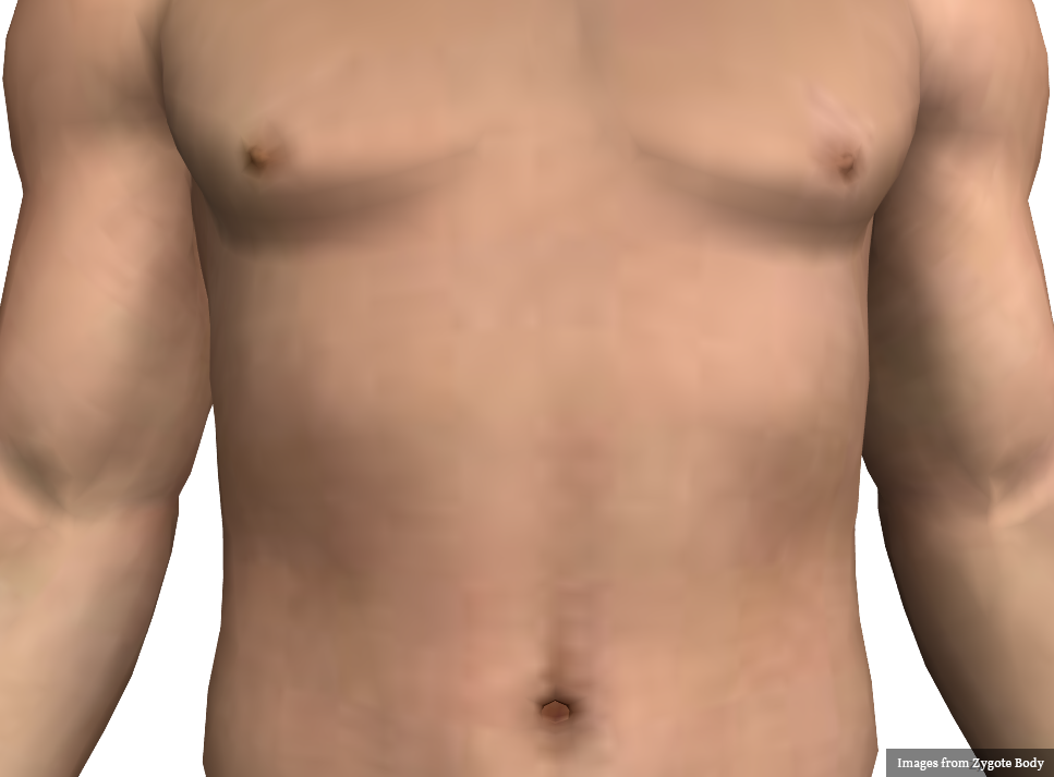
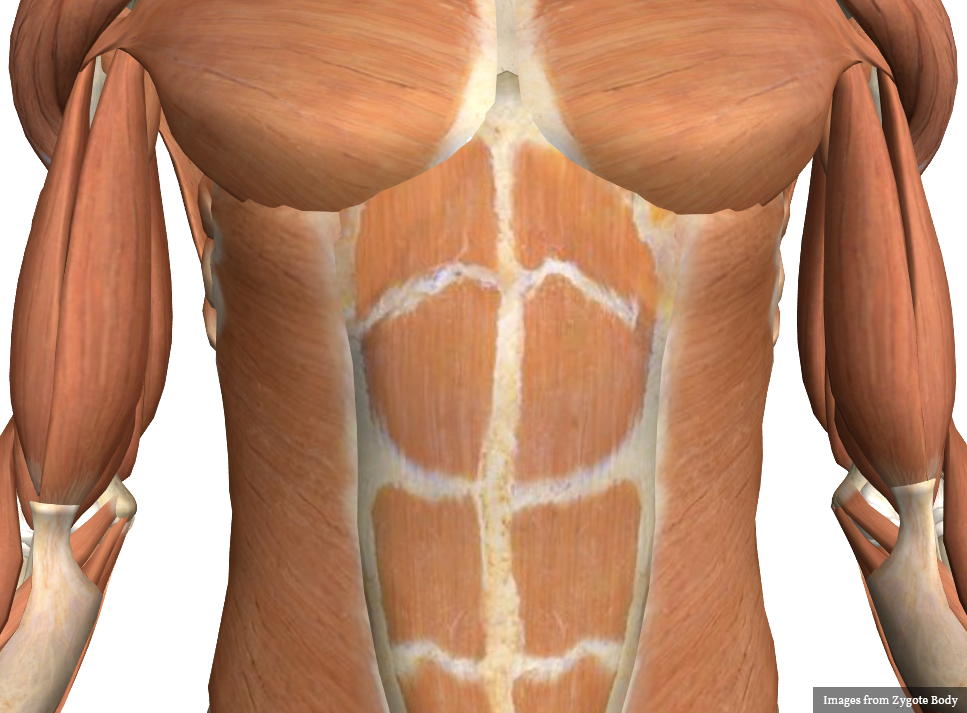
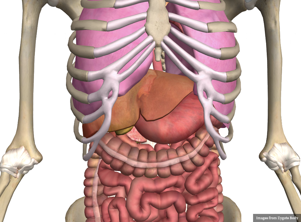
Abdominal Pain by Location

References:

  1. Scorza, K., Williams, A., Phillips, J. D., & Shaw, J. (2007). Evaluation of nausea and vomiting. American family physician, 76(1), 76–84.
  2. Bork S, Ditkoff J, Hang BS. Chapter 75. Nausea and Vomiting. In: Tintinalli JE, Stapczynski JS, Cline DM, Ma OJ, Cydulka RK, Meckler GD, eds. Tintinalli’s Emergency Medicine: A Comprehensive Study Guide. 7th ed. New York: McGraw-Hill; 2011. http://www.accessmedicine.com/content.aspx?aID=6360091. Accessed June 15, 2013.
  3. Koch, K. L., Stern, R. M., Vasey, M. W., Seaton, J. F., Demers, L. M., & Harrison, T. S. (1990). Neuroendocrine and gastric myoelectrical responses to illusory self-motion in humans. The American journal of physiology, 258(2 Pt 1), E304–10.
  4. Longstreth, G. F. Approach to the adult with nausea and vomiting. In: UpToDate, Basow, DS (Ed), UpToDate, Waltham, MA, 2013.
  5. Costanzo, L. (2011). Physiology. Philadelphia: Wolters Kluwer Health/Lippincott Williams & Wilkins.
  6. Patanwala, A. E., Amini, R., Hays, D. P., & Rosen, P. (2010). Antiemetic therapy for nausea and vomiting in the emergency department. The Journal of emergency medicine, 39(3), 330–336. doi:10.1016/j.jemermed.2009.08.060