Hypoglycemia

Brief HPI:

A 40 year-old male with a history of diabetes presents after accidental insulin overdose. He reports inadvertently administering 50 units of rapid-acting insulin when intending to take his long-acting insulin 25 minutes prior to presentation. He is currently asymptomatic, has a normal physical examination and his point-of-care glucose is 65mg/dL.

ED Course

The patient’s insulin regimen was clarified:

  • Insulin glargine 50 units SQ once daily
  • Insulin aspart 1 unit per 5g carbohydrates SQ before meals

The patient administered an insulin dose sufficient to dispose of 250g of carbohydrates with an impending peak action within minutes. The required dose of parenteral dextrose would require potentially-toxic1,2 solution concentrations (one liter of D25 or 10 ampules of D50) and there was insufficient time for central venous catheter placement. Moreover, the patient was awake and asymptomatic – able to tolerate oral intake. Juice boxes containing 15g of carbohydrates each were located and the patient drank 16-17 boxes while undergoing serial glucose measurements. His blood glucose ranged between 70mg/dL and 150mg/dL and he was discharged after an uneventful 6-hour observation.

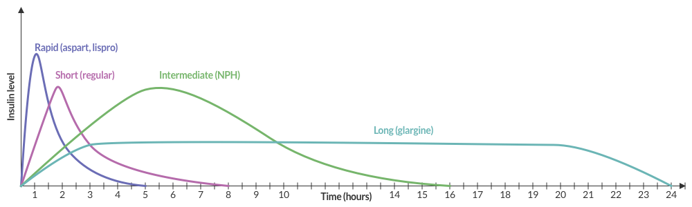
Insulin Pharmacokinetics3

Graph of insulin pharmacokinetics

Insulin and Carbohydrates

The effects of both insulin and carbohydrate ingestion on blood glucose are individualized. If known, the patient’s insulin:carbohydrate ratio can be used to anticipate the effect of overdose. Alternatively, their mealtime dose and carbohydrate allowance can be used.

Very broadly, one gram of carbohydrates increases blood glucose by 3-5mg/dL and one unit of insulin disposes of 10 grams of carbohydrates (or reduces blood glucose by 30-50mg/dL).4

Symptoms

  • Autonomic: tremor, palpitations, anxiety, diaphoresis
  • Neuroglycopenic: cognitive impairment, psychomotor, seizure, coma

Diagnosis

  • Serum glucose <60mg/dL
  • Generally symptomatic at <55mg/dL though threshold is variable depending on chronicity
  • Whipple Triad:
    • Symptoms suggestive of hypoglycemia
    • Low glucose
    • Resolution of symptoms after administration of glucose

Differential Diagnosis of Hypoglycemia

Differential Diagnosis of Hypoglycemia

Common Anti-hyperglycemic Drugs and Pharmacology

Drug Pharmacology
Onset Peak Duration
Rapid-acting insulin

  • Aspart (Novolog)
  • Lispro (Humalog)
15-30min 1-2h 3-5h
Short-acting insulin

  • Regular
30-60min 2-4h 6-10h
Intermediate-acting insulin

  • NPH
1-3h 4-12h 18-24h
Long-acting insulin

  • Glargine (Lantus)
2-4h None 24h
Sulfonylurea

  • Glimepiride
  • Glipizide (Glucotrol)
  • Glyburide (Glycron, Micronase)
2-6h 12-24h

Evaluation of Hypoglycemia

Patients with known diabetes who are not systemically ill and can identify a clear precipitant, no extensive workup is required. In severely ill patients, consider:

  • BMP
  • LFT
  • EtOH
  • Infectious workup: CXR, UA, urine and blood cultures
  • ECG, troponin
  • Other studies: insulin, C-peptide, pro-insulin, glucagon, growth hormone, cortisol, B-OH, insulin antibodies

Management and Monitoring

An algorithm for the management and monitoring of hypoglycemia

Special thanks to Dr. Ryan Pedigo, Associate Residency Program Director at Harbor-UCLA Medical Center for his case submission and content review.
Twitter

References:

  1. Kuwahara T, Asanami S, Kubo S. Experimental infusion phlebitis: tolerance osmolality of peripheral venous endothelial cells. Nutrition. 1998;14(6):496-501.
  2. Wiegand R, Brown J. Hyaluronidase for the management of dextrose extravasation. Am J Emerg Med. 2010;28(2):257.e1-2.
  3. Hirsch IB. Insulin analogues. N Engl J Med. 2005;352(2):174-183.
  4. Your Insulin Therapy. (2004). Am Fam Physician, 70(3), 511-512.
  5. Self, W. H., & McNaughton, C. D. (2013). Hypoglycemia. In Emergency Medicine (2nd ed., pp. 1379-1390). Elsevier.
  6. Service, FJ. Hypoglycemia in adults: Clinical manifestations, definition, and causes. In: UpToDate, Post TW (Ed), UpToDate, Waltham, MA. (Accessed on March 18, 2016.)
  7. Service FJ. Hypoglycemic disorders. N Engl J Med. 1995;332(17):1144–1152. doi:10.1056/NEJM199504273321707.
  8. Krinsley JS, Grover A. Severe hypoglycemia in critically ill patients: risk factors and outcomes. Critical Care Medicine. 2007;35(10):2262–2267. doi:10.1097/01.CCM.0000282073.98414.4B.
  9. Lacherade J-C, Jacqueminet S, Preiser J-C. An overview of hypoglycemia in the critically ill. J Diabetes Sci Technol. 2009;3(6):1242–1249.

Hyperammonemia

Brief H&P

A 38 year-old male with unknown medical history is brought to the emergency department by EMS with agitation and bizarre behavior. According to prehospital report, the patient was acting erratically – shouting incomprehensibly in the middle of a busy street with possible associated seizure activity.

On evaluation, the patient was found to be tachycardic, hypertensive, and markedly agitated. Physical examination with a focus on toxidromes was notable for the presence of rotary nystagmus suggestive of hallucinogen including phencyclidine toxicity. The patient required pharmacologic sedation to allow for a broad evaluation of altered mental status.

ED Course

The patient’s workup including core temperature, head imaging and laboratory tests (including AST/ALT, albumin, INR) were unremarkable with the exception of an ammonia level of 142 umol/L (normal range 16-53), slightly elevated CK, and urine toxicology screen with multiple positive agents. Over the course of several hours in the emergency department, the patient’s mental status gradually improved reaching normal level of alertness and orientation with normal neurological examination. He acknowledged PCP use as well as a prior history of seizures (possibly related to ethanol withdrawal) without routine anti-epileptic drug use. He denied known history of liver disease.

The patient’s hyperammonemia was attributed to a hypercatabolic state secondary to phencyclidine-induced agitation with possible seizure. He was discharged with resources for assistance with substance cessation.

An Algorithm for the Differential Diagnosis of Hyperammonemia:

Algorithm for the Differential Diagnosis of Hyperammonemia

References:

  1. Kalra A, Norvell JP. Cause for Confusion: Noncirrhotic Hyperammonemic Encephalopathy. Clin Liver Dis. 2020;15(6):223-227. doi:10.1002/cld.929
  2. Mallet M, Weiss N, Thabut D, Rudler M. Why and when to measure ammonemia in cirrhosis? Clin Res Hepatol Gas. 2018;42(6):505-511. doi:10.1016/j.clinre.2018.01.004
  3. Hassan AAI, Ibrahim W, Subahi A, Mohamed A. ‘All that glitters is not gold’: when hyperammonaemia is not from hepatic aetiology. Bmj Case Reports. 2017;2017:bcr-2017-219441. doi:10.1136/bcr-2017-219441
  4. Odigwe CC, Khatiwada B, Holbrook C, et al. Noncirrhotic Hyperammonemia Causing Relapsing Altered Mental Status. Bayl Univ Medical Cent Proc. 2017;28(4):472-474. doi:10.1080/08998280.2015.11929312
  5. Upadhyay R, Bleck TP, Busl KM. Hyperammonemia: What Urea-lly Need to Know: Case Report of Severe Noncirrhotic Hyperammonemic Encephalopathy and Review of the Literature. Case Reports Medicine. 2016;2016:1-10. doi:10.1155/2016/8512721
  6. Walker V. Severe hyperammonaemia in adults not explained by liver disease. Ann Clin Biochem. 2011;49(3):214-228. doi:10.1258/acb.2011.011206
  7. Laish I, Ari ZB. Noncirrhotic hyperammonaemic encephalopathy. Liver Int. 2011;31(9):1259-1270. doi:10.1111/j.1478-3231.2011.02550.x
  8. LaBuzetta JN, Yao JZ, Bourque DL, Zivin J. Adult Nonhepatic Hyperammonemia: A Case Report and Differential Diagnosis. Am J Medicine. 2010;123(10):885-891. doi:10.1016/j.amjmed.2010.02.029
  9. Clay AS, Hainline BE. Hyperammonemia in the ICU. Chest. 2007;132(4):1368-1378. doi:10.1378/chest.06-2940
  10. Weng T-I, Shih FF-Y, Chen W-J. Unusual causes of hyperammonemia in the ED. Am J Emerg Medicine. 2004;22(2):105-107. doi:10.1016/j.ajem.2003.12.011
  11. Hawkes ND, Thomas GAO, Jurewicz A, et al. Non-hepatic hyperammonaemia: an important, potentially reversible cause of encephalopathy. Postgrad Med J. 2001;77(913):717. doi:10.1136/pmj.77.913.717

Hyponatremia

HPI:

A 62 year-old male with a history of hepatitis C cirrhosis complicated by hepatocellular carcinoma s/p radiofrequency ablation presenting after referral from hepatology clinic for hyponatremia. One week ago, the patient developed abdominal distension and shortness of breath that resolved after large-volume paracentesis and was started on furosemide 40mg p.o. daily and aldactone 100mg p.o. daily. After initiating diuretics, the patient noted worsening lower extremity edema, and increased thirst/fluid intake.

He reports two days of fatigue and intermittent confusion supported by family members who reported slowed speech. He otherwise denies abdominal pain, distension, nausea/vomiting, diarrhea/constipation, chest pain or shortness of breath. In the ED, the patient received 1L NS bolus.

PMH:

  • Hepatitis C cirrhosis c/b HCC s/p RFA
  • Rheumatoid arthritis

PSH:

  • None

Family History:

  • Non-contributory.

Social History:

  • Lives with partner, denies current or prior t/e/d abuse
  • HepC contracted from blood transfusions

Medications:

  • Furosemide 40mg p.o. daily
  • Spironolactone 100mg p.o. daily
  • Rifaximin 550mg p.o. b.i.d.

Allergies:

  • NKDA

Physical Exam:

VS: T 98.2 HR 80 RR 14 BP 95/70 O2 98% RA
Gen: Elderly male in no acute distress, alert and answering questions appropriately.
HEENT: NC/AT, PERRL, EOMI, faint scleral icterus, MMM.
CV: RRR, normal S1/S2, no murmurs. JVP 8cm.
Lungs: Faint basilar crackles on bilateral lung bases.
Abd: Normoactive bowel sounds, mildly distended but non-tender, without rebound/guarding.
Ext: 2+ pitting edema in lower extremities to knees bilaterally. 2+ peripheral pulses, warm and well perfused.
Neuro: AAOx3. CN II-XII intact. No asterixis. Normal gait. Normal FTN/RAM.

Labs/Studies:

  • BMP (admission): 112/5.6/88/22/28/1.1/97
  • BMP (+10h): 118/5.4/93/23/26/1.0/133
  • sOsm: 264
  • Urine: Na <20, K 26, Osm 453
  • BNP: 40
  • AST/ALT/AP/TB/Alb: 74/57/91/2.4/2.2

Assessment/Plan:

62M with HepC cirrhosis, with e/o decompensation (new-onset ascites) and hyponatremia.

  1. #Hyponatremia: Sodium 114, likely chronic, patient currently asymptomatic without concerning findings on neurological exam. Clinical findings suggestive of hypervolemic hyponatremia 2/2 decompensated cirrhosis resulting in decreased effective arterial blood volume and volume retention. However, the recent initiation of diuretics, mild AKI and early response to isotonic fluids in the ED suggests possible hypovolemic component.
    • 1L fluid restriction
    • q.4.h. sodium check, goal increase of 8mEq per 24h
    • hold diuretics
  2. #Hyperkalemia: Potassium 5.6, asymptomatic, AKI vs. medication-induced (aldactone). Continue monitoring.
  3. #AKI: Elevated creatinine 1.1 from baseline 0.7. Likely pre-renal given recent initiation of diuretics. Consider hepatorenal syndrome given decompensated cirrhosis. Follow-up repeat creatinine after 1L NS bolus in ED.
  4. #Hepatitis C: decompensated with new-onset ascites. No e/o encephalopathy, continue home rifaximin.

Physiology of Hyponatremia: 1,2,3,4

Physiology of Hyponatremia

Differential Diagnosis of Hyponatremia: 5

Differential Diagnosis of Hyponatremia

Evaluation of Hyponatremia: 2

  1. Identification of onset (acute vs. chronic)
  2. Presence of symptoms (HA, nausea, confusion, seizures)
  3. Assessment of volume status (edema, JVD, skin turgor, postural BP)
  4. Medical history (cardiac, liver, renal disease), drug history

References:

  1. Freda BJ, Davidson MB, Hall PM. Evaluation of hyponatremia: a little physiology goes a long way. Cleve Clin J Med. 2004;71(8):639–650.
  2. Biswas M, Davies JS. Hyponatraemia in clinical practice. Postgrad Med J. 2007;83(980):373–378. doi:10.1136/pgmj.2006.056515.
  3. Adrogué HJ, Madias NE. Hyponatremia. N. Engl. J. Med. 2000;342(21):1581–1589. doi:10.1056/NEJM200005253422107.
  4. Marx JA, Hockberger RS, Walls RM, Adams JG. Rosen’s emergency medicine: concepts and clinical practice. 2010;1.
  5. Milionis HJ, Liamis GL, Elisaf MS. The hyponatremic patient: a systematic approach to laboratory diagnosis. CMAJ. 2002;166(8):1056–1062.

Hypocalcemia

Brief H&P:

34M with a history of HTN, polysubstance abuse, presenting with muscle cramps. He reported onset of diffuse muscle cramping 1-hour prior to presentation while showering. Symptoms involved bilateral upper and lower extremities and resolved spontaneously.

On initial evaluation, the patient was tachycardic and hypertensive. Examination was notable for tremors in bilateral upper extremities with outstretched hands, as well as of extended tongue. Other notable findings included spasm of the upper extremity during blood pressure measurement, hyperreflexia and clonus.

Laboratory evaluation was notable for normal total calcium level, low ionized calcium level, primary respiratory alkalosis, and elevated anion gap metabolic acidosis.

The patient was treated with intravenous fluids, benzodiazepines for alcohol withdrawal, and calcium gluconate 4g IV and was admitted.

Calcium Homeostasis1

  • Fraction
    • 15% bound to anions (phosphate, lactate, citrate)
    • 40% bound to albumin
    • 45% free (regulated by PTH, Vit-D)
  • Conditions causing changes in total calcium (without affecting ionized calcium)
    • Low albumin causes hypocalcemia. Corrected = measured + [0.8 x (4-albumin)]
    • Elevated albumin causes hypercalcemia
    • Multiple myeloma causes hypercalcemia
  • Conditions causing changes in ionized calcium (without affecting total calcium)
    • Alkalemia causes increased ionized calcium binding to albumin and decreases ionized calcium levels
    • Hyperphosphatemia causes increased ionized calcium binding to phosphate and decreases ionized calcium levels
    • Hyperparathyroidism causes decreased ionized calcium binding to albumin and increases ionized calcium levels

Causes of Hypocalcemia1,2,3

Algorithm for the Evaluation of Hypocalcemia

Symptoms1

Acute Chronic

Neuromuscular

  • Paresthesia
  • Tetany
  • Carpopedal spasm
  • Trousseau
  • Chvostek
  • Seizure
  • Laryngospasm

Cardiac

  • QT prolongation
  • Hypotension
  • Heart failure
  • Arrhythmia

CNS

  • Basal ganglia calcifications
  • EPS
  • Parkinsonism
  • Dementia

Ophthalmologic

  • Cataracts

Management

  • Severe (symptomatic, QT prolongation)
    • Calcium gluconate 1-2g IV in 50mL of D5W over 10-20min followed by slow infusion of additional 2g over 2 hours.
  • Asymptomatic
    • Calcium gluconate 1g PO q6h
    • Calcitriol 0.2mcg PO BID

References:

  1. Yu, AS. Relation between total and ionized serum calcium concentrations. In: UpToDate, Post TW (Ed), UpToDate, Waltham, MA. (Accessed on October 6th, 2016.)
  2. Cooper MS, Gittoes NJL. Diagnosis and management of hypocalcaemia. BMJ. 2008;336(7656):1298-1302. doi:10.1136/bmj.39582.589433.BE.
  3. Hannan FM, Thakker RV. Investigating hypocalcaemia. BMJ. 2013;346(may09 1):f2213-f2213. doi:10.1136/bmj.f2213.

Oncologic Emergencies

Overview

  •  Complications
    • Airway obstruction
    • PNA
    • Pleural effusion
    • Pericardial effusion
    • VTE
    • SVC syndrome
      • Symptoms: dyspnea (airway edema), chest fullness, blurred vision, headache (increased ICP)
    • Massive hemoptysis
      • Management: ETT (large-bore for bronschoscopy), affected side down
  • Brain Metastases
    • Cancers: melanoma, lung, breast, colorectal
    • Management: dexamethasone 10mg IV load, elevated HOB, hypertonic saline or mannitol, prophylactic anti-eplipetics
  • Meningitis
    • Pathogens: Listeria (ampicillin), Cryptococcus (amphotericin)
    • Evaluation: CSF sampling with cytology (diagnose leptomeningeal metastases)

Metabolic Disturbances

  • Hypercalcemia
    • Cancers: MM, RCC, lymphoma, bone metastases (breast, lung, prostate)
    • Mechanism: metastatic destruction, PTH-RP, tumor calcitriol
    • Prognosis: 50% 30-day mortality
    • Symptoms
      • Chronic: anorexia, nausea/vomiting, constipation, fatigue, memory loss
      • Acute: CNS (lethargy, somnolence)
    • Findings
      • Calcium: >13.0mg/dL
      • ECG: QT shortening
    • Treatment
      • Mild: IVF
      • Severe: IVF, loop diuretics, bisophosphanate (pamidronate 90mg IV infused over 4 hours), consider calcitriol, consider hemodialysis if cannot tolerate fluids or unlikely to respond to diuretics
  • Hyponatremia
    • Cancers: lung (small-cell), pancreatic, ovarian, lymphoma, thymoma, CNS
    • Mechanism: SIADH
    • Symptoms: muscle twitching, seizure, coma
    • Management: fluid restriction, if seizing administer 3% hypertonic saline at 100cc/hr until resolution
  • Hypernatremia
    • Mechanism: decreased intake, increased GI losses from chemotherapy
    • Management: cautious fluid resuscitation
  • Tumor Lysis Syndrome (TLS)
    • Cancers: hematologic, rapid-growth solid tumors
    • Mechanism: release of intracellular contents (uric acid, K, PO4, Ca)
    • Timing: 1-4 days after therapy (chemo, radiation)
    • Diagnosis
      • Uric acid >8mg/dL
      • Potassium >6mEq/L
      • Calcium <7mg/dL
      • PO4 >4.5mg/dL
      • Acute kidney injury
    • Management
      • IVF, allopurinol, rasburicase, urinary alkalinization
      • Consider hemodialysis if volume overloaded

Localized Complications

  • Musculoskeletal Complications
    • Spinal cord compression
      • Cancers: prostate, breast, lung, RCC, non-Hodgkin lymphoma, MM (5-10% of all cancer patients)
      • Sites: thoracic (60%), lumbosacral (30%), cervical (10%)
      • Symptoms: pain (worse lying flat, cough/sneeze, heavy lifting)
      • Evaluation: MRI (se 93%, sp 97%)
      • Management: dexamethasone 10mg IV load, 4mg q6h, neurosurgical consultation, radiation oncology consultation
    • Pathologic fracture
      • Features: sudden onset, low-force mechanism
  • Therapy Complications
    • Neutropenic fever
      • Definition: ANC <500 or ANC <1000 with expected nadir <500 (nadir typically occurs 5-10d after chemotherapy) with Tmax >38.3°C or >38.0°C for >1h
      • Examination: subtle signs of infection, thorough examination is critical (skin, catheter, perineum)
      • Treatment: carbapenem monotherapy, vancomycin if indwelling catheter, oncology consultation for colony stimulating factors
    • Chemotherapy-induced vomiting
      • Management: ondansetron with dexamethasone, consider NK-1 antagonist (aprepitant)

Hematologic Malignancies

  • Acute leukemia
    • Signs/Symptoms: leukopenia (infection), anemia (weakness/fatigue), thrombocytopenia (bleeding)
    • Diagnosis: >5% blasts
  • Thrombocytopenia
    • Management
      • No bleeding, goal >10,000
      • Fever, coagulopathy, hyperleukoctosis, goal >20,000
      • One unit of platelets increases count by 5,000
  • Hyperleukocytosis
    • Definition: WBC > 50-100k
    • Complications: microvascular congestion (pulmonary, cerebral, coronary)
    • Symptoms
      • CNS: confusion, somnolence, coma
      • Pulmonary: dyspnea, respiratory alkalosis
    • Management: cytoreduction (induction chemotherapy, increased risk TLS)
  • Hyperviscosity
    • Cancer: macroglobulinemia, MM
    • Symptoms: epistaxis, purpura, GIB, neuro deficits
    • Diagnosis: serum viscosity > 1.4-1.8
    • Management: emergent plasmapheresis
  • Polycythemia
    • Diagnosis: Hb >17
    • Differential: dehydration, hypoxia, smoking, altitude
    • Symptoms: HA, vertigo, angina, claudication, pruritus (after showering)
    • Complications: thrombosis (stroke), bleeding
    • Management: emergent phlebotomy (500cc if otherwise healthy)
  • Thrombocytosis
    • Diagnosis: platelet >1,000,000
    • Symptoms: vasomotor (HA, lightheadedness, syncope, chest pain, paresthesias)
    • Management: low-dose aspirin

Acid-Base Disturbances

Method

  • Primary disturbance (acidemia/alkalemia)
  • Primary process (metabolic/respiratory)
  • Presence of mixed disorder
    • Increase PCO2 of 10, increases HCO3 by 1 (acute) or 3 (chronic)
    • Decreased PCO2 of 10, decreases HCO3 by 2 (acute) or 5 (chronic)
    • Increase HCO3 of 1, increases PCO2 by 0.7
    • Decreased HCO3, add 15, result should equal PCO2 and number after decimal of pH
  • Anion gap

Causes

  • Anion Gap
    • Methanol
    • Uremia
    • DKA/AKA
    • Paraldehyde, propylene glycol
    • INH
    • Lactate
    • Ethylene glycol
    • Salicylate
  • Non-Anion Gap
    • Fistulae
    • Ureteral fistulae
    • Saline
    • Diarrhea
    • Carbonic anhydrase inhibitors
    • Spironolactone
    • RTA
  • Metabolic Alkalosis
    • Vomiting
    • Volume depletion
    • Diuretics
    • Steroids
  • Respiratory Acidosis
    • CNS lesion
    • Myopathies
    • Chest wall abnormalities
    • Obstructive lung disease
  • Respiratory Alkalosis
    • Anxiety
    • Fever
    • Hyperthyroidism
    • Hypoxia
    • Sympathomimetic

See Also

Altered Mental Status

Components of Consciousness

Components of Consciousness

Causes of Altered Mental Status

Causes of Altered Mental Status

History

Rate of onset
Abrupt: CNS
Gradual: Systemic

Physical Examination

  • Vital Signs

    • Blood Pressure: low (shock), high (SAH, stroke, ICP)
    • Heart Rate: low (medication overdose, conduction block), high (hypovolemia, infection, anemia, thyrotoxicosis, drug/toxin)
    • Temperature: low/high (infection, drug/toxin, environmental)
    • Respiratory Rate: low/high (CNS, drug/toxin, metabolic derangement)
  • Eyes

    • Unilateral dilation: CNS/structural cause
    • Papilledema: ICP
    • EOM: cranial nerve dysfunction
    • Oculocephalic: brainstem function
  • Head: trauma
  • Mucous membranes: hydration, laceration
  • Neck: meningeal irritation
  • Pulmonary: respiratory effort
  • CV: murmur, arrhythmia, CO
  • Abdomen: pulsatile mass, sequelae of liver failure
  • Skin: rash, needle tracks

Labs

  • Glucose
  • ECG: arrhythmia, ischemia, electrolyte abnormalities
  • BMP: electrolytes, renal failure, anion gap
  • ABG: hypoxemia, hypercarbia
  • Urinalysis: infection, SG
  • Utox
  • CBC: leukocytosis, leukopenia, severe anemia, thrombocytopenia
  • Ammonia: hepatic encephalopathy
  • TFT: thyrotoxicosis, myxedema coma
  • CSF: meningitis, encephalitis

Imaging

  • CT head: Non-contrast sufficient to identify ICH. Use contrast if mass/infection suspected
  • CTA head/neck: If aneurysm, AVM, venous sinus thrombosis or vertebrobasilar insufficiency suspected
  • CXR: PNA

References

  1. Bassin, B., & Cooke, J. (2013). Depressed Consciousness and Coma. In Rosen’s Emergency Medicine – Concepts and Clinical Practice (8th ed., Vol. 1, pp. 142-150). Elsevier Health Sciences.

Lactic Acidosis

HPI:

59F with a reported history of congestive heart failure, presenting with intermittent chest discomfort for three days.

She characterized this discomfort as “heartburn”, describing a mid-epigastric burning sensation radiating up her neck, not associated with exertion, lasting 1-2 hours and resolving with antacids. The patient has poor exercise tolerance at baseline and for the past several years has been able to ambulate only short distances around her home, and states that these symptoms have been worsening in the past week. She denies chest pain on exertion, orthopnea or paroxysmal nocturnal dyspnea. She states that she was diagnosed with congestive heart failure five years ago, but was never prescribed medications.

On further questioning, the patient reports several weeks of mouth and lip pain which has limited oral intake, though no dysphagia to solids or liquids. She otherwise denies fevers/chills, abdominal pain, nausea/vomiting, cough, changes in urinary or bowel habits.

In the emergency department, the patient was noted to have an elevated serum troponin, though ECG showed no changes of acute ischemia/infarction.

PMH:

  • Congestive heart failure

PSH:

  • None

FH:

  • Mother with diabetes
  • Father with MI at age 65

SHx:

  • 4-5 drinks of alcohol/day
  • No tobacco or drug use

Meds:

  • None

Allergies:

NKDA

Physical Exam:

VS: T 37.4 HR 106 RR 18 BP 145/82 O2 100% RA
Gen: Morbidly obese female, lying in bed, in no acute respiratory distress, speaking in complete sentences.
HEENT: Dry, cracked lips, slightly erythematous, otherwise moist mucous membranes, poor dentition. Mild scleral icterus. No cervical lymphadenopathy.
CV: Rapid rate, regular rhythm, normal S1/S2, II/VI systolic ejection murmur at LUSB, no radiation appreciated. No jugular venous distension.
Lungs: Clear to auscultation in posterior lung fields bilaterally, no crackles appreciated.
Chest: Well-circumscribed erythematous patch in folds beneath left breast, no underlying fluctuance, no significant tenderness to palpation. On contralateral breast, some hyperpigmentation but no erythema.
Abdomen: Obese, non-tender, non-distended. Patch of erythema below pannus, mildly tender to palpation.
Ext: Bilateral lower extremities with marked edema and overlying scaly plaques, some slightly ulcerated weeping serous fluid. Peripheral pulses are difficult to palpate, capillary refill difficult to assess.

Labs/Studies:

  • CBC: 11.1/11.1/34.5/212 (MCV 114.2)
  • BMP: 140/4.5/97/20/10/1.14/64
  • Anion Gap: 23
  • LFT: AST: 73, ALT: 26, AP: 300, TB: 4.6, DB: 2.1, Alb: 3.0, INR 1.3
  • BNP: 158
  • Troponin: 1.284
Sinus tachycardia, LVH, secondary repolarization abnormalities

Sinus tachycardia, LVH, secondary repolarization abnormalities

Imaging:

CT Pulmonary Angiography:
No evidence of central pulmonary embolism, thoracic aortic dissection, or thoracic aortic aneurysm. Evaluation of the peripheral vessels is limited due to motion artifact. No focal consolidation or pneumothorax.

CT Abdomen/Pelvis non-contrast:
No evidence of intra-abdominal abscess or definite source of infection. Marked hepatic steatosis.

CT Lower Extremity non-contrast:
Diffuse circumferential subcutaneous edema involving both lower extremities from the level of the mid thighs distally through the feet. There are bilateral subcutaneous calcifications which are likely venous calcifications in the setting of chronic venous stasis disease. There is some overlying skin thickening.

TTE:
There is moderate concentric left ventricular hypertrophy with hyperdynamic LV wall motion. The Ejection Fraction estimate is >70%. Grade I/IV (mild) LV diastolic dysfunction. No hemodynamically significant valve abnormalities.

US Abdomen:
Hepatomegaly, echogenic liver suggesting fatty infiltration. Moderately blunted hepatic vein waveforms suggesting decreased hepatic parenchymal compliance.

Assessment/Plan:

The patient was admitted to the cardiology service for management of NSTEMI and evaluation of undiagnosed CHF. She was started on a heparin continuous infusion. In addition, a CT pulmonary angiogram was obtained to evaluate for pulmonary embolism as an explanation of her progressive dyspnea on exertion. No PE, consolidation or effusion was identified.

Despite the patient’s reported history of congestive heart failure, there was no evidence that her symptoms were a result of an acute exacerbation with only a mildly elevated BNP but no jugular venous distension or evidence of pulmonary edema. The patient’s significant lower extremity edema was more suggestive of chronic venous stasis.

One notable laboratory abnormality that was explored was her elevated anion gap metabolic acidosis. Studies submitted included serum lactate, salicylates, osmolarity, CK, and urinalysis for ketonuria. This evaluation was notable for an elevated serum lactate of 13.2mmol/L and an arterial blood gas that showed adequate respiratory compensation (and no A-a gradient). Given the patient’s modest leukocytosis (with neutrophil predominance), and tachycardia, the concern for sepsis was increased though the source remained unclear. Prominent possibilities included a skin and soft-tissue infection vs. less likely intra-abdominal source though the patient’s physical examination was not suggestive of a process that would produce such a substantial lactic acidosis. Blood cultures were drawn and the patient was started on empiric antibiotics for the suspected sources. In addition, the patient was cautiously volume resuscitated given her reported history of CHF while pending a transthoracic echocardiogram to evaluate cardiac function. Additional imaging including CT abdomen/pelvis and lower extremities was obtained (though without contrast due to the patient’s recent exposure), and no obvious source was identified.

Over the next two days, the patient’s serum lactate downtrended to normal range, as did the serum troponin. A transthoracic echocardiogram showed an LVEF >70% with mild concentric hypertrophy and diastolic dysfunction. Blood and urine cultures were without growth.

Additional issues managed during the hospitalization included elevated serum transaminases (AST > ALT), conjugated hyperbilirubinemia and evidence of decreased hepatic synthetic function with hypoalbuminemia and elevated INR. Given the patient’s history of EtOH use, as well as other corroborating findings including macrocytic anemia, hypomagnesemia, folate and B12 deficiency, this was attributed to alcoholic hepatitis (discriminant function <32). Infectious hepatitis serologies were negative. The patient was started on nutritional supplements. Finally, the patient persistently complained of lip and oral mucosal pain. Examination was without discrete lesions but some mucosal redness was identified. Despite poor dentition, there was no evidence of abscess and HSV/HIV testing was negative. This was thought to be stomatitis caused by her identified nutritional deficiencies.

Differential Diagnosis of Elevated Serum Lactate 1,2

Differential Diagnosis of Elevated Serum Lactate

Algorithm for Evaluation of Acidemia 3,4

Algorithm for Evaluation of Acidemia

Algorithm for Evaluation of Alkalemia 3,4

Algorithm for Evaluation of Alkalemia

References:

  1. Fall, P. J., & Szerlip, H. M. (2005). Lactic acidosis: from sour milk to septic shock. Journal of intensive care medicine, 20(5), 255–271. doi:10.1177/0885066605278644
  2. Luft, F. C. (2001). Lactic acidosis update for critical care clinicians. Journal of the American Society of Nephrology : JASN, 12 Suppl 17, S15–9.
  3. Ingelfinger, J. R., Berend, K., de Vries, A. P. J., & Gans, R. O. B. (2014). Physiological Approach to Assessment of Acid–Base Disturbances. The New England journal of medicine, 371(15), 1434–1445. doi:10.1056/NEJMra1003327
  4. Ingelfinger, J. R., & Seifter, J. L. (2014). Integration of Acid–Base and Electrolyte Disorders. The New England journal of medicine, 371(19), 1821–1831. doi:10.1056/NEJMra1215672

Electrolyte Abnormalities

Routine laboratory studies are common in the intensive care unit; abnormalities are even more common. Typically these studies include a chemistry panel (Chem 10). The differential diagnoses of the most frequent and clinically relevant electrolyte abnormalities are detailed below.

Differential Diagnosis and Evaluation of Hyponatremia

Differential Diagnosis and Evaluation of Hyponatremia

Differential Diagnosis and Evaluation of Hypernatremia

Differential Diagnosis and Evaluation of Hypernatremia

Differential Diagnosis and Evaluation of Hypokalemia

Differential Diagnosis and Evaluation of Hypokalemia

Differential Diagnosis of Hyperkalemia

Differential Diagnosis of Hyperkalemia

Differential Diagnosis of Hypo and Hypercalcemia

Differential Diagnosis of Hypo and Hypercalcemia

Differential Diagnosis of Hypo and Hypermagnesemia

Differential diagnosis of hypo and hypermagnesemia

Differential Diagnosis of Hypo and Hyperphosphatemia

Differential diagnosis of hypo and hyperphosphatemia

References:

  1. Marino, P. (2014). Marino’s the ICU book. Philadelphia: Wolters Kluwer Health/Lippincott Williams & Wilkins.
  2. Fulop, M. (1998). Algorithms for diagnosing some electrolyte disorders. American Journal of Emergency Medicine, 16(1), 76–84.
  3. WikEM: Hypokalemia
  4. WikEM: Hyponatremia

Hyperglycemic Crises

CC:

Blurred vision, numbness

HPI:

56 year-old male with a history of DM, questionable HTN presenting with blurred vision, numbness of fingertips/toes for 2wks. Associated symptoms include dry mouth, polydipsia/polyuria. He states that these symptoms coincide with elevated measurements of blood glucose at home (>500). He ran out of his diabetes medication (metformin) 8mo ago but states his BG was typically between 100-200 with diet/exercise until 2wks ago. He reports recent dietary indiscretions on a trip to Las Vegas.

He denies fevers/chills, CP/SOB, cough, abdominal pain, N/V, or dysuria.

PMH:

  • DM II
  • HTN

PSH:

None

FH:

Several maternal family members with DM.

SHx:

  • No tobacco/drug use
  • 5-6 alcoholic drinks/wk

Meds:

  • Metformin 500mg p.o. b.i.d.

Allergies:

NKDA

Physical Exam:

VS: T 37.8 HR 60 RR 14 BP 165/90 O2 99% RA
Gen: Well-appearing, no acute distress, obese
HEENT: PERRL, EOMI, optic discs sharp b/l, no abnormalities visualized
CV: RRR, normal S1/S2, no M/R/G, no additional heart sounds
Lungs: CTAB, no wheezes/crackles
Abd: +BS, soft, NT/ND, no rebound/guarding
Ext: Warm, well-perfused, 2+ pulses, no clubbing/cyanosis/edema
Neuro: AAOx3, CN II-XII intact

Labs/Studies:

  • BMP: 135/3.8/102/24/18/1.1/378
  • CBC: 7.4/14.1/42.0/403
  • UA: + glucose, – ketones

Assessment/Plan:

56M, hx DM with poor medication adherence presenting with vision changes and stocking/glove paresthesias for 2wks after reported dietary indiscretion found to be hyperglycemic. DKA/HHS unlikely given stable vital signs, normal metabolic panel with exception of isolated hyperglycemia (slight hyponatremia likely related to osmotic effect of elevated serum glucose). Also, no evidence of concerning precipitates for hyperglycemic crisis (no CP/SOB, no F/C, no cough, no abdominal pain, no change in mental status). Patient was discharged home with education on importance of medication adherence, refill of metformin, and follow-up with primary care physician for further management of DM and possible hypertension.

Evaluation of hyperglycemic crises in patients with diabetes:1,2

Evaluation of Hyperglycemic Crises in Patients with Diabetes

Key signs/symptoms of HHS/DKA:

  • Both: Polyuria, polydipsia, weight loss, hypovolemia (dry MM, skin turgor, tachycardia, hypotension)
  • DKA: Short course (<24h), N/V, diffuse abdominal pain, Kussmaul respirations
  • HHS: Longer course (days/weeks), altered mental status (lethargy, coma, seizure)

Admission Laboratory Data of Patients with HHS vs. DKA:1

DKA HHS
Glucose (mg/dl) 616 930
pH 7.12 7.30
3-β-hydroxybutyrate (mmol/l) 9.1 1.0
Serum osmolality 323 380
Delta gap (AG-12) 17 11
Na (mEq/l) 134 149
K (mEq/l) 4.5 3.9
Bicarbonate (mEq/l) 9 18

References:

  1. Kitabchi, A. E., Umpierrez, G. E., Miles, J. M., & Fisher, J. N. (2009). Hyperglycemic crises in adult patients with diabetes. Diabetes care, 32(7), 1335–1343. doi:10.2337/dc09-9032
  2. De Beer, K., Michael, S., Thacker, M., Wynne, E., Pattni, C., Gomm, M., Ball, C., et al. (2008). Diabetic ketoacidosis and hyperglycaemic hyperosmolar syndrome – clinical guidelines. Nursing in critical care, 13(1), 5–11. doi:10.1111/j.1478-5153.2007.00259.x
  3. Stoner, G. D. (2005). Hyperosmolar hyperglycemic state. American family physician, 71(9), 1723–1730.

Delirium

ID:

A 70 year-old female with a PMH of HTN, DM, hyperlipidemia and stage I breast cancer s/p lumpectomy with sentinel LN biopsy several years ago presented for elective surgery complicated by post-operative bleeding. She is now 4 days post-op and was found to be confused, somnolent and occasionally agitated.

HPI:

The patient could not be interviewed.

PE:

  • VS: Stable and within normal limits
  • General: unremarkable except for crackles in bilateral lung bases
  • MSE: only arouses to sternal rub and becomes agitated, moving all four extremities spontaneously and symmetrically.
  • Reflexes: corneal and gag reflexes present, suppresses eye movements with head turn, deep tendon reflexes 3+ throughout UE/LE bilaterally.

Assessment:

70 year-old woman with a history of HTN, DM, hyperlipidemia and breast cancer presents with worsening confusion, somnolence and occasional agitation four days after surgery. The combination of significantly altered consciousness and absence of focal neurological findings, all in the setting of a complicated surgical course suggest delirium.

Differential Diagnosis of Altered Mental Status:

Levels of consciousness

There are different levels of consciousness, they are named in the diagram below but are better described by the characteristics observed.

Initial assessment

Differential Diagnosis for Altered Mental Status

References:

  1. Inouye, S. K. (2006). Delirium in Older Persons. The New England journal of medicine, 354(11), 1157–1165. doi:10.1056/NEJMra052321
  2. Blueprints neurology. Philadelphia: Wolters Kluwer Health/Lippincott William & Wilkins, 2009.
  3. Tindall SC. Level of Consciousness. In: Walker HK, Hall WD, Hurst JW, editors. Clinical Methods: The History, Physical, and Laboratory Examinations. 3rd edition. Boston: Butterworths; 1990. Chapter 57. Available from: http://www.ncbi.nlm.nih.gov/books/NBK380/