Lower Extremity Edema Ultrasound

Brief H&P:

Ultrasound image of heart with depressed ejection fraction

Depressed ejection fraction, image from The POCUS Atlas

An 44 year-old male with no reported medical history (though limited access to medical care) presents with lower extremity swelling. He states that the symptoms have been gradually worsening over the past 3 months. He notes occasional fatigue while at work but denies chest pain, shortness of breath, leg pain or changes in urination.

A point-of-care ultrasound is performed showing decreased left ventricular ejection fraction. The patient was admitted for further evaluation and management of new-onset congestive heart failure.

Algorithm for the Evaluation of Lower Extremity Edema with Ultrasound

An algorithm for the evaluation of lower extremity edema with ultrasound

Gallery

The POCUS Atlas
The ultrasound images and videos used in this post come from The POCUS Atlas, a collaborative collection focusing on rare, exotic and perfectly captured ultrasound images.
The POCUS Atlas
Nodular liver contour, ascites

Nodular liver contour, ascites

Ascites

Ascites

Cobblestoning

Cobblestoning

Cobblestoning

Cobblestoning

Longitudinal view of a ruptured Baker cyst

Longitudinal view of a ruptured Baker cyst

References

  1. Trayes KP, Studdiford JS, Pickle S, Tully AS. Edema: diagnosis and management. Am Fam Physician. 2013;88(2):102-110.
  2. Goyal A, Cusick AS, Bhutta BS. Peripheral Edema. [Updated 2022 Nov 19]. In: StatPearls [Internet]. Treasure Island (FL): StatPearls Publishing; 2022 Jan-. Available from: https://www.ncbi.nlm.nih.gov/books/NBK554452/
  3. Smith, C. Clinical manifestations and evaluation of edema in adults. Post TW, ed. UpToDate. Waltham, MA: UpToDate Inc. http://www.uptodate.com. Accessed 2/11/2023.
This algorithm was developed by Dr. Huakang Huang. Huakang is an emergency medicine resident at UTHealth Houston.

Monocular Painless Vision Loss

Brief H&P:

A 29 year-old female with no known medical history presents with vision changes for two weeks. She describes blurring and darkening of her vision in the left eye without associated pain after which her vision returns to normal. She notes occasional headaches which she describes as her usual migraines.

Physical examination including a detailed neurological examination, visual acuity, and visual fields is normal with the exception of bilateral optic disc edema. A non-contrast head CT is performed and is similarly normal. The patient underwent uncomplicated lumbar puncture with identification of elevated opening pressure with otherwise unremarkable cerebrospinal fluid analyses.

The patient’s evaluation was consistent with idiopathic intracranial hypertension, she was started on acetazolamide 500mg p.o. b.i.d and was discharged with close neurology follow-up.

An Algorithm for the Evaluation of Painless Monocular Vision Loss

An algorithm for the evaluation of painless monocular vision loss

This algorithm was developed by Dr. Lara Samarneh. Lara is an emergency medicine resident at UTHealth Houston. She attended medical school at UTHealth Houston McGovern Medical School and graduated from The University of Texas at Austin with a Bachelor’s degree in Biomedical Engineering. She is passionate about education and is currently serving as Vice Chief of Education.
Special thanks to Dr. Ore-ofe O. Adesina, MD , Associate Professor, Department of Ophthalmology and Visual Science – McGovern Medical School, Medical Director, Cizik Eye Clinic for his expertise and review of this algorithm.

References

  1. Fundoscopy images in the algorithm are sourced from Ophthalmic Atlas Images by EyeRounds.org, The University of Iowa which are licensed under a Creative Commons Attribution-NonCommercial-NoDerivs 3.0 Unported License.
  2. Guluma, K, Lee, J. Ophthalmology. In Rosen’s Emergency Medicine: Concepts and Clinical Practice (10th ed., pp. 750-780). Philadelphia, PA: Elsevier.
  3. Sharma P, Sridhar J, Mehta S. Flashes and floaters. Prim Care. 2015;42(3):425-435.
  4. Bagheri N, Mehta S. Acute vision loss. Prim Care. 2015;42(3):347-361.
  5. Pula JH, Kwan K, Yuen CA, Kattah JC. Update on the evaluation of transient vision loss. Clin Ophthalmol. 2016;10:297-303.
  6. Dehghani A, Giti M, Akhlaghi MR, Karami M, Salehi F. Ultrasonography in distinguishing optic neuritis from nonarteritic anterior ischemic optic neuropathy. Adv Biomed Res. 2012;1:3.
  7. Prasad S, Galetta SL. Approach to the patient with acute monocular visual loss. Neurol Clin Pract. 2012;2(1):14-23.
  8. Abbatemarco JR, Patell R, Buccola J, Willis MA. Acute monocular vision loss: Don’t lose sight of the differential. Rutecki GW, ed. CCJM. 2017;84(10):779-787.
  9. Current management of amaurosis fugax. The amaurosis fugax study group. Stroke. 1990;21(2):201-208.

Flank Pain Ultrasound

Brief H&P:

A 63 year-old male with a history of hypertension, tobacco use, and nephrolithiasis presents with left-sided flank pain. He notes sudden-onset approximately 4 hours prior to presentation and describes radiation to his groin. Pain was associated with nausea, denies hematuria and states pain is different compared to prior kidney stones. On evaluation, vital signs are notable for tachycardia (114bpm), blood pressure measured at 112/65mmHg. The patient appears uncomfortable and examination is notable for a pulsatile abdominal mass above the umbilicus.

ED Course

Normal

Normal

Abdominal Aortic Aneurysm

Abdominal Aortic Aneurysm

A point-of-care ultrasound is performed which shows no hydronephrosis and an 8cm infrarenal abdominal aortic aneurysm. Vascular surgery was consulted and a CTA was performed revealing ruptured abdominal aortic aneurysm. Uncrossmatched blood products were administered en route to the operating room due to hypotension.

Algorithm for the Evaluation of Flank Pain with Ultrasound

Algorithm for the Evaluation of Flank Pain with Ultrasound

Gallery

The POCUS Atlas
The ultrasound images and videos used in this post come from The POCUS Atlas, a collaborative collection focusing on rare, exotic and perfectly captured ultrasound images.
The POCUS Atlas
Normal

Normal

Abdominal Aortic Aneurysm

Abdominal Aortic Aneurysm

Abdominal Aortic Aneurysm

Abdominal Aortic Aneurysm

Dissection Flap

Dissection Flap

  View Gallery

Normal

Normal

Subpleural Consolidation

Subpleural Consolidation

Pleural Effusion

Pleural Effusion

Pneumothorax

Pneumothorax

  View Gallery   View Algorithm

Normal

Normal

Mild Hydronephrosis

Mild Hydronephrosis

Moderate Hydronephrosis

Moderate Hydronephrosis

Severe Hydronephrosis

Severe Hydronephrosis

  View Gallery

Gallstones

Many gallstones

Gallbladder wall thickening

Pericholecystic fluid

Choledocholithiasis

Common bile duct dilation

  View Gallery   View Algorithm

References

  1. Moore CL, Daniels B, Singh D, Luty S, Molinaro A. Prevalence and clinical importance of alternative causes of symptoms using a renal colic computed tomography protocol in patients with flank or back pain and absence of pyuria. Acad Emerg Med. 2013;20(5):470-478.
  2. ​​Prince L.A., & Johnson G.A. (2020). Aneurysmal disease. Tintinalli J.E., & Ma O, & Yealy D.M., & Meckler G.D., & Stapczynski J, & Cline D.M., & Thomas S.H.(Eds.),Tintinalli’s Emergency Medicine: A Comprehensive Study Guide, 9e. McGraw Hill.
  3. Bueschen AJ. Flank Pain. In: Walker HK, Hall WD, Hurst JW, editors. Clinical Methods: The History, Physical, and Laboratory Examinations. 3rd edition. Boston: Butterworths; 1990. Chapter 182. Available from: https://www.ncbi.nlm.nih.gov/books/NBK292/
  4. Carter MR, Green BR. Renal calculi: emergency department diagnosis and treatment. Emerg Med Pract. 2011;13(7):1-17;
  5. https://wikem.org/wiki/Abdominal_aortic_aneurysm
This algorithm was developed by Dr. Timothy George. Timothy is an emergency medicine resident at UTHealth Houston.

Anticoagulant Reversal in Intracranial Hemorrhage

Brief HPI:

A 65-year-old male with a past medical history of hypertension, diabetes mellitus, and atrial fibrillation presents after a mechanical fall with a posterior scalp hematoma and altered mental status. The patient’s family reports that the patient is taking apixaban with his last dose 4 hours prior to arrival. Physical examination reveals a GCS of 13, blood pressure of 175/99, and asymmetric pupils. The patient is taken to CT where head imaging reveals left sided subdural hematoma with midline shift and developing uncal herniation.

00_subdural
02_subdural
04_subdural
06_subdural
08_subdural
10_subdural
12_subdural
14_subdural
16_subdural
18_subdural
20_subdural
22_subdural
24_subdural
26_subdural
28_subdural
30_subdural
32_subdural
34_subdural
36_subdural
38_subdural
40_subdural
42_subdural
44_subdural
46_subdural

CT Head:

Left cerebral convexity acute subdural hematoma producing substantial mass effect with midline shift and left uncal herniation.
Case courtesy of Dr Andrew Dixon, Radiopaedia.org. From the case rID: 32395

A nicardipine infusion is initiated and the head of the bed is elevated. Andexanet Alfa is not available, therefore an infusion of 4-Factor PCC is initiated. The patient is taken emergently to the operating room by neurosurgery for craniotomy and hematoma evacuation.

An Algorithm for the Reversal of Anticoagulation for Intracranial Hemorrhage 1-4

An Algorithm for Anticoagulant Reversal in Intracranial Hemorrhage


All Agents

For all agents, discontinue anticoagulation. Patients may require blood pressure control including anti-hypertensive infusions (goal SBP <140). Avoid reversal for intracranial hemorrhage associated with cerebral venous thrombosis. Use cautiously in patients with concomitant life-threatening ischemia, thrombosis, or severe DIC.

Vitamin K Antagonists (ex. warfarin)

Initial Dose

A fixed dose of 4F-PCC 1500 to 2000 units can be given as an initial dose with repeat dosing based on INR measurement 15 minutes after completion of infusion. Follow local institution guidelines if available.

Monitoring and Repeat Dosing

  • Vitamin K: if INR ≥1.4 at 12 hours 5
  • 4F-PCC: May consider repeat PCC dosing based on INR, though with increased DIC and thrombotic risk, it is recommended to correct further with FFP if INR remains ≥1.4 6

Direct Factor Xa Inhibitors (ex. rivaroxaban, apixaban)

Activated charcoal may be effective for up to six hours for apixaban 7 and eight hours for rivaroxaban 8.

*Andexanet alfa Regimens 9,10

  • Low-dose: rivaroxaban <10mg, apixaban <5mg, edoxaban <30mg or 8 or more hours since last dose
  • High-dose: If greater than above thresholds, or dose/timing unknown

Pentasaccharides (ex. fondaparinux)

Use high-dose Andexanet alfa regimen 12

Direct Thrombin inhibitors (ex. dabigatran)

Monitoring and Repeat Dosing

If ongoing significant bleeding after treatment, consider redosing idarucizumab and/or hemodialysis.

Alternative Regimens

If idarucizumab is not available, aPCC (50-80 units/kg) , 4F-PCC or 3F-PCC (50 units/kg) can be used in order of preference.

Unfractionated Heparin

Dosing

Determination of units of heparin is based on estimated active agent (half-life 1-2 hours)

  • Protamine sulfate 1mg/100 units IV, maximum dose 50mg
  • Alternatively, can give fixed dose of 25-50mg

Monitoring and Repeat Dosing

If aPTT is persistently elevated, repeat 0.5 mg/100 units

Low-Molecular Weight Heparin 13

Reversal is not indicated if more than 3-5 half-lives have passed since administration:

  • Enoxaparin mean half-life: 4-5 hours
  • Dalteparin mean half-life: 2.8 hours
  • Nadroparin mean half-life: 3.7 hours

If bleeding persists, or renal insufficiency, repeat dose .5 mg/1 mg enoxaparin or .5 mg/100 anti-Xa units.

This algorithm was developed by Dr. Taylor Martin. Taylor is an emergency medicine resident at McGovern Medical School at UTHealth Houston.

References

Guidelines & Reviews

  1. Greenberg SM, Ziai WC, Cordonnier C, et al. 2022 guideline for the management of patients with spontaneous intracerebral hemorrhage: a guideline from the american heart association/american stroke association. Stroke. Published online May 17, 2022:101161STR0000000000000407.
  2. Tomaselli GF, Mahaffey KW, Cuker A, et al. 2020 acc expert consensus decision pathway on management of bleeding in patients on oral anticoagulants: a report of the american college of cardiology solution set oversight committee. J Am Coll Cardiol. 2020;76(5):594-622.
  3. Frontera JA, Lewin JJ, Rabinstein AA, et al. Guideline for reversal of antithrombotics in intracranial hemorrhage: a statement for healthcare professionals from the neurocritical care society and society of critical care medicine. Neurocrit Care. 2016;24(1):6-46.
  4.  Freeman, W. David, Weitz, Jeffrey. “Reversal of anticoagulation in intracranial hemorrhage.” UpToDate. (2022) https://www.uptodate.com/contents/reversal-of-anticoagulation-in-intracranial-hemorrhage?search=anticoagulation%20reversal (Accessed on May 26, 2022)

Vitamin K Antagonists

  1. Ansell J, Hirsh J, Hylek E, Jacobson A, Crowther M, Palareti G. Pharmacology and management of the vitamin k antagonists: american college of chest physicians evidence-based clinical practice guidelines(8th edition). Chest. 2008;133(6 Suppl):160S-198S.
  2. Pabinger I, Brenner B, Kalina U, et al. Prothrombin complex concentrate (Beriplex p/n) for emergency anticoagulation reversal: a prospective multinational clinical trial. J Thromb Haemost. 2008;6(4):622-631.

Direct Factor Xa Inhibitors

  1. http://packageinserts.bms.com/pi/pi_eliquis.pdf
  2. https://www.bayer.com/sites/default/files/2020-11/xarelto-pm-en.pdf
  3. Demchuk AM, Yue P, Zotova E, et al. Hemostatic efficacy and anti-fxa (Factor xa) reversal with andexanet alfa in intracranial hemorrhage: annexa-4 substudy. Stroke. 2021;52(6):2096-2105.
  4. Cohen AT, Lewis M, Connor A, et al. Thirty-day mortality with andexanet alfa compared with prothrombin complex concentrate therapy for life-threatening direct oral anticoagulant-related bleeding. J Am Coll Emerg Physicians Open. 2022;3(2):e12655.
  5. Scaglione F. New oral anticoagulants: comparative pharmacology with vitamin K antagonists. Clin Pharmacokinet. 2013;52(2):69-82.

Pentasaccharides (ex. fondaparinux)

  1. Lu G, DeGuzman FR, Hollenbach SJ, et al. A specific antidote for reversal of anticoagulation by direct and indirect inhibitors of coagulation factor Xa. Nat Med. 2013;19(4):446-451.

Low-Molecular Weight Heparin

  1. Fareed J, Hoppensteadt D, Walenga J, et al. Pharmacodynamic and pharmacokinetic properties of enoxaparin : implications for clinical practice. Clin Pharmacokinet. 2003;42(12):1043-1057.

Wheezing and Stridor

Brief HPI:

A 66 year-old male with a history of hypertension and COPD presents with shortness of breath. He states that his symptoms are unimproved with home nebulizer treatments and denies fever, cough or new sputum production. On examination, he has stridor appreciated during inspiratory and expiratory phases.

01_stridor
02_stridor
03_stridor
04_stridor
05_stridor
06_stridor
07_stridor
08_stridor
09_stridor
10_stridor
11_stridor
12_stridor
13_stridor
14_stridor
15_stridor
16_stridor
17_stridor
18_stridor
19_stridor
20_stridor
21_stridor
22_stridor
23_stridor
24_stridor
25_stridor
26_stridor
27_stridor
28_stridor
29_stridor
30_stridor
31_stridor
32_stridor

CT Chest:

1.9cm soft tissue thickening of the left tracheal wall at the level of the inferior thyroid gland. Luminal narrowing to 4 mm at this level.
Case courtesy of Dr Ian Bickle from Radiopaedia.org: 47677

Sound Characteristics

Stridor

An inspiratory, expiratory, or continuous monophonic sound that is loudest over the central airways.

Wheezing

A musical, high-pitched sound – more commonly expiratory. Requires sufficient airflow to induce airway oscillations.

Respiratory Phase

Inspiratory

Supraglottic: negative intratracheal pressure during inspiration causes airway collapse.

Biphasic

Glottic/Subglottic: fixed obstruction not impacted by changes in luminal/thoracic pressure.

Expiratory

Intrathoracic: increased pleural pressure compresses the narrowed airway.

An Algorithm for the Diagnosis of Wheezing and Stridor

An Algorithm for the Diagnosis of Wheezing and Stridor

Special thanks to Dr. Denna Zebda, Assistant Professor, Department of Otorhinolaryngology – McGovern Medical School for her expertise and review of this algorithm.

References

  1. Sicari V, Zabbo CP. Stridor. [Updated 2021 Jul 10]. In: StatPearls [Internet]. Treasure Island (FL): StatPearls Publishing; 2022 Jan-. Available from: https://www.ncbi.nlm.nih.gov/books/NBK525995/
  2. Patel PH, Mirabile VS, Sharma S. Wheezing. [Updated 2021 May 12]. In: StatPearls [Internet]. Treasure Island (FL): StatPearls Publishing; 2022 Jan-. Available from: https://www.ncbi.nlm.nih.gov/books/NBK482454/
  3. Bohadana A, Izbicki G, Kraman SS. Fundamentals of lung auscultation. N Engl J Med. 2014;370(21):2053.
  4. Orient JM, Sapira JD. Sapira’s Art & Science of Bedside Diagnosis. 4th ed. Wolters Kluwer Health/Lippincott Williams & Wilkins; 2010.

Infographic: CT Chest

Examples

01_dissection-a
03_dissection-a
05_dissection-a
07_dissection-a
09_dissection-a
11_dissection-a
13_dissection-a
15_dissection-a
17_dissection-a
19_dissection-a
21_dissection-a
23_dissection-a
25_dissection-a
27_dissection-a
29_dissection-a
31_dissection-a
33_dissection-a
35_dissection-a
37_dissection-a
39_dissection-a
41_dissection-a
43_dissection-a
45_dissection-a
47_dissection-a
49_dissection-a
51_dissection-a
53_dissection-a
55_dissection-a
57_dissection-a
59_dissection-a
61_dissection-a
63_dissection-a
65_dissection-a
67_dissection-a
69_dissection-a
71_dissection-a
73_dissection-a
75_dissection-a
77_dissection-a
79_dissection-a

This slideshow requires JavaScript.

Circulation: Dissection

Stanford type A aortic dissection. Flap extends cranially into the common trunk of the innominate/left common carotid arteries and proximal right subclavian artery. Caudally, extends to the abdominal aorta below the level of the superior mesenteric artery.

000-9e30a83281d8cd8b5062cd7be8d3eb_big_gallery
001-172d1633087011c5900b7faa83069f_big_gallery
002-84594cad1b7e3b76fd014e2da99855_big_gallery
003-a80fbdd3d2e19f66370a446f468f8b_big_gallery
004-4aa094723a791bf588e0ed264f772a_big_gallery
005-e4a226a62a909d7c8a047a1b872932_big_gallery
006-7610851bcf1015fed3eb355dd0220e_big_gallery
007-97331e0d369db7c99451d6bc75af21_big_gallery
008-e56c78dad0d766defff840f57c1af6_big_gallery
009-3e42941ea7dce29cec2f9d14cde6d2_big_gallery
010-cee7d6a234bc4e7595279a7a0c35c3_big_gallery
011-9fb3e31fc01b644f00cce6ca6140f5_big_gallery
012-605b792fab054e4f7b0481c1a06688_big_gallery
013-4c11a1e32a569a5942ec14b3225eed_big_gallery
014-c99896363aa54419052802b8208e3e_big_gallery
015-27f195ccc055804f9bf882373988a1_big_gallery
016-af2b52df0aa2f1685ad9ee2ba8e8a4_big_gallery
017-935b535c046a12bab59275019c274d_big_gallery
018-16799bc0f6fad834572ba90aae52d4_big_gallery
019-db2333f97bab39b8bca54366c53a91_big_gallery
020-2bd374348e04799e2ca37a89e64065_big_gallery
021-390d24d581cbf1b544657f4958af87_big_gallery
022-88b6ca275e0757a4096606aa51fa4d_big_gallery
023-4dcda55685c682ad956c7f57f1705c_big_gallery
024-42ab779bbb194554d63fd3892f299c_big_gallery
025-ede3ecd889fbfcda094f809d993ce6_big_gallery
026-0f011a15b3f598caa3df0fb8efb4ea_big_gallery
027-7b67ed104cca44641750d9678f32b5_big_gallery
028-00b5878757c2fee0ae0a0e17a7df2e_big_gallery
029-c08985fc8ea295ab1940932e6a0d0c_big_gallery
030-a6acc1260e0007ce3d6f2e5a9dc068_big_gallery
031-fe6405537d165f3c79c4f9e761adb6_big_gallery
032-7d6ec7fd098546e70179a974e50e7f_big_gallery
033-9ee5dbb0d2d5b31624c611a02eb97e_big_gallery

This slideshow requires JavaScript.

Circulation: Penetrating Atherosclerotic Ulcer

Penetrating atherosclerotic ulcer of the ascending aorta with mediastinal hematoma and compression of the pulmonary artery.
Case courtesy of Dr Jens Christian Fischer, Radiopaedia.org. From the case rID: 12810

000-867587463d372d24b2b85250801c08_big_gallery
001-86341bc69038d7dba6c8b214dbd7c6_big_gallery
002-3ecd031c687b50c7f638b89a07a6b2_big_gallery
003-fc7ae33a714c317405cbdcad8ba928_big_gallery
004-df10ff6032d8778870f4d1bc324239_big_gallery
005-d25bd376a013076542b5e13a3632b7_big_gallery
006-bf4c7f76c923fe9a347ce79834bb8d_big_gallery
007-5644688715e4a142836c83974912b7_big_gallery
008-a4d612629b3734055830a7fba49ce3_big_gallery
009-270b688772d018c5df629238469775_big_gallery
010-cb4fcb36bec3df294eae04e2757bdb_big_gallery
011-e336de8e0329a3a505c03c54af1873_big_gallery
012-209baf0df18e74ac5b8b1d03ae97f5_big_gallery
013-b5de9e8d295cdd23824b6fc6bc2f20_big_gallery
014-59749360eb2b2fe9345dcc4c9ebdb3_big_gallery
015-82a894b7759091f1976ee585abc77f_big_gallery
016-b007313a5e7cf19fee5d6bdcd16c00_big_gallery
017-ba78a9eca498b2d3f269b2b91d0184_big_gallery
018-5017f99f8870a75b17818af58ae3b8_big_gallery
019-c483f0793ac01098b7f623a00ee1cd_big_gallery
020-276239983571b1bda2d7e23c72f317_big_gallery
021-80a2615431ba2c78788120c881e681_big_gallery
022-d042500418422c7c11b19023e1c88e_big_gallery
023-0c098e5f4411c67b2b7695f4d08b7e_big_gallery
024-fe3cbc3a671ec8533d578dc5884430_big_gallery
025-d5b58c845a93a9ecb1297f3da236c5_big_gallery
026-1c9ef17a044b21ffac2708e2dc5941_big_gallery

This slideshow requires JavaScript.

Circulation: Intramural Hematoma

Intramural hematoma surrounding aortic root and ascending aorta with small hemopericardium.
Case courtesy of Dr David Preston, Radiopaedia.org. From the case rID: 27746

01_pe
02_pe
03_pe
04_pe
05_pe
06_pe
07_pe
08_pe
09_pe
10_pe
11_pe
12_pe
13_pe
14_pe
15_pe

This slideshow requires JavaScript.

Circulation: Filling Defects

Extensive bilateral pulmonary emboli involving nearly all branches distal to left/right main pulmonary arteries.

01_pe-saddle
02_pe-saddle
03_pe-saddle
04_pe-saddle
05_pe-saddle
06_pe-saddle
07_pe-saddle
08_pe-saddle
09_pe-saddle
10_pe-saddle
11_pe-saddle
12_pe-saddle
13_pe-saddle
14_pe-saddle
15_pe-saddle
16_pe-saddle
17_pe-saddle
18_pe-saddle
19_pe-saddle
20_pe-saddle
21_pe-saddle
22_pe-saddle
23_pe-saddle

This slideshow requires JavaScript.

Circulation: Complications

Saddle pulmonary embolism with pulmonary artery dilation and flattening of the interventricular septum suggestive of right ventricular strain.

01_ptx
02_ptx
03_ptx
04_ptx
05_ptx
06_ptx
07_ptx
08_ptx
09_ptx
10_ptx
11_ptx
12_ptx
13_ptx
14_ptx

Breathing: Pleura

Pneumothorax

LVAD

Brief H&P:

A 48 year-old male with a history of congestive heart failure s/p left ventricular assist device is brought in by EMS with low-flow alarms. According to prehospital report, the patient had otherwise been in his usual state of health and had been shocked by his ICD multiple times prior to their arrival. No vital signs could be obtained en route.

On arrival in the emergency department, the patient was awake and responding appropriately to questions. His MAP was 80mmHg, an audible whir was auscultated from his device and the skin surrounding the percutaneous exit site appeared normal.

ECG

ECG with Ventricular Fibrillation

POCUS

Ultrasound showing parasternal long axis view of fibrillating heart

The patient’s device was inactivated with a magnet to prevent further ineffectual shocks. An arterial line was placed for continuous blood pressure measurement. He was sedated and externally defibrillated with return to normal sinus rhythm prior to admission to the CCU.

An Algorithm for the Evaluation of Unstable LVAD1

Algorithm for the Evaluation of Unstable LVAD

Reference

Stenberg R, Shenvi C. Targeted evaluation of patients with left ventricular assist devices and shock or hypotension. Ann Emerg Med. 2020;76(1):34-41.

BRUE

Brief H&P:

A 4 month-old male with no past medical history and fully-immunized is brought to the emergency department by her mother after an episode of breathing difficulty. She describes that just prior to presentation she noted her child had stopped breathing. She lifted him from the bed and noted some blue discoloration to the mouth with limp extremities. She began to stimulate him by rubbing his chest and abdomen and he began crying after approximately 30 seconds.

She states that he has since returned to normal and she was able to feed him upon arrival to the emergency department without apparent difficulty or vomiting. Prior to the episode, the patient had been in his usual state of health (normal oral intake, urine/stool). No family history of sudden death.

On physical examination, vital signs are normal. The child appears comfortable. Head is normocephalic and atraumatic with normal anterior fontanelle. Mucous membranes are moist, heart sounds are normal and lungs are clear. The abdomen is soft and without organomegaly. The remainder of a detailed physical examination is unremarkable.

The patient was placed on continuous pulse oximetry, remained well-appearing on serial reassessments and had no further episodes while continuing to feed normally. An ECG was obtained:

The patient’s mother was counseled regarding the diagnosis of low-risk BRUE and the reassuring evaluation and ED observation period. She states that she is able to present to her pediatrician the subsequent morning for evaluation. She was counseled regarding return precautions prior to discharge.

An Algorithm for the Evaluation and Management of Brief Resolved Unexplained Events (BRUE)1,2

An Algorithm for the Evaluation and Management of Brief Resolved Unexplained Events (BRUE)

Differential Diagnosis for BRUE3

Category Causes
Environmental Abuse/trauma
Toxicological
CNS Seizure
Intracranial mass
Cardiovascular Congenital heart disease
Arrhythmia
Pulmonary Airway obstruction
Central apnea
Apnea of prematurity
Pneumonia
Bronchiolitis
Pertussis
Gastrointestinal GERD
IEM Glycogen storage disease
Hyperinsulinism
Fatty acid oxidation defects
This algorithm was developed by Dr. Ali Sina Mirab. Dr. Mirab is a PGY-3 emergency medicine resident at the McGovern Medical School at the University of Texas Health Science Center at Houston (UTHealth).

Special thanks to Dr. Thomas McCarty, Assistant Professor and Pediatric Emergency Medicine Fellowship Director in the Department of Emergency Medicine at McGovern Med EM for his review of the algorithm.

References:

  1. Tieder JS, Bonkowsky JL, Etzel RA, et al. Clinical Practice Guideline: Brief Resolved Unexplained Events (Formerly Apparent Life-Threatening Events) and Evaluation of Lower-Risk Infants: Executive Summary. Pediatrics. 2016:137(5):e20160591.
  2. Merritt JL, Quinonez RA, Bonkowsky JL, et al. A framework for evaluation of the higher-risk infant after a brief resolved unexplained event. Pediatrics. 2019;144(2):e20184101.
  3. McGovern MC, Smith MBH. Causes of apparent life threatening events in infants: a systematic review. Arch Dis Child. 2004;89(11):1043-1048.

Headache

Brief HPI:

A 48 year-old male with hypertension and hyperlipidemia presents with headache. Notes onset of symptoms 8 hours prior to presentation, reaching maximal severity within seconds. Headache improved with over-the-counter analgesics. On examination, there are no neurological deficits, neck is supple. A CT head non-contrast is obtained:

01_normal
02_normal
03_normal
04_normal
05_normal
06_normal
07_normal
08_normal
09_normal
10_normal
11_normal
12_normal
13_normal
14_normal
15_normal
16_normal
18_normal
19_normal
20_normal

CT Head:

No acute intracranial process. Case courtesy of Assoc Prof Craig Hacking, Radiopaedia.org, rID: 37118

ED Course:

A lumbar puncture is performed, CSF sampling reveals xanthochromia – neurosurgery is consulted and the patient is admitted for angiography and possible intervention.


An Algorithm for the Evaluation of Headache

An Algorithm for the Evaluation of Headache

High-Risk Historical Features

  • Sudden onset (seconds/minutes), patient recalls activity at onset
  • Worst in life or change in character from established headache
  • Fever, neck pain/stiffness
  • Altered mental status
  • Malignancy
  • Coagulopathy: iatrogenic, hepatopathy, dialysis
  • Immunocompromised
  • Rare: CO exposure, jaw claudication, PCKD

Location of Pain

Headache Location

  1. Unilateral: migraine
  2. Periorbital: glaucoma, CVT, optic neuritis, cluster
  3. Facial/maxillary: trigeminal neuralgia, sinusitis
  4. Temporal: GCA
  5. Occipital: cerebellar stroke
  6. Nuchal: meningitis

Characteristics of Primary Headaches

Type Location Duration Quality Associated symptoms Comment
Migraine Unilateral Hours to days Throbbing Photophobia, phonophobia Atypical migraines with neurological findings (basilar, ophthalmoplegic, ophthalmic, hemiplegic)
Tension Bilateral Minutes to days Constricting None
Cluster Unilateral, periorbital Minutes to hours Throbbing Conjunctival injection, lacrimation, rhinorrhea, miosis, eyelid edema Males 90%, triggered by EtOH.

Physical Examination Findings

Vital Signs
Fever: present in 95% of patients with meningitis
Head
Trauma: signs of basilar skull fracture
Temporal artery tenderness/induration: GCA
Pericranial muscle tenderness: tension headache
Trigger point, Tinnel sign: occipital neuralgia
Eyes
Pupillary defects: aneurysm with CN III compression
Papilledema, absence of spontaneous venous pulsations: elevated intracranial pressure
EOM abnormalities: ICH, mass lesion, neuropathy (DM, Lyme)
Horner syndrome (ptosis, miosis, anhidrosis): carotid dissection
Visual field defect: stroke, atypical migraine
Conjunctival injection: glaucoma (fixed, mid-size pupil, elevated intraocular pressure), cluster headache
Mouth
Thrush: immunocompromise
Sinuses
Tenderness to palpation, abnormal transillumination: sinusitis
Neck
Resistance to supine neck flexion: meningitis
Kernig: supine position, hip flexed, knee flexed, resistance to knee extension
Brudzinski: supine position, neck flexion results in knee flexion
Jolt accentuation: patient rotates head side-to-side, 2-3 times/sec exacerbates headache

References:

  1. Russi, C. (2013). Headache. In Rosen’s Emergency Medicine – Concepts and Clinical Practice (8th ed., Vol. 1, pp. 170-175). Elsevier Health Sciences.
  2. Godwin SA, Villa J. “Acute headache in the ED: Evidence-Based Evaluation and Treatment Options.” Emerg Med Pract 2001; 3(6): 1-32.
  3. Edlow, J. A., Panagos, P. D., Godwin, S. A., Thomas, T. L., & Decker, W. W. (2008). Clinical Policy: Critical Issues in the Evaluation and Management of Adult Patients Presenting to the Emergency Department With Acute Headache. Annals of emergency medicine, 52(4), 407–436. doi:10.1016/j.annemergmed.2008.07.001
  4. WikEM: Headache

Ear Pain

Brief HPI:

A 48 year-old female with a history of hypertension, diabetes, and hyperlipidemia presents with ear pain and discharge. She notes an associated headache and fevers. Examination demonstrates external auditory canal edema with granulation tissue along the floor. No cranial nerve abnormalities identified.

CT suggestive of malignant otitis externa

Image courtesy of Dr. Charlie Chia-Tsong Hsu, Radiopaedia.org case rID: 19938.

ED Course:

A CT was obtained which demonstrated edema of the external auditory meatus, pinna and periauricular soft tissue with fluid in the left mastoid sinuses without evidence of bone erosion. The patient was diagnosed with malignant otitis externa, started on intravenous ciprofloxacin and admitted with otolaryngology consultation.

An Algorithm for the Differential Diagnosis of Ear Pain1-5

An Algorithm for the Differential Diagnosis of Ear Pain

Otalgia Gallery

References

  1. Earwood JS, Rogers TS, Rathjen NA. Ear pain: diagnosing common and uncommon causes. Am Fam Physician. 2018;97(1):20-27.
  2. Coulter J, Kwon E. Otalgia. [Updated 2020 Aug 15]. In: StatPearls [Internet]. Treasure Island (FL): StatPearls Publishing; 2021 Jan-. Available from: https://www.ncbi.nlm.nih.gov/books/NBK549830/
  3. Pfaff, J. A., & Moore, G. P. (2018). Rosen’s Emergency Medicine: Concepts and Clinical Practice. In 1325336653 972907711 R. M. Walls (Author), Rosen’s Emergency Medicine: Concepts and Clinical Practice (9th ed., Vol. 1, pp. 820-831). Philadelphia, PA: Elsevier.
  4. Safavi Naini A, Ghorbani J, Montazer Lotfe Elahi S, Beigomi M. Otologic manifestations and progression in patients with wegener’s granulomatosis: a survey in 55 patients. Iran J Otorhinolaryngol. 2017;29(95):327-331.
  5. Conover K. Earache. Emerg Med Clin North Am. 2013;31(2):413-442.

Infographic: CT Cervical Spine Interpretation

Header: CT Interpretation C-SpineAlignmentSpacingArticulationsFractures: PrimaryFractures: SecondaryLung Apices

Examples

01
02
03
04
05
06
07
08
09
10
11
12
13
14
15
16
17
18
19
20
21
23
24
25
26
27
28
29
30
31
32
33
34
35
36
37
38
39
40
41
42
43
44
45
46
47
48
49
50
51
52
53
54
55
56
57
58
59
60

Alignment

Note the sharp angle of the anterior vertebral line and associated asymmetric widening of the interspinous space.

01
03
04
05
07
08
09
10
11
12
13
14
15
16
17
18
19
20
21
22
23

Articulations: Craniocervical

Powers ratio >0.9, increased basion-dens interval (BDI). Case courtesy of Assoc Prof Craig Hacking, Radiopaedia.org. From the case rID: 87570

01
02
03
04
10
12
14
15
16
17
18
19
20
21
22
23
24
25
26
27
28
29
30
31
32
33
34
35
36
37
38

Articulations: Facets

Facet dislocation. Case courtesy of RMH Core Conditions, Radiopaedia.org. From the case rID: 33851

02
03
04
05
06
07
08
09
10
11
12
13
14
15
16
17

Fractures: Primary

Vertebral body height variance, posterior longitudinal ligament malalignment. Case courtesy of Dr Jeremy Jones, Radiopaedia.org. From the case rID: 6150

02
03
04
05
06
07
08
09
10
11
12
13
14
15
16
17
18
19
20
21
22
23
24
25
26
28
29
30
31
32
33
34
35
36
37
38
39
40
41
42
43
44
45
46
47
48

Fractures: Secondary

Prevertebral edema

CSF Shunt Complications

Brief HPI:

A 33 year-old female with a history of idiopathic intracranial hypertension and ventriculoperitoneal shunt placement presents with headache and confusion. She denies fever, trauma, neck pain or stiffness. She has not had symptoms like this since her shunt was placed 2 years ago. Imaging was obtained which showed ventriculomegaly and a fracture of the shunt at the level of the cervical spine. Neurosurgery was consulted and the patient was admitted for shunt repair.

00_ct_csf_shunt
01_ct_csf_shunt
02_ct_csf_shunt
03_ct_csf_shunt
04_ct_csf_shunt
05_ct_csf_shunt
06_ct_csf_shunt
07_ct_csf_shunt
08_ct_csf_shunt
09_ct_csf_shunt
10_ct_csf_shunt
11_ct_csf_shunt
12_ct_csf_shunt

CT Head

Ventriculomegaly with dilatation of the temporal horns in particular. Right parietal approach ventricular drain. Case courtesy of Dr. Henry Knipe, Radiopaedia.org, rID: 39615

XR Shunt Series

XR Shunt Series

Shunt tubing fractured at the level of the upper cervical spine

An Algorithm for CSF Shunt Complications

An Algorithm for CSF Shunt Complications

References:

  1. Madsen MA. Emergency department management of ventriculoperitoneal cerebrospinal fluid shunts. Ann Emerg Med. 1986;15(11):1330-1343.
  2. Ferras M, McCauley N, Stead T, Ganti L, Desai B. Ventriculoperitoneal shunts in the emergency department: a review. Cureus. 2020;12(2):e6857.
  3. Paff M, Alexandru-Abrams D, Muhonen M, Loudon W. Ventriculoperitoneal shunt complications: A review. Interdisciplinary Neurosurgery. 2018;13:66-70.
  4. Pitetti R. Emergency department evaluation of ventricular shunt malfunction: is the shunt series really necessary? Pediatr Emerg Care. 2007;23(3):137-141.
  5. Fowler JB, De Jesus O, Mesfin FB. Ventriculoperitoneal Shunt. [Updated 2021 Feb 7]. In: StatPearls [Internet]. Treasure Island (FL): StatPearls Publishing; 2021 Jan-. Available from: https://www.ncbi.nlm.nih.gov/books/NBK459351/
  6. Broggi M, Zattra CM, Schiariti M, et al. Diagnosis of ventriculoperitoneal shunt malfunction: a practical algorithm. World Neurosurg. 2020;137:e479-e486.

Hypoglycemia

Brief HPI:

A 40 year-old male with a history of diabetes presents after accidental insulin overdose. He reports inadvertently administering 50 units of rapid-acting insulin when intending to take his long-acting insulin 25 minutes prior to presentation. He is currently asymptomatic, has a normal physical examination and his point-of-care glucose is 65mg/dL.

ED Course

The patient’s insulin regimen was clarified:

  • Insulin glargine 50 units SQ once daily
  • Insulin aspart 1 unit per 5g carbohydrates SQ before meals

The patient administered an insulin dose sufficient to dispose of 250g of carbohydrates with an impending peak action within minutes. The required dose of parenteral dextrose would require potentially-toxic1,2 solution concentrations (one liter of D25 or 10 ampules of D50) and there was insufficient time for central venous catheter placement. Moreover, the patient was awake and asymptomatic – able to tolerate oral intake. Juice boxes containing 15g of carbohydrates each were located and the patient drank 16-17 boxes while undergoing serial glucose measurements. His blood glucose ranged between 70mg/dL and 150mg/dL and he was discharged after an uneventful 6-hour observation.

Insulin Pharmacokinetics3

Graph of insulin pharmacokinetics

Insulin and Carbohydrates

The effects of both insulin and carbohydrate ingestion on blood glucose are individualized. If known, the patient’s insulin:carbohydrate ratio can be used to anticipate the effect of overdose. Alternatively, their mealtime dose and carbohydrate allowance can be used.

Very broadly, one gram of carbohydrates increases blood glucose by 3-5mg/dL and one unit of insulin disposes of 10 grams of carbohydrates (or reduces blood glucose by 30-50mg/dL).4

Symptoms

  • Autonomic: tremor, palpitations, anxiety, diaphoresis
  • Neuroglycopenic: cognitive impairment, psychomotor, seizure, coma

Diagnosis

  • Serum glucose <60mg/dL
  • Generally symptomatic at <55mg/dL though threshold is variable depending on chronicity
  • Whipple Triad:
    • Symptoms suggestive of hypoglycemia
    • Low glucose
    • Resolution of symptoms after administration of glucose

Differential Diagnosis of Hypoglycemia

Differential Diagnosis of Hypoglycemia

Common Anti-hyperglycemic Drugs and Pharmacology

Drug Pharmacology
Onset Peak Duration
Rapid-acting insulin

  • Aspart (Novolog)
  • Lispro (Humalog)
15-30min 1-2h 3-5h
Short-acting insulin

  • Regular
30-60min 2-4h 6-10h
Intermediate-acting insulin

  • NPH
1-3h 4-12h 18-24h
Long-acting insulin

  • Glargine (Lantus)
2-4h None 24h
Sulfonylurea

  • Glimepiride
  • Glipizide (Glucotrol)
  • Glyburide (Glycron, Micronase)
2-6h 12-24h

Evaluation of Hypoglycemia

Patients with known diabetes who are not systemically ill and can identify a clear precipitant, no extensive workup is required. In severely ill patients, consider:

  • BMP
  • LFT
  • EtOH
  • Infectious workup: CXR, UA, urine and blood cultures
  • ECG, troponin
  • Other studies: insulin, C-peptide, pro-insulin, glucagon, growth hormone, cortisol, B-OH, insulin antibodies

Management and Monitoring

An algorithm for the management and monitoring of hypoglycemia

Special thanks to Dr. Ryan Pedigo, Associate Residency Program Director at Harbor-UCLA Medical Center for his case submission and content review.
Twitter

References:

  1. Kuwahara T, Asanami S, Kubo S. Experimental infusion phlebitis: tolerance osmolality of peripheral venous endothelial cells. Nutrition. 1998;14(6):496-501.
  2. Wiegand R, Brown J. Hyaluronidase for the management of dextrose extravasation. Am J Emerg Med. 2010;28(2):257.e1-2.
  3. Hirsch IB. Insulin analogues. N Engl J Med. 2005;352(2):174-183.
  4. Your Insulin Therapy. (2004). Am Fam Physician, 70(3), 511-512.
  5. Self, W. H., & McNaughton, C. D. (2013). Hypoglycemia. In Emergency Medicine (2nd ed., pp. 1379-1390). Elsevier.
  6. Service, FJ. Hypoglycemia in adults: Clinical manifestations, definition, and causes. In: UpToDate, Post TW (Ed), UpToDate, Waltham, MA. (Accessed on March 18, 2016.)
  7. Service FJ. Hypoglycemic disorders. N Engl J Med. 1995;332(17):1144–1152. doi:10.1056/NEJM199504273321707.
  8. Krinsley JS, Grover A. Severe hypoglycemia in critically ill patients: risk factors and outcomes. Critical Care Medicine. 2007;35(10):2262–2267. doi:10.1097/01.CCM.0000282073.98414.4B.
  9. Lacherade J-C, Jacqueminet S, Preiser J-C. An overview of hypoglycemia in the critically ill. J Diabetes Sci Technol. 2009;3(6):1242–1249.

Hyperammonemia

Brief H&P

A 38 year-old male with unknown medical history is brought to the emergency department by EMS with agitation and bizarre behavior. According to prehospital report, the patient was acting erratically – shouting incomprehensibly in the middle of a busy street with possible associated seizure activity.

On evaluation, the patient was found to be tachycardic, hypertensive, and markedly agitated. Physical examination with a focus on toxidromes was notable for the presence of rotary nystagmus suggestive of hallucinogen including phencyclidine toxicity. The patient required pharmacologic sedation to allow for a broad evaluation of altered mental status.

ED Course

The patient’s workup including core temperature, head imaging and laboratory tests (including AST/ALT, albumin, INR) were unremarkable with the exception of an ammonia level of 142 umol/L (normal range 16-53), slightly elevated CK, and urine toxicology screen with multiple positive agents. Over the course of several hours in the emergency department, the patient’s mental status gradually improved reaching normal level of alertness and orientation with normal neurological examination. He acknowledged PCP use as well as a prior history of seizures (possibly related to ethanol withdrawal) without routine anti-epileptic drug use. He denied known history of liver disease.

The patient’s hyperammonemia was attributed to a hypercatabolic state secondary to phencyclidine-induced agitation with possible seizure. He was discharged with resources for assistance with substance cessation.

An Algorithm for the Differential Diagnosis of Hyperammonemia:

Algorithm for the Differential Diagnosis of Hyperammonemia

References:

  1. Kalra A, Norvell JP. Cause for Confusion: Noncirrhotic Hyperammonemic Encephalopathy. Clin Liver Dis. 2020;15(6):223-227. doi:10.1002/cld.929
  2. Mallet M, Weiss N, Thabut D, Rudler M. Why and when to measure ammonemia in cirrhosis? Clin Res Hepatol Gas. 2018;42(6):505-511. doi:10.1016/j.clinre.2018.01.004
  3. Hassan AAI, Ibrahim W, Subahi A, Mohamed A. ‘All that glitters is not gold’: when hyperammonaemia is not from hepatic aetiology. Bmj Case Reports. 2017;2017:bcr-2017-219441. doi:10.1136/bcr-2017-219441
  4. Odigwe CC, Khatiwada B, Holbrook C, et al. Noncirrhotic Hyperammonemia Causing Relapsing Altered Mental Status. Bayl Univ Medical Cent Proc. 2017;28(4):472-474. doi:10.1080/08998280.2015.11929312
  5. Upadhyay R, Bleck TP, Busl KM. Hyperammonemia: What Urea-lly Need to Know: Case Report of Severe Noncirrhotic Hyperammonemic Encephalopathy and Review of the Literature. Case Reports Medicine. 2016;2016:1-10. doi:10.1155/2016/8512721
  6. Walker V. Severe hyperammonaemia in adults not explained by liver disease. Ann Clin Biochem. 2011;49(3):214-228. doi:10.1258/acb.2011.011206
  7. Laish I, Ari ZB. Noncirrhotic hyperammonaemic encephalopathy. Liver Int. 2011;31(9):1259-1270. doi:10.1111/j.1478-3231.2011.02550.x
  8. LaBuzetta JN, Yao JZ, Bourque DL, Zivin J. Adult Nonhepatic Hyperammonemia: A Case Report and Differential Diagnosis. Am J Medicine. 2010;123(10):885-891. doi:10.1016/j.amjmed.2010.02.029
  9. Clay AS, Hainline BE. Hyperammonemia in the ICU. Chest. 2007;132(4):1368-1378. doi:10.1378/chest.06-2940
  10. Weng T-I, Shih FF-Y, Chen W-J. Unusual causes of hyperammonemia in the ED. Am J Emerg Medicine. 2004;22(2):105-107. doi:10.1016/j.ajem.2003.12.011
  11. Hawkes ND, Thomas GAO, Jurewicz A, et al. Non-hepatic hyperammonaemia: an important, potentially reversible cause of encephalopathy. Postgrad Med J. 2001;77(913):717. doi:10.1136/pmj.77.913.717

Sedative-Hypnotic Withdrawal

Brief H&P

A 24 year-old male with no reported medical history is transferred from jail for altered mental status. The patient had been in jail for two days and was noted to develop worsening confusion and agitation.

On arrival in the emergency department, the patient was agitated, moving wildly requiring physical restraints and speaking incomprehensibly. Initial vital signs were notable for tachycardia (HR 145bpm) and hypertension (BP 140/100mmmHg), afebrile core temperature. Examination revealed dilated pupils and dry skin. Point-of-care glucose measured 70mg/dL. Midazolam 5mg IV was administered due to his severe agitation and inability to cooperate with detailed evaluation.

After administration of midazolam, the patient became somewhat more lucid though he appeared to be hallucinating. He reported a history of alprazolam use. The patient was treated with nutritional supplementation and escalating doses of benzodiazepines. The patient remained persistently agitated after administration of midazolam 40mg IV as a single dose so phenobarbital was administered and the patient was intubated for airway protection and anticipated clinical course. The patient was started on a midazolam infusion at 40mg/hour after which he became more calm and vital signs normalized. Laboratory tests were unremarkable with the exception of elevated CK. Head imaging was negative for acute intracranial processes. The patient was admitted to the intensive care unit where he was transitioned to propofol and dexmedetomidine infusions. He was eventually extubated and discharged on hospital day #3 with a chlordiazepoxide taper.

Algorithm for the Management of Sedative-Hypnotic Withdrawal

Algorithm for the Management of Sedative-Hypnotic Withdrawal

Special thanks to David A. Tanen, MD FAAEM FACMT, Professor of Emergency Medicine, Harbor-UCLA Medical Center for his expertise and review of this algorithm.

References

Treatment Algorithms

  1. Gold, J. A., Rimal, B., Nolan, A. & Nelson, L. S. A strategy of escalating doses of benzodiazepines and phenobarbital administration reduces the need for mechanical ventilation in delirium tremens Crit Care Med 35, 724–730 (2007).
  2. Schmidt, K. J. et al. Treatment of Severe Alcohol Withdrawal. Ann Pharmacother 50, 389–401 (2016).
  3. Santos, C., Olmedo, R. E. & Kim, J. Sedative-hypnotic drug withdrawal syndrome: recognition and treatment. Emerg Medicine Pract 19, S1–S2 (2017).

Gabapentin

  1. Myrick, H. et al. A Double‐Blind Trial of Gabapentin Versus Lorazepam in the Treatment of Alcohol Withdrawal. Alcohol Clin Exp Res 33, 1582–1588 (2009).
  2. Stock, C. J., Carpenter, L., Ying, J. & Greene, T. Gabapentin Versus Chlordiazepoxide for Outpatient Alcohol Detoxification Treatment. Ann Pharmacother 47, 961–969 (2013).

Barbiturates

  1. Hendey, G. W., Dery, R. A., Barnes, R. L., Snowden, B. & Mentler, P. A prospective, randomized, trial of phenobarbital versus benzodiazepines for acute alcohol withdrawal. Am J Emerg Medicine 29, 382–385 (2011).
  2. Rosenson, J. et al. Phenobarbital for Acute Alcohol Withdrawal: A Prospective Randomized Double-blind Placebo-controlled Study. J Emerg Medicine 44, 592-598.e2 (2013).

Textbook Chapters

  1. Cohen, J. P. et al. Alcohols. in Tintinalli’s Emergency Medicine: A Comprehensive Study Guide, 9e (McGraw-Hill Education, 2020).
  2. Quan, D. et al. Benzodiazepines. in Tintinalli’s Emergency Medicine: A Comprehensive Study Guide, 9e (McGraw-Hill Education, 2020).

Reviews

  1. Mayo-Smith, M. F. Pharmacological Management of Alcohol Withdrawal: A Meta-analysis and Evidence-Based Practice Guideline. Jama 278, 144–151 (1997).
  2. Mayo-Smith, M. F. et al. Management of Alcohol Withdrawal Delirium: An Evidence-Based Practice Guideline. Arch Intern Med 164, 1405–1412 (2004).
  3. DeBellis, R., Smith, B. S., Choi, S. & Malloy, M. Management of Delirium Tremens. J Intensive Care Med 20, 164–173 (2005).
  4. Amato, L., Minozzi, S., Vecchi, S. & Davoli, M. Benzodiazepines for alcohol withdrawal. Cochrane Db Syst Rev CD005063 (2010) doi:10.1002/14651858.cd005063.pub3.

Adrenal Insufficiency

Brief H&P:

A 65 year-old female with a history of hypertension, hyperlipidemia and rheumatoid arthritis presents with generalized weakness and nausea/vomiting for 3 days. She denies fever, focal weakness, numbness or speech difficulty.

HPA Axis

HPA Axis

Vital signs were notable for hypotension (72/48mmHg), her examination revealed diffuse weakness but no focal deficits and her abdominal examination was unremarkable. Laboratory tests were notable for hyponatremia (117 mEq/L) and new renal dysfunction. She received 2L of intravenous fluids but remained hypotensive.

Additional history revealed that the patient had been out of her home medications (including prednisone) for the past 1 week. Samples for laboratory tests (cortisol, ACTH) were drawn and she was treated with hydrocortisone 100mg IV with subsequent improvement in blood pressure – she was admitted to the ICU for adrenal crisis.

An Algorithm for the Evaluation and Management of Adrenal Insufficiency and Crisis

Algorithm for the Evaluation and Management of Adrenal Insufficiency and Crisis

Special thanks to Dr. Katrin Takenaka, Professor, Department of Emergency Medicine and Dr. Reem Al-Dallal, Assistant Professor, Division of Endocrinology, Department of Internal Medicine – McGovern Medical School for their expertise and review of this algorithm.

References:

  1. Idrose, A., Tintinalli, J., Ma, O., Yealy, D., Meckler, G., Stapczynski, J., Cline, D., Thomas, S. (2020). Adrenal Insufficiency Tintinalli’s Emergency Medicine: A Comprehensive Study Guide, 9e
  2. Carroll, T., Aron, D., Findling, J., Tyrrell, J., Gardner, D., Shoback, D. (2017). Glucocorticoids and Adrenal Androgens Greenspan’s Basic & Clinical Endocrinology, 10e
  3. Dineen, R., Thompson, C., Sherlock, M. (2019). Adrenal crisis: prevention and management in adult patients Therapeutic Advances in Endocrinology and Metabolism 10(), 2042018819848218. https://dx.doi.org/10.1177/2042018819848218
  4. Rushworth, R., Torpy, D., Falhammar, H. (2019). Adrenal Crisis New England Journal of Medicine 381(9), 852-861. https://dx.doi.org/10.1056/nejmra1807486
  5. Amrein, K., Martucci, G., Hahner, S. (2018). Understanding adrenal crisis Intensive Care Medicine 44(5), 652-655. https://dx.doi.org/10.1007/s00134-017-4954-2
  6. Oelkers, W. (1996). Adrenal Insufficiency The New England Journal of Medicine 335(16), 1206-1212. https://dx.doi.org/10.1056/nejm199610173351607
  7. Tucci, V., Sokari, T. (2014). The Clinical Manifestations, Diagnosis, and Treatment of Adrenal Emergencies Emergency Medicine Clinics of North America 32(2), 465-484. https://dx.doi.org/10.1016/j.emc.2014.01.006
  8. Bleicken, B., Hahner, S., Ventz, M., Quinkler, M. (2010). Delayed Diagnosis of Adrenal Insufficiency Is Common: A Cross-Sectional Study in 216 Patients The American Journal of the Medical Sciences 339(6), 525-531. https://dx.doi.org/10.1097/maj.0b013e3181db6b7a
  9. Broersen, L., Pereira, A., Jørgensen, J., Dekkers, O. (2015). Adrenal Insufficiency in Corticosteroids Use: Systematic Review and Meta-Analysis The Journal of Clinical Endocrinology & Metabolism 100(6), 2171-2180. https://dx.doi.org/10.1210/jc.2015-1218
  10. Joseph, R., Hunter, A., Ray, D., Dixon, W. (2016). Systemic glucocorticoid therapy and adrenal insufficiency in adults: A systematic review Seminars in Arthritis and Rheumatism 46(1), 133-141. https://dx.doi.org/10.1016/j.semarthrit.2016.03.001

Toxidromes

Case 1:

A 20 year-old male with a history of polysubstance use, depression and seasonal allergies presents via ambulance for altered mental status. According to prehospital report, EMS were contacted by the patient’s roommate who noted that he had been acting strangely after being alone in his room for several hours. Vital signs are notable for fever (T 103.2°F) and tachycardia. The patient was confused, unable to follow commands – pupils were dilated.

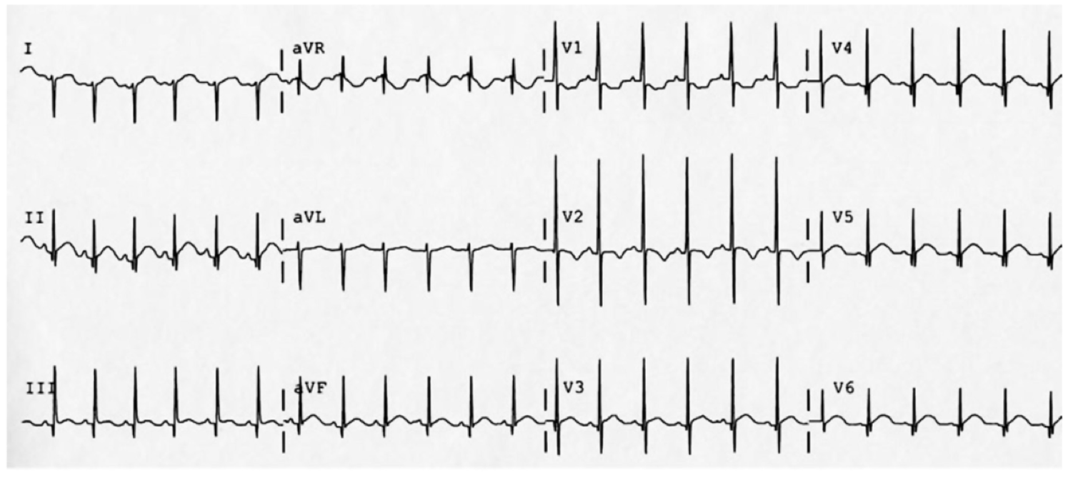
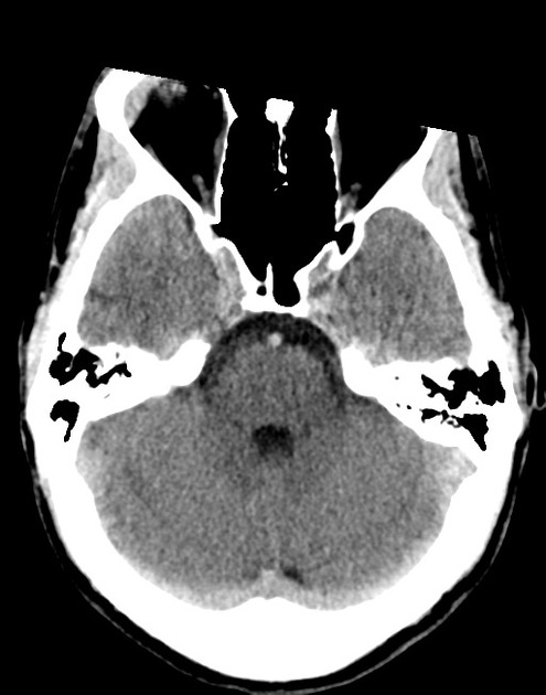
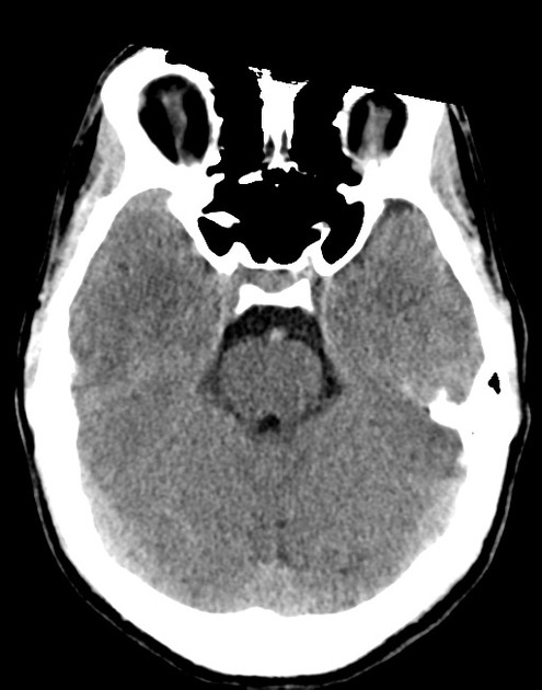
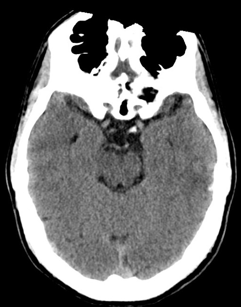
The initial impression was concerning for sympathomimetic toxicity, the patient was treated with cooled intravenous fluids and required pharmacologic sedation and physical restraints to obtain blood samples. ECG, initial laboratory tests and urine toxicology screen were unremarkable. A non-contrast CT head was normal.

The patient remained altered and a repeat examination was performed which revealed multiple, opened blister packs of diphenhydramine and dry, flushed skin.

Anti-cholinergic toxicity was presumed, likely exacerbated by the administration of butyrophenones for sedation. He was treated with benzodiazepines, additional evaporative cooling measures and was admitted to the intensive care unit.

Case 2:

A 32 year-old female with a history of depression was brought to the emergency department by family members who were concerned about bizarre behavior and muscle stiffness. They note that the patient was recently started on a new antidepressant though they are unsure of the name. They describe occasional alcohol consumption but no illicit drug use.

In the emergency department, vital signs were notable for fever and hypertension. Examination demonstrated increased muscle tone and sustained clonus in bilateral lower extremities.

The patient’s presentation was concerning for serotonin syndrome, she was treated with benzodiazepines and intravenous fluids with gradual improvement in mental status and hypertonicity. Upon awakening, she reported doubling her medication dose recently due to persistent feelings of hopelessness as well as increased wine consumption.

An Algorithm for the Evaluation of Toxidromes

An Algorithm for the Evaluation of Toxidromes

Diagnostic Tests

All Patients Most Patients Critical Patients
POC glucose

Core temperature

ECG

Urine hCG

BMP

UA

Acetaminophen

Salicylate

Ethanol

LFT

Lipase

Serum osmolarity

Ionized calcium

Magnesium

GI Decontamination

Activated charcoal

Activated charcoal (1g/kg) within 1-hour post-ingestion and if the patient is awake and cooperative (or via enteric tube if intubated).

Not recommended

  1. Heavy metals
  2. Ions (ex. lithium)
  3. Corrosives
  4. Hydrocarbons
  5. Alcohols

Whole-bowel irrigation

Indicated for sustained-release formulations, expulsion of body packing materials, or ingestion of agent not absorbed by activated charcoal.

Serum alkalinization

For certain ingestions (salicylate, phenobarbital, methotrexate), serum alkalinization through infusion of sodium bicarbonate targeting serum pH 7.5 (and urine pH 8.0) may promote elimination.

Intralipid emulsion

May be useful for local anesthetic toxicity, b-blocker, and calcium channel blocker overdose.

Electrocardiographic Toxidromes


QT Prolongation QRS Prolongation
Anti-emetic Diphenhydramine
Anti-psychotic Cocaine
Anti-microbials (fluoroquinolone, macrolide) Diltiazem, verapamil
Anti-depressant (TCA, SSRI) Propranolol
Anti-arrhythmic Amantadine
Carbamazepine

Gap-producing Toxidromes

Osmolar Gap

  • Toxic alcohol
    • Ethanol
    • Methanol
    • Ethylene glycol
    • Isopropyl alcohol
  • Drug stabilizing agents
    • Mannitol
    • Propylene glycol
    • Glycerol

Anion Gap

  • Salicylate
  • Iron
  • Isoniazid
  • Methanol
  • Ethylene glycol
  • Cyanide

References

  1. Meehan, T. J. (2018). Approach to the Poisoned Patient. In Rosens emergency medicine: concepts and clinical practice (pp. 1813–1822). Philadelphia, PA: Elsevier.
  2. Holstege, C., Borek, H. (2012). Toxidromes Critical Care Clinics 28(4), 479-498. https://dx.doi.org/10.1016/j.ccc.2012.07.008
  3. Mégarbane, B. (2014). Toxidrome-based Approach to Common Poisonings Asia Pacific Journal of Medical Toxicology 3(1), 2-12. https://dx.doi.org/10.22038/apjmt.2014.2463
  4. Rasimas, J., Sinclair, C. (2017). Assessment and Management of Toxidromes in the Critical Care Unit. Critical care clinics 33(3), 521-541. https://dx.doi.org/10.1016/j.ccc.2017.03.002
  5. Thompson, T., Theobald, J., Lu, J., Erickson, T. (2014). The general approach to the poisoned patient Disease-a-Month 60(11), 509-524. https://dx.doi.org/10.1016/j.disamonth.2014.10.002
This algorithm was co-developed by Dr. Chigozie Dike, and Dr. Katrina Nemri.

Dr. Dike is a Houstonian true and true, born across the street at Ben Taub Hospital and proud to have the support of her family and friends through her medical school and emergency medicine training at McGovern Med EM at UT Health. She is a big foodie and loves music. In her free time she’s exploring local restaurants, traveling to new cities, and practicing yoga.

Dr. Nemri is currently a second year emergency medicine resident at McGovern Med EM at UT Health. Her interests are in medical education and critical care. She is a local Houstonian who hopes to stay in the city after residency.

Isolation Precautions

An Algorithm for the Determination of Isolation Precautions

An Algorithm for the Determination of Isolation Precautions

References

  1. Siegel, J., Rhinehart, E., Jackson, M., Chiarello, L., Committee, H. (2007). 2007 Guideline for Isolation Precautions: Preventing Transmission of Infectious Agents in Health Care Settings American Journal of Infection Control 35(10), S65-S164. https://dx.doi.org/10.1016/j.ajic.2007.10.007
  2. Liang, S., Theodoro, D., Schuur, J., Marschall, J. (2014). Infection Prevention in the Emergency Department Annals of Emergency Medicine 64(3), 299-313. https://dx.doi.org/10.1016/j.annemergmed.2014.02.024
  3. Liang, S., Riethman, M., Fox, J. (2018). Infection Prevention for the Emergency Department: Out of Reach or Standard of Care? Emergency medicine clinics of North America 36(4), 873-887. https://dx.doi.org/10.1016/j.emc.2018.06.013
  4. Gottenborg, E., Barron, M. (2016). Isolation Precautions in the Inpatient Setting Hospital Medicine Clinics 5(1), 30-42. https://dx.doi.org/10.1016/j.ehmc.2015.08.004
  5. Harding, A., Almquist, L., Hashemi, S. (2011). The use and need for standard precautions and transmission-based precautions in the emergency department. Journal of emergency nursing: JEN : official publication of the Emergency Department Nurses Association 37(4), 367-73; quiz 424-5. https://dx.doi.org/10.1016/j.jen.2010.11.017

Opioid Withdrawal

Brief HPI:

A 28 year-old female with a history of IV drug use presents to the emergency department with back pain and fever. During evaluation for spinal epidural abscess, she develops vomiting and diarrhea. Examination reveals diaphoresis, mydriasis and hyperactive bowel sounds – she states that her last heroin use was 18-hours ago.

The patient was interested in guidance with cessation of opioid dependence and was evaluated by a recovery support specialist in the emergency department and provided with an appointment for outpatient follow-up. She was treated with buprenorphine-naloxone 8mg sublingual and her symptoms resolved. Her diagnostic evaluation was normal and she was discharged with a prescription for buprenorphine-naloxone 16mg daily until follow-up.

An Algorithm for the Management of Opioid Withdrawal1-4

An Algorithm for the Management of Opioid Withdrawal

Signs

  • Mydriasis
  • Piloerection
  • Diaphoresis
  • Hyperactive bowel sounds

COWS Calculator

Symptoms

  • Dysphoria
  • Rhinorrhea
  • Myalgias, arthralgias
  • Nausea, vomiting, diarrhea
  • Abdominal cramps

Buprenorphine Considerations

Buprenorphine is a high-affinity, opioid partial agonist. The administration of buprenorphine may displace lower-affinity opioids.5 When used for the treatment of acute opioid withdrawal, special care must be taken to ensure that sufficient time has elapsed since last use (evidenced by the presence of moderate withdrawal symptoms) as the immediate displacement of existing opioids can precipitate severe withdrawal. In addition to provoking the maximum severity of the symptoms for which treatment was sought, this can generate mistrust in an otherwise effective medication and the healthcare system more broadly. The combination of buprenorphine with naloxone is intended to deter parenteral abuse – oral/sublingual naloxone is poorly bioavailable.

The initiation of medication-assisted treatment for opioid dependence from the emergency department should be dependent on the availability of outpatient follow-up and addiction treatment programs.6

Supportive Care4,6-7

Symptom Agent Dose
Nausea, Vomiting Promethazine 25mg IM
Diarrhea Loperamide 4mg PO
Octreotide 50mcg SQ
Muscle cramps Baclofen 5mg PO
Anxiety, Dysphoria Lorazepam 1-2mg IV
Diazepam 2-10mg PO, IM, IV
Pain, Myalgia Acetaminophen 650mg – 1,000mg PO
Ibuprofen 600mg PO

Unobserved Induction Guide8

The following guide is adapted from the Yale Department of Emergency Medicine ED-Initiated Buprenorphine Program and is available free for use.
Home Induction Guide Preview

This algorithm was developed with Dr. Drew Silver. Drew is an emergency medicine resident at McGovern Medical School at the University of Texas Health Science Center at Houston (UTHealth).

References

  1. Strayer R, Hawk K, Hayes B, Herring A et al. Management of Opiate Misuse Disorder in the Emergency Department: A White Paper Prepared for the American Academy of Emergency Medicine. American Academy of Emergency Medicine.
  2. ED-Initiated Buprenorphine. Retrieved July 17, 2020, from https://medicine.yale.edu/edbup/Algorithm_338052_5_v2.pdf
  3. Su, M., Lopez, J., Crossa, A., Hoffman, R. (2018). Low dose intramuscular methadone for acute mild to moderate opioid withdrawal syndrome The American Journal of Emergency Medicine 36(11), 1951-1956. https://dx.doi.org/10.1016/j.ajem.2018.02.019
  4. Stolbach A, Hoffman R. Opioid withdrawal in the emergency setting. Post TW, ed. UpToDate. Waltham, MA: UpToDate Inc.
  5. Boas, R., Villiger, J. (1985). Clinical actions of fentanyl and buprenorphine. The significance of receptor binding. British journal of anaesthesia 57(2), 192-6. https://dx.doi.org/10.1093/bja/57.2.192
  6. D’Onofrio, G., Chawarski, M., O’Connor, P., Pantalon, M., Busch, S., Owens, P., Hawk, K., Bernstein, S., Fiellin, D. (2017). Emergency Department-Initiated Buprenorphine for Opioid Dependence with Continuation in Primary Care: Outcomes During and After Intervention Journal of General Internal Medicine 32(6), 660-666. https://dx.doi.org/10.1007/s11606-017-3993-2
  7. Gowing, L., Farrell, M., Ali, R., White, J. (2016). Alpha2‐adrenergic agonists for the management of opioid withdrawal Cochrane Database of Systematic Reviews https://dx.doi.org/10.1002/14651858.cd002024.pub5
  8. Home Initiated Buprenorphine. Retrieved July 17, 2020, from https://medicine.yale.edu/edbup/quickstart/Home_Buprenorphine_Initiation_338574_42801_v1.pdf
  9. Wesson DR, Ling W. The Clinical Opiate Withdrawal Scale (COWS). J Psychoactive Drugs. 2003;35(2):253-259. doi:10.1080/02791072.2003.10400007