BRUE

Brief H&P:

A 4 month-old male with no past medical history and fully-immunized is brought to the emergency department by her mother after an episode of breathing difficulty. She describes that just prior to presentation she noted her child had stopped breathing. She lifted him from the bed and noted some blue discoloration to the mouth with limp extremities. She began to stimulate him by rubbing his chest and abdomen and he began crying after approximately 30 seconds.

She states that he has since returned to normal and she was able to feed him upon arrival to the emergency department without apparent difficulty or vomiting. Prior to the episode, the patient had been in his usual state of health (normal oral intake, urine/stool). No family history of sudden death.

On physical examination, vital signs are normal. The child appears comfortable. Head is normocephalic and atraumatic with normal anterior fontanelle. Mucous membranes are moist, heart sounds are normal and lungs are clear. The abdomen is soft and without organomegaly. The remainder of a detailed physical examination is unremarkable.

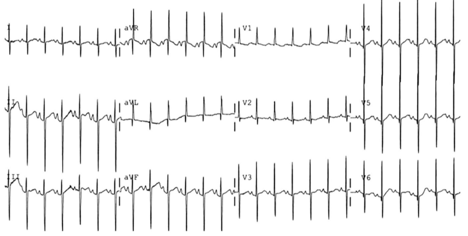
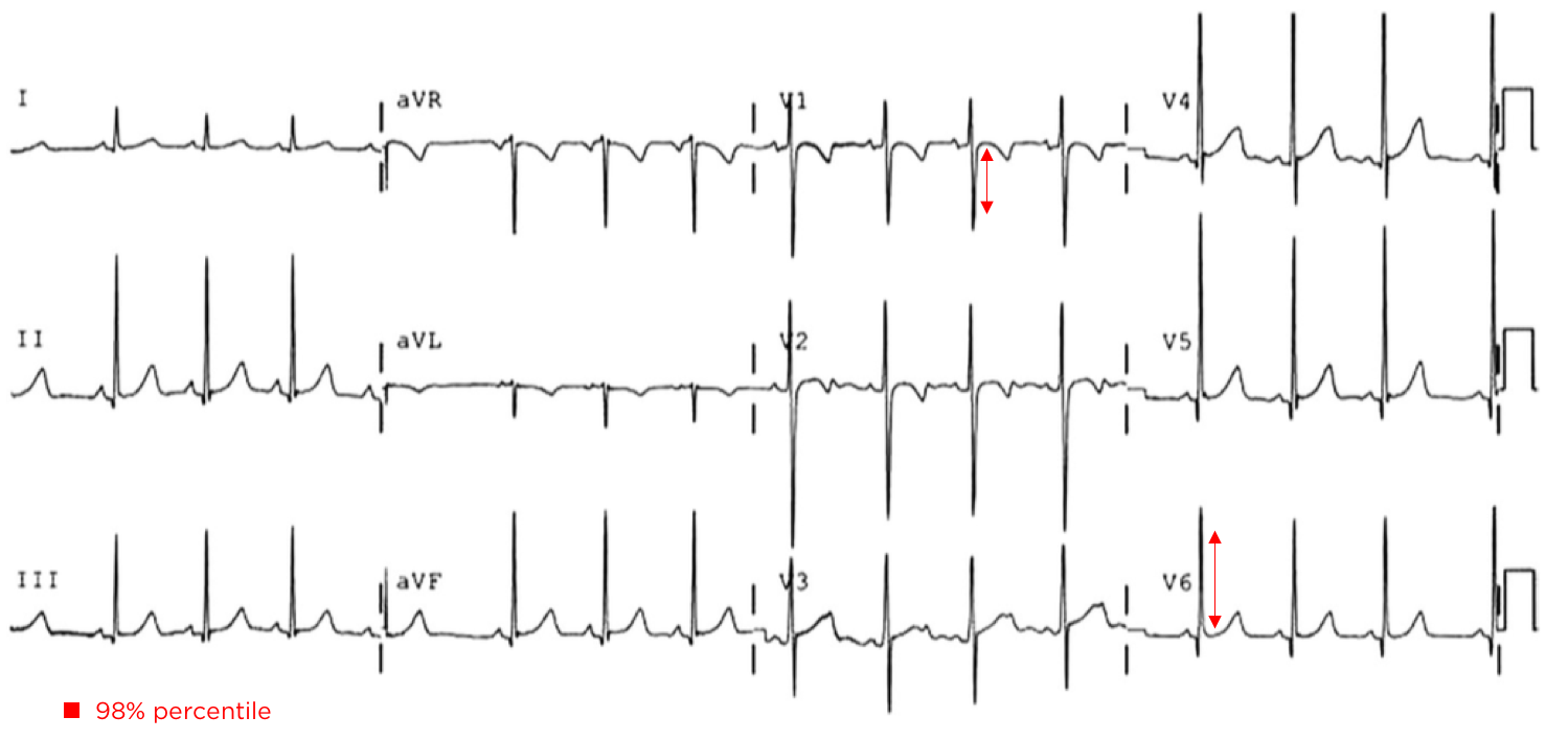
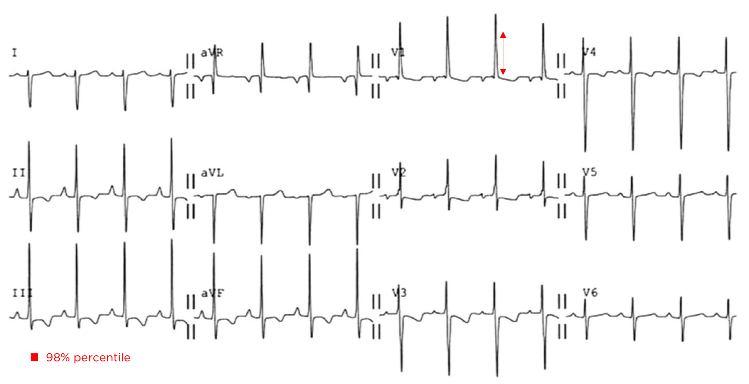
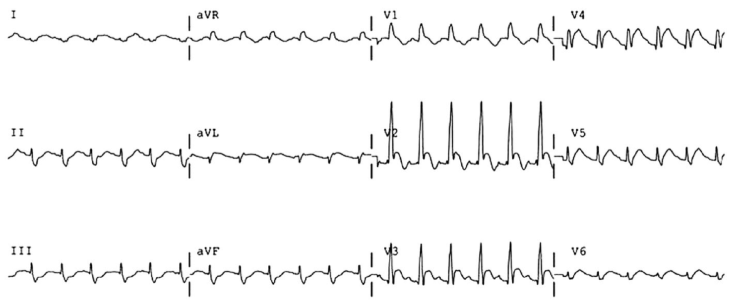
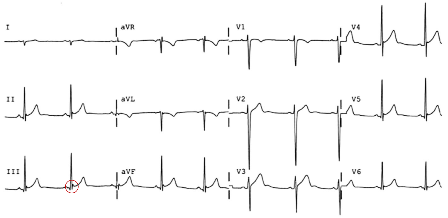
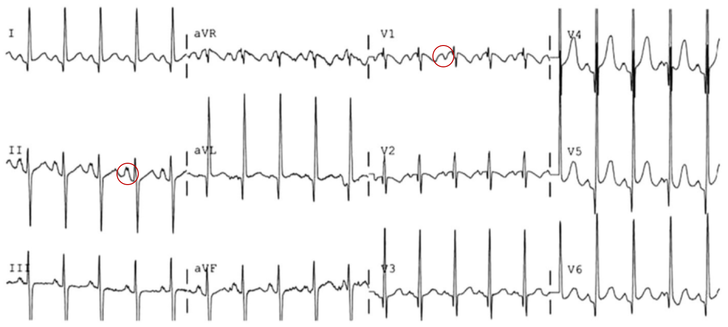
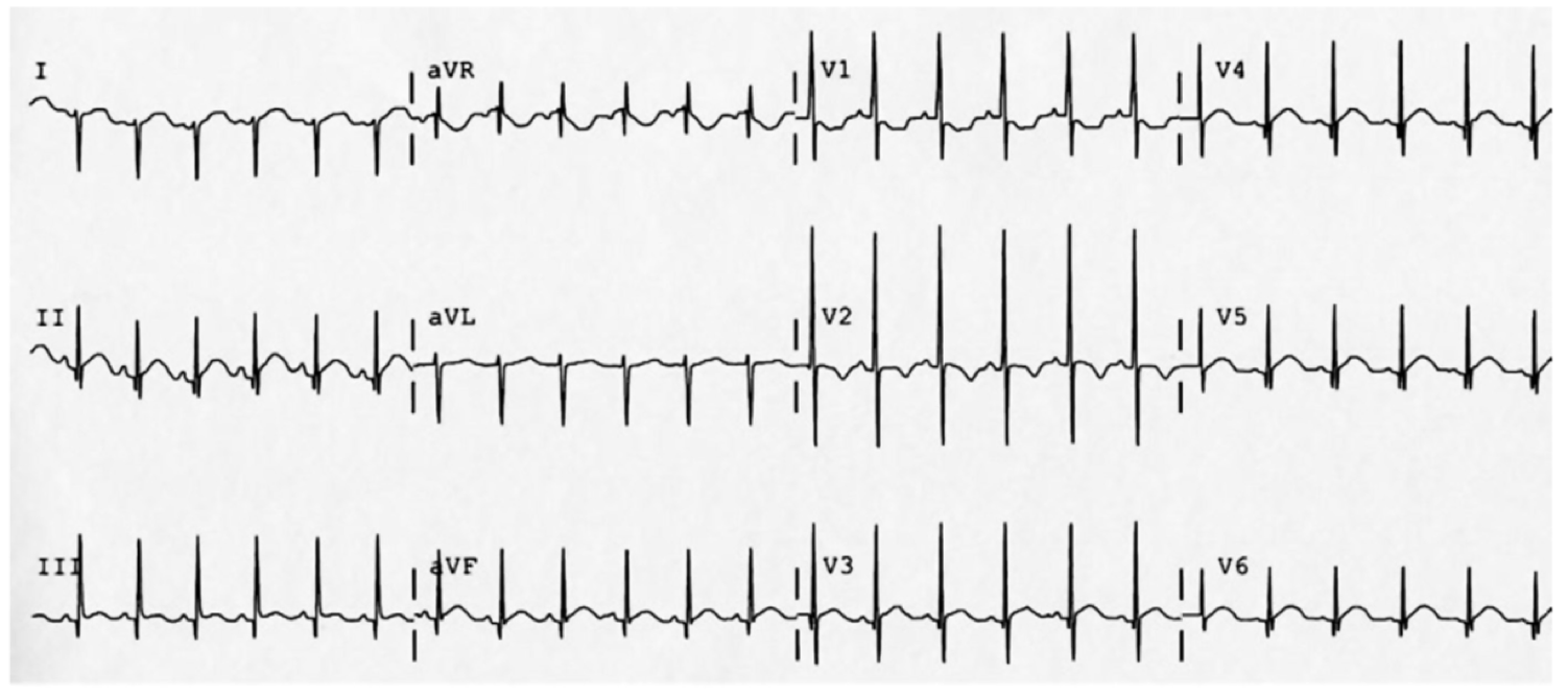
The patient was placed on continuous pulse oximetry, remained well-appearing on serial reassessments and had no further episodes while continuing to feed normally. An ECG was obtained:

The patient’s mother was counseled regarding the diagnosis of low-risk BRUE and the reassuring evaluation and ED observation period. She states that she is able to present to her pediatrician the subsequent morning for evaluation. She was counseled regarding return precautions prior to discharge.

An Algorithm for the Evaluation and Management of Brief Resolved Unexplained Events (BRUE)1,2

An Algorithm for the Evaluation and Management of Brief Resolved Unexplained Events (BRUE)

Differential Diagnosis for BRUE3

Category Causes
Environmental Abuse/trauma
Toxicological
CNS Seizure
Intracranial mass
Cardiovascular Congenital heart disease
Arrhythmia
Pulmonary Airway obstruction
Central apnea
Apnea of prematurity
Pneumonia
Bronchiolitis
Pertussis
Gastrointestinal GERD
IEM Glycogen storage disease
Hyperinsulinism
Fatty acid oxidation defects
This algorithm was developed by Dr. Ali Sina Mirab. Dr. Mirab is a PGY-3 emergency medicine resident at the McGovern Medical School at the University of Texas Health Science Center at Houston (UTHealth).

Special thanks to Dr. Thomas McCarty, Assistant Professor and Pediatric Emergency Medicine Fellowship Director in the Department of Emergency Medicine at McGovern Med EM for his review of the algorithm.

References:

  1. Tieder JS, Bonkowsky JL, Etzel RA, et al. Clinical Practice Guideline: Brief Resolved Unexplained Events (Formerly Apparent Life-Threatening Events) and Evaluation of Lower-Risk Infants: Executive Summary. Pediatrics. 2016:137(5):e20160591.
  2. Merritt JL, Quinonez RA, Bonkowsky JL, et al. A framework for evaluation of the higher-risk infant after a brief resolved unexplained event. Pediatrics. 2019;144(2):e20184101.
  3. McGovern MC, Smith MBH. Causes of apparent life threatening events in infants: a systematic review. Arch Dis Child. 2004;89(11):1043-1048.

Pediatric Status Asthmaticus

Brief HPI:

A 6 year-old boy with a history of asthma presents to the emergency department via EMS for dyspnea. The patient is agitated on exam with nasal flaring and intercostal retractions. The parents report that his difficulty breathing started two days ago. The first day his MDI inhaler provided transient relief; however, over the next 24 hours he required nebulized albuterol 3 times with no significant relief. They deny any recent infections or steroid use and state that his immunizations are up-to-date.

On evaluation, vital signs are notable for BP 93/61, HR 140, RR 47, and SpO2 90%. He is afebrile; capillary glucose 113mg/dL. On examination, the patient is agitated with nasal flaring, intercostal retractions, shallow breathing with diminished breath sounds throughout.

Algorithm for the Management of Pediatric Asthma1-11

Algorithm for the Management of Pediatric Asthma

PASS12

Wheezing Work of Breathing Prolonged Expiration
Mild (0) None or end-expiration Normal or minimal retractions Normal or minimally prolonged
Moderate (1) Throughout expiration Intercostal retractions Moderately prolonged
Severe (2) Severe wheezing or absent Suprasternal retractions, abdominal wall movement Severely prolonged
This algorithm was developed by Dr. Joshua Niforatos. Joshua is an emergency medicine resident at The Johns Hopkins School of Medicine and an alumnus of the Cleveland Clinic Lerner College of Medicine.

Special thanks to Dr. Kelly Young, Director of the Pediatric Emergency Medicine Fellowship at Harbor-UCLA Medical Center and Dr. Adeola Kosoko, Assistant Professor, Assistant Residency Program Director, Director Of Diversity, Inclusion, And Mission at McGovern Medical School for their review of the algorithm.

References

  1. Rowe, B., Bretzlaff, J., Bourdon, C., Bota, G., Camargo, C. (2000). Magnesium sulfate for treating exacerbations of acute asthma in the emergency department. The Cochrane database of systematic reviews https://dx.doi.org/10.1002/14651858.cd001490
  2. Camargo, C., Spooner, C., Rowe, B. (2003). Continuous versus intermittent beta-agonists in the treatment of acute asthma. The Cochrane database of systematic reviews https://dx.doi.org/10.1002/14651858.cd001115
  3. Camargo, C., Rachelefsky, G., Schatz, M. (2009). Managing asthma exacerbations in the emergency department: summary of the National Asthma Education And Prevention Program Expert Panel Report 3 guidelines for the management of asthma exacerbations. Proceedings of the American Thoracic Society 6(4), 357 – 366. https://dx.doi.org/10.1513/pats.p09st2
  4. Gouin, S., Robidas, I., Gravel, J., Guimont, C., Chalut, D., Amre, D. (2010). Prospective evaluation of two clinical scores for acute asthma in children 18 months to 7 years of age. Academic emergency medicine : official journal of the Society for Academic Emergency Medicine 17(6), 598 – 603. https://dx.doi.org/10.1111/j.1553-2712.2010.00775.x
  5. Travers, A., Milan, S., Jones, A., Camargo, C., Rowe, B. (2012). Addition of intravenous beta(2)-agonists to inhaled beta(2)-agonists for acute asthma. The Cochrane database of systematic reviews 12(), CD010179. https://dx.doi.org/10.1002/14651858.cd010179
  6. Jat, K., Chawla, D. (2012). Ketamine for management of acute exacerbations of asthma in children. The Cochrane database of systematic reviews 11(), CD009293. https://dx.doi.org/10.1002/14651858.cd009293.pub2
  7. Ortiz-Alvarez, O., Mikrogianakis, A., Committee, C. (2012). Managing the paediatric patient with an acute asthma exacerbation. Paediatrics & child health 17(5), 251 – 262. https://dx.doi.org/10.1093/pch/17.5.251
  8. Jones, B., Paul, A. (2013). Management of acute asthma in the pediatric patient: an evidence-based review. Pediatric emergency medicine practice 10(5), 1 – 23- quiz 23-4.
  9. Nievas, I., Anand, K. (2013). Severe acute asthma exacerbation in children: a stepwise approach for escalating therapy in a pediatric intensive care unit. The journal of pediatric pharmacology and therapeutics : JPPT : the official journal of PPAG 18(2), 88 – 104. https://dx.doi.org/10.5863/1551-6776-18.2.88
  10. Rehder, K. (2017). Adjunct Therapies for Refractory Status Asthmaticus in Children. Respiratory care 62(6), 849 – 865. https://dx.doi.org/10.4187/respcare.05174
  11. Carroll, C., Sala, K. (2013). Pediatric status asthmaticus. Critical care clinics 29(2), 153 – 166. https://dx.doi.org/10.1016/j.ccc.2012.12.001
  12. Gorelick, M., Stevens, M., Schultz, T., Scribano, P. (2004). Performance of a novel clinical score, the Pediatric Asthma Severity Score (PASS), in the evaluation of acute asthma. Academic Emergency Medicine 11(1), 10 – 18. https://dx.doi.org/10.1197/s1069-6563(03)00579-7

Febrile Seizure

Brief HPI:

An 8-month old female, fully-immunized, otherwise healthy is brought in by paramedics after 1 minute of witnessed generalized tonic-clonic shaking. The patient had otherwise been well, eating and behaving normally earlier that day. On EMS arrival, the patient was post-ictal but grew increasingly responsive en-route and upon presentation to the pediatric emergency department she was crying and appeared normal to her parents. Capillary glucose was 118g/dL. On examination the patient was noted to be febrile with a rectal temperature of 39.4°C. The remainder of the physical examination was normal.

ED Course:

The patient received anti-pyretics and a urinalysis was obtained which was not suggestive of urinary tract infection. During the 3-hour period of observation in the emergency department the patient remained at her normal baseline, had no further seizure activity, and tolerated oral intake with difficulty. The patient was suspected to have a simple febrile seizure and was discharged home.

Algorithm for the Diagnosis of Febrile Seizure

Algorithm for the Evaluation of Febrile Seizure

References

  1. Syndi Seinfeld DO, Pellock JM. Recent Research on Febrile Seizures: A Review. J Neurol Neurophysiol. 2013;4(165). doi:10.4172/2155-9562.1000165.
  2. Whelan H, Harmelink M, Chou E, et al. Complex febrile seizures-A systematic review. Dis Mon. 2017;63(1):5-23. doi:10.1016/j.disamonth.2016.12.001.
  3. Millichap JJ, Gordon Millichap J. Methods of investigation and management of infections causing febrile seizures. Pediatr Neurol. 2008;39(6):381-386. doi:10.1016/j.pediatrneurol.2008.07.017.
  4. Subcommittee on Febrile Seizures, American Academy of Pediatrics. Neurodiagnostic evaluation of the child with a simple febrile seizure. Pediatrics. 2011;127(2):389-394. doi:10.1542/peds.2010-3318.

Neonatal Congenital Heart Disease

Brief H&P

An 8-day old male infant, ex-full term, born by normal spontaneous vaginal delivery and discharged home 2 days after birth without identified complications or maternal infections presents with parents to the emergency department due to decreased activity. Starting on day-of-life six, the family noted that feeding appeared to be taking longer and the mother felt her infant was breathing faster.

On presentation, the patient was pale, dusky, lethargic and with mottled skin. Temperature 36.3°C (rectal), HR 170, RR 60, BP 62/35, SpO2 70%. Physical examination demonstrated flat fontanelle, coarse breath sounds, regular rate and rhythm without additional heart sounds or murmurs, and hepatomegaly with liver edge 3cm below costal margin. Capillary refill was delayed at 5-6 seconds. Supplemental oxygen was applied without effect.

Algorithm for the Evaluation and Management of Suspected Congenital Heart Disease in Neonates

Algorithm for the Evaluation of Neonatal Congenital Heart Disease

Neonates with undiagnosed congenital heart disease may present to the emergency department with nonspecific symptoms, and may be considerably unstable requiring immediate life-saving interventions.

Key Historical Features

  • Respiratory difficulty
  • Feeding difficulty (small quantities, diaphoresis during feeding)
  • Poor weight gain
  • Chromosomal abnormalities, syndromes
  • Maternal risk factors: diabetes, teratogen exposure, substance use
  • Sibling of affected child

Key Examination Findings

  • Vital signs: tachycardia, tachypnea, hypotension
  • Blood pressure differential (RUE vs. LE >8mmHg difference)
  • Pulse oximetry differential (RUE vs. LE >4% difference, <95%)
  • Cardiac examination: murmur, thrill, pulse differential, capillary refill, hepatomegaly

Workup

  • CXR: Evaluate for cardiomegaly, pulmonary vascular congestion
  • ECG: Evaluate for axis deviation (right axis deviation is normal for neonate)
  • ABG with co-oximetry

References

  1. Special thanks to Dr. Kelly Young, MD, MS, FAAP. Director, Pediatric Emergency Medicine Fellowship. Harbor-UCLA Medical Center Department of Emergency Medicine.
  2. Association AAOPAAH. Textbook of Neonatal Resuscitation. 2016.
  3. Lissauer T, Fanaroff AA, Miall L, Fanaroff J. Neonatology at a Glance. John Wiley & Sons; 2015.
  4. Steinhorn RH. Evaluation and Management of the Cyanotic Neonate. Clinical Pediatric Emergency Medicine. 2008;9(3):169-175. doi:10.1016/j.cpem.2008.06.006.
  5. MD MR. Chapter 7 – Cardiology. Twenty First Edition. Elsevier Inc.; 2018:156-202. doi:10.1016/B978-0-323-39955-5.00007-7.
  6. Gomella T, Cunningham M. Neonatology 7/E. McGraw-Hill Prof Med/Tech; 2013.
  7. Yee L. Cardiac emergencies in the first year of life. Emergency Medicine Clinics of NA. 2007;25(4):981–1008–vi. doi:10.1016/j.emc.2007.08.001.
  8. Yates MC, Rao PS. Pediatric cardiac emergencies. Emerg Med. 2013. doi:10.4172/2165-7548.1000164.
  9. Silberbach M, Hannon D. Presentation of congenital heart disease in the neonate and young infant. Pediatr Rev. 2007;28(4):123-131.
  10. Mastropietro CW, Tourner SP, Sarnaik AP. Emergency presentation of congenital heart disease in children. Pediatric Emergency …. 2008.
  11. Brousseau T, Sharieff GQ. Newborn Emergencies: The First 30 Days of Life. Pediatric Clinics of North America. 2006;53(1):69-84. doi:10.1016/j.pcl.2005.09.011.

ECG Guide: Pediatrics

ECG Standard

  • Full standard: no adjustment
  • Half-standard: commensurate reduction in amplitude (usually 50%)
  • Mixed: reduction in amplitude of precordial leads

Atrial Abnormalities

Right Atrial Abnormality (P pulmonale)
Peaked P-wave in II (>3mm from 0-6mo or >2.5mm >6mo)
Causes: right atrial volume overload, ASD, Ebstein, Fontan
Left Atrial Abnormality (P mitrale)
Wide, notched P-wave in II or biphasic in V1
Causes: MS, MR

Axis

  • Anatomical dominance of right ventricle until approximately 6mo
  • RAD normal
  • eRAD suggests AV canal defect

T-waves

  • 1st week of life: Upright
  • Adolescent: Inverted
  • Adult: Upright

Ventricular Hypertrophy

Right Ventricular Hypertrophy
R-wave height >98% for age in lead V1
S-wave depth >98% for age in lead V6
T-wave abnormality (ex. upright in childhood)
Causes: pHTN, PS, ToF
Left Ventricular Hypertrophy
R-wave height >98% for age in lead V6
S-wave depth >98% for age in lead V1
Adult-pattern R-wave progression in newborn (no large R-waves and small S-waves in right precordial leads)
Left-axis deviation
Causes: AS, coarctation, VSD, PDA

Examples


Normal Neonatal ECG

  • 2mo old
  • RAD
  • Inverted T-waves (normal)
  • Tall R-waves in V1-V3


Extreme Axis Deviation

  • Neonate with Down syndrome
  • Isoelectric in I, Negative in aVF negative in II  mean QRS vector -87°
  • Extreme RAD suggestive of AV canal defect


LVH:

  • Unrepaired Coarctation
  • Deep S-wave in V1 (>98%)
  • Tall R-wave in V6 (>98%)


RVH:

  • 10 year-old boy with pulmonary Hypertension
  • RAD after expected age for normal RAD
  • Tall R-waves in V1 (>98%)
  • Deep S-wave in V6 (>98%)


STEMI

  • ALCAPA (anomalous origin of the left coronary artery from the pulmonary artery): coronary artery arises anomalously from the pulmonary artery; as pulmonary arterial pressure falls during the first 6 months of infancy, prograde flow through the left coronary artery ceases and may even reverse.
  • HLHS (hypoplastic left heart syndrome): coronary arteries are perfused from a hypoplastic, narrow aorta that is susceptible to flow disruption
  • Orthotopic heart transplant with allograft vasculopathy
  • Kawasaki: coronary artery aneurysm with subsequent thrombosis


Benign early repolarization

  • 14 year-old male
  • Concave ST-segment elevation


Left Atrial Abnormality:

  • 9mo female with mitral insufficiency
  • Broad biphasic P-wave in V1
  • Tall, notched P-wave in II


Prolonged QT interval

  • 18-year-old female
  • Familial long QT syndrome and a history of cardiac arrest


WPW:

  • Delta wave, shortened PR interval

References

  1. O’Connor M, McDaniel N, Brady WJ. The pediatric electrocardiogram. Part I: Age-related interpretation. Am J Emerg Med. 2008;26(2):221-228. doi:10.1016/j.ajem.2007.08.003.
  2. Goodacre S, McLeod K. ABC of clinical electrocardiography: Paediatric electrocardiography. BMJ. 2002;324(7350):1382-1385.
  3. O’Connor M, McDaniel N, Brady WJ. The pediatric electrocardiogram Part II: Dysrhythmias. Am J Emerg Med. 2008;26(3):348-358. doi:10.1016/j.ajem.2007.07.034.
  4. O’Connor M, McDaniel N, Brady WJ. The pediatric electrocardiogram Part III: Congenital heart disease and other cardiac syndromes. Am J Emerg Med. 2008;26(4):497-503. doi:10.1016/j.ajem.2007.08.004.
  5. Schwartz P. Guidelines for the interpretation of the neonatal electrocardiogram. Eur Heart J. 2002;23(17):1329-1344. doi:10.1053/euhj.2002.3274.

Pediatric Foreign Body Ingestion

Brief H&P

XR Chest: Circular radioopaque foreign body likely in the antrum of the stomach.

A healthy 5 year-old boy is brought to the pediatric emergency department after he informed his parents that he accidentally swallowed a coin just prior to presentation. He has no complaints and on evaluation appears to be breathing comfortably and is tolerating secretions normally. A plain radiograph was obtained and is shown below.

The patient remained well-appearing and was discharged with primary care follow-up.


Indications for Emergent Endoscopy

  • Esophageal button battery
  • Severe symptoms
  • Sharp foreign body in esophagus
  • Multiple magnets in esophagus or stomach

Radiographic Findings


Esophageal foreign bodies typically orient coronally. For example, a coin will appear as a circle on an anteroposterior projection.

Tracheal foreign bodies typically orient sagitally. For example a coin will appear as a line on an anteroposterior projection.

Algorithm for the Evaluation and Management of Pediatric Foreign Body Aspiration

Algorithm for the Management of Pediatric Foreign Body Ingestion

References

  1. Sahn, B, et al. Foreign Body Ingestion Clinical Pathway. 1 Aug. 2016, www.chop.edu/clinical-pathway/foreign-body-ingestion-clinical-pathway. Accessed 26 Aug. 2017.
  2. Wyllie R. Foreign bodies in the gastrointestinal tract. Current Opinion in Pediatrics. 2006;18 N2 -(5).
  3. Uyemura MC. Foreign body ingestion in children. Am Fam Physician. 2005;72(2):287-291.
  4. Chung S, Forte V, Campisi P. A Review of Pediatric Foreign Body Ingestion and Management. Vol 11. 2010:225-230.
  5. Louie MC, Bradin S. Foreign Body Ingestion and Aspiration. Pediatrics in Review. 2009;30(8):295-301. doi:10.1542/pir.30-8-295.
  6. Green SS. Ingested and Aspirated Foreign Bodies. Pediatrics in Review. 2015;36(10):430-437. doi:10.1542/pir.36-10-430.

Principles of Neonatal Resuscitation

The following resource for neonatal resuscitation and neonatal critical care was developed with the guidance of Dr. Agrawal (Neonatology) while on rotation at the White Memorial Medical Center Neonatal Intensive Care Unit.

Endotracheal Tube Size1-3

Simplified Formula
Estimated gestational age in weeks ÷ 10 = round to nearest half-size uncuffed tube

NRP Recommendation

Gestation age (weeks) Weight (kg) ETT Size (ID, mm) Depth (cm from lip)
<28 <1.0 2.5 6-7
28-34 1.0-2.0 3.0 7-8
34-38 2.0-3.0 3.5 8-9
>38 >3.0 3.5-4.0 9-10

Laryngoscope Blade Size

Age Blade
Preterm 0
Term 1

Umbilical Vein Catheter Placement4

ED Indications
Unstable neonate
Contraindications
Omphalocele
Gastroschisis
Necrotizing enterocolitis
Depth
4-5cm or until blood return (for emergent placement)

Umbilical artery/vein catheter position on plain radiograph.

Umbilical catheter size

Weight (kg) Size (F)
<1.5 3.5
1.5-3.5 5
>3.5 8

Umbilical catheter positioning on plain radiographs

Umbilical venous catheter position can be verified with a plain radiograph. Positioning within the umbilical vein can be confirmed by tracing a cephalad trajectory from the insertion point at the umbilicus. An umbilical artery catheter will first pass caudally into the internal iliac artery before travelling cephalad into a common iliac artery and the abdominal aorta.

Medications5

Medication Dose
Epinephrine 0.1mL/kg (1:10,000) IV, 0.01mg/kg
Volume Expansion 10mL/kg (normal saline, blood)
Naloxone 0.1-0.2mg/kg
Dopamine 5-20mcg/kg/min IV infusion

Neonatal Physiology and Transition to Extrauterine Life6

An important principle in neonatal resuscitation is supporting the appropriate transition from intra- to extra-uterine life which is dependent on several key anatomic and physiologic changes occurring in an optimal environment.

Anatomy7

Fetal Circulation
Neonatal Circulation

Fetal Circulation

In the fetal circulatory system, oxygenated blood is delivered via the umbilical vein, entering the inferior vena cava via the ductus venosus. The majority of this oxygenated blood passes through the right atrium and into the left atrium through the foramen ovale to enter the systemic circulation.

Meanwhile, high pulmonary pulmonary vascular resistance (due to hypoxic vasoconstriction in fluid-filled alveoli) means that most of the deoxygenated right ventricular output is routed through the ductus arteriosus and enters into the systemic circulation – mixing with oxygenated blood distal to the highest priority end-organs (brain and heart), to be reoxygenated at the placenta.

Post-transition Circulation

The transition to extra-uterine life involves several key steps detailed below and is supported by appropriate ventilation, oxygenation and temperature regulation.

  1. Alveolar Fluid Clearance
    Catecholamine and hormone changes (predominantly corticosteroids) during the process of labor induce changes in enzymatic expression that result in the resorption of alveolar fluid into the interstitial space. At the time of delivery, negative intra-thoracic pressure from inspiration further promotes the resorption of alveolar fluid. Mechanical thoracic compression from delivery may also contribute.
  2. Respiration and Breathing
    Disconnection from the placenta ceases the transfer of placenta-derived factors including prostaglandins. The withdrawal of tonic inhibition of central respiratory drive from prostaglandins with cord clamping stimulates rhythmic breathing. The infant’s initial breaths and resultant lung expansion promotes alveolar expansion and stimulates surfactant production – this decreases alveolar surface tension, increases lung compliance and further facilitates breathing.
  3. Circulatory Changes
    At delivery, clamping the umbilical cord removes a large bed of low-resistance circulation, increasing systemic vascular resistance and systemic blood pressure. At the same time, lung expansion and alveolar aeration decreases pulmonary vascular resistance and pulmonary arterial pressures. At the ductus arteriosus, increased systemic vascular resistance combined with decreased pulmonary vascular resistance decreases shunting and contributes to closure. Similarly, as left atrial pressure approaches and exceeds right atrial pressure, right-to-left flow across the foramen ovale ceases. Collectively, these changes serve to effectively separate the left- and right-sided circulations.

NRP Resuscitation Algorithm5,8

Neonatal Resuscitation Algorithm

References

  1. Luten R, Kahn N, Wears R, Kissoon N. Predicting Endotracheal Tube Size by Length in Newborns. J Emerg Med. 2007;32(4):343-347. doi:10.1016/j.jemermed.2007.02.035.
  2. Peterson J, Johnson N, Deakins K, Wilson-Costello D, Jelovsek JE, Chatburn R. Accuracy of the 7-8-9 Rule for endotracheal tube placement in the neonate. J Perinatol. 2006;26(6):333-336. doi:10.1038/sj.jp.7211503.
  3. Kempley ST, Moreiras JW, Petrone FL. Endotracheal tube length for neonatal intubation. Resuscitation. 2008;77(3):369-373. doi:10.1016/j.resuscitation.2008.02.002.
  4. Anderson J, Leonard D, Braner DAV, Lai S, Tegtmeyer K. Videos in Clinical Medicine. Umbilical Vascular Catheterization. Vol 359. 2008:e18. doi:10.1056/NEJMvcm0800666.
  5. Association AAOPAAH. Textbook of Neonatal Resuscitation. 2016.
  6. Caraciolo J Fernandes MD. Physiologic transition from intrauterine to extrauterine life. UpToDate.
  7. Sadler TW. Langman’s Medical Embryology. Lippincott Williams & Wilkins; 2011.
  8. Perlman JM, Wyllie J, Kattwinkel J, et al. Part 7: Neonatal Resuscitation: 2015 International Consensus on Cardiopulmonary Resuscitation and Emergency Cardiovascular Care Science With Treatment Recommendations. In: Vol 132. American Heart Association, Inc.; 2015:S204-S241. doi:10.1161/CIR.0000000000000276.

Kawasaki Disease

Brief H&P:

An 8-month old male is brought to the emergency department with fever. He has had four days of fever (temperature ranging from 37-40°C), rash on trunk and extremities, white-colored tongue discoloration, and irritability with decreased oral intake. Temperature on presentation was 39.4°C, examination revealed an erythematous maculopapular rash on the extremities and trunk including soles of the feet. Mucous membrane involvement was noted with oropharyngeal erythema and bilateral conjunctival injection. Neck examination demonstrated right-sided cervical adenopathy.

Labs:

  • WBC: 23.4 (N: 59%, B: 21%)
  • ESR: 100mm/hr
  • CRP: 7.59mg/dL
  • Albumin: 3.3g/dL
  • AST/ALT: 78U/L, 65U/L
  • UA: 7WBC, no bacteria

Hospital Course

The patient was admitted with a diagnosis of Kawasaki Disease and was treated with IVIG and high-dose aspirin. The patient demonstrated marked improvement with treatment and had a normal echocardiogram. He was discharged on hospital day three.

Epidemiology1,2

  • Age: 6 months to 5 years
  • Northeast Asian
  • Possible heritable component
  • Seasonal (winter/spring)

Course

  • Acute febrile (T > 39°C refractory to anti-pyretics)
  • Subacute (coronary vasculitis)
  • Convalescent

Diagnosis

  • Fever >5d
  • Criteria (4/5)
    • Conjunctivitis (bilateral, non-exudative)
    • Oropharynx changes (strawberry tongue, erythema, perioral)
    • Cervical lymphadenopathy (unilateral, >1.5cm)
    • Rash
    • Extremity changes (erythema, edema, palm/sole involvement)
  • Incomplete (2-3 criteria)

Labs

  • CBC: Elevated WBC (neutrophil predominant)
  • Urinalysis: Sterile pyuria
  • Acute phase reactants: Elevated ESR (>40-60mm/hr), CRP (>3.0-3.5mg/dL)
  • CMP: Hyponatremia, hypoalbuminemia, hypoproteinemia, elevated transaminases
  • ECG: AV block, ischemia/infarction (aneurysm/thrombosis)
  • Echocardiography: Decreased LVEF, MR, pericardial effusion

Management

  • Hospital admission
  • IVIG (2g/kg)
  • Aspirin (80mg/kg/day)

Algorithm for the Evaluation of Kawasaki and Incomplete Kawasaki Disease3,4

Algorithm for the Evaluation of Kawasaki and Incomplete Kawasaki Disease

References:

  1. Shiari R. Kawasaki Disease; A Review Article. Arch Pediatr Infect Dis. 2014;2(1 SP 154-159).
  2. Yu JJ. Diagnosis of incomplete Kawasaki disease. Korean J Pediatr. 2012;55(3):83-87. doi:10.3345/kjp.2012.55.3.83.
  3. Newburger JW, Takahashi M, Gerber MA, et al. Diagnosis, treatment, and long-term management of Kawasaki disease: a statement for health professionals from the Committee on Rheumatic Fever, Endocarditis, and Kawasaki Disease, Council on Cardiovascular Disease in the Young, American Heart Association. Pediatrics. 2004;114(6):1708-1733. doi:10.1542/peds.2004-2182.
  4. Yellen ES, Gauvreau K, Takahashi M, et al. Performance of 2004 American Heart Association recommendations for treatment of Kawasaki disease. Pediatrics. 2010;125(2):e234-e241. doi:10.1542/peds.2009-0606.

Pediatric Head Trauma

Brief H&P:

A young child, otherwise healthy, is brought to the pediatric emergency department after a fall. The parents report a fall from approximately 2 feet after which the patient cried immediately and without apparent loss of consciousness. Over the course of the day, the patient developed an enlarging area of swelling over the left head. The parents were concerned about a progressive decrease in activity and interest in oral intake by the child, and they were brought to the emergency department for evaluation. Examination demonstrated a well-appearing and interactive child – appropriate for age. Head examination was notable for a 5x5cm hematoma over the left temporoparietal skull with an underlying palpable skull irregularity not present on the contralateral side. Non-contrast head computed tomography was obtained.

Imaging

ct_head_0007_Screen-Shot-2017-06-05-at-4.18.59-PM
ct_head_0006_Screen-Shot-2017-06-05-at-4.19.02-PM
ct_head_0005_Screen-Shot-2017-06-05-at-4.19.04-PM
ct_head_0004_Screen-Shot-2017-06-05-at-4.19.08-PM
ct_head_0003_Screen-Shot-2017-06-05-at-4.19.10-PM
ct_head_0002_Screen-Shot-2017-06-05-at-4.19.15-PM
ct_head_0001_Screen-Shot-2017-06-05-at-4.19.18-PM
ct_head_0000_Screen-Shot-2017-06-05-at-4.19.22-PM

CT Head

Fracture of the left temporal and parietal bone with overlying scalp hematoma.

Algorithm for the Evaluation of Pediatric Head Trauma (PECARN)1,2,3

Algorithm for the evaluation of pediatric head trauma

References

  1. Kuppermann N, Holmes JF, Dayan PS, et al. Identification of children at very low risk of clinically-important brain injuries after head trauma: a prospective cohort study. Lancet. 2009;374(9696):1160-1170. doi:10.1016/S0140-6736(09)61558-0.
  2. Brenner D, Elliston C, Hall E, Berdon W. Estimated risks of radiation-induced fatal cancer from pediatric CT. American Journal of Roentgenology. 2001;176(2):289-296. doi:10.2214/ajr.176.2.1760289.
  3. Schonfeld D, Bressan S, Da Dalt L, Henien MN, Winnett JA, Nigrovic LE. Pediatric Emergency Care Applied Research Network head injury clinical prediction rules are reliable in practice. Archives of Disease in Childhood. 2014;99(5):427-431. doi:10.1136/archdischild-2013-305004.

Pediatric Emergencies

Diseases by Age

  • 1 week – 1 month: Ductal dependent cardiac lesions
  • 1st month: Malrotation with volvulus
  • 1 – 2 months: Pyloric Stenosis
  • 2 – 6 months: CHF
  • 3 months – 2 years: Intussusception
  • 6 months – 2 years: Croup
  • <2 years: Bronchiolitis
  • 2 years: Meckel’s
  • 2 years – 6 years: Epiglottitis

Cardiology

Ductal Dependent Lesions

  • Present 1st week to 1st month
  • Normal duct seals by 3 weeks
  • If dependent on shunt for pulmonary flow  cyanosis
  • If dependent on shunt for systemic flow cold shock (may be worse w/ fluids)
  • Prostaglandin E1
    • 1 mg/kg/min
    • Side effects include apnea, bradycardia, hypotension, seizure
      • Consider intubating prior to administration
    • IVF, cover for sepsis

Congestive Heart Failure

  • Present 2nd to 6th month
  • Presents with respiratory symptoms (wheezing, retractions, tachypnea)
  • Difficulty with feeding (the infant stress test)
  • Treatment: Supportive

 Tetrology of Fallot

  1. Calm the child, knee to chest
  2. O2 = reduction in PVR
  3. Analgesia: morphine 0.1mg/kg, fentanyl 1.5 mcg/kg, ketamine 0.25 mg/kg
  4. Establish Access: 10-20cc/kg bolus
  5. Phenylephrine 0.2 mg/kg IV (to increase SVR)
  6. +/- HCO3 1mmol/kg (if acidosis)
  7. +/- beta blocker (with cardiology consultation)
  8. PGE1 0.05mcg/kg/min titrating to 0.1mcg/kg/min

Dermatology

Slapped Cheek/5th Disease

  • Parvo B19
  • Slapped cheeks, lacy reticular pattern of rash on body
  • Complications:
    • Pregnancy hydrops
    • Sickle Cell Disease  aplastic crisis

Measles

  • Koplik spots, conjunctivitis, fever
  • Can cause blindness

VZV

  • Different stages of development
  • Treat with acyclovir if > 12 years old
  • Give VZIG in neonates and immunocompromised

Scarlet Fever

  • Erythematous rash, palatal petechiae, pastia’s lines
  • Strawberry tongue
  • Trunk to periphery
  • Treat with Pen VK: 50mg/kg BID x10d or Amox 20mg/kg BID x10d
  • Pen allergic: Azithro 10mg/kg day 1 then 5mg/kg 2-5

Staphylococcal Scalded Skin Syndrome

  • Toxin mediated, negative Nikolsky, good prognosis
  • Treatment: Anti-staphylococcal antibiotics
    • Nafcillin 25mg/kg/d IV
    • Augmentin 45mg/kg/d PO in 2 divdied doses 7-10d
    • Keflex 10mg/kg/d QID x7-10d

Henoch-Schonlein Purpura

  • Palpable purpura in dependent areas
  • Arthralgia/Arthritis (50-84%)
  • Abdominal pain (50%): vascular lesions in bowel, may be intussusception lead point
  • Renal Disease (20-50%) may develop within 2 months
  • Treatment: Supportive, NSAIDs

Kawasaki Disease

  • 5 days of fever + 4/5 of criteria
    • Diffuse polymorphous diffuse rash
    • Conjunctivitis
    • Mucous membrane change (strawberry tongue)
    • Cervical LAD (usually unilateral)
    • Extremity changes
  • Incomplete and atypical forms more common in infants
  • Treatment (drop complications from 25% to 4-5%)
    • Aspirin 20mg/kg/dose Q6H
    • IVIG 2gm/kg over 12H

Gastroenterology

Bilious Vomiting

  • Bilious vomiting malrotation with volvulus until proven otherwise  surgical emergency
  • 1st month of life “pre-verbal child’s disease”
  • Dx: Upper GI Series (10-15%) false positive rate

Necrotizing Enterocolitis

  • 10% of cases full term
  • XR w/ pneumatosis intestinalis

Hirschsprung’s

  • No meconium, slightly distended abdomen
  • Less severe  later presentation, p/w constipation

Pyloric Stenosis

  • Presents around 6 wks: vomiting but very hungry
  • Diagnosis
    • US pylorus > 4mm thick, >15mm long
    • NGT aspiration 5cc is abnormal
  • Treatment
    • Resuscitate
    • Correct metabolic abnormalities
    • Consult surgery

Intussusception

  • Most common infant emergency
    • 3 months – 2 years
  • Abdominal pain, currant jelly, palpable mass (30% only)
  • Typical presentation
    • Lethargy (may be only sign)
    • Vomiting
    • Paroxysms of pain
    • SBO
    • PO intolerance
  • Diagnosis: US
  • Treatment: Enema (80-95% successful), 10% recurrence

Meckel’s Diverticulum

  • Around 2 years of age, boys > girls
  • Obstruction, intussusception
  • Diagnose with technetium scan

Appendicitis

  • 1/3rd with vomiting and diarrhea (AGE-type syndrome)

Hemolytic Uremic Syndrome

  • Watery/bloody diarrhea
  • Three components
    • Acute renal failure
    • Thrombocytopenia
    • Microangiopathic hemolytic anemia (MAHA)
  • Signs
    • Pallor
    • Abdominal Pain
    • Decreased urine output
    • Low energy/AMS
    • Hypertension
    • Edema
    • Petechiae
    • Icterus
  • Treatment: Supportive vs. Dialysis (50%)

GI Bleed by Age

Age Well-Appearing Ill-Appearing
Neonate Allergic Proctocolitis Malrotation with Volvulus
Anal Fissure Necrotizing Enterocolitis
Swallowed Maternal Blood Coagulopathy
Infant/Young Child Allergic Proctocolitis Meckel’s
Gastritis Intussusception
Infectious Colitis Vascular Malformation
Older Child/Adolescent Gastritis IBD
Esophageal Bleeding Cryptic Liver Disease
Juvenile Polyps Intestinal Ulceration

Congenital Disorders

Congenital Adrenal Hyperplasia

  • Presents in first two weeks of life
  • Chief complaint may be vomiting
  • Lyte: HyperK, HypoNa, Hypoglycemia  dysrhythmias, seizures
  • Treatment
    • IVF (usual dose)
    • Glucose (usual dose)
    • Hydrocortisone: 25mg (neonate/infant), 50mg child, adolescent/adult 100mg

Inborn Errors of Metabolism

  • Possible CC: Vomiting, Lethargy, Seizures, Hepatomegaly, Metab Acidosis, Odor
  • May have normal labs and imaging
  • Life-threatening: Metabolic acidosis, Hypoglycemia, Hyperammonemia, Sepsis
  • Labs
    • VBG (acidosis),
    • CMP (liver, kidney, anion gap)
    • Ammonia, lactate, urine (ketones, reducing substance)
    • Bunch of extra tubes for labs later
  • Treatment
    • NPO
    • IVF bolus
    • D10 at 1.5x maintenance
    • Treat Sepsis
    • Control seizures PRN, correct hyperammonemia/acid/lyte (may need dialysis)

Pulmonary

Croup

  • Toddlers (6-24 months), 5% of all children, boys > girls
    • PIV #1
    • Rhinovirus, Metapneumovirus, PIV II-IV, RSV, Flu A/B
    • Frequent co-infections with one or more viruses
  • Sx: 1-3 days of URI Sx  Abrupt cough/stridor worse for one day, then better
  • Signs: Nontoxic, if wheezing likely RSV
  • Studies: XR to r/o FB (steeple sign if positive)
  • Treatment: Racemic Epi: 0.25-0.75 cc in 3 cc Q 20 minutes, lasts < 2 hours
  • Disposition: If stridor at rest then treat if no improvement, then admit
Stridor Steroids Racemic Epi Dispo
Mild 0.15 mg/kg No Home
At rest with WOB 0.30 mg/kg Yes Admit
Severe at rest 0.60 mg/kg Yes ICU

Bronchiolitis

  • Children < 2 years old, November through April (peak Jan/Feb)
    • Apnea in neonates and ex-premies < 2 months
    • Bacterial superinfection is very rare
  • Presentation: Desat, tachypnea, nasal flaring, intercostal retractions, secretions
  • Exam: Fine rales, diffuse/fine wheezing
  • Treatment: Suction, O2 (if < 90%), NPPV
  • Maybe albuterol, but no steroids/epi/abx

Epiglottitis

  • Bimodal (2-6, 20-40y), < 1% URI with stridor, boys = girls, al year
    • Non-typable H.flu, staph/strep, Moraxella
    • Candida, HSV, VZV, crack cocaine
  • Symptoms: Muffled voice, drooling rapid progression in hours
  • Signs: No pharyngeal findings with severely tender anterior neck
  • Studies: XR w/ thumb sign
  • Treatment: Laryngoscopy, airway management

Bacterial Tracheitis

  • Preschool (1-10y), boys = girls, Downs
  • Symptoms: Several days’ URI  toxic in hours, rapid progression
  • Signs: Subglottic diffuse inflammation, edema with exudates and pseudomembranes
  • Studies: CXR demonstrates narrow trachea
  • Treatment: Emergent intubation, 3rd generation cephalosporin

Rapid Pediatric Assessment

This post presents a tool for the rapid assessment of the cardiopulmonary status and cerebral/metabolic function of critically ill pediatric patients. The purpose is not to establish a diagnosis, rather to identify the particular physiological derangements to prioritize initial interventions. The tool was initially designed as a “triangle” – it has been adapted here (with permission) as a Venn diagram.1

Pediatric Assessment Diagram

Pediatric Assessment Diagram

Assessment of Appearance

  • Tone: Moves spontaneously, resists examination
  • Interactivity: Interacts with environment, reaches for items
  • Consolability: Comforted by caregiver
  • Gaze: Makes eye contact

Assessment of Work of Breathing

  • Airway Sounds: Stridor, grunting, wheezing
  • Position: Tripod
  • Retractions

Assessment of Circulation

  • Pallor
  • Mottling
  • Cyanosis

Management

Impression Interventions
Respiratory distress
  • Position of comfort
  • Oxygen, suction
  • Therapy as appropriate (albuterol, epinephrine, etc)
  • Labs/radiographs as indicated
Respiratory failure
  • Head/airway positioning
  • 100% oxygen
  • Ventilation support (BVM)
  • Advanced airway
Shock (compensated and decompensated)
  • Oxygen
  • Access
  • Fluid resuscitation
  • Specific therapy (antibiotics, surgery)
  • Labs/radiographs as indicated
CNS/Metabolic
  • Pulse oximetry
  • Rapid glucose
  • Labs/radiographs as indicated
Cardiopulmonary Failure
  • Head/airway positioning
  • 100% oxygen
  • Ventilation support (BVM)
  • Chest compressions as needed
  • Specific therapy (defibrillation, epinephrine, amiodarone)
  • Labs/radiographs as indicated

References:

  1. The pediatric assessment triangle: a novel approach for the rapid evaluation of children. Pediatr Emerg Care. 2010;26(4):312-315. doi:10.1097/PEC.0b013e3181d6db37.

Pediatric Sizes and Doses

Below is a rapid reference for essential information related to the care of pediatric patients including sizing estimates for endotracheal tubes and weight-based dosing for critical/common medications (rapid sequence intubation, pediatric advanced life support, seizure management), compiled by Dr. Kelly Young1.

Airway

ETT
4 + Age/4 = uncuffed
Subtract 0.5 for cuffed
Gestational age (weeks) / 10 if premature
Depth = ETTx3
Blade
Newborn: 0
<2yo: 1
2-8yo: 2
>8yo: 3
Other Tubes
NGT = ETT x 2
Chest tube = ETT x 4

Estimating Weight

Age (years) 1 3 5 7 9
Weight (kg) 10 15 20 25 30

Vital Signs

Blood Pressure

Age Measure
Neonate 60mmHg
<1yo 70mmHg
1-10yo 70 + (Age x2)
>10yo 90mmHg

Heart/Respiratory Rate

Age (yrs) HR RR
0-1 140 40
1-4 120 30
4-12 100 20
>12 80 15

Medications

Name Dose
RSI (Paralysis)
Succinylcholine 1mg/kg (x2 infant, x3 neonate)
Rocuronium 1-1.2mg/kg
RSI (Sedation)
Etomidate 0.3mg/kg
Ketamine 2mg/kg
Midazolam 0.1mg/kg
Fentantyl 1mcg/kg
PALS
Defibrillation 2, 4, 10J/kg
Cardioversion 0.5, 1J/kg
Epinephrine 0.01mg/kg (0.1mL/kg of 1:10,000)
Atropine 0.02mg/kg (minimum dose 0.1mg, maximum 0.5mg)
Adenosine 0.1mg/kg (max 6mg), 0.2 mg/kg (max 12mg)
Amiodarone 5mg/kg
Calcium gluconate (10%) 1mL/kg
Calcium chloride (10%) 0.2mL/kg
Magnesium sulfate 25mg/kg
Sodium bicarbonate 1mEq/kg
3% saline 5cc/kg
Mannitol 1g/kg
Fluids
Normal saline (0.9%) 20cc/kg
PRBC 10cc/kg
Maintenance 4cc/kg (first 10kg), 2cc/kg (second 10kg), 1cc/kg thereafter
Dextrose
<1yo D10, 5cc/kg
1-10yo D25, 2cc/kg
>10yo D50, 1cc/kg
Anti-epileptics
Lorazepam, Midazolam 0.1mg/kg x3
Fosphenytoin 20 PE/kg
Keppra 20-40mg/kg
Valproate 20mg/kg
Phenobarbital 20mg/kg
Midazolam infusion 0.1mg/kg/h
Midazolam IN 0.2mg/kg (max 10mg)
Antibiotics
Ceftriaxone 50mg/kg
Amoxicillin 90mg/kg divided BID
Azithromycin 10mg/kg day 1, 5mg/kg days 2-5
Common Medications
Acetaminophen 15mg/kg
Ibuprofen 10mg/kg
Diphenhydramine 1.25mg/kg
Ondansetron 0.15mg/kg
Intranasal Medications
Fentanyl 1.5mcg/kg (max 100mcg)
Midazolam 0.5mg/kg (max 10mg)

Reference:

  1. Young, K. D. (2016, April 18). Pediatric Doses and Sizes. Lecture presented at Harbor-UCLA Medical Center in CA, Torrance.

Failure to Thrive

Failure to ThriveID:

5mo female with a history of multiple food allergies, GERD and FTT admitted from clinic for persistent failure to gain weight.

HPI:

The patient’s mother states that the current diet is 3oz of Neocate 20cal/oz q3h, and that the baby sleeps through the night. The child has a history of reflux, but no emesis in the past few weeks since starting Reglan. There was a history of bloody diarrhea, however none since age 2mo after a change of formula. Mother reports known allergies to milk, soy, protein, and egg. No recent fevers/chills, emesis, diarrhea, fussiness.

The patient was born at 27wks via emergency Cesarean for non-reassuring fetal heart tracings, was intubated in the DR and remained in the NICU for one week.

PE:

  • VS: 98/65mmHg, 114bpm, 98.1°, 33/min, 100% RA
  • Gen: Small for age, smiling and interactive
  • HEENT: PERRL, MMM, no lesions
  • CV: RRR, no M/R/G, Lungs: CTAB
  • Abdomen: +BS, soft, NT/ND, no masses, no hepatosplenomegaly
  • Ext: Normal capillary refill

Assessment & Plan:

5mo female, ex-27wks with a history of multiple food allergies, GERD, FTT. Persistent failure to gain weight, admitted for evaluation of feeding habits and observed weight gain. The patient was determined to not be receiving adequate intake and was advanced to a high-calorie formula and parental education was provided. After two days of observed (and appropriate) weight gain, the patient was discharged with follow-up at multiple specialty clinics including GI, FTT, and A&I.

Differential Diagnosis for Failure to Thrive:

A System for Failure to Thrive


Sore Throat

Oropharynx AnatomyID:

17 year-old female presenting to the pediatric ED with sore throat for 2 days.

HPI:

The patient reports steadily worsening sore throat over the past 2 days, associated with a sensation of swelling. The pain is described as sharp, 4/10 in severity, located on the left side of her throat, and worsened with swallowing. She denies inability to swallow or difficulty breathing, she also denies fever, cough, new skin rashes or genital lesions.

She has no PMH/PSH, takes no medications, denies t/e/d use and is not currently sexually active.

PE:

  • VS: 111/65mmHg, 80bpm, 97.8°, 16/min, 100% RA
  • Gen: Well-appearing, NAD
  • HEENT: PERRL, no conjunctival injection, TM clear b/l, minimal pharyngeal erythema on left with 6mm white circular lesion on left tonsil, no tonsillar enlargement, no uvular deviation, no cervical LAD, neck supple no masses, normal neck ROM
  • CV: RRR, no M/R/G, Lungs: CTAB
  • Abdomen: +BS, soft, NT/ND
  • Ext: Warm, well-perfused, normal peripheral pulses

Assessment & Plan:

17yo female with no significant PMH with acute pharyngitis for 2 days. The most likely cause of the patient’s symptoms is viral pharyngitis, potentially herpangina (given the appearance of the tonsillar lesion). A more serious viral/bacterial pharyngitis is less likely given the absence of fever or significant erythema/exudate. There was no uvular deviation to suggest peritonsillar abscess and no evidence of airway obstruction to suggest other acute processes (epiglottitis, retropharyngeal abscess). The plan is to recommend supportive care and ibuprofen for symptomatic relief. The patient will be discharged home in good condition with precautions to return if symptoms worsen or she begins to have difficulty swallowing/breathing.

Differential Diagnosis of Acute Pharyngitis:

Acute Pharyngitis


Evaluation (history):

  • Respiratory distress: epiglottitis, retropharyngeal abscess, peritonsillar abscess, EBV (obstruction in or near pharynx)
  • Fatigue: infectious mononucleuosis
  • Abrupt onset: epiglottitis

Evaluation (physical examination):

  • Vesicles anterior: herpetic stomatitis, SJS, Behcet
  • Vesicles posterior: herpangina (± involvement of extremities)
  • Asymmetry: peritonsillar abscess
  • Stridor, drooling, respiratory distress: airway obstruction
  • Generalized inflammation: Kawasaki

Pediatric Fever

CXR with infiltrates

ID:

5yo girl brought to the pediatric emergency department by her mother due to 3 days of fever.

HPI:

The patient’s fever was first noted 3 days ago, measured at home to 103°F. It is associated with a moist cough, vomiting, and decreased PO intake. Her mother reports that she appears lethargic and has been urinating less frequently. The patient denies headache, changes in vision, burning with urination, or ear pain. No known sick contacts, attends day care.

PMH (Birth History):

No significant medical/surgical history. Ex-term born NSVD with no complications.

PE:

  • VS: 95/65mmHg, 100bpm, 102.6°, 22/min
  • General: Well-appearing, mildly irritated but consolable
  • HEENT: NC/AT, PERRL, oropharynx without erythema, no cervical LAD
  • CV: RRR, no M/G/R
  • Lungs: No evidence of respiratory distress (retractions, flaring), faint crackles over right inferior lung fields
  • Abd: +BS, soft, non-distended, TTP RLQ > LLQ, no rebound/guarding
  • Back: No CVAT

Labs/Imaging:

  • CXR PA/Lateral: RML/RLL infiltrate

Assessment:

5yo with 3 days persistent high fever and cough. These symptoms along with examination findings of crackles warranted further imaging (CXR) which revealed infiltrate in the right inferior lung field. The patient appeared clinically stable and was tolerating PO intake in the ED and was discharged home with azithromycin 5mg/kg/dose (with loading dose), clinic follow-up and strict return precautions.

Evaluation and Management of Pediatric Fever

Algorithm for the Evaluation of Pediatric Fever

A System for Pediatric Fever:

Pathophysiology:

Pathophysiology

Diagnosis:

  • <3mo: 38.0°C, 100.4°F
  • 3-36mo: 39.0°C, 102.2°F
  • Rectal > oral > axillary

Differential Diagnosis of Pediatric Fever:

Causes Of Fever

Serious Bacterial Illness (SBI):

1) UTI and pyelonephritis

  • Most common cause of SBI
  • Accounts for 3-8% of uncharacterized fevers
  • Female > male, uncircumcised > circumcised
  • Consider BCx, CSF evaluation as 5-10% bacteremic at presentation
  • Urinalysis: LE 75% specificity, Nitrites 97% specificity

2) Pneumonia and sinusitis

  • Sinusitis uncommon <3yo (sinuses unformed)
  • PNA diagnosed with CXR, obtain if findings of respiratory distress (grunting, tachypnea, hypoxemia) or rales on exam

3) Meningitis

  • Diagnose with LP
  • Meningitis suggested if:
    • ANC > 1,000
    • Protein > 80
    • Seizure (particularly complex febrile seizure)

Diagnosis by Age Group:

<3mo

  • Physical exam findings:
    • Tachypnea, hypoxemia → LRT infection
    • Irritability, inconsolability, bulging anterior fontanelle → meningitis
    • Vomiting/diarrhea → non-specific, GE, AOM, UTI, meningitis
  • History
    • Recent immunization: increased risk of SBI (usually UTI) 24-72h after immunization
    • Confirmed bronchiolitis (viral): enterovirus/parainfluenza associated with SBI

3-36mo

  • Physical exam findings:
    • Viral (URTI, GE) → vomiting, diarrhea, rhinorrhea, cough, rash; still playful and responsive
    • UTI → fever, foul-smelling urine, crying when urinating
    • Meningitis → irritability with handling, vomiting, bulging anterior fontanelle, complex febrile seizures

>36mo

  • Physical exam findings: presentation more adult-like
  • Watch for:
    • Group A Streptococcal pharyngitis
    • Infectious mononulceosis
    • Kawasaki: high fever (>5d), strawberry tongue, conjunctivitis, desquamating rash on palms/soles

External Links