Resuscitative Thoracotomy

Brief HPI:

A call is received from pre-hospital providers regarding an inbound trauma patient. An estimated 30 year-old male with unknown history sustained a penetrating wound to the right flank. On EMS arrival the patient was unresponsive but had a weakly-palpable radial pulse which was lost en-route. Their estimated time of arrival is 5 minutes.

Algorithm for the Selection of Patients for Resuscitative Thoracotomy

Algorithm for the Selection of Patients for Resuscitative Thoracotomy

References:

  1. Seamon MJ, Haut ER, Van Arendonk K, Barbosa RR, Chiu WC, Dente CJ, et al. An evidence-based approach to patient selection for emergency department thoracotomy: A practice management guideline from the Eastern Association for the Surgery of Trauma. J Trauma Acute Care Surg. 2015 Jul;79(1):159–73.
  2. Burlew CC, Moore EE, Moore FA, Coimbra R, McIntyre RC, Davis JW, et al. Western Trauma Association critical decisions in trauma: resuscitative thoracotomy. J Trauma Acute Care Surg. 2012 Dec;73(6):1359–63.
  3. Sherren PB, Reid C, Habig K, Burns BJ. Algorithm for the resuscitation of traumatic cardiac arrest patients in a physician-staffed helicopter emergency medical service. Crit Care. 2013 Mar 12;17(2):308.
  4. Cothren CC, Moore EE. Emergency department thoracotomy for the critically injured patient: Objectives, indications, and outcomes. World J Emerg Surg. 2006 Mar 24;1:4.

Blunt Cardiac Injury

Case Presentation

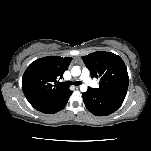
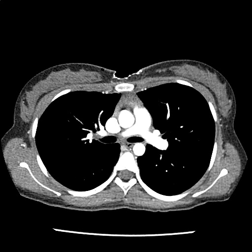
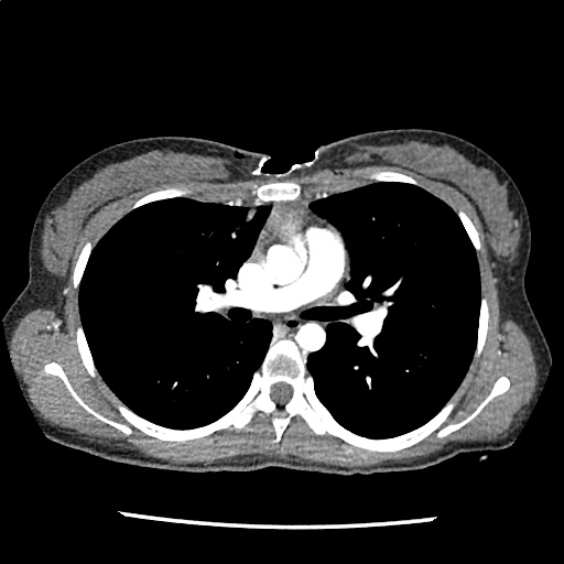
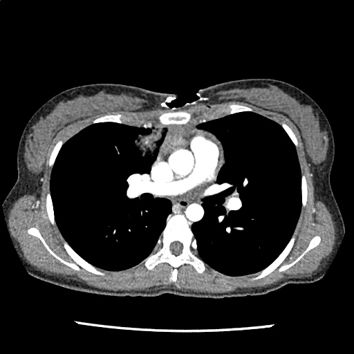
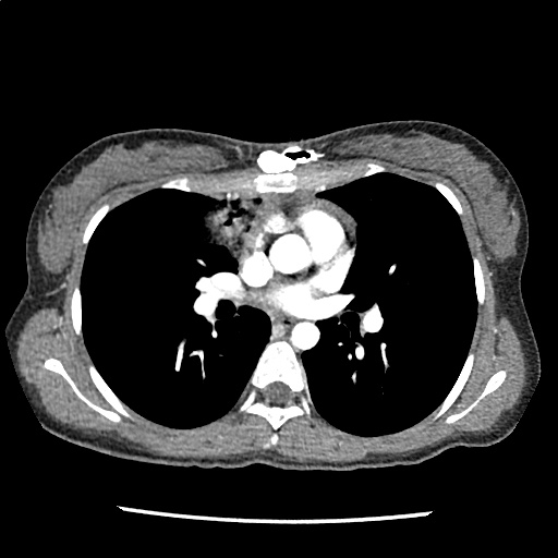
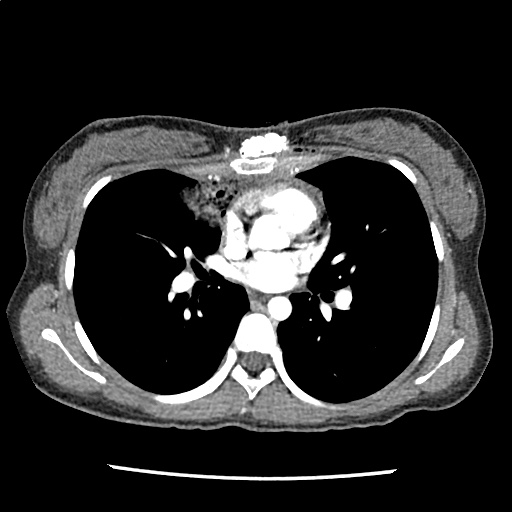
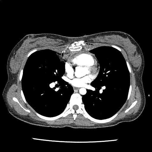
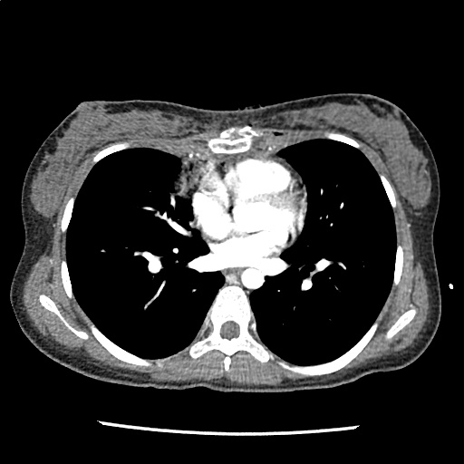
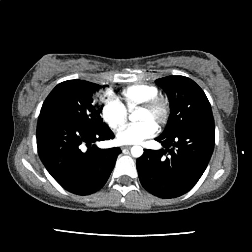
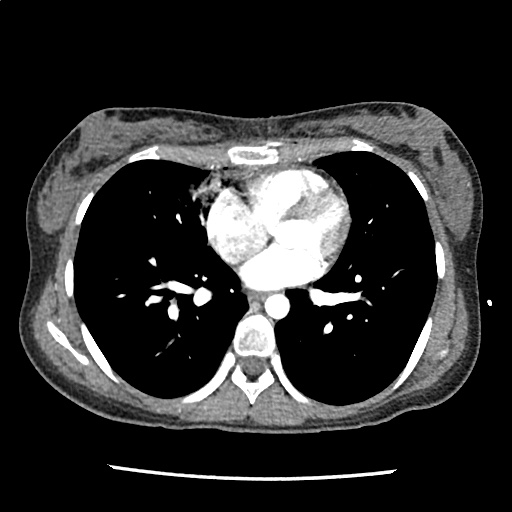
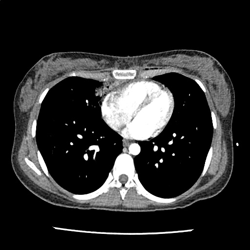
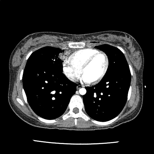
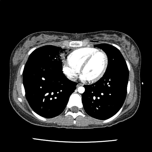
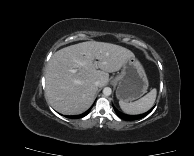
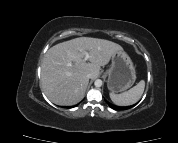
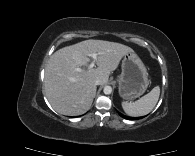
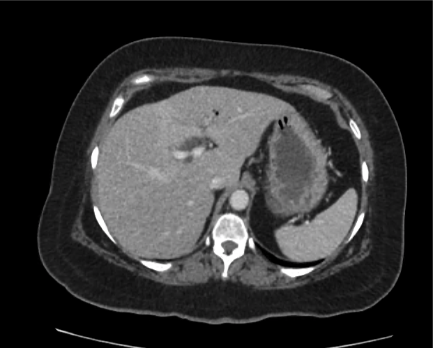
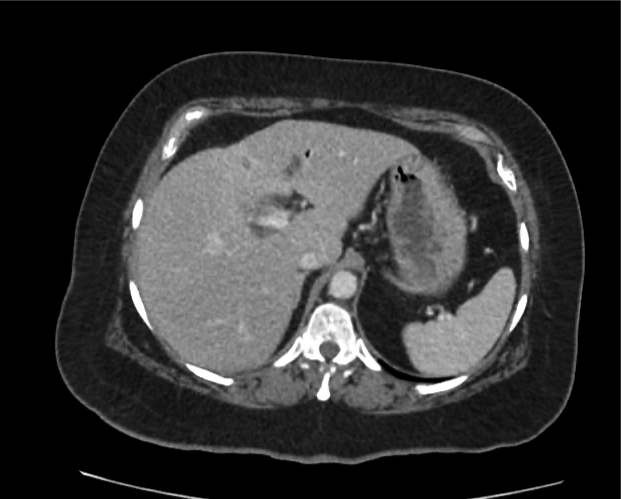
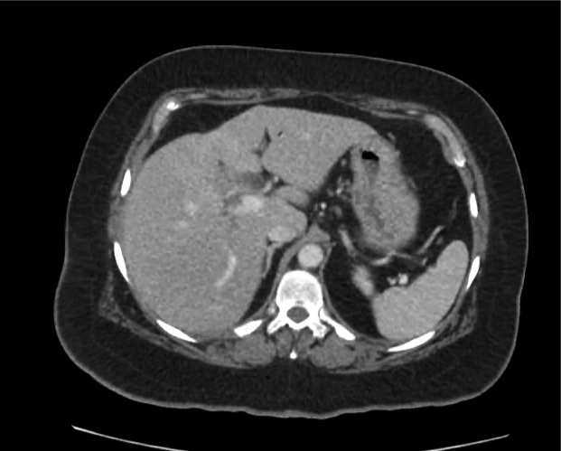
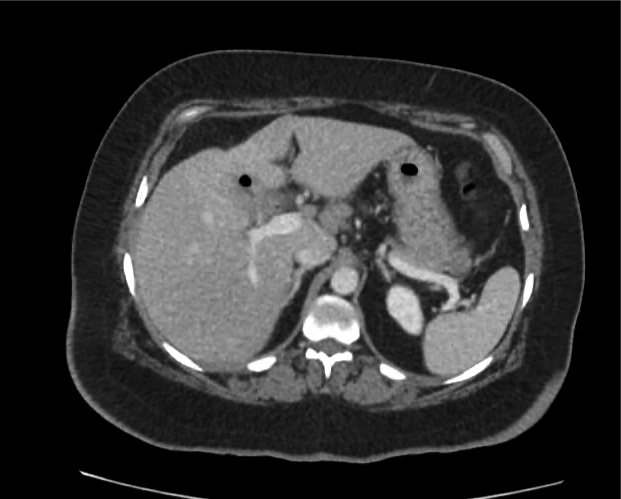
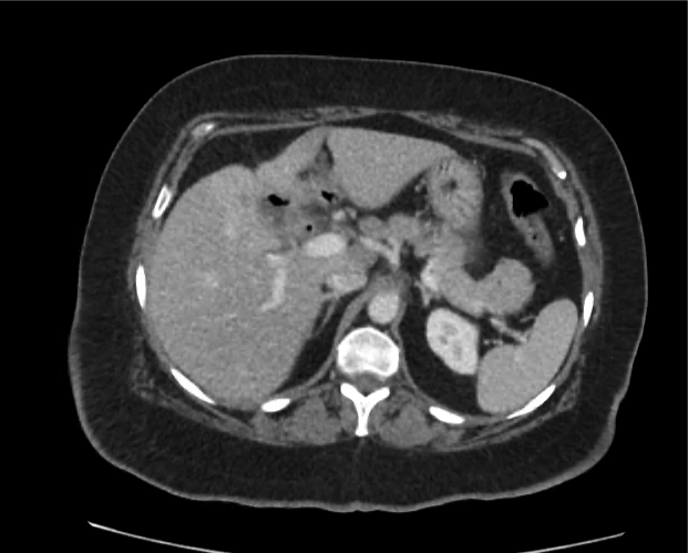
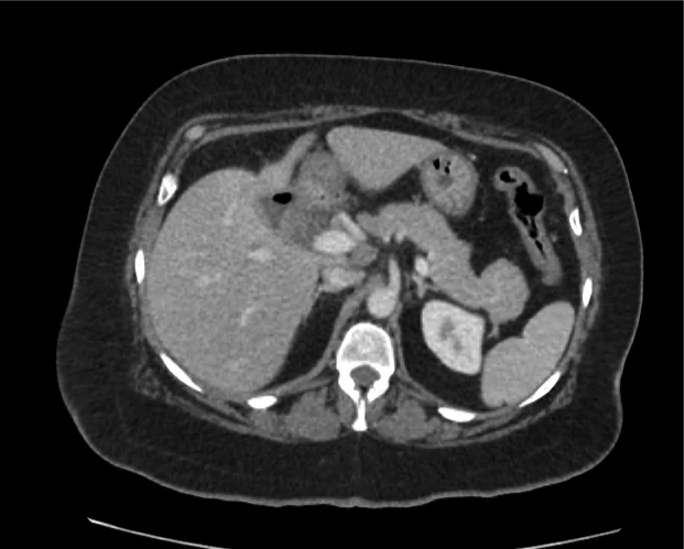
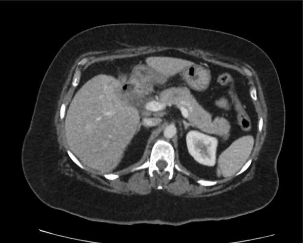
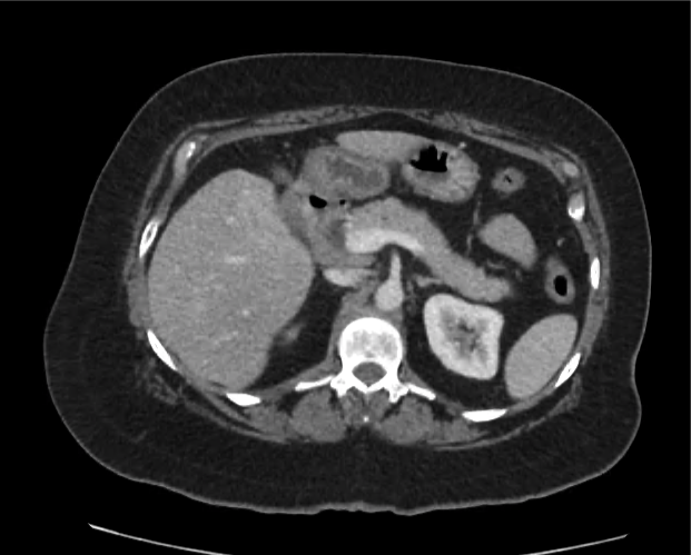
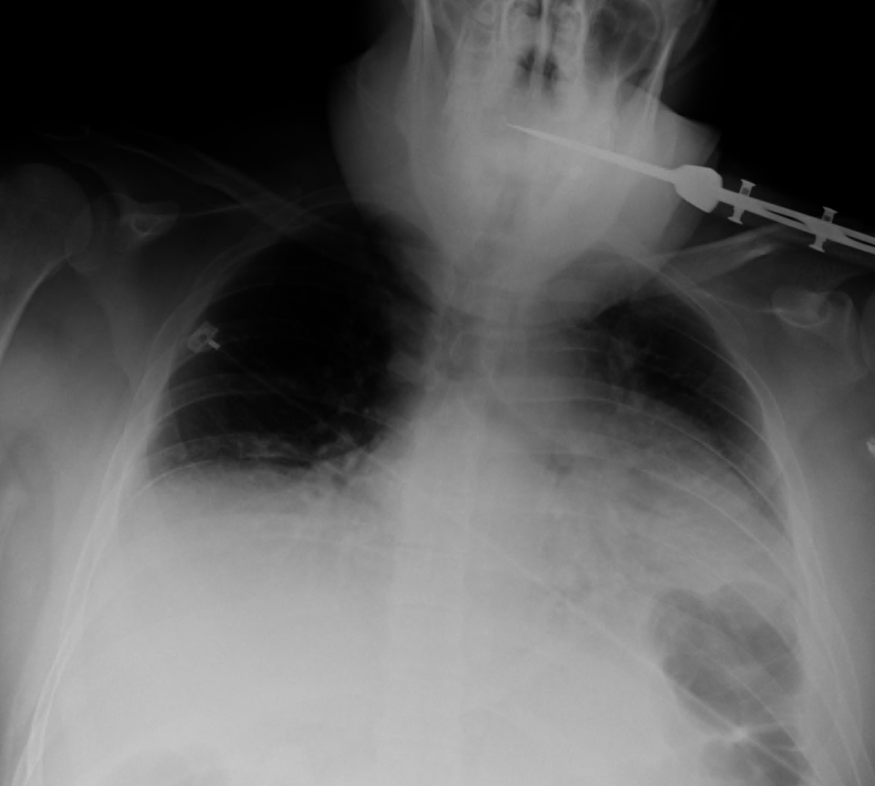
A 35-year-old female with no past medical history is brought in by ambulance to the emergency department. She was struck by a firework (“Roman Candle”) which lodged in her mid-chest until the propellant was consumed. She transiently lost consciousness but was awake upon EMS arrival. She complains of pleuritic chest pain. Examination reveals a circular 4x4cm full-thickness burn to the mid-chest with surrounding deep and superficial partial-thickness burns. Her ECG shows normal sinus rhythm, the initial serum troponin I is 32.9 (normal <0.012). CT angiography of the thorax is obtained.

bci_01
bci_02
bci_03
bci_04
bci_05
bci_06
bci_07
bci_08
bci_09
bci_10
bci_11
bci_12
bci_13

Mechanisms

Blunt cardiac injury (BCI) may be induced by multiple forces including direct thoracic trauma, crush injury of mediastinal contents between the sternum and thoracic spine, rapid deceleration causing tears at venous-atrial confluences, abrupt pressure changes from rapid compression of abdominal contents, blast injury, or laceration from bone fracture fragments1. The most common mechanisms of injury are motor vehicle collisions (50%), auto versus pedestrian (35%), motorcycle accidents (9%) and falls from significant height (>6m)2.

BCI represents a spectrum of conditions. Diagnosis is both challenging and critical as clinical manifestations can be absent or rapidly fatal.

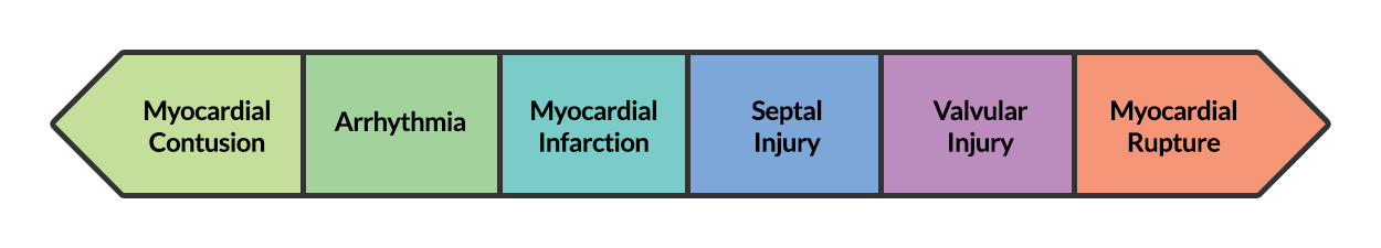
At one end of the spectrum is myocardial contusion. The lack of a gold-standard for the diagnosis of this clinical entity has led to a preference for describing associated abnormalities if present3,4, including cardiac dysfunction (identified on echocardiography) or the next entity along the spectrum – arrhythmia.

The most common arrhythmia identified in blunt cardiac injury is sinus tachycardia, followed by premature atrial or ventricular contractions, T-wave changes, and atrial fibrillation or flutter5. Commotio cordis is a unique arrhythmia induced by untimely precordial impact (often in sports) during a vulnerable phase of ventricular excitability, resulting in ventricular fibrillation2.

ST-segment elevations after blunt cardiac injury should raise concern for myocardial infarction due to coronary artery dissection, laceration or thrombosis (often in already-diseased vessels) 5,6.

The remaining disease entities are increasingly rare, require careful examination or imaging for diagnosis, and are more likely to be non-survivable. Septal injury can range from small tears to rupture. Valvular injury most commonly affects the aortic valve (followed by mitral and tricuspid valves) and involves damage to leaflets, or rupture of papillary muscles or chordae tendineae. The clinical presentation is of acute valvular insufficiency, including acute heart failure and murmur2,7,8. A widened pulse pressure may be noted with aortic valve injury, and the manifestations of valvular injury may be delayed9. Finally, myocardial wall rupture is unlikely to be survivable, though patients may present with cardiac tamponade if rupture is small or contained2.

Evaluation

The primary diagnostic modalities for the assessment of BCI in the emergency department include assessment for pericardial fluid during the Focused Assessment with Sonography for Trauma (FAST), electrocardiography and cardiac enzymes.

While specific for identifying patients at risk of complications of BCI, electrocardiography alone is not sufficient to exclude BCI. In one study, only 59% of patient with echocardiographic evidence of BCI (wall-motion abnormalities, other chamber abnormalities) had initially abnormal ECG’s10. In another study, 41% of patients with initially normal ECG’s developed clinically significant abnormalities11. The use of specialized electrocardiography including right-sided ECG (proposed to better detect right-ventricular abnormalities which are more commonly associated with BCI) and signal-averaged ECG is not supported11,12.

Several studies have supported the use of serum troponin for the detection of clinically significant BCI – particularly in combination with electrocardiography. A prospective study in 2001 evaluating patients with blunt thoracic trauma using ECG at admission and 8-hours, as well as troponin I at admission, 4- and 8-hours had a negative predictive value of 100% for significant BCI (arrhythmia requiring treatment, shock, or structural cardiac abnormalities) in patients with initially normal ECG and troponin13.

Another prospective study adding to the population evaluated by Salim et al. included 41 patients with normal ECG’s and troponin levels at admission and 8-hours who were admitted for significant mechanisms, none developed significant BCI (again described as arrhythmia requiring treatment, shock, or structural cardiac abnormalities) after 1 to 3 days of observation14. The precise timing of serum troponin analysis remains unclear.

While FAST may detect hemopericardium warranting immediate intervention, formal echocardiography is indicated for patients with unexplained hypotension (to evaluate for valvular injury or regional wall-motion abnormalities) or persistent arrhythmias (to evaluate for arrhythmogenic intramural hematomas)15. The presence of sternal fractures was previously thought to increase risk of BCI and mandate echocardiography, however this notion is no longer supported16-18. The role of advanced imaging including helical CT (cardiac-gated), and MRI remains unclear19.

Algorithm for the Evaluation of Blunt Thoracic Trauma

Notes:
† Arrest in ED, immediate chest tube output >20ml/kg (>1.5L) or >200mL/hr for 2-4hr.

Management

Management of BCI depends on the pathologic process localized along the spectrum defined above. Persistent hypotension after appropriate evaluation for alternative etiologies may represent myocardial contusion with cardiac dysfunction and should be evaluated with echocardiography. Similarly, echocardiography and observation with continuous telemetry monitoring is indicated for any new arrhythmia or persistent and unexplained tachycardia. Patients with only elevation of the serum troponin without electrocardiographic abnormalities or obvious cardiac dysfunction should also be admitted for observation and serial cardiac enzymes. Traumatic myocardial infarction, valvular injury, or post-traumatic structural myocardial defects should be managed in consultation with cardiothoracic surgery5,19-21.

Case Conclusion

The CT interpretation noted the soft-tissue defect identified on examination as well as associated pulmonary contusions and a non-displaced sternal fracture. The patient went to the operating room for washout and debridement. A transthoracic echocardiogram demonstrated trace mitral regurgitation and a small pericardial effusion. She remained hemodynamically stable and serial troponin measures downtrended – no dysrhythmias were noted on telemetry monitoring. She was discharged on hospital day four with a negative-pressure wound dressing.

References

  1. Schultz JM, Trunkey DD. Blunt cardiac injury. Crit Care Clin. 2004;20(1):57-70.
  2. Yousef R, Carr JA. Blunt cardiac trauma: a review of the current knowledge and management. Ann Thorac Surg. 2014;98(3):1134-1140. doi:10.1016/j.athoracsur.2014.04.043.
  3. Mattox KL, Flint LM, Carrico CJ, et al. Blunt cardiac injury. The Journal of Trauma: Injury, Infection, and Critical Care. 1992;33(5):649-650.
  4. Sybrandy KC, Cramer MJM, Burgersdijk C. Diagnosing cardiac contusion: old wisdom and new insights. Heart. 2003;89(5):485-489.
  5. Elie M-C. Blunt cardiac injury. Mt Sinai J Med. 2006;73(2):542-552.
  6. Edouard AR, Felten M-L, Hebert J-L, Cosson C, Martin L, Benhamou D. Incidence and significance of cardiac troponin I release in severe trauma patients. Anesthesiology. 2004;101(6):1262-1268.
  7. Cordovil A, Fischer CH, Rodrigues ACT, et al. Papillary Muscle Rupture After Blunt Chest Trauma. Journal of the American Society of Echocardiography. 2006;19(4):469.e1-469.e3. doi:10.1016/j.echo.2005.12.005.
  8. Pasquier M, Sierro C, Yersin B, Delay D, Carron P-N. Traumatic Mitral Valve Injury After Blunt Chest Trauma: A Case Report and Review of the Literature. The Journal of Trauma: Injury, Infection, and Critical Care. 2010;68(1):243-246. doi:10.1097/TA.0b013e3181bb881e.
  9. Ismailov RM, Weiss HB, Ness RB, Lawrence BA, Miller TR. Blunt cardiac injury associated with cardiac valve insufficiency: trauma links to chronic disease? Injury. 2005;36(9):1022-1028. doi:10.1016/j.injury.2005.05.028.
  10. García-Fernández MA, López-Pérez JM, Pérez-Castellano N, et al. Role of transesophageal echocardiography in the assessment of patients with blunt chest trauma: correlation of echocardiographic findings with the electrocardiogram and creatine kinase monoclonal antibody measurements. Am Heart J. 1998;135(3):476-481.
  11. Fulda GJ, Giberson F, Hailstone D, Law A, Stillabower M. An evaluation of serum troponin T and signal-averaged electrocardiography in predicting electrocardiographic abnormalities after blunt chest trauma. The Journal of Trauma: Injury, Infection, and Critical Care. 1997;43(2):304–10–discussion310–2.
  12. Walsh P, Marks G, Aranguri C, et al. Use of V4R in patients who sustain blunt chest trauma. The Journal of Trauma: Injury, Infection, and Critical Care. 2001;51(1):60-63.
  13. Salim A, Velmahos GC, Jindal A, et al. Clinically significant blunt cardiac trauma: role of serum troponin levels combined with electrocardiographic findings. The Journal of Trauma: Injury, Infection, and Critical Care. 2001;50(2):237-243.
  14. Velmahos GC, Karaiskakis M, Salim A, et al. Normal electrocardiography and serum troponin I levels preclude the presence of clinically significant blunt cardiac injury. The Journal of Trauma: Injury, Infection, and Critical Care. 2003;54(1):45–50–discussion50–1. doi:10.1097/01.TA.0000046315.73441.D8.
  15. Nagy KK, Krosner SM, Roberts RR, Joseph KT, Smith RF, Barrett J. Determining which patients require evaluation for blunt cardiac injury following blunt chest trauma. World J Surg. 2001;25(1):108-111.
  16. Roy-Shapira A, Levi I, Khoda J. Sternal fractures: a red flag or a red herring? The Journal of Trauma: Injury, Infection, and Critical Care. 1994;37(1):59-61.
  17. Hills MW, Delprado AM, Deane SA. Sternal fractures: associated injuries and management. The Journal of Trauma: Injury, Infection, and Critical Care. 1993;35(1):55-60.
  18. Rashid MA, Ortenwall P, Wikström T. Cardiovascular injuries associated with sternal fractures. Eur J Surg. 2001;167(4):243-248. doi:10.1080/110241501300091345.
  19. Clancy K, Velopulos C, Bilaniuk JW, et al. Screening for blunt cardiac injury: an Eastern Association for the Surgery of Trauma practice management guideline. J Trauma Acute Care Surg. 2012;73(5 Suppl 4):S301-S306. doi:10.1097/TA.0b013e318270193a.
  20. El-Menyar A, Thani Al H, Zarour A, Latifi R. Understanding traumatic blunt cardiac injury. Ann Card Anaesth. 2012;15(4):287-295. doi:10.4103/0971-9784.101875.
  21. Hockberger RS, Walls RM. Rosen’s Emergency Medicine. Mosby Incorporated; 2002.

Arterial Pressure Indices

Indications

  • Assess for peripheral arterial disease (PAD)
  • Assess for risk of arterial injury in trauma

Test characteristics

  • PAD: ABI <0.90 sensitivity 95%, specificity 100% for >50% stenosis on arteriography1
  • Trauma: API <0.90 sensitivity 95%, specificity 97% for major arterial injury2

Technique for obtaining arterial pressure indices3

  1. Patient lies supine with extremities at level of the heart for 10 minutes
  2. Ankle: cuff positioned just above malleolus
  3. Brachial: cuff positioned just above antecubital fossa
  4. Doppler SBP
  5. Sequence: first arm, first PT, first DP, other PT, other DP, other arm. If SBP of first arm >10mmHg compared to second arm, repeat first arm and disregard first measure
  6. Calculation: ABI = higher of DP or PT / higher arm

Interpretation of ABI for PAD3

Value Interpretation
0-0.40 Severe PAD, rest pain, gangrene
0.41-0.90 PAD, claudication
0.91-1.30 Normal
>1.30 Non-compressible, severely calcified

Algorithm for the Evaluation of Arterial Injury4, 5

Algorithm for the Evaluation of Arterial Injury

Notes:

  • † If unable to palpate pulses due to patient habitus or shock/hypothermia, reattempt with oversized cuff and after appropriate rewarming/resuscitation. If remains challenging, proceed with imaging.
  • ‡ Proximal LE arterial injuries refers to the major named arteries of the thigh (excluding the profunda femoris) and proximal to the anterior tibial artery and tibioperoneal bifurcation.

Arteries of the Lower Leg

References:

  1. Mohler ER. Peripheral arterial disease: identification and implications. Arch Intern Med. 2003;163(19):2306-2314. doi:10.1001/archinte.163.19.2306.
  2. Johansen K, Lynch K, Paun M, Copass M. Non-invasive vascular tests reliably exclude occult arterial trauma in injured extremities. The Journal of Trauma: Injury, Infection, and Critical Care. 1991;31(4):515–9–discussion519–22.
  3. Aboyans V, Criqui MH, Abraham P, et al. Measurement and interpretation of the ankle-brachial index: a scientific statement from the American Heart Association. Circulation. 2012;126(24):2890-2909. doi:10.1161/CIR.0b013e318276fbcb.
  4. Feliciano DV, Moore FA, Moore EE, et al. Evaluation and management of peripheral vascular injury. Part 1. Western Trauma Association/critical decisions in trauma. J Trauma. 2011;70(6):1551-1556. doi:10.1097/TA.0b013e31821b5bdd.
  5. Fox N, Rajani RR, Bokhari F, et al. Evaluation and management of penetrating lower extremity arterial trauma: an Eastern Association for the Surgery of Trauma practice management guideline. J Trauma Acute Care Surg. 2012;73(5 Suppl 4):S315-S320. doi:10.1097/TA.0b013e31827018e4.
  6. Inaba K, Branco BC, Reddy S, et al. Prospective evaluation of multidetector computed tomography for extremity vascular trauma. J Trauma. 2011;70(4):808-815. doi:10.1097/TA.0b013e3182118384.

Thromboelastography

Thromboelastography (TEG) is an assessment of hemostatic function intended to evaluate in vivo coagulation parameters, guiding the targeted correction of coagulopathy1. TEG has predominantly been studied in cardiac surgery, though research has extended to other peri-operative and peri-procedural transfusion management2-5.

Recently, a randomized trial explored the use of TEG to guide transfusion in trauma patients requiring massive transfusion6. 111 patients meeting requirements for massive transfusion protocol activation were randomized to a conventional coagulation assay (CCA) or TEG-guided transfusion algorithm. Patients in the TEG group demonstrated significantly decreased mortality at 28 days and reductions in plasma and platelet transfusion requirements.

More research is needed before TEG can be recommended for use in trauma resuscitation or other common emergency department applications7,8, however it may be useful to prepare by becoming familiar with the most basic aspects of thromboelastography.

Thromboelastography Summary

Thromboelastography Summary

Examples

Normal

Normal

Anti-coagulants

Anti-coagulants

R,K: Increased
Angle: Decreased

Anti-Platelet

Anti-Platelet

R: Normal
K: Increased
MA: Decreased

Hypercoagulable

Hypercoagulable

R,K: Decreased
MA: Increased

FIbrinolysis

FIbrinolysis

MA: Decreasing
LY30: Increased

DIC (Phase 1)

DIC (Phase 1)

R,K: Decreased
MA: Increased
LY30: Increased

DIC (Phase 2)

DIC (Phase 2)

R,K: Increased
MA: Decreased

References

  1. Bolliger D, Seeberger MD, Tanaka KA. Principles and Practice of Thromboelastography in Clinical Coagulation Management and Transfusion Practice. Transfusion Medicine Reviews. 2012;26(1):1-13. doi:10.1016/j.tmrv.2011.07.005.
  2. Porte RJ, Bontempo FA, Knot EA, Lewis JH, Kang YG, Starzl TE. Systemic effects of tissue plasminogen activator-associated fibrinolysis and its relation to thrombin generation in orthotopic liver transplantation. Transplantation. 1989;47(6):978-984.
  3. Rahe-Meyer N, Solomon C, Hanke A, et al. Effects of fibrinogen concentrate as first-line therapy during major aortic replacement surgery: a randomized, placebo-controlled trial. Anesthesiology. 2013;118(1):40-50. doi:10.1097/ALN.0b013e3182715d4d.
  4. Weber CF, Klages M, Zacharowski K. Perioperative coagulation management during cardiac surgery. Current Opinion in Anaesthesiology. 2013;26(1):60-64. doi:10.1097/ACO.0b013e32835afd28.
  5. De Pietri L, Bianchini M, Montalti R, et al. Thrombelastography-guided blood product use before invasive procedures in cirrhosis with severe coagulopathy: A randomized, controlled trial. Hepatology. 2016;63(2):566-573. doi:10.1002/hep.28148.
  6. Gonzalez E, Moore EE, Moore HB, et al. Goal-directed Hemostatic Resuscitation of Trauma-induced Coagulopathy. Ann Surg. 2016;263(6):1051-1059. doi:10.1097/SLA.0000000000001608.
  7. Afshari A, Wikkelsø A, Brok J, Møller AM, Wetterslev J. Thrombelastography (TEG) or Thromboelastometry (ROTEM) to Monitor Haemotherapy Versus Usual Care in Patients with Massive Transfusion. Vol 24. (Afshari A, ed.). Chichester, UK: John Wiley & Sons, Ltd; 1996:404–3. doi:10.1002/14651858.CD007871.pub2.
  8. da Luz LT, Nascimento B, Rizoli S. Thrombelastography (TEG®): practical considerations on its clinical use in trauma resuscitation. Scand J Trauma Resusc Emerg Med. 2013;21(1):29. doi:10.1186/1757-7241-21-29.

Pneumobilia: Hepatic Gas Applied

Brief HPI

A 45 year-old female with a history of pre-diabetes and gastroesophageal reflux disease presents with 3 days of epigastric abdominal pain. She describes constant, burning abdominal pain which worsened on the day of presentation associated with two episodes of non-bloody and non-bilious emesis. The patient was tender to palpation in the epigastrium and right upper quadrant.

Right upper quadrant ultrasound

Laboratory studies were largely normal. A complete blood count demonstrated minimal leukocytosis (11.6 with normal differential), and liver function tests were normal.

A right-upper quadrant ultrasound was obtained which demonstrated “strongly shadowing structures in the gallbladder fossa which might represent a wall-echo-shadow, calcified gallbladder wall, or air within the gallbladder”.

The patient underwent contrast-enhanced computed tomography of the abdomen and pelvis which is shown below.

Imaging

cyst-gastric_01
cyst-gastric_02
cyst-gastric_03
cyst-gastric_04
cyst-gastric_05
cyst-gastric_06
cyst-gastric_07
cyst-gastric_08
cyst-gastric_09
cyst-gastric_10
cyst-gastric_11
cyst-gastric_12
cyst-gastric_13
cyst-gastric_14
cyst-gastric_15

CT Abdomen/Pelvis with Contrast

Pneumobilia, intra- and extra-hepatic biliary duct dilation, pericholecystic fat stranding, and an air-fluid level within a contracted gallbladder. Mildly dilated loops of ileal bowel with a possible transition point in the right lower quadrant. Findings suggestive of possible gallstone ileus.

The patient was taken to the operating room for exploratory laparotomy, possible cholecystectomy and possible small bowel resection for presumed gallstone ileus. Intra-operative findings were notable for a cholecystogastric fistula which was repaired.

Differentiation between Portal Venous Gas and Pneumobilia

The patient’s CT demonstrated mostly central hepatic gas. This finding combined with the presence of an air-fluid level in the gallbladder was most consistent with pneumobilia. This case demonstrates an application of the previously-developed algorithm for the evaluation of hepatic gas in a relatively unique pathologic process.
Hepatic Gas: Pneumobilia vs. Portal Venous Gas

Penetrating Neck Trauma

Brief H&P

A young male presents to the emergency department after a self-inflicted stab wound to the neck. Examination revealed a knife handle protruding from the left lateral neck. A plain radiograph is shown below.

CXR: Radiopaque foreign body in left neck.

The patient was initially stable but developed shortness of breath upon attempting to lie flat for advanced imaging and was taken emergently to the operating room. Neck exploration showed no obvious neurovascular injuries, and the course of the 6cm blade was posterior to the trachea and esophagus. The knife was removed with “considerable force” as it was likely lodged within a portion of vertebral bone. The patient underwent esophagoscopy and bronchoscopy without identified tracheoesophageal injuries. The patient did well post-operatively and was discharged home.

Zones of Injury1-3

Previously, the evaluation and management of hemodynamically stable patients with penetrating neck injury was guided by the anatomic “zone” of injury. The affected zone guided the performance of additional diagnostic procedures including potentially morbid neck explorations.

Neck Zones of Injury

Understanding zone definitions remains important for the emergency physician to appreciate potentially implicated underlying structures. However, the advent of modern imaging modalities, specifically computed tomography with angiography, provides appropriate sensitivity for vascular and tracheoesophageal injuries when combined with detailed physical examination and maintenance of an appropriate threshold for the performance of additional studies if warranted by the clinical presentation (suboptimal imaging, concerning projectile trajectory, etc).

Zone Definition
I Clavicles/sternum to cricoid cartilage
II Cricoid cartilage to the angle of mandible
III Superior to the angle of mandible to the skull base


Algorithm for the Evaluation of Penetrating Neck Trauma


References

  1. Sperry JL, Moore EE, Coimbra R, et al. Western Trauma Association critical decisions in trauma: penetrating neck trauma. J Trauma Acute Care Surg. 2013;75(6):936-940. doi:10.1097/TA.0b013e31829e20e3.
  2. Brywczynski JJ, Barrett TW, Lyon JA, Cotton BA. Management of penetrating neck injury in the emergency department: a structured literature review. Emerg Med J. 2008;25(11):711-715. doi:10.1136/emj.2008.058792.
  3. Shiroff AM, Gale SC, Martin ND, et al. Penetrating neck trauma: a review of management strategies and discussion of the “No Zone” approach. Am Surg. 2013;79(1):23-29. doi:10.1007/978-3-662-49859-0_29.


Pediatric Head Trauma

Brief H&P:

A young child, otherwise healthy, is brought to the pediatric emergency department after a fall. The parents report a fall from approximately 2 feet after which the patient cried immediately and without apparent loss of consciousness. Over the course of the day, the patient developed an enlarging area of swelling over the left head. The parents were concerned about a progressive decrease in activity and interest in oral intake by the child, and they were brought to the emergency department for evaluation. Examination demonstrated a well-appearing and interactive child – appropriate for age. Head examination was notable for a 5x5cm hematoma over the left temporoparietal skull with an underlying palpable skull irregularity not present on the contralateral side. Non-contrast head computed tomography was obtained.

Imaging

ct_head_0007_Screen-Shot-2017-06-05-at-4.18.59-PM
ct_head_0006_Screen-Shot-2017-06-05-at-4.19.02-PM
ct_head_0005_Screen-Shot-2017-06-05-at-4.19.04-PM
ct_head_0004_Screen-Shot-2017-06-05-at-4.19.08-PM
ct_head_0003_Screen-Shot-2017-06-05-at-4.19.10-PM
ct_head_0002_Screen-Shot-2017-06-05-at-4.19.15-PM
ct_head_0001_Screen-Shot-2017-06-05-at-4.19.18-PM
ct_head_0000_Screen-Shot-2017-06-05-at-4.19.22-PM

CT Head

Fracture of the left temporal and parietal bone with overlying scalp hematoma.

Algorithm for the Evaluation of Pediatric Head Trauma (PECARN)1,2,3

Algorithm for the evaluation of pediatric head trauma

References

  1. Kuppermann N, Holmes JF, Dayan PS, et al. Identification of children at very low risk of clinically-important brain injuries after head trauma: a prospective cohort study. Lancet. 2009;374(9696):1160-1170. doi:10.1016/S0140-6736(09)61558-0.
  2. Brenner D, Elliston C, Hall E, Berdon W. Estimated risks of radiation-induced fatal cancer from pediatric CT. American Journal of Roentgenology. 2001;176(2):289-296. doi:10.2214/ajr.176.2.1760289.
  3. Schonfeld D, Bressan S, Da Dalt L, Henien MN, Winnett JA, Nigrovic LE. Pediatric Emergency Care Applied Research Network head injury clinical prediction rules are reliable in practice. Archives of Disease in Childhood. 2014;99(5):427-431. doi:10.1136/archdischild-2013-305004.

Spontaneous Intracranial Hemorrhage

Brief HPI

An approximately 40 year-old male with a history of aortic stenosis s/p mechanical aortic valve replacement (on Coumadin) as well as hypertension presented to the emergency department with a chief complaint of severe headache. The patient was in severe distress on arrival and was unable to provide detailed history, he complained of two days of severe left-sided headache while clutching his head and groaning. Examination was notable for sensory localization with directed movements of right hemibody, and no apparent response on the left. He was taken to emergently for CT head non-contrast.

Imaging

ich_0000_Layer-Comp-1
ich_0001_Layer-Comp-2
ich_0002_Layer-Comp-3
ich_0003_Layer-Comp-4
ich_0004_Layer-Comp-5
ich_0005_Layer-Comp-6
ich_0006_Layer-Comp-7
ich_0007_Layer-Comp-8
ich_0008_Layer-Comp-9
ich_0009_Layer-Comp-10
ich_0010_Layer-Comp-11
ich_0011_Layer-Comp-12

CT Head non-contrast

57 mm right posterior parenchymal hemorrhage with intraventricular component. Moderate edema, mass effect and 9 mm of midline shift.

ED Course

Admission INR was 2.9, the patient received 25 units/kg of PCC as well as vitamin K 10mg IV x1. Neurosurgery was consulted and the patient was taken to the operating room for management.

Management of Supratherapeutic INR and Complications of Anti-Coagulation

Management of Supratherapeutic INR

References

  1. Ansell J, Hirsh J, Hylek E, et al. Pharmacology and management of the vitamin K antagonists: American College of Chest Physicians Evidence-Based Clinical Practice Guidelines (8th Edition). Chest 2008; (6 Suppl):160s

Severe Burns

ED Presentation

34F with no reported medical history BIBA with severe burns after house fire with estimated 70% TBSA involvement. On arrival, the patient was hypoxic, striderous, and unable to provide history. She was intubated for airway protection with some difficulty. Examination revealed deep partial and full-thickness burns to 70% of total body surface area including circumferential burns to bilateral upper extremities and extensive neck and anterior chest involvement. Initial fluid resuscitation and warming measures were instituted. Emergent bedside bronchoscopy revealed copious carbonaceous material throughout with attempts at lavage. Urine output was minimal despite aggressive resuscitation. Critical care transport to local burn facility was arranged where the patient ultimately expired.

Algorithm for the Management of Severe Burns

Algorithm for the Management of Severe Burns

Assessment of Burn Depth

Depth Cause Appearance Sensation
Superficial UV exposure Dry, red
Blanching
Painful
Superficial partial-thickness Scald (splash)
Short flash
Blisters, moist, red
Blanching
Painful to temperature/air
Deep partial-thickness Scald (spill)
Flame, oil, grease
Blisters, waxy dry, white/red
Non-blanching
Pressure
Full-thickness Scald (immersion)
Flame, steam, oil, grease, chemical, electrical
Waxy white, leathery grey, black
Non-blanching
Deep pressure

Estimating Burn Surface Area

Burn TBSA

Image from UWHealth.org

  • Trunk: 18% anterior, 18% posterior
  • Lower extremity (each): 9% anterior, 9% posterior
  • Upper extremity (each): 9%
  • Head/neck: 9%
  • Perineum: 1%

Burn Transfer Criteria

  • Partial thickness > 20% TBSA
  • Partial thickness > 10% TBSA for extremes of age (<10 or >50 years-old)
  • Any full-thickness
  • Burns involving face, hands, feet, genitalia, major joints
  • Electrical/chemical
  • Inhalation injury
  • Medical comorbidities impacting management/healing

See Also

References

  1. Monafo WW. Initial management of burns. N Engl J Med. 1996;335(21):1581–1586. doi:10.1056/NEJM199611213352108.
  2. Hettiaratchy S, Papini R. Initial management of a major burn: I–overview. BMJ. 2004;328(7455):1555–1557. doi:10.1136/bmj.328.7455.1555.
  3. Singer AJ, Della-Giustina D. Thermal Burns: Rapid Assessment and Treatment. Emergency Medicine Practice; 2000.
  4. Rice, PL. Emergency care of moderate and severe thermal burns in adults. In: UpToDate, Moreira ME (Ed), UpToDate, Waltham, MA. (Accessed on March 29, 2016)
  5. Gauglitz, GG. Overview of the management of the severely burned patient. In: UpToDate, Jeschke MG (Ed), UpToDate, Waltham, MA. (Accessed on March 29, 2016)

Necrotizing Soft-Tissue Infection (NSTI)

HPI:

40 year-old male with a history of diabetes presents with right foot pain and swelling. His symptoms began 3 days ago with pain on the lateral surface of his right foot, described as aching, non-radiating and exacerbated with walking. Yesterday, he noted more prominent swelling and redness involving 4th and 5th toes. He denies trauma, fevers, and discharge.

PMH:

  • Diabetes mellitus, diagnosed 8yrs ago

PSH:

  • None

FH:

  • Non-contributory

SHx:

  • Lives with wife and 2 children and works an office job.
  • Ten year history of tobacco use, quit 3 years ago.
  • No EtOH or drug abuse.

Meds:

  • Metformin 500mg p.o. b.i.d.
  • Ibuprofen p.r.n. joint pain

Allergies:

NKDA

Physical Exam:

VS: T 101.2 HR 88 RR 14 BP 147/71 O2 100% RA
Gen: Obese male, pleasant and in no acute distress, lying in bed with right foot raised.
HEENT: PERRL, EOMI, dry mucous membranes.
CV: RRR, normal S1/S2, no extra heart sounds, no murmurs.
Lungs: CTAB
Abd: +BS, non-tender.
Ext: Right lower extremity with 8x8cm area of erythema predominantly involving lateral aspect of foot, dorsum of foot and 3-5th digits. There is a shallow, 1x1cm ulcer on the plantar surface of foot near 5th MTP. Area is also notable for ecchymosis and palpable crepitus. There is minimal tenderness to palpation or with active/passive range of motion.
Skin: The remainder of the skin exam is unremarkable.
Neuro: AAOx3.

Labs/Studies:

  • BMP: 134/4.3/104/26/18/1.4/206
  • WBC: 27.3/13.1/40/189 (90% neutrophils)
  • Lactate: 1.2
  • CRP: [pending]

Imaging:

CT Lower Extremity

  1. Calf cellulitis and gas-producing cellulitis in the lateral foot and toes.
  2. Thigh and inguinal lymphadenopathy.
  3. Although gas is seen down to the level of the bone, no definite bony changes are identified to establish a diagnosis of osteomyelitis. Please note that MRI is more sensitive for detection of early osteomyelitis.

Assessment/Plan:

40M with DM and diabetic foot ulcer resulting in a necrotizing soft tissue infection as evidenced by gas on imaging. Recommended surgical debridement and started on broad-spectrum antibiotics including:

  • vancomycin 1g i.v. q.12.h.
  • cefepime 2g i.v. q.8.h.
  • metronidazole 500mg i.v. q.8.h.

The patient underwent amputation of 3-5th digits with good surgical margins and was discharged on post-operative day three in good condition.

Skin and soft-tissue layers and their infections: 1

Skin and soft-tissue layers and their infections

Necrotizing Soft-Tissue Infections (NSTI):2,3,4

Risk Factors

  • IVDA
  • Comorbid conditions
    • DM
    • Obesity
    • Immunosuppression

Physical Exam

  • Early (non-specific)
    • Swelling
    • Erythema
    • Pain
  • Late (non-sensitive)
    • Tense edema outside affected skin perimeter
    • Disproportionate pain
    • Ecchymosis
    • Bullae
    • Crepitus
    • Systemic signs (fever, tachycardia, hypotension)

Treatment

  • Surgical debridement
  • Antimicrobials
    • Carbapenem, combination B-lactam B-lactamase
    • Vancomycin, linezolid (MRSA coverage)
    • Clindamycin (inhibit protein synthesis)
  • Supportive therapy

LRINEC score 5

Name Value Score
CRP ≥150 4
WBC 15-25
>25
1
2
Hb 11-13.5
<11
1
2
Na <135 2
Creatinine >1.6 2
Glucose >180 1

<5 Low risk, 6-7 Intermediate risk, >8 High risk

References:

  1. Morchi, R. (2/18/14). Emergency Medicine Procedures Cadaver Lab. Clinical Clerkship at UCLA. Los Angeles, CA.
  2. Goldstein, E. J. C., Anaya, D. A., & Dellinger, E. P. (2007). Necrotizing Soft-Tissue Infection: Diagnosis and Management. Clinical infectious diseases, 44(5), 705–710. doi:10.1086/511638
  3. Headley, A. J. (2003). Necrotizing soft tissue infections: a primary care review. American family physician, 68(2), 323–328.
  4. McHenry, C. R., Piotrowski, J. J., Petrinic, D., & Malangoni, M. A. (1995). Determinants of mortality for necrotizing soft-tissue infections. Annals of surgery, 221(5), 558–63.
  5. Wong, C.-H., Khin, L.-W., Heng, K.-S., Tan, K.-C., & Low, C.-O. (2004). The LRINEC (Laboratory Risk Indicator for Necrotizing Fasciitis) score: A tool for distinguishing necrotizing fasciitis from other soft tissue infections. Critical Care Medicine, 32(7), 1535–1541. doi:10.1097/01.CCM.0000129486.35458.7D

Skull Fracture

Frontal bone fractureID:

14 year-old female, previously healthy, brought in by ambulance s/p auto vs. pedestrian.

HPI:

Incident unwitnessed, paramedics report no LOC with GCS 15 at scene. GCS 10 upon arrival to ED, with 2min GTC seizure. Patient intubated for airway protection and CT head showed non-displaced frontal bone fracture and small frontal SAH. Patient self-extubated, returned to baseline mental status and was transferred to PICU.

PE:

  • VS: 128/76mmHg, 120bpm, 22 R/min, 100% RA, 37.6°C
  • General: Alert and responsive young female with multiple bandages on extremities
  • HEENT: Right frontal hematoma, no bony defect palpated, multiple facial abrasions, no otorrhea, no rhinorrhea, TM clear b/l, no other ecchymosis.
  • CV: RRR, normal S1/S2, no M/R/G
  • Lungs: CTAB
  • Abdomen: +BS, soft, NT/ND, no rebound/guarding, no flank ecchymoses
  • Neuro: AAOx3, CN II-XII intact, sensation/motor/reflexes symmetric and intact.
  • Extremities: Well-perfused with good pulses, no focal bony tenderness, no joint effusions, multiple abrasions on extensor surfaces of all four extremities.

Assessment & Plan:

14yo female, previously healthy, s/p auto vs. peds followed by GTC seizure and CT head showing small SAH and non-displaced frontal bone skull fracture. No evidence of basilar skull fracture on examination or imaging. Seizure likely 2/2 irritation from SAH. Patient was followed closely in PICU with q1h neuro checks with low threshold for repeat CT if change in mental status or more seizures occurred. The patient was eventually transferred to the general ward and was discharged with neurology follow-up and Keppra for seizure prophylaxis for 6mo.

Types of Skull Fractures:

A system for skull fractures