Anticoagulant Reversal in Intracranial Hemorrhage

Brief HPI:

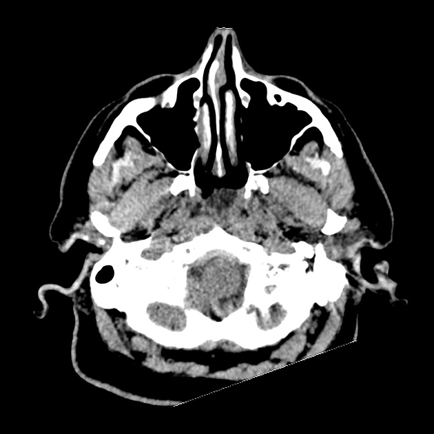
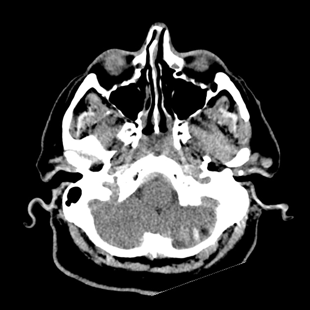
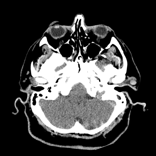
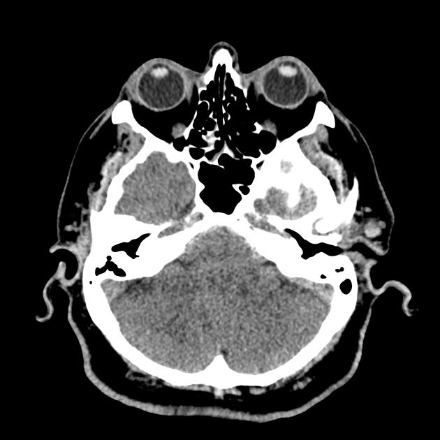
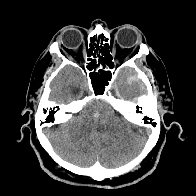
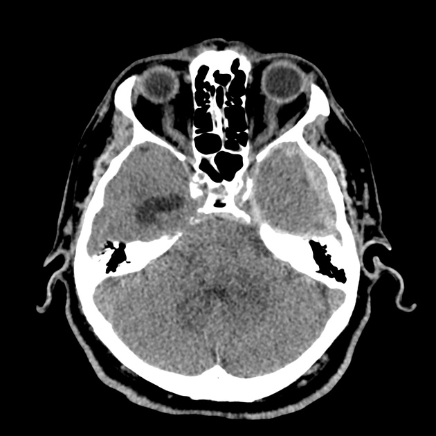
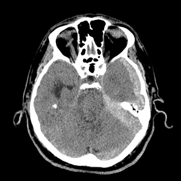
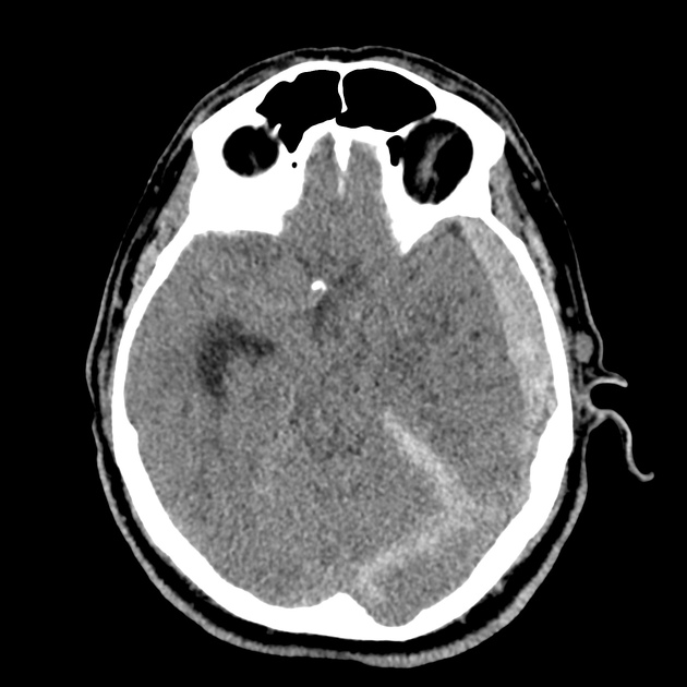
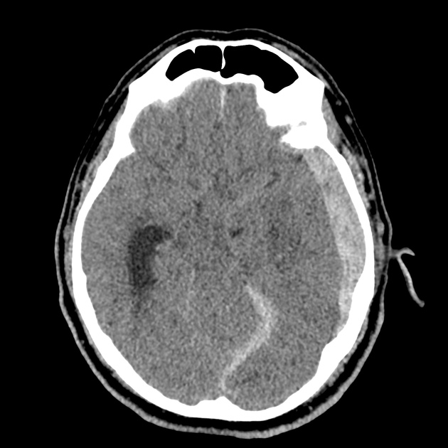
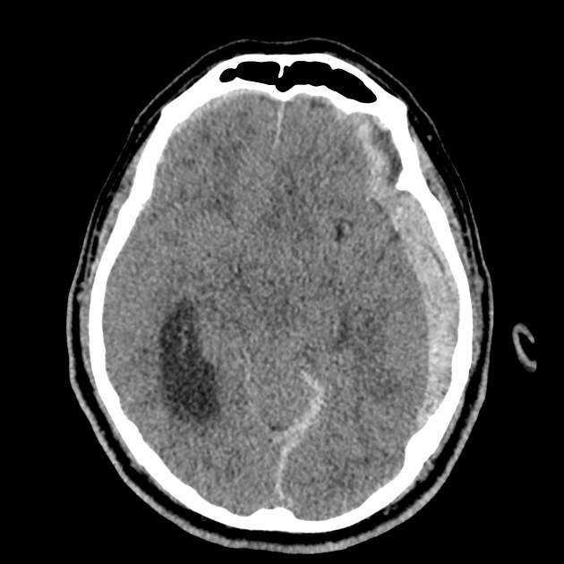
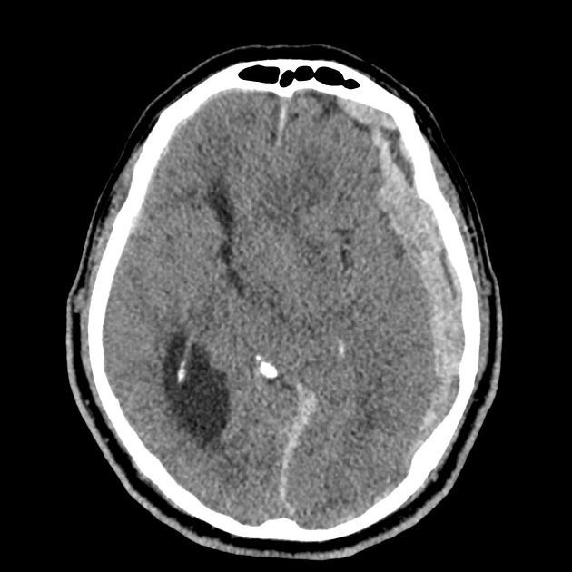
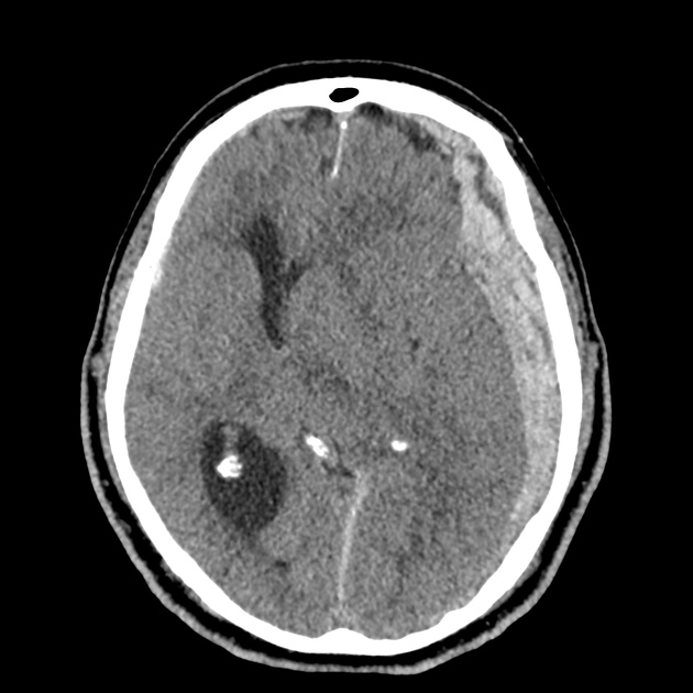
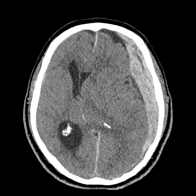
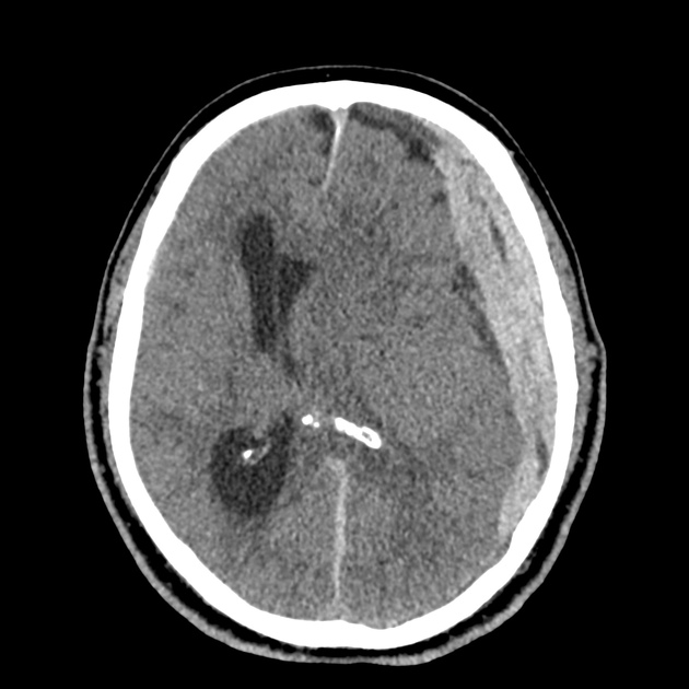
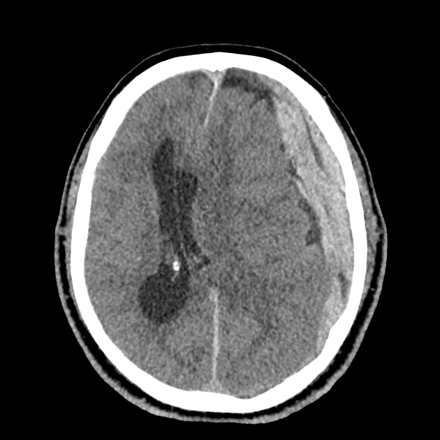
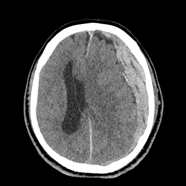
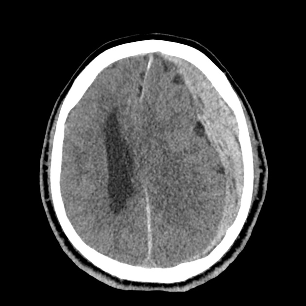
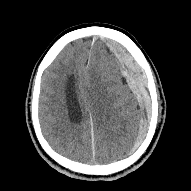
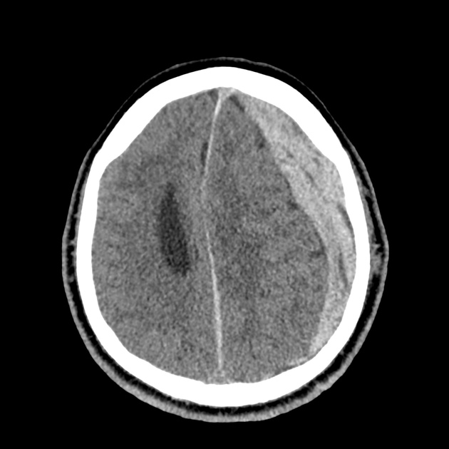
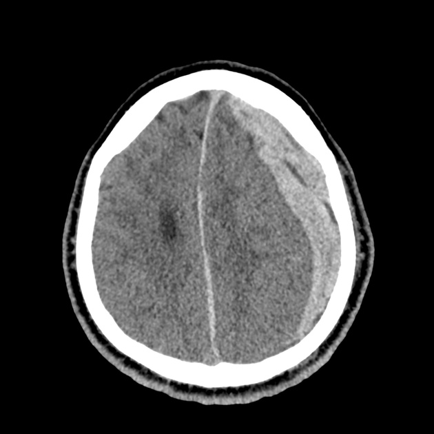
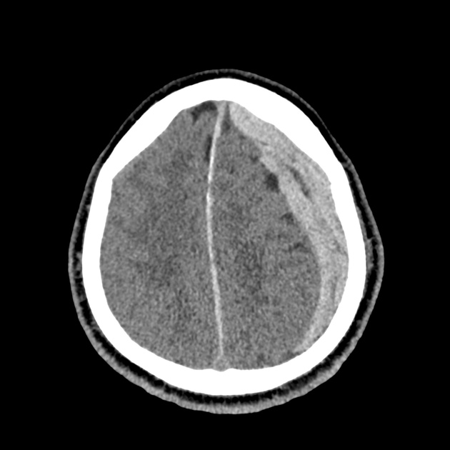
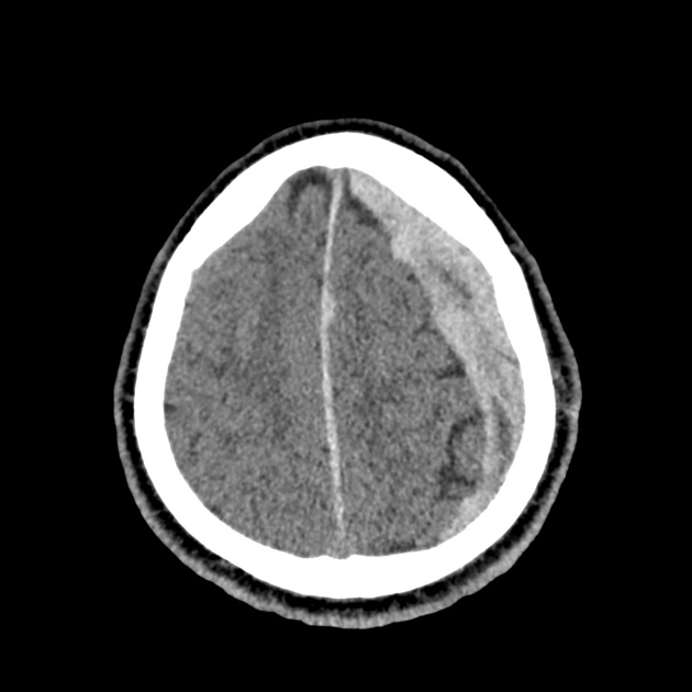
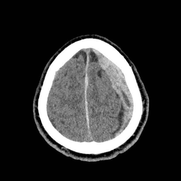
A 65-year-old male with a past medical history of hypertension, diabetes mellitus, and atrial fibrillation presents after a mechanical fall with a posterior scalp hematoma and altered mental status. The patient’s family reports that the patient is taking apixaban with his last dose 4 hours prior to arrival. Physical examination reveals a GCS of 13, blood pressure of 175/99, and asymmetric pupils. The patient is taken to CT where head imaging reveals left sided subdural hematoma with midline shift and developing uncal herniation.

00_subdural
02_subdural
04_subdural
06_subdural
08_subdural
10_subdural
12_subdural
14_subdural
16_subdural
18_subdural
20_subdural
22_subdural
24_subdural
26_subdural
28_subdural
30_subdural
32_subdural
34_subdural
36_subdural
38_subdural
40_subdural
42_subdural
44_subdural
46_subdural

CT Head:

Left cerebral convexity acute subdural hematoma producing substantial mass effect with midline shift and left uncal herniation.
Case courtesy of Dr Andrew Dixon, Radiopaedia.org. From the case rID: 32395

A nicardipine infusion is initiated and the head of the bed is elevated. Andexanet Alfa is not available, therefore an infusion of 4-Factor PCC is initiated. The patient is taken emergently to the operating room by neurosurgery for craniotomy and hematoma evacuation.

An Algorithm for the Reversal of Anticoagulation for Intracranial Hemorrhage 1-4

An Algorithm for Anticoagulant Reversal in Intracranial Hemorrhage


All Agents

For all agents, discontinue anticoagulation. Patients may require blood pressure control including anti-hypertensive infusions (goal SBP <140). Avoid reversal for intracranial hemorrhage associated with cerebral venous thrombosis. Use cautiously in patients with concomitant life-threatening ischemia, thrombosis, or severe DIC.

Vitamin K Antagonists (ex. warfarin)

Initial Dose

A fixed dose of 4F-PCC 1500 to 2000 units can be given as an initial dose with repeat dosing based on INR measurement 15 minutes after completion of infusion. Follow local institution guidelines if available.

Monitoring and Repeat Dosing

  • Vitamin K: if INR ≥1.4 at 12 hours 5
  • 4F-PCC: May consider repeat PCC dosing based on INR, though with increased DIC and thrombotic risk, it is recommended to correct further with FFP if INR remains ≥1.4 6

Direct Factor Xa Inhibitors (ex. rivaroxaban, apixaban)

Activated charcoal may be effective for up to six hours for apixaban 7 and eight hours for rivaroxaban 8.

*Andexanet alfa Regimens 9,10

  • Low-dose: rivaroxaban <10mg, apixaban <5mg, edoxaban <30mg or 8 or more hours since last dose
  • High-dose: If greater than above thresholds, or dose/timing unknown

Pentasaccharides (ex. fondaparinux)

Use high-dose Andexanet alfa regimen 12

Direct Thrombin inhibitors (ex. dabigatran)

Monitoring and Repeat Dosing

If ongoing significant bleeding after treatment, consider redosing idarucizumab and/or hemodialysis.

Alternative Regimens

If idarucizumab is not available, aPCC (50-80 units/kg) , 4F-PCC or 3F-PCC (50 units/kg) can be used in order of preference.

Unfractionated Heparin

Dosing

Determination of units of heparin is based on estimated active agent (half-life 1-2 hours)

  • Protamine sulfate 1mg/100 units IV, maximum dose 50mg
  • Alternatively, can give fixed dose of 25-50mg

Monitoring and Repeat Dosing

If aPTT is persistently elevated, repeat 0.5 mg/100 units

Low-Molecular Weight Heparin 13

Reversal is not indicated if more than 3-5 half-lives have passed since administration:

  • Enoxaparin mean half-life: 4-5 hours
  • Dalteparin mean half-life: 2.8 hours
  • Nadroparin mean half-life: 3.7 hours

If bleeding persists, or renal insufficiency, repeat dose .5 mg/1 mg enoxaparin or .5 mg/100 anti-Xa units.

This algorithm was developed by Dr. Taylor Martin. Taylor is an emergency medicine resident at McGovern Medical School at UTHealth Houston.

References

Guidelines & Reviews

  1. Greenberg SM, Ziai WC, Cordonnier C, et al. 2022 guideline for the management of patients with spontaneous intracerebral hemorrhage: a guideline from the american heart association/american stroke association. Stroke. Published online May 17, 2022:101161STR0000000000000407.
  2. Tomaselli GF, Mahaffey KW, Cuker A, et al. 2020 acc expert consensus decision pathway on management of bleeding in patients on oral anticoagulants: a report of the american college of cardiology solution set oversight committee. J Am Coll Cardiol. 2020;76(5):594-622.
  3. Frontera JA, Lewin JJ, Rabinstein AA, et al. Guideline for reversal of antithrombotics in intracranial hemorrhage: a statement for healthcare professionals from the neurocritical care society and society of critical care medicine. Neurocrit Care. 2016;24(1):6-46.
  4.  Freeman, W. David, Weitz, Jeffrey. “Reversal of anticoagulation in intracranial hemorrhage.” UpToDate. (2022) https://www.uptodate.com/contents/reversal-of-anticoagulation-in-intracranial-hemorrhage?search=anticoagulation%20reversal (Accessed on May 26, 2022)

Vitamin K Antagonists

  1. Ansell J, Hirsh J, Hylek E, Jacobson A, Crowther M, Palareti G. Pharmacology and management of the vitamin k antagonists: american college of chest physicians evidence-based clinical practice guidelines(8th edition). Chest. 2008;133(6 Suppl):160S-198S.
  2. Pabinger I, Brenner B, Kalina U, et al. Prothrombin complex concentrate (Beriplex p/n) for emergency anticoagulation reversal: a prospective multinational clinical trial. J Thromb Haemost. 2008;6(4):622-631.

Direct Factor Xa Inhibitors

  1. http://packageinserts.bms.com/pi/pi_eliquis.pdf
  2. https://www.bayer.com/sites/default/files/2020-11/xarelto-pm-en.pdf
  3. Demchuk AM, Yue P, Zotova E, et al. Hemostatic efficacy and anti-fxa (Factor xa) reversal with andexanet alfa in intracranial hemorrhage: annexa-4 substudy. Stroke. 2021;52(6):2096-2105.
  4. Cohen AT, Lewis M, Connor A, et al. Thirty-day mortality with andexanet alfa compared with prothrombin complex concentrate therapy for life-threatening direct oral anticoagulant-related bleeding. J Am Coll Emerg Physicians Open. 2022;3(2):e12655.
  5. Scaglione F. New oral anticoagulants: comparative pharmacology with vitamin K antagonists. Clin Pharmacokinet. 2013;52(2):69-82.

Pentasaccharides (ex. fondaparinux)

  1. Lu G, DeGuzman FR, Hollenbach SJ, et al. A specific antidote for reversal of anticoagulation by direct and indirect inhibitors of coagulation factor Xa. Nat Med. 2013;19(4):446-451.

Low-Molecular Weight Heparin

  1. Fareed J, Hoppensteadt D, Walenga J, et al. Pharmacodynamic and pharmacokinetic properties of enoxaparin : implications for clinical practice. Clin Pharmacokinet. 2003;42(12):1043-1057.

Sedative-Hypnotic Withdrawal

Brief H&P

A 24 year-old male with no reported medical history is transferred from jail for altered mental status. The patient had been in jail for two days and was noted to develop worsening confusion and agitation.

On arrival in the emergency department, the patient was agitated, moving wildly requiring physical restraints and speaking incomprehensibly. Initial vital signs were notable for tachycardia (HR 145bpm) and hypertension (BP 140/100mmmHg), afebrile core temperature. Examination revealed dilated pupils and dry skin. Point-of-care glucose measured 70mg/dL. Midazolam 5mg IV was administered due to his severe agitation and inability to cooperate with detailed evaluation.

After administration of midazolam, the patient became somewhat more lucid though he appeared to be hallucinating. He reported a history of alprazolam use. The patient was treated with nutritional supplementation and escalating doses of benzodiazepines. The patient remained persistently agitated after administration of midazolam 40mg IV as a single dose so phenobarbital was administered and the patient was intubated for airway protection and anticipated clinical course. The patient was started on a midazolam infusion at 40mg/hour after which he became more calm and vital signs normalized. Laboratory tests were unremarkable with the exception of elevated CK. Head imaging was negative for acute intracranial processes. The patient was admitted to the intensive care unit where he was transitioned to propofol and dexmedetomidine infusions. He was eventually extubated and discharged on hospital day #3 with a chlordiazepoxide taper.

Algorithm for the Management of Sedative-Hypnotic Withdrawal

Algorithm for the Management of Sedative-Hypnotic Withdrawal

Special thanks to David A. Tanen, MD FAAEM FACMT, Professor of Emergency Medicine, Harbor-UCLA Medical Center for his expertise and review of this algorithm.

References

Treatment Algorithms

  1. Gold, J. A., Rimal, B., Nolan, A. & Nelson, L. S. A strategy of escalating doses of benzodiazepines and phenobarbital administration reduces the need for mechanical ventilation in delirium tremens Crit Care Med 35, 724–730 (2007).
  2. Schmidt, K. J. et al. Treatment of Severe Alcohol Withdrawal. Ann Pharmacother 50, 389–401 (2016).
  3. Santos, C., Olmedo, R. E. & Kim, J. Sedative-hypnotic drug withdrawal syndrome: recognition and treatment. Emerg Medicine Pract 19, S1–S2 (2017).

Gabapentin

  1. Myrick, H. et al. A Double‐Blind Trial of Gabapentin Versus Lorazepam in the Treatment of Alcohol Withdrawal. Alcohol Clin Exp Res 33, 1582–1588 (2009).
  2. Stock, C. J., Carpenter, L., Ying, J. & Greene, T. Gabapentin Versus Chlordiazepoxide for Outpatient Alcohol Detoxification Treatment. Ann Pharmacother 47, 961–969 (2013).

Barbiturates

  1. Hendey, G. W., Dery, R. A., Barnes, R. L., Snowden, B. & Mentler, P. A prospective, randomized, trial of phenobarbital versus benzodiazepines for acute alcohol withdrawal. Am J Emerg Medicine 29, 382–385 (2011).
  2. Rosenson, J. et al. Phenobarbital for Acute Alcohol Withdrawal: A Prospective Randomized Double-blind Placebo-controlled Study. J Emerg Medicine 44, 592-598.e2 (2013).

Textbook Chapters

  1. Cohen, J. P. et al. Alcohols. in Tintinalli’s Emergency Medicine: A Comprehensive Study Guide, 9e (McGraw-Hill Education, 2020).
  2. Quan, D. et al. Benzodiazepines. in Tintinalli’s Emergency Medicine: A Comprehensive Study Guide, 9e (McGraw-Hill Education, 2020).

Reviews

  1. Mayo-Smith, M. F. Pharmacological Management of Alcohol Withdrawal: A Meta-analysis and Evidence-Based Practice Guideline. Jama 278, 144–151 (1997).
  2. Mayo-Smith, M. F. et al. Management of Alcohol Withdrawal Delirium: An Evidence-Based Practice Guideline. Arch Intern Med 164, 1405–1412 (2004).
  3. DeBellis, R., Smith, B. S., Choi, S. & Malloy, M. Management of Delirium Tremens. J Intensive Care Med 20, 164–173 (2005).
  4. Amato, L., Minozzi, S., Vecchi, S. & Davoli, M. Benzodiazepines for alcohol withdrawal. Cochrane Db Syst Rev CD005063 (2010) doi:10.1002/14651858.cd005063.pub3.

Toxidromes

Case 1:

A 20 year-old male with a history of polysubstance use, depression and seasonal allergies presents via ambulance for altered mental status. According to prehospital report, EMS were contacted by the patient’s roommate who noted that he had been acting strangely after being alone in his room for several hours. Vital signs are notable for fever (T 103.2°F) and tachycardia. The patient was confused, unable to follow commands – pupils were dilated.

The initial impression was concerning for sympathomimetic toxicity, the patient was treated with cooled intravenous fluids and required pharmacologic sedation and physical restraints to obtain blood samples. ECG, initial laboratory tests and urine toxicology screen were unremarkable. A non-contrast CT head was normal.

The patient remained altered and a repeat examination was performed which revealed multiple, opened blister packs of diphenhydramine and dry, flushed skin.

Anti-cholinergic toxicity was presumed, likely exacerbated by the administration of butyrophenones for sedation. He was treated with benzodiazepines, additional evaporative cooling measures and was admitted to the intensive care unit.

Case 2:

A 32 year-old female with a history of depression was brought to the emergency department by family members who were concerned about bizarre behavior and muscle stiffness. They note that the patient was recently started on a new antidepressant though they are unsure of the name. They describe occasional alcohol consumption but no illicit drug use.

In the emergency department, vital signs were notable for fever and hypertension. Examination demonstrated increased muscle tone and sustained clonus in bilateral lower extremities.

The patient’s presentation was concerning for serotonin syndrome, she was treated with benzodiazepines and intravenous fluids with gradual improvement in mental status and hypertonicity. Upon awakening, she reported doubling her medication dose recently due to persistent feelings of hopelessness as well as increased wine consumption.

An Algorithm for the Evaluation of Toxidromes

An Algorithm for the Evaluation of Toxidromes

Diagnostic Tests

All Patients Most Patients Critical Patients
POC glucose

Core temperature

ECG

Urine hCG

BMP

UA

Acetaminophen

Salicylate

Ethanol

LFT

Lipase

Serum osmolarity

Ionized calcium

Magnesium

GI Decontamination

Activated charcoal

Activated charcoal (1g/kg) within 1-hour post-ingestion and if the patient is awake and cooperative (or via enteric tube if intubated).

Not recommended

  1. Heavy metals
  2. Ions (ex. lithium)
  3. Corrosives
  4. Hydrocarbons
  5. Alcohols

Whole-bowel irrigation

Indicated for sustained-release formulations, expulsion of body packing materials, or ingestion of agent not absorbed by activated charcoal.

Serum alkalinization

For certain ingestions (salicylate, phenobarbital, methotrexate), serum alkalinization through infusion of sodium bicarbonate targeting serum pH 7.5 (and urine pH 8.0) may promote elimination.

Intralipid emulsion

May be useful for local anesthetic toxicity, b-blocker, and calcium channel blocker overdose.

Electrocardiographic Toxidromes


QT Prolongation QRS Prolongation
Anti-emetic Diphenhydramine
Anti-psychotic Cocaine
Anti-microbials (fluoroquinolone, macrolide) Diltiazem, verapamil
Anti-depressant (TCA, SSRI) Propranolol
Anti-arrhythmic Amantadine
Carbamazepine

Gap-producing Toxidromes

Osmolar Gap

  • Toxic alcohol
    • Ethanol
    • Methanol
    • Ethylene glycol
    • Isopropyl alcohol
  • Drug stabilizing agents
    • Mannitol
    • Propylene glycol
    • Glycerol

Anion Gap

  • Salicylate
  • Iron
  • Isoniazid
  • Methanol
  • Ethylene glycol
  • Cyanide

References

  1. Meehan, T. J. (2018). Approach to the Poisoned Patient. In Rosens emergency medicine: concepts and clinical practice (pp. 1813–1822). Philadelphia, PA: Elsevier.
  2. Holstege, C., Borek, H. (2012). Toxidromes Critical Care Clinics 28(4), 479-498. https://dx.doi.org/10.1016/j.ccc.2012.07.008
  3. Mégarbane, B. (2014). Toxidrome-based Approach to Common Poisonings Asia Pacific Journal of Medical Toxicology 3(1), 2-12. https://dx.doi.org/10.22038/apjmt.2014.2463
  4. Rasimas, J., Sinclair, C. (2017). Assessment and Management of Toxidromes in the Critical Care Unit. Critical care clinics 33(3), 521-541. https://dx.doi.org/10.1016/j.ccc.2017.03.002
  5. Thompson, T., Theobald, J., Lu, J., Erickson, T. (2014). The general approach to the poisoned patient Disease-a-Month 60(11), 509-524. https://dx.doi.org/10.1016/j.disamonth.2014.10.002
This algorithm was co-developed by Dr. Chigozie Dike, and Dr. Katrina Nemri.

Dr. Dike is a Houstonian true and true, born across the street at Ben Taub Hospital and proud to have the support of her family and friends through her medical school and emergency medicine training at McGovern Med EM at UT Health. She is a big foodie and loves music. In her free time she’s exploring local restaurants, traveling to new cities, and practicing yoga.

Dr. Nemri is currently a second year emergency medicine resident at McGovern Med EM at UT Health. Her interests are in medical education and critical care. She is a local Houstonian who hopes to stay in the city after residency.

Opioid Withdrawal

Brief HPI:

A 28 year-old female with a history of IV drug use presents to the emergency department with back pain and fever. During evaluation for spinal epidural abscess, she develops vomiting and diarrhea. Examination reveals diaphoresis, mydriasis and hyperactive bowel sounds – she states that her last heroin use was 18-hours ago.

The patient was interested in guidance with cessation of opioid dependence and was evaluated by a recovery support specialist in the emergency department and provided with an appointment for outpatient follow-up. She was treated with buprenorphine-naloxone 8mg sublingual and her symptoms resolved. Her diagnostic evaluation was normal and she was discharged with a prescription for buprenorphine-naloxone 16mg daily until follow-up.

An Algorithm for the Management of Opioid Withdrawal1-4

An Algorithm for the Management of Opioid Withdrawal

Signs

  • Mydriasis
  • Piloerection
  • Diaphoresis
  • Hyperactive bowel sounds

COWS Calculator

Symptoms

  • Dysphoria
  • Rhinorrhea
  • Myalgias, arthralgias
  • Nausea, vomiting, diarrhea
  • Abdominal cramps

Buprenorphine Considerations

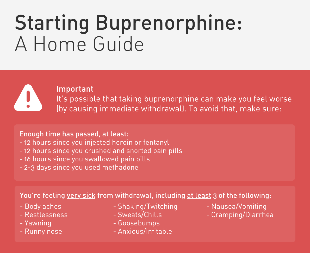
Buprenorphine is a high-affinity, opioid partial agonist. The administration of buprenorphine may displace lower-affinity opioids.5 When used for the treatment of acute opioid withdrawal, special care must be taken to ensure that sufficient time has elapsed since last use (evidenced by the presence of moderate withdrawal symptoms) as the immediate displacement of existing opioids can precipitate severe withdrawal. In addition to provoking the maximum severity of the symptoms for which treatment was sought, this can generate mistrust in an otherwise effective medication and the healthcare system more broadly. The combination of buprenorphine with naloxone is intended to deter parenteral abuse – oral/sublingual naloxone is poorly bioavailable.

The initiation of medication-assisted treatment for opioid dependence from the emergency department should be dependent on the availability of outpatient follow-up and addiction treatment programs.6

Supportive Care4,6-7

Symptom Agent Dose
Nausea, Vomiting Promethazine 25mg IM
Diarrhea Loperamide 4mg PO
Octreotide 50mcg SQ
Muscle cramps Baclofen 5mg PO
Anxiety, Dysphoria Lorazepam 1-2mg IV
Diazepam 2-10mg PO, IM, IV
Pain, Myalgia Acetaminophen 650mg – 1,000mg PO
Ibuprofen 600mg PO

Unobserved Induction Guide8

The following guide is adapted from the Yale Department of Emergency Medicine ED-Initiated Buprenorphine Program and is available free for use.
Home Induction Guide Preview

This algorithm was developed with Dr. Drew Silver. Drew is an emergency medicine resident at McGovern Medical School at the University of Texas Health Science Center at Houston (UTHealth).

References

  1. Strayer R, Hawk K, Hayes B, Herring A et al. Management of Opiate Misuse Disorder in the Emergency Department: A White Paper Prepared for the American Academy of Emergency Medicine. American Academy of Emergency Medicine.
  2. ED-Initiated Buprenorphine. Retrieved July 17, 2020, from https://medicine.yale.edu/edbup/Algorithm_338052_5_v2.pdf
  3. Su, M., Lopez, J., Crossa, A., Hoffman, R. (2018). Low dose intramuscular methadone for acute mild to moderate opioid withdrawal syndrome The American Journal of Emergency Medicine 36(11), 1951-1956. https://dx.doi.org/10.1016/j.ajem.2018.02.019
  4. Stolbach A, Hoffman R. Opioid withdrawal in the emergency setting. Post TW, ed. UpToDate. Waltham, MA: UpToDate Inc.
  5. Boas, R., Villiger, J. (1985). Clinical actions of fentanyl and buprenorphine. The significance of receptor binding. British journal of anaesthesia 57(2), 192-6. https://dx.doi.org/10.1093/bja/57.2.192
  6. D’Onofrio, G., Chawarski, M., O’Connor, P., Pantalon, M., Busch, S., Owens, P., Hawk, K., Bernstein, S., Fiellin, D. (2017). Emergency Department-Initiated Buprenorphine for Opioid Dependence with Continuation in Primary Care: Outcomes During and After Intervention Journal of General Internal Medicine 32(6), 660-666. https://dx.doi.org/10.1007/s11606-017-3993-2
  7. Gowing, L., Farrell, M., Ali, R., White, J. (2016). Alpha2‐adrenergic agonists for the management of opioid withdrawal Cochrane Database of Systematic Reviews https://dx.doi.org/10.1002/14651858.cd002024.pub5
  8. Home Initiated Buprenorphine. Retrieved July 17, 2020, from https://medicine.yale.edu/edbup/quickstart/Home_Buprenorphine_Initiation_338574_42801_v1.pdf
  9. Wesson DR, Ling W. The Clinical Opiate Withdrawal Scale (COWS). J Psychoactive Drugs. 2003;35(2):253-259. doi:10.1080/02791072.2003.10400007

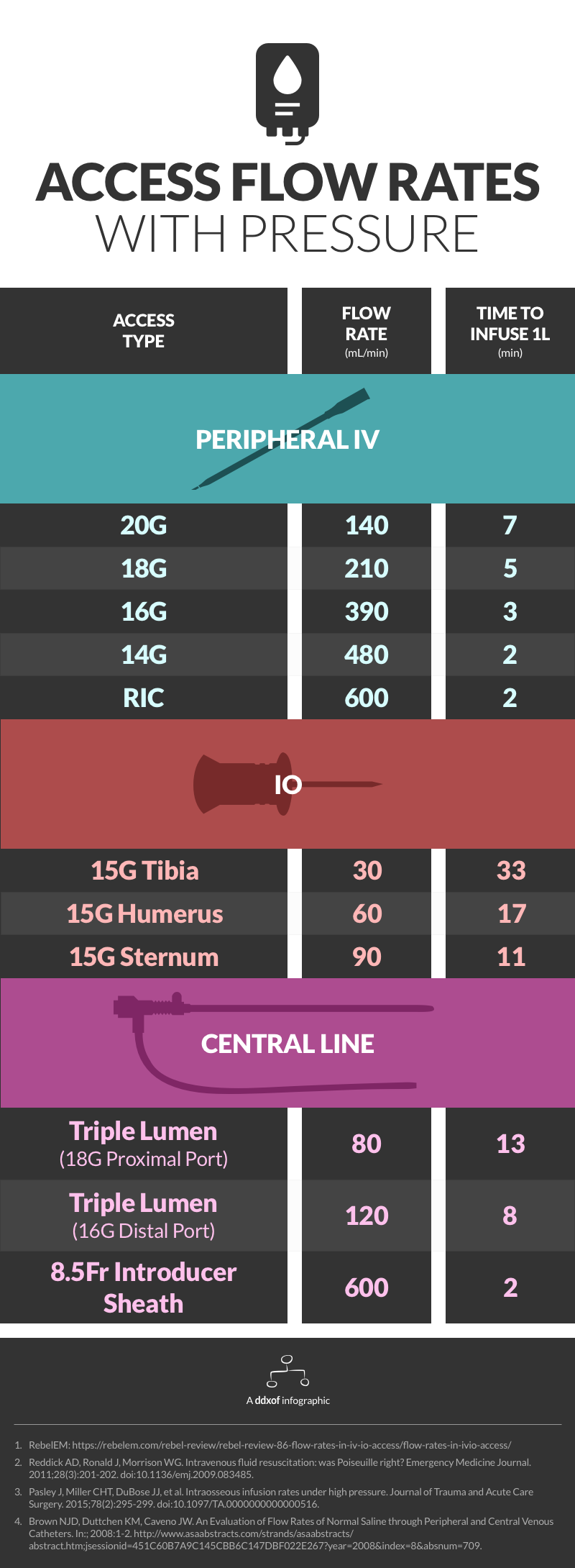
Infographic: Access Flow Rates

Infographic for IV flow rates

Notes

Data on access flow rates are highly variable. This infographic uses flow rates achieved through dedicated rapid infusers (ex. Level 1 ®) or at a pressure of 300mmHg. It is possible that rapid infusers and specialized pressure tubing achieve higher flow rates. The main references and additional sources are listed below. See this post from REBEL EM for gravity flow rates.

References

  1. Reddick AD, Ronald J, Morrison WG. Intravenous fluid resuscitation: was Poiseuille right? Emergency Medicine Journal. 2011;28(3):201-202. doi:10.1136/emj.2009.083485.
  2. Pasley J, Miller CHT, DuBose JJ, et al. Intraosseous infusion rates under high pressure. Journal of Trauma and Acute Care Surgery. 2015;78(2):295-299. doi:10.1097/TA.0000000000000516.
  3. Brown NJD, Duttchen KM, Caveno JW. An Evaluation of Flow Rates of Normal Saline through Peripheral and Central Venous Catheters. In:; 2008:1-2. http://www.asaabstracts.com/strands/asaabstracts/abstract.htm;jsessionid=451C60B7A9C145CBB6C147DBF022E267?year=2008&index=8&absnum=709.

Additional Sources

  1. Ngo AS-Y, Oh JJ, Chen Y, Yong D, Ong MEH. Intraosseous vascular access in adults using the EZ-IO in an emergency department. Int J Emerg Med. 2009;2(3):155-160. doi:10.1007/s12245-009-0116-9.
  2. Traylor S, Bastani A, Emergency NB-DAO, 2016. 311 Are Three Ports Better Than One? an Evaluation of Flow Rates Using All Ports of a Triple Lumen Central Venous Catheter in Volume Resuscitation. doi:10.1016/j.annemergmed.2016.08.327.
  3. Hammer N, Möbius R, Gries A, Hossfeld B, Bechmann I, Bernhard M. Comparison of the Fluid Resuscitation Rate with and without External Pressure Using Two Intraosseous Infusion Systems for Adult Emergencies, the CITRIN (Comparison of InTRaosseous infusion systems in emergency medicINe)-Study. Raju R, ed. PLoS ONE. 2015;10(12):e0143726–15. doi:10.1371/journal.pone.0143726.
  4. Ong MEH, Chan YH, Oh JJ, Ngo AS-Y. An observational, prospective study comparing tibial and humeral intraosseous access using the EZ-IO. Am J Emerg Med. 2009;27(1):8-15. doi:10.1016/j.ajem.2008.01.025.
  5. Philbeck TE, Miller LJ, Montez D, Puga T. Hurts so good. Easing IO pain and pressure. JEMS. 2010;35(9):58–62–65–6–68–quiz69. doi:10.1016/S0197-2510(10)70232-1.
  6. FRCA SIK, MRCA PRG, FRCA KP, MBChB SW, FRCA TS, MRCP PRG. Flow rates through intravenous access devices: an in vitro study. J Clin Anesth. 2016;31:101-105. doi:10.1016/j.jclinane.2016.01.048.
  7. Puga T, Montez D, Care TPC, 2016. 263: ADEQUACY OF INTRAOSSEOUS VASCULAR ACCESS INSERTION SITES FOR HIGH-VOLUME FLUID INFUSION. journalslwwcom
  8. Tan BKK, Chong S, Koh ZX, Ong MEH. EZ-IO in the ED: an observational, prospective study comparing flow rates with proximal and distal tibia intraosseous access in adults. Am J Emerg Med. 2012;30(8):1602-1606. doi:10.1016/j.ajem.2011.10.025.

Hypertensive Emergency

Brief HPI:

A 62 year-old female with a history of hypertension, diabetes and coronary artery disease is brought to the emergency department with altered mental status. The patient is confused and unable to provide history. Her family note that symptoms have been gradually worsening for the past one day and she had previously been in her usual state of good health. There was no history of recent illness, medication changes, recreational substance use, sick contacts, or travel.

On evaluation, vital signs were notable for hypertension (224/120mmHg, comparable on all extremities) though otherwise normal including afebrile core temperature – capillary glucose was 114mg/dL. On examination, the patient was awake and alert, making coordinated movements symmetrically in all four extremities without hyperreflexia or increased tone. Speech was unintelligible and the patient was unable to follow simple commands.

Labs/Imaging

Laboratory tests were notable for a serum creatinine of 1.2mg/dL (baseline unknown) but otherwise normal including CBC, troponin, TSH, and UA. ECG demonstrated left ventricular hypertrophy without acute ischemic changes. Imaging including chest radiograph and CT head non-contrast and CTA brain/neck were normal. Lumbar puncture was performed and CSF was normal.

Hospital Course

The patient was initiated on a continuous infusion of nicardipine for presumed hypertensive encephalopathy and admitted to the medical intensive care unit. An MRI was performed on hospital day 1 and demonstrated chronic microvascular ischemic changes. The patient’s mental status gradually improved over the course of her hospitalization and she was discharged home on hospital day 4.

An Algorithm for the Evaluation and Management of Hypertensive Emergencies

An Algorithm for the Evaluation and Management of Hypertensive Emergencies

References

General

  1. Lloyd-Jones DM, Morris PB, Ballantyne CM, et al. 2017 Focused Update of the 2016 ACC Expert Consensus Decision Pathway on the Role of Non-Statin Therapies for LDL-Cholesterol Lowering in the Management of Atherosclerotic Cardiovascular Disease Risk: A Report of the American College of Cardiology Task Force on Expert Consensus Decision Pathways. In: Vol 70. 2017:1785-1822. doi:10.1016/j.jacc.2017.07.745.
  2. Janke AT, McNaughton CD, Brody AM, Welch RD, Levy PD. Trends in the Incidence of Hypertensive Emergencies in US Emergency Departments From 2006 to 2013. J Am Heart Assoc. 2016;5(12). doi:10.1161/JAHA.116.004511.
  3. Rodriguez MA, Kumar SK, De Caro M. Hypertensive crisis. Cardiology in Review. 2010;18(2):102-107. doi:10.1097/CRD.0b013e3181c307b7.
  4. Katz JN, Gore JM, Amin A, et al. Practice patterns, outcomes, and end-organ dysfunction for patients with acute severe hypertension: the Studying the Treatment of Acute hyperTension (STAT) registry. Am Heart J. 2009;158(4):599–606.e1. doi:10.1016/j.ahj.2009.07.020.
  5. Elliott WJ. Clinical features in the management of selected hypertensive emergencies. Prog Cardiovasc Dis. 2006;48(5):316-325. doi:10.1016/j.pcad.2006.02.004.
  6. Aggarwal M, Khan IA. Hypertensive crisis: hypertensive emergencies and urgencies. Cardiol Clin. 2006;24(1):135-146. doi:10.1016/j.ccl.2005.09.002.
  7. Varon J, Marik PE. Clinical review: the management of hypertensive crises. Crit Care. 2003;7(5):374-384. doi:10.1186/cc2351.
  8. Shayne PH, Pitts SR. Severely increased blood pressure in the emergency department. YMEM. 2003;41(4):513-529. doi:10.1067/mem.2003.114.
  9. Vaughan CJ, Delanty N. Hypertensive emergencies. The Lancet. 2000;356(9227):411-417. doi:10.1016/S0140-6736(00)02539-3.

Ischemic Stroke

  1. Powers WJ, Rabinstein AA, Ackerson T, et al. 2018 Guidelines for the Early Management of Patients With Acute Ischemic Stroke: A Guideline for Healthcare Professionals From the American Heart Association/American Stroke Association. Stroke. 2018;49(3):e46-e110. doi:10.1161/STR.0000000000000158.

Hemorrhagic Stroke

  1. Hemphill JC, Greenberg SM, Anderson CS, et al. Guidelines for the Management of Spontaneous Intracerebral Hemorrhage: A Guideline for Healthcare Professionals From the American Heart Association/American Stroke Association. Stroke. 2015;46(7):2032-2060. doi:10.1161/STR.0000000000000069.

Subarachnoid Hemorrhage

  1. Connolly ES, Rabinstein AA, Carhuapoma JR, et al. Guidelines for the management of aneurysmal subarachnoid hemorrhage: a guideline for healthcare professionals from the American Heart Association/american Stroke Association. Stroke. 2012;43(6):1711-1737. doi:10.1161/STR.0b013e3182587839.

Renal

  1. Gillies MA, Kakar V, Parker RJ, Honoré PM, Ostermann M. Fenoldopam to prevent acute kidney injury after major surgery-a systematic review and meta-analysis. Crit Care. 2015;19(1):449. doi:10.1186/s13054-015-1166-4.
  2. Tumlin JA, Dunbar LM, Oparil S, et al. Fenoldopam, a dopamine agonist, for hypertensive emergency: a multicenter randomized trial. Fenoldopam Study Group. Academic Emergency Medicine. 2000;7(6):653-662.
  3. Shusterman NH, Elliott WJ, White WB. Fenoldopam, but not nitroprusside, improves renal function in severely hypertensive patients with impaired renal function. Am J Med. 1993;95(2):161-168.

Aortic Disease

  1. Hiratzka LF, Bakris GL, Beckman JA, et al. 2010 ACCF/AHA/AATS/ACR/ASA/SCA/SCAI/SIR/STS/SVM guidelines for the diagnosis and management of patients with Thoracic Aortic Disease: a report of the American College of Cardiology Foundation/American Heart Association Task Force on Practice Guidelines, American Association for Thoracic Surgery, American College of Radiology, American Stroke Association, Society of Cardiovascular Anesthesiologists, Society for Cardiovascular Angiography and Interventions, Society of Interventional Radiology, Society of Thoracic Surgeons, and Society for Vascular Medicine. Circulation. 2010;121(13):e266-e369. doi:10.1161/CIR.0b013e3181d4739e.

Pregnancy

  1. Townsend R, O’Brien P, Khalil A. Current best practice in the management of hypertensive disorders in pregnancy. Integr Blood Press Control. 2016;9:79-94. doi:10.2147/IBPC.S77344.
  2. Al-Safi Z, Imudia AN, Filetti LC, Hobson DT, Bahado-Singh RO, Awonuga AO. Delayed Postpartum Preeclampsia and Eclampsia. Obstet Gynecol. 2011;118(5):1102-1107. doi:10.1097/AOG.0b013e318231934c.
  3. Hypertension in pregnancy: diagnosis and management. National Institute for Health and Care Excellence. https://www.nice.org.uk/guidance/cg107. Published August 1, 2010. Accessed May 20, 2019.

Cardiac Arrest

Brief HPI:

An overhead page alerts you to an arriving patient with cardiac arrest. An approximately 35-year-old male was running away from police officers and collapsed after being shot with a stun gun. The patient was found to be pulseless, CPR was started by police officers and the patient is en route.

An Algorithm for the Evaluation and Management of Cardiac Arrest with Ultrasonography

An Algorithm for the Evaluation and Management of Cardiac Arrest with Ultrasonography

Causes of Cardiac (and non-cardiac) Arrest

Sudden cardiac arrest (SCA) leading to sudden cardiac death (SCD) if not successfully resuscitated, refers to the unexpected collapse of circulatory function. Available epidemiologic data for in-hospital and out-of-hospital cardiac arrest (OHCA) point appropriately to cardiac processes as the most common cause, though extra-cardiac processes (most frequently respiratory), comprise up to 40% of cases1-3.

Identifying the underlying cause is critical as several reversible precipitants require rapid identification. However, the usual diagnostic techniques may be challenging, limited or absent – including patient history, detailed examination, and diagnostic studies.

The initial rhythm detected upon evaluation is most suggestive of the etiologic precipitant. Pulseless ventricular tachycardia (pVT) or ventricular fibrillation (VF) is suggestive of a cardiac process – most commonly an acute coronary syndrome although heart failure or other structural and non-structural heart defects associated with dysrhythmias may be at fault4.

Pulseless electrical activity (PEA) presents a broader differential diagnosis as it essentially represents severe shock. The most common extra-cardiac cause is hypoxia – commonly secondary to pulmonary processes including small and large airway obstruction (bronchospasm, aspiration, foreign body, edema). Other causes include substance intoxication, medication adverse effect5,6, or electrolyte disturbances7. Finally, any precipitant of shock may ultimately lead to PEA, including hypovolemia/hemorrhage, obstruction (massive pulmonary embolus8, tamponade, tension pneumothorax), and distribution (sepsis).

Asystole is the absence of even disorganized electrical discharge and is the terminal degeneration of any of the previously-mentioned rhythms if left untreated.

Management of Cardiac Arrest

Optimizing survival outcomes in patients with cardiac arrest is dependent on early resuscitation with the prioritization of interventions demonstrated to have survival benefit. When advanced notice is available, prepare the resuscitation area including airway equipment (with adjuncts to assist ventilation and waveform capnography devices). Adopt the leadership position and assign roles for chest compressions, airway support, application of monitor/defibrillator, and establishment of peripheral access.

High-quality chest compressions with minimal interruptions are the foundation of successful resuscitation – and guideline changes prioritizing compressions have demonstrated detectable improvements in rates of successful resuscitation9,10. Measurement of quantitative end-tidal capnography can guide adequacy of chest compressions11,12 and an abrupt increase may signal restoration of circulation without necessitating interruptions of chest compressions13,14. Sustained, low measures of end-tidal CO2 despite appropriate resuscitation may signal futility and (alongside other factors) guides termination of resuscitation11,12.

The next critical step in restoring circulation is prompt defibrillation of eligible rhythms (pVT/VF) when detected. The immediate delivery of 200J (uptitrated to the device maximum for subsequent shocks) of biphasic energy and restoration of a perfusing rhythm is one of few interventions with clear benefits. For pVT/VF that persists despite multiple countershocks (more than three), the addition of an intravenous antiarrhythmic appears to improve survival to hospital admission. The ARREST trial was a randomized controlled study comparing amiodarone to its diluent as placebo for OHCA with refractory pVT/VF showing significant improvement in survival to hospital admission for the amiodarone group15. This was followed by the ALIVE trial comparing amiodarone and lidocaine which showed significantly higher rates of survival to hospital admission in the amiodarone group16. However, a more recent randomized trial comparing amiodarone (in a novel diluent less likely to cause hypotension), lidocaine, and placebo in a similar patient population showed less convincing results, with no detected difference in survival or the secondary outcome of favorable neurological outcome for either amiodarone or lidocaine compared with placebo17. The heterogeneity of available data contributed to current guidelines which recommend that either amiodarone or lidocaine may be used for shock-refractory pVT/VF18.

Current guidelines also recommend the administration of vasopressors (epinephrine 1mg every 3-5 minutes). In one randomized controlled trial exploring the long-standing guideline recommendations, epinephrine was associated with increased rates of restoration of spontaneous circulation, though no significant impact on the primary outcome of survival to hospital discharge was identified19. Physiologically, increased systemic vascular resistance combined with positive beta-adrenergic impact on cardiac output would be expected to complement resuscitative efforts. However, more recent studies have suggested that arrest physiology and unanticipated pharmacologic effects may complicate this simplistic interpretation – particularly when patient-centered outcomes are emphasized. Research exploring the timing and amount of epinephrine suggest that earlier administration and higher cumulative doses are associated with negative impacts on survival to hospital discharge and favorable neurological outcomes20-22.

Ultimately, treatment should focus on optimal execution of measures with clear benefits (namely chest compressions and early defibrillation of eligible rhythms). Other management considerations with which the emergency physician is familiar with including establishing peripheral access and definitive airway management can be delayed.

Rapid Diagnostic Measures for the Identification of Reversible Processes

Traditional diagnostic measures are generally unavailable during an ongoing cardiac arrest resuscitation. The emergency medicine physician must rely on the physical examination and point-of-care tests with the objective of identifying potentially reversible processes. Measurement of capillary blood glucose can exclude hypoglycemia as a contributor. Point-of-care chemistry and blood gas analyzers can identify important electrolyte derangements, as well as clarifying the primary impulse in acid-base disturbances.

End-tidal capnography was discussed previously for the guidance of ongoing resuscitation, but it may have diagnostic utility in patients with SCD. In one study the initial EtCO2 was noted to be significantly higher for primary pulmonary processes (with PEA/asystole as presenting rhythm) compared to primary cardiac processes (with pVT/VF as presenting rhythm)23.

The use of point-of-care ultrasonography, particularly in PEA arrest where non-cardiac etiologies dominate, may help identify the etiology of arrest and direct therapy. Bedside ultrasonography should be directed first at assessment of cardiac function – examining the pericardial sac and gross abnormalities in chamber size. A pericardial effusion may suggest cardiac tamponade, ventricular collapse can be seen with hypovolemia, and asymmetric right-ventricular dilation points to pulmonary embolus where thrombolysis should be considered8. If cardiac ultrasound is unrevealing, thoracic ultrasound can identify pneumothorax24-27.

In the absence of ultrasonographic abnormalities, attention turns to other rapidly reversible precipitants first. If opioid toxicity is a consideration, an attempt at reversal with naloxone has few adverse effects. If any detected rhythm is a polymorphic ventricular tachycardia characteristic of torsades de pointes – rapid infusion of magnesium sulfate should follow defibrillation. Other potentially reversible medications or toxins should be managed as appropriate.

Post-Resuscitation Steps

After successful restoration of circulation, the next management steps are critical to the patient’s long-term outcomes. A definitive airway should be established if not already secured (and if restoration of circulation was not associated with neurological recovery sufficient for independent airway protection). Circulatory support should continue with fluid resuscitation and vasopressors to maintain end-organ perfusion.

An immediate ECG should be performed to identify infarction, ischemia or precipitants of dysrhythmia. ST-segment elevation after return of spontaneous circulation (ROSC) warrants emergent angiography and possible intervention. However, given the prevalence of cardiac causes (of which coronary disease is most common) for patients with pVT/VF arrest, the presence of ST elevations is likely of insufficient sensitivity to identify all patients who would benefit from angiography. Several studies and meta-analyses have explored a more inclusive selection strategy for angiography (patients without obvious non-cardiac causes for arrest), all of which identified survival benefits with angiography and successful angioplasty when possible28-30.

Finally, the induction of hypothermia (or targeted temperature management) has significant benefits in survivors of cardiac arrest and can be instituted in the emergency department. Studies first targeted a core temperature of 32-24°C, with a randomized controlled trial demonstrating higher rates of favorable neurological outcome and reduced mortality31. More recent studies suggest that a more liberal temperature target does not diffuse the positive effects of induced hypothermia. A randomized trial of 939 patients with OHCA comparing a targeted temperature of 33°C vs 36°C suggested that a lower temperature target did not confer higher benefit to mortality or recovery of neurological function32. The more liberal temperature target may alleviate adverse effects associated with hypothermia which include cardiovascular effects (bradycardia), electrolyte derangements (during induction and rewarming), and possible increased risk of infections33. Targeted temperature management is achieved with external cooling measures or infusion of cooled fluids, rarely requiring more invasive measures34. Aggregate review of available data in a recent meta-analysis further supports the use of targeted temperature management after cardiac arrest as standard-of-care35.

References

  1. Bergum D, Nordseth T, Mjølstad OC, Skogvoll E, Haugen BO. Causes of in-hospital cardiac arrest – Incidences and rate of recognition. Resuscitation. 2015;87:63-68. doi:10.1016/j.resuscitation.2014.11.007.
  2. Wallmuller C, Meron G, Kurkciyan I, Schober A, Stratil P, Sterz F. Causes of in-hospital cardiac arrest and influence on outcome. Resuscitation. 2012;83(10):1206-1211. doi:10.1016/j.resuscitation.2012.05.001.
  3. Vaartjes I, Hendrix A, Hertogh EM, et al. Sudden death in persons younger than 40 years of age: incidence and causes. European Journal of Cardiovascular Prevention & Rehabilitation. 2009;16(5):592-596. doi:10.1097/HJR.0b013e32832d555b.
  4. Zheng ZJ, Croft JB, Giles WH, Mensah GA. Sudden cardiac death in the United States, 1989 to 1998. Circulation. 2001;104(18):2158-2163.
  5. Hoes AW, Grobbee DE, Lubsen J, Man in ‘t Veld AJ, van der Does E, Hofman A. Diuretics, beta-blockers, and the risk for sudden cardiac death in hypertensive patients. Ann Intern Med. 1995;123(7):481-487.
  6. Siscovick DS, Raghunathan TE, Psaty BM, et al. Diuretic therapy for hypertension and the risk of primary cardiac arrest. N Engl J Med. 1994;330(26):1852-1857. doi:10.1056/NEJM199406303302603.
  7. Gettes LS. Electrolyte abnormalities underlying lethal and ventricular arrhythmias. Circulation. 1992;85(1 Suppl):I70-I76.
  8. Kürkciyan I, Meron G, Sterz F, et al. Pulmonary embolism as a cause of cardiac arrest: presentation and outcome. Arch Intern Med. 2000;160(10):1529-1535.
  9. Callaway CW, Soar J, Aibiki M, et al. Part 4: Advanced Life Support: 2015 International Consensus on Cardiopulmonary Resuscitation and Emergency Cardiovascular Care Science With Treatment Recommendations. In: Vol 132. American Heart Association, Inc.; 2015:S84-S145. doi:10.1161/CIR.0000000000000273.
  10. Kudenchuk PJ, Redshaw JD, Stubbs BA, et al. Impact of changes in resuscitation practice on survival and neurological outcome after out-of-hospital cardiac arrest resulting from nonshockable arrhythmias. Circulation. 2012;125(14):1787-1794. doi:10.1161/CIRCULATIONAHA.111.064873.
  11. Touma O, Davies M. The prognostic value of end tidal carbon dioxide during cardiac arrest: a systematic review. Resuscitation. 2013;84(11):1470-1479. doi:10.1016/j.resuscitation.2013.07.011.
  12. Levine RL, Wayne MA, Miller CC. End-tidal carbon dioxide and outcome of out-of-hospital cardiac arrest. N Engl J Med. 1997;337(5):301-306. doi:10.1056/NEJM199707313370503.
  13. Garnett AR, Ornato JP, Gonzalez ER, Johnson EB. End-tidal carbon dioxide monitoring during cardiopulmonary resuscitation. JAMA. 1987;257(4):512-515.
  14. Falk JL, Rackow EC, Weil MH. End-tidal carbon dioxide concentration during cardiopulmonary resuscitation. N Engl J Med. 1988;318(10):607-611. doi:10.1056/NEJM198803103181005.
  15. Kudenchuk PJ, Cobb LA, Copass MK, et al. Amiodarone for resuscitation after out-of-hospital cardiac arrest due to ventricular fibrillation. N Engl J Med. 1999;341(12):871-878. doi:10.1056/NEJM199909163411203.
  16. Dorian P, Cass D, Schwartz B, Cooper R, Gelaznikas R, Barr A. Amiodarone as compared with lidocaine for shock-resistant ventricular fibrillation. N Engl J Med. 2002;346(12):884-890. doi:10.1056/NEJMoa013029.
  17. Kudenchuk PJ, Brown SP, Daya M, et al. Amiodarone, Lidocaine, or Placebo in Out-of-Hospital Cardiac Arrest. N Engl J Med. 2016;374(18):1711-1722. doi:10.1056/NEJMoa1514204.
  18. Neumar RW, Shuster M, Callaway CW, et al. Part 1: Executive Summary: 2015 American Heart Association Guidelines Update for Cardiopulmonary Resuscitation and Emergency Cardiovascular Care. In: Vol 132. American Heart Association, Inc.; 2015:S315-S367. doi:10.1161/CIR.0000000000000252.
  19. Jacobs IG, Finn JC, Jelinek GA, Oxer HF, Thompson PL. Effect of adrenaline on survival in out-of-hospital cardiac arrest: A randomised double-blind placebo-controlled trial. Resuscitation. 2011;82(9):1138-1143. doi:10.1016/j.resuscitation.2011.06.029.
  20. Hagihara A, Hasegawa M, Abe T, Nagata T, Wakata Y, Miyazaki S. Prehospital epinephrine use and survival among patients with out-of-hospital cardiac arrest. JAMA. 2012;307(11):1161-1168. doi:10.1001/jama.2012.294.
  21. Dumas F, Bougouin W, Geri G, et al. Is epinephrine during cardiac arrest associated with worse outcomes in resuscitated patients? J Am Coll Cardiol. 2014;64(22):2360-2367. doi:10.1016/j.jacc.2014.09.036.
  22. Andersen LW, Kurth T, Chase M, et al. Early administration of epinephrine (adrenaline) in patients with cardiac arrest with initial shockable rhythm in hospital: propensity score matched analysis. BMJ. 2016;353:i1577. doi:10.1136/bmj.i1577.
  23. Grmec S, Lah K, Tusek-Bunc K. Difference in end-tidal CO2 between asphyxia cardiac arrest and ventricular fibrillation/pulseless ventricular tachycardia cardiac arrest in the prehospital setting. Crit Care. 2003;7(6):R139-R144. doi:10.1186/cc2369.
  24. Rose JS, Bair AE, Mandavia D, Kinser DJ. The UHP ultrasound protocol: a novel ultrasound approach to the empiric evaluation of the undifferentiated hypotensive patient. American Journal of Emergency Medicine. 2001;19(4):299-302. doi:10.1053/ajem.2001.24481.
  25. Hernandez C, Shuler K, Hannan H, Sonyika C, Likourezos A, Marshall J. C.A.U.S.E.: Cardiac arrest ultra-sound exam—A better approach to managing patients in primary non-arrhythmogenic cardiac arrest. Resuscitation. 2008;76(2):198-206. doi:10.1016/j.resuscitation.2007.06.033.
  26. Chardoli M, Heidari F, Shuang-ming S, et al. Echocardiography integrated ACLS protocol versus con- ventional cardiopulmonary resuscitation in patients with pulseless electrical activity cardiac arrest. Chinese Journal of Traumatology. 2012;15(5):284-287. doi:10.3760/cma.j.issn.1008-1275.2012.05.005.
  27. Zengin S, Yavuz E, Al B, et al. Benefits of cardiac sonography performed by a non-expert sonographer in patients with non-traumatic cardiopulmonary arrest. Resuscitation. 2016;102:105-109. doi:10.1016/j.resuscitation.2016.02.025.
  28. Spaulding CM, Joly LM, Rosenberg A, et al. Immediate coronary angiography in survivors of out-of-hospital cardiac arrest. N Engl J Med. 1997;336(23):1629-1633. doi:10.1056/NEJM199706053362302.
  29. Dumas F, Cariou A, Manzo-Silberman S, et al. Immediate Percutaneous Coronary Intervention Is Associated With Better Survival After Out-of-Hospital Cardiac Arrest: Insights From the PROCAT (Parisian Region Out of Hospital Cardiac Arrest) Registry. Circulation: Cardiovascular Interventions. 2010;3(3):200-207. doi:10.1161/CIRCINTERVENTIONS.109.913665.
  30. Millin MG, Comer AC, Nable JV, et al. Patients without ST elevation after return of spontaneous circulation may benefit from emergent percutaneous intervention: A systematic review and meta-analysis. Resuscitation. 2016;108:54-60. doi:10.1016/j.resuscitation.2016.09.004.
  31. Hypothermia after Cardiac Arrest Study Group. Mild therapeutic hypothermia to improve the neurologic outcome after cardiac arrest. N Engl J Med. 2002;346(8):549-556. doi:10.1056/NEJMoa012689.
  32. Nielsen N, Wetterslev J, Cronberg T, et al. Targeted Temperature Management at 33°C versus 36°C after Cardiac Arrest. N Engl J Med. 2013;369(23):2197-2206. doi:10.1056/NEJMoa1310519.
  33. Polderman KH, Peerdeman SM, Girbes AR. Hypophosphatemia and hypomagnesemia induced by cooling in patients with severe head injury. J Neurosurg. 2001;94(5):697-705. doi:10.3171/jns.2001.94.5.0697.
  34. Polderman KH, Herold I. Therapeutic hypothermia and controlled normothermia in the intensive care unit: Practical considerations, side effects, and cooling methods*. Critical Care Medicine. 2009;37(3):1101-1120. doi:10.1097/CCM.0b013e3181962ad5.
  35. Schenone AL, Cohen A, Patarroyo G, et al. Therapeutic hypothermia after cardiac arrest: A systematic review/meta-analysis exploring the impact of expanded criteria and targeted temperature. Resuscitation. 2016;108:102-110. doi:10.1016/j.resuscitation.2016.07.238.

Simplified Airway Management Algorithm

An airway management algorithm, developed with Dr. Diane Birnbaumer, has been previously developed on ddxof: airway management algorithm. The algorithm provides detailed, step-by-step recommendations for specific airway classifications – divided into normal, anticipated difficult, crash, and failed airways.

While helpful as an educational tool, the algorithm is likely too complex for rapid review or bedside application. Admittedly sacrificing some detail, this simplified airway management algorithm highlights the critical steps, incorporates only the most commonly-used airway adjuncts, assumes imminent respiratory decompensation and failure of progressive intubation attempts.

Simplified Airway Management Algorithm

Simplified Airway Management Algorithm

Hyperthermia

Brief H&P

A young male with unknown medical history is brought in by ambulance with altered mental status. EMS reports that the patient was agitated, requiring restraints for transportation. On arrival, the patient is agitated, uncooperative and unable to provide history. Vital signs are notable for tachycardia, tachypnea and hypertension. Physical examination demonstrates diaphoresis and mydriasis, as well as increased muscle tone – particularly in the lower extremities with ankle clonus. A core temperature is obtained and noted to be elevated at 41.5°C. Point-of-care glucose is normal.

Rapid external cooling measures were instituted and several doses of intravenous benzodiazepines were administered with improvement in agitation. Laboratory studies were notable for a modest leukocytosis (WBC 18.4 without immature forms), serum sodium was 135 without osmolar gap, creatine kinase was slightly elevated without renal dysfunction, and thyroid function tests were normal. Toxicology screen was negative. ECG revealed sinus tachycardia but was otherwise normal and non-contrast computed tomography of the head was normal.

After a brief admission in the intensive care unit, the patient’s mental status improved and he reported MDMA use on the evening of presentation, he also described a history of major depression and was taking paroxetine.

Evaluation of Elevated Temperature

The designation of 38°C as “suspicious” for fever dates to 1868 and the analysis of over one million (axillary) temperature measurements by Carl Wunderlich1. Any cutoff is arbitrary and requires recognition of the clinical context and normal daily variations (with nadir in the morning and peak in evening) 2,3. What is clear is that peripheral thermometry (unless demonstrating fever) is unreliable and a core temperature should be sought4.

Thermoregulation

Temperature homeostasis is a balance between heat production and dissipation maintained by the anterior hypothalamus. Heat production is a byproduct of normal metabolic processes and skeletal muscle activity. Conservation, maintenance or dissipation of heat is aided by cutaneous vasodilation, sweating, or behavioral responses.

Fever is caused by endogenous or exogenous pyrogens which alter the homeostatic set-point, inducing thermogenesis and elevating the body temperature. Precipitants of fever are usually infectious, however non-infectious processes (ex. malignancy, tissue ischemia/infarction, auto-immune disease) resulting in inflammation can provoke a similar response 5-7.

There is no explicit temperature distinction to diagnose hyperthermia, instead the physiologic mechanism is different. In hyperthermia, the body’s homeostatic mechanisms are dysfunctional or overwhelmed due to heat exposure, excess production, ineffective dissipation or hypothalamic malfunction 8.

Algorithm for the Evaluation of Hyperthermia 8-15

Algorithm for the Evaluation of Hyperthermia

Implicated Agents in Drug-Induced Hyperthermic Syndromes 9,10

Serotonin Syndrome

Class Examples
SSRI sertraline, fluoxetine, paroxetine
Other anti-depressants trazodone, venlafaxine, lithium
MAOI phenelzine, isocarboxazid
Anti-epileptic drugs valproate
Analgesics meperidine, fentanyl, tramadol
Anti-emetic ondansetron, metoclopramide
Anti-migraine sumatriptan
Antimicrobial linezolid, ritonavir
Illicit substances MDMA, LSD

Neuroleptic Malignant Syndrome (NMS)

Class Examples
Typical anti-psychotic haloperidol, prochlorperazine
Atypical anti-psychotic risperidone, olanzapine, quetiapine, aripiprazole
Anti-dopaminergic metoclopramide, droperidol

References:

  1. Wunderlich CA. Das Verhalten Der Eigenwärme in Krankheiten. 1870.
  2. Mackowiak PA, Wasserman SS, Levine MM. A critical appraisal of 98.6 degrees F, the upper limit of the normal body temperature, and other legacies of Carl Reinhold August Wunderlich. JAMA. 1992;268(12):1578-1580.
  3. Lee-Chiong TL, Stitt JT. Disorders of temperature regulation. Compr Ther. 1995;21(12):697-704.
  4. Niven DJ, Gaudet JE, Laupland KB, Mrklas KJ, Roberts DJ, Stelfox HT. Accuracy of peripheral thermometers for estimating temperature: a systematic review and meta-analysis. Ann Intern Med. 2015;163(10):768-777. doi:10.7326/M15-1150.
  5. Dinarello CA. Infection, fever, and exogenous and endogenous pyrogens: some concepts have changed. J Endotoxin Res. 2004;10(4):201-222. doi:10.1179/096805104225006129.
  6. Greisman LA, Mackowiak PA. Fever: beneficial and detrimental effects of antipyretics. Curr Opin Infect Dis. 2002;15(3):241-245.
  7. Dinarello CA. Thermoregulation and the pathogenesis of fever. Infect Dis Clin North Am. 1996;10(2):433-449.
  8. Simon HB. Hyperthermia. N Engl J Med. 1993;329(7):483-487. doi:10.1056/NEJM199308123290708.
  9. Boyer EW, Shannon M. The serotonin syndrome. N Engl J Med. 2005;352(11):1112-1120. doi:10.1056/NEJMra041867.
  10. Berman BD. Neuroleptic malignant syndrome: a review for neurohospitalists. Neurohospitalist. 2011;1(1):41-47. doi:10.1177/1941875210386491.
  11. Hayes BD, Martinez JP, Barrueto F. Drug-induced hyperthermic syndromes: part I. Hyperthermia in overdose. Emerg Med Clin North Am. 2013;31(4):1019-1033. doi:10.1016/j.emc.2013.07.004.
  12. Oruch R, Pryme IF, Engelsen BA, Lund A. Neuroleptic malignant syndrome: an easily overlooked neurologic emergency. Neuropsychiatr Dis Treat. 2017;13:161-175. doi:10.2147/NDT.S118438.
  13. Musselman ME, Saely S. Diagnosis and treatment of drug-induced hyperthermia. Am J Health Syst Pharm. 2013;70(1):34-42. doi:10.2146/ajhp110543.
  14. Ahuja N, Cole AJ. Hyperthermia syndromes in psychiatry. Adv psychiatr treat (Print). 2018;15(03):181-191. doi:10.1192/apt.bp.107.005090.
  15. Tomarken JL, Britt BA. Malignant hyperthermia. Ann Emerg Med. 1987;16(11):1253-1265. doi:10.1016/S0196-0644(87)80235-4.

Technique for Video Laryngoscopy

Overview

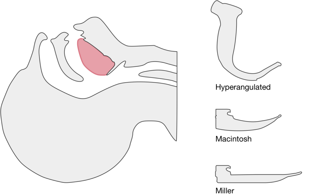
Laryngoscopy had until recently remained relatively unchanged since the introduction of the Macintosh and Miller blades in the 1940’s. Advancements in fiberoptic technology have led to the development of multiple novel devices obviating line-of-sight requirements for direct laryngoscopy which may aid with endotracheal intubation in some populations. While there is no evidence to suggest that video laryngoscopy improves patient-centered outcomes, familiarity with the technique of video laryngoscopy is a critical educational objective for trainees who should be comfortable in the use of this and other rescue airway management adjuncts.

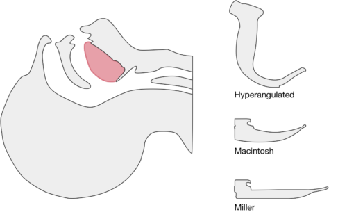
The image above shows a comparison of the curvature of straight (Miller), curved (Macintosh) and hyperangulated blades relative to the oral, pharyngeal and laryngeal axes. The hyperangulated blade easily positions a fiberoptic camera over the glottic opening, allowing easy visualization of the vocal cords. Passing the endotracheal tube without direct line-of-sight becomes the challenge of intubation.

The purpose of this guide is to explore the differences in technique for performing video laryngoscopy compared to direct laryngoscopy. Specifically, the guide is focused on video laryngoscopes with hyper-angulated blades (Verathon Glidescope, Storz C-MAC D-Blade) as these devices pose more challenges for appropriate use while potentially offering the greatest advantages for glottic visualization in challenging airway management.


Advantages and Disadvantages

Advantages1

  • Improved glottic visualization especially in scenarios with limited neck mobility as there is no need to align the oral-pharyngeal-laryngeal axes.
  • Allows others to view the screen and facilitate endotracheal intubation.

Disadvantages1

  • Difficulty passing endotracheal tube despite improved glottic visualization.
  • Obscured view by fogging or contaminated airway (blood, secretions, vomit).
  • Possibility for deterioration of skills in direct laryngoscopy.

Technique

Blade Insertion

Because of it’s unique shape, special care should be taken when inserting a hyperangulated blade to avoid dental/oropharyngeal injury. The handle of the blade may need to be angled towards the patient’s chest and turned slightly to facilitate atraumatic insertion.

Blade Advancement

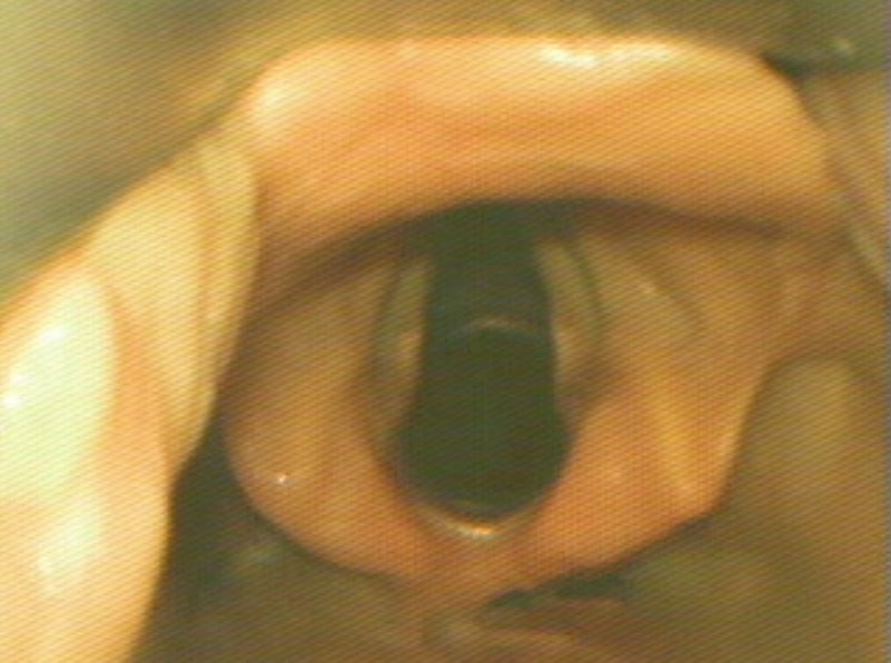
Transition to viewing the screen once the tip of the blade is past the visualized posterior portion of the tongue. Advance the blade along the base of the tongue while monitoring the view on screen. Identify the epiglottis and guide the tip of the blade into the vallecula, the objective is to center the glottic aperture in the upper 2/3 of the screen.

Optimal View

The optimal view shows the vocal cords centered in the upper 2/3 of the screen. This provides adequate screen “real estate” to show the endotracheal tube entering the screen and provide more visual feedback as the tube position is manipulated.

Blade Adjustment

The shape of the hyperangulated blade means that visualizing the airway is less dependent on the lifting motion applied with direct laryngoscopy. Instead, angulating the tip of the blade upward by gently rocking the handle back (taking great care to avoid dental/oropharyngeal trauma) will dramatically improve the view.

Endotracheal Tube Insertion

Directly visualize the insertion of the endotracheal tube into the oral cavity. The endotracheal tube should be loaded with an appropriately angulated rigid stylet. Follow the curvature of the laryngoscope blade entering from the right corner of the mouth. When the endotracheal tube is passed along the edge of the laryngoscope blade, few adjustments will be required for successful endotracheal intubation.

Endotracheal Tube Position Adjustments

If the view is optimized but the endotracheal tube is off-center and does not directly pass the vocal cords into the trachea, subtle adjustments to the endotracheal tube can help guide it into position. Hold the endotracheal tube like a pencil, turning it slightly clockwise or counter-clockwise between your index finger and thumb to manipulate the distal tip left or right above the laryngeal opening.

Stylet Removal

Once past the vocal cords, the hyperangulated rigid stylet will limit continued passage of the endotracheal tube into the trachea as it will get stuck on the anterior surface of the trachea. To facilitate easy passage into the trachea, the stylet should be pulled back slightly to allow passage into the trachea before being fully removed and the tube secured in position.


Review of Evidence

Emergency Department:

  • C-MAC associated with higher rate of successful intubation and greater proportion of grade I-II Cormack-Lehane views compared to direct laryngoscopy.5
  • Glidescope associated with higher rate of successful intubation (and fewer esophageal complications) compared to direct laryngoscopy. 6
  • Glidescope associated with higher rate of successful intubation compared to direct laryngoscopy. 7

Adjustments to Challenging Tube Placement:

  • Combination of flexible-tipped endotracheal tube with rigid stylet (Verathon GlideRight stylet) may increase intubation success.8
  • No apparent benefit to “reverse camber” loading of stylet into endotracheal tube. Reverse camber loading references placing the angled stylet 180° opposite to the natural curvature of the endotracheal tube.9
  • If a malleable stylet is used, 90° angulation was associated with higher rates of successful intubation. 10

Recent Data:

  • Propensity score-matched analysis of multicenter emergency department airway registry – no significant difference in rates of successful intubation comparing Glidescope to direct laryngoscopy. 11
  • Randomized trial comparing direct and video laryngoscopy using C-MAC showed no difference in rates of successful intubation, duration of intubation attempt, aspiration events or length of hospitalization. For interventions randomized to direct laryngoscopy, the video laryngoscope screen was covered. 12

Meta-Analyses:

  • Meta-analysis (predominantly for intubations in the operating room) suggests video laryngoscopy associated with fewer failed intubations, particularly among patients with anticipated difficult airway. Insufficient evidence to analyze effects on incidence of hypoxia, respiratory complications, time to intubation, or mortality.13

References

  1. Maldini B, Hodžović I, Goranović T, Mesarić J. CHALLENGES IN THE USE OF VIDEO LARYNGOSCOPES. Acta Clin Croat. 2016;55 Suppl 1:41-50. doi:10.20471/acc.2016.55.s1.05.
  2. Levitan RM, Heitz JW, Sweeney M, Cooper RM. The complexities of tracheal intubation with direct laryngoscopy and alternative intubation devices. Ann Emerg Med. 2011;57(3):240-247. doi:10.1016/j.annemergmed.2010.05.035.
  3. Hurford WE. The video revolution: a new view of laryngoscopy. Respir Care. 2010;55(8):1036-1045.
  4. Cheyne DR, Doyle P. Advances in laryngoscopy: rigid indirect laryngoscopy. F1000 Med Rep. 2010;2:61. doi:10.3410/M2-61.
  5. Sakles JC, Mosier J, Chiu S, Cosentino M, Kalin L. A Comparison of the C-MAC Video Laryngoscope to the Macintosh Direct Laryngoscope for Intubation in the Emergency Department. Ann Emerg Med. 2012;60(6):739-748. doi:10.1016/j.annemergmed.2012.03.031.
  6. Sakles JC, Mosier JM, Chiu S, Keim SM. Tracheal Intubation in the Emergency Department: A Comparison of GlideScope® Video Laryngoscopy to Direct Laryngoscopy in 822 Intubations. J Emerg Med. 2012;42(4):400-405. doi:10.1016/j.jemermed.2011.05.019.
  7. MD JMM, MPH USP, BA SC, MD JCS. Difficult Airway Management in the Emergency Department: GlideScope Videolaryngoscopy Compared to Direct Laryngoscopy. J Emerg Med. 2012;42(6):629-634. doi:10.1016/j.jemermed.2011.06.007.
  8. Radesic BP, Winkelman C, Einsporn R, Kless J. Ease of intubation with the Parker Flex-Tip or a standard Mallinckrodt endotracheal tube using a video laryngoscope (GlideScope). AANA J. 2012;80(5):363-372.
  9. Jones PM, Turkstra TP, Armstrong KP, et al. Effect of stylet angulation and endotracheal tube camber on time to intubation with the GlideScope. Can J Anesth/J Can Anesth. 2007;54(1):21-27.
  10. Turkstra TP, Harle CC, Armstrong KP, et al. The GlideScope-specific rigid stylet and standard malleable stylet are equally effective for GlideScope use. Can J Anesth/J Can Anesth. 2007;54(11):891-896.
  11. Choi HJ, Kim Y-M, Oh YM, et al. GlideScope video laryngoscopy versus direct laryngoscopy in the emergency department: a propensity score-matched analysis. BMJ Open. 2015;5(5):e007884-e007884. doi:10.1136/bmjopen-2015-007884.
  12. Driver BE, Prekker ME, Moore JC, Schick AL, Reardon RF, Miner JR. Direct Versus Video Laryngoscopy Using the C-MAC for Tracheal Intubation in the Emergency Department, a Randomized Controlled Trial. Acad Emerg Med. 2016;23(4):433-439. doi:10.1111/acem.12933.
  13. Lewis SR, Butler AR, Parker J, Cook TM, Smith AF. Videolaryngoscopy versus direct laryngoscopy for adult patients requiring tracheal intubation. Cochrane Database Syst Rev. 2016;11:CD011136. doi:10.1002/14651858.CD011136.pub2.

Hypotension

Brief H&P:

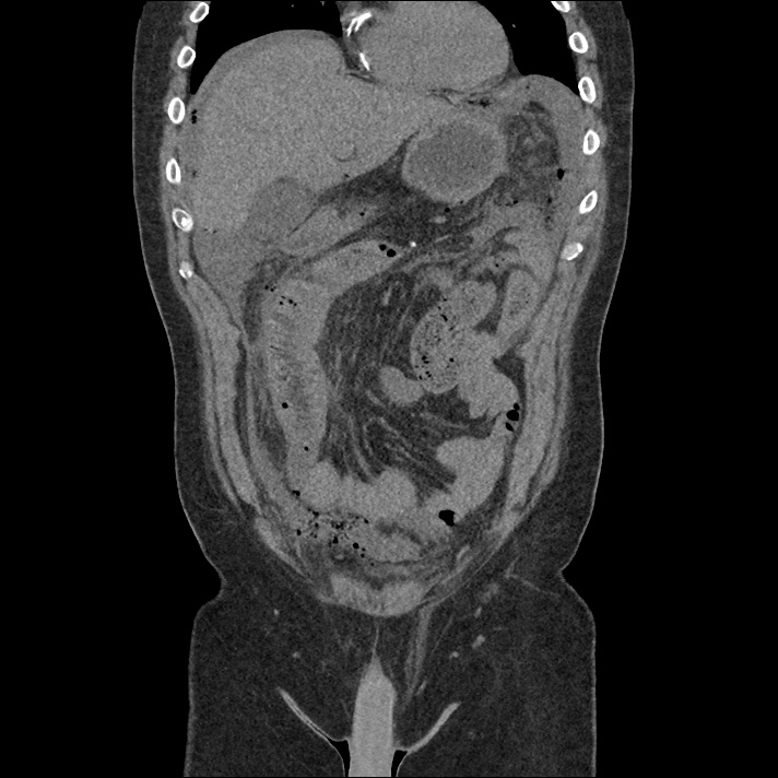
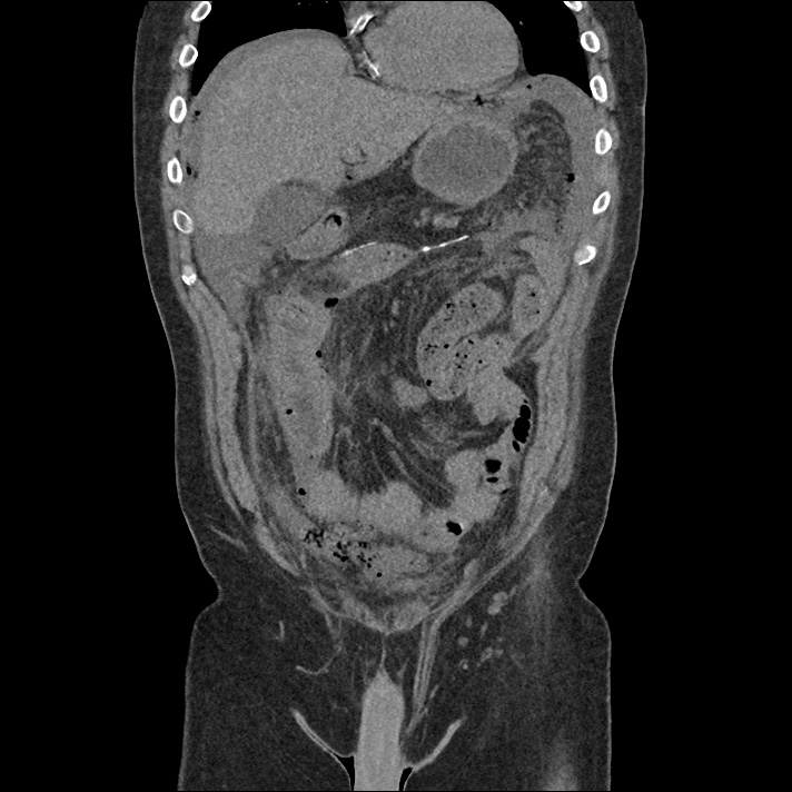
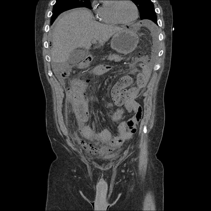
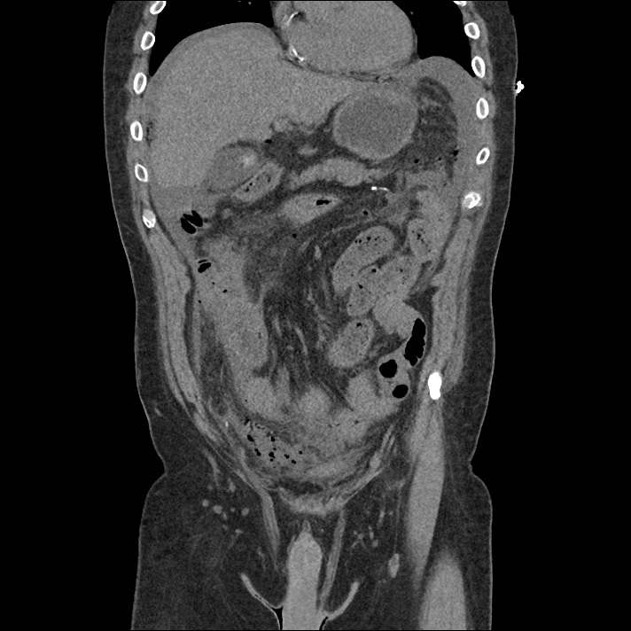
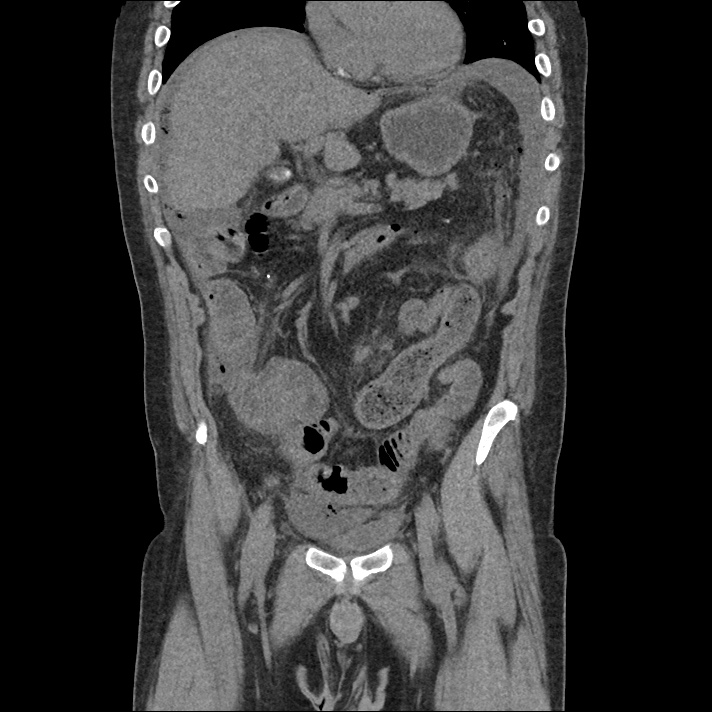
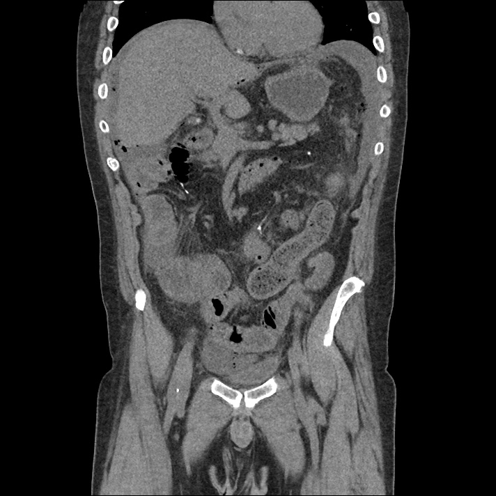
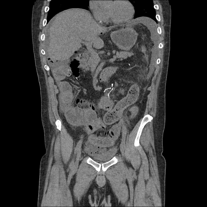
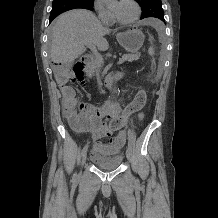
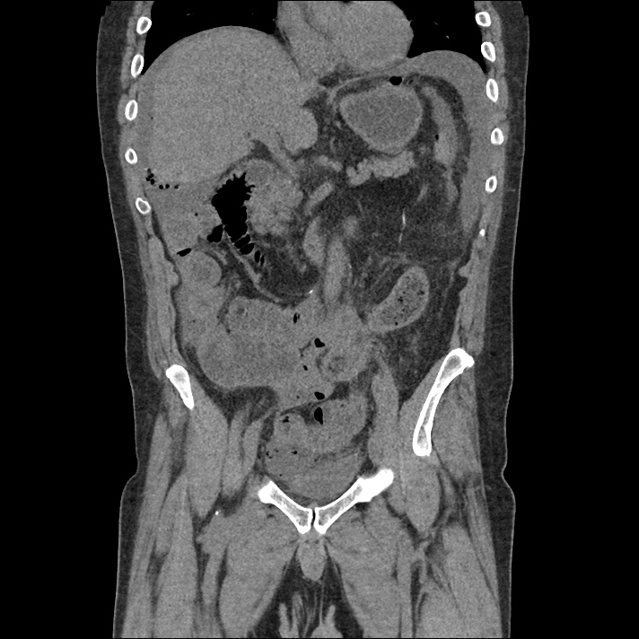
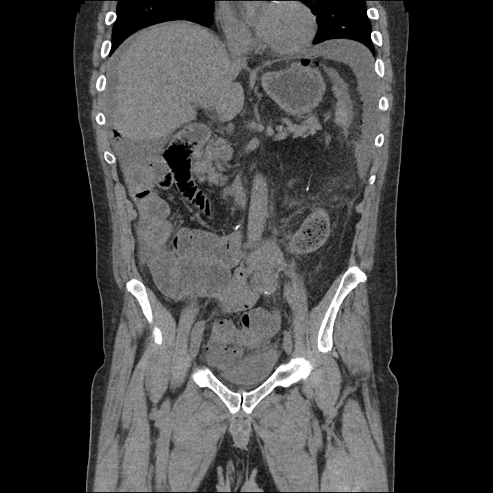
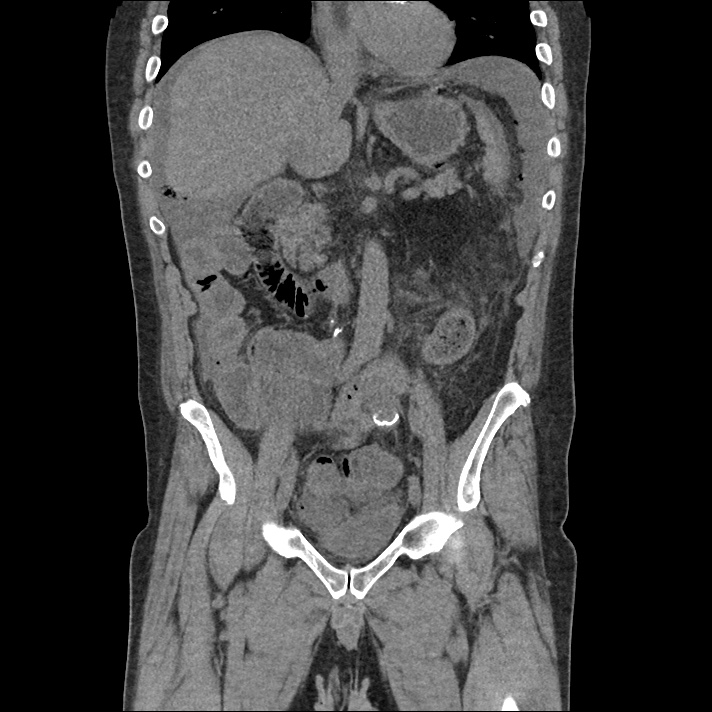
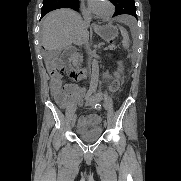
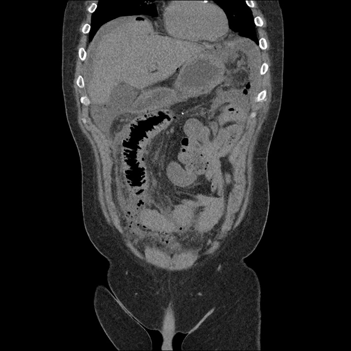
A 50 year-old male with a history of colonic mucinous adenocarcinoma on chemotherapy presented with a chief complaint of “vomiting”. He was unwilling to provide further history, repeating that he had vomited blood prior to presentation. His initial vital signs were notable for tachycardia. Physical examination showed some dried vomitus, brown in color, at the nares and lips; left upper quadrant abdominal tenderness to palpation; and guaiac-positive stool. Point-of-care hemoglobin was 3g/dL below the most recent measure two months prior. As his evaluation progressed, he developed hypotension and was transfused two units of uncrossmatched blood with adequate blood pressure response – he was started empirically on broad-spectrum antibiotics for an intra-abdominal source. Notable laboratory findings included a normal hemoglobin/hematocrit, acute kidney injury, and elevated anion gap metabolic acidosis presumably attributable to serum lactate of 10.7mmol/L. Computed tomography of the abdomen and pelvis demonstrated pneumoperitoneum with complex ascites concerning for bowel perforation. The patient deteriorated, was intubated, started on vasopressors and admitted to the surgical intensive care unit. The initial operative report noted extensive adhesions and perforated small bowel with feculent peritonitis. He has since undergone multiple further abdominal surgeries and remains critically ill.

Imaging

IM-0001-0032
IM-0001-0033
IM-0001-0034
IM-0001-0035
IM-0001-0036
IM-0001-0037
IM-0001-0038
IM-0001-0039
IM-0001-0040
IM-0001-0041
IM-0001-0042
IM-0001-0043
IM-0001-0044
IM-0001-0045
IM-0001-0046
IM-0001-0047
IM-0001-0048
IM-0001-0049
IM-0001-0050

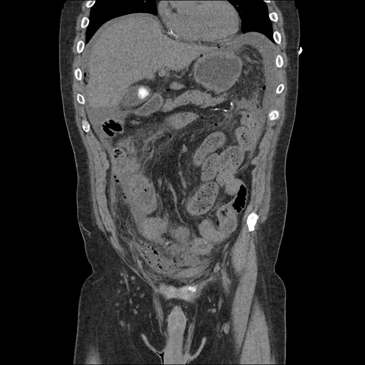
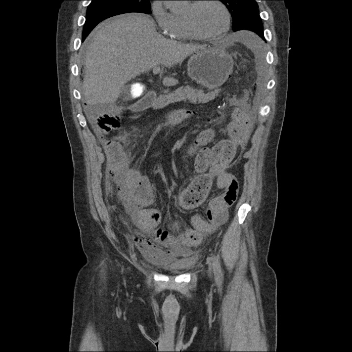
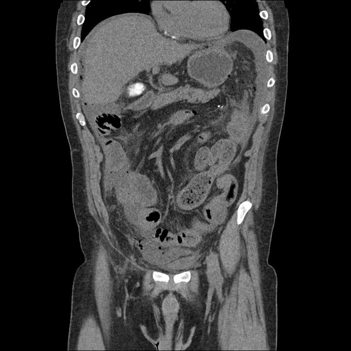
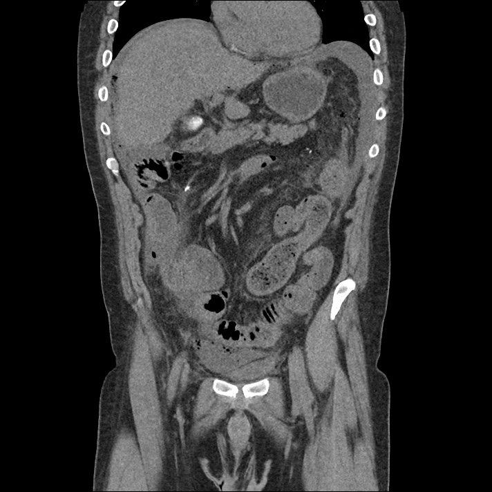
CT Abdomen/Pelvis

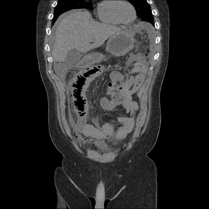
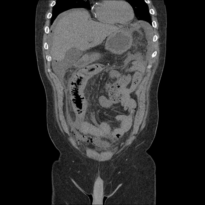
Free air is seen diffusely in the non-dependent portions of the abdomen: in the anterior abdomen and pelvis, inferior to the diaphragm, and in the perisplenic region. There is complex free fluid in the abdomen.

Algorithm for the Evaluation of Hypotension1

This process for the evaluation of hypotension in the emergency department was developed by Dr. Ravi Morchi. In the case above, a systematic approach to the evaluation of hypotension using ultrasonography and appropriately detailed physical examination may have expedited the patient’s care. The expertly-designed algorithm traverses the cardiovascular system, halting at evaluable checkpoints that may contribute to hypotension.

  1. The process begins with the cardiac conduction system to identify malignant dysrhythmias (bradycardia, or non-sinus tachycardia >170bpm), which, in unstable patients are managed with electricity.
  2. The next step assesses intravascular volume with physical examination or bedside ultrasonography of the inferior vena cava. Decreased right atrial pressure (whether due to hypovolemia, hemorrhage, or a distributive process) is evidenced by a small and collapsible IVC. If hemorrhage is suspected, further ultrasonography with FAST and evaluation of the abdominal aorta may identify intra- or retroperitoneal bleeding.
  3. If a normal or elevated right atrial pressure is identified, evaluate for dissociation between the RAP and left ventricular end-diastolic volume. This is typically caused by a pre- or intra-pulmonary obstructive process such as tension pneumothorax, cardiac tamponade, massive pulmonary embolism, pulmonary hypertension, or elevated intra-thoracic pressures secondary to air-trapping. Thoracic ultrasonography can identify pneumothorax, pericardial effusion, or signs of elevated right ventricular systolic pressures (RV:LV, septal flattening).
  4. Assuming adequate intra-vascular volume is arriving at the left ventricle, rapid echocardiography can be used to provide a gross estimate of cardiac contractility and point to a cardiogenic process. If there is no obvious pump failure, auscultation may reveal murmurs that would suggest systolic output is refluxing to lower-resistance routes (ex. mitral insufficiency, aortic insufficiency, or ventricular septal defect).
  5. Finally, if the heart rate is suitable, volume deficits are not grossly at fault, no obstructive process is suspected, and cardiac contractility is adequate and directed appropriately through the vascular tree, the cause may be distributive. Physical examination may reveal dilated capillary beds and low systemic vascular resistance.

Algorithm for the Evaluation of Hypotension

Guided Lecture

EM Ed
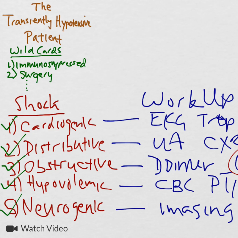
Watch “The Transiently Hypotensive Patient: Who Cares?” from EM Ed. In this lecture Dr. Basrai reviews the diagnostic pathway for a patient who presents with transient hypotension.

References

  1. Morchi R. Diagnosis Deconstructed: Solving Hypotension in 30 Seconds. Emergency Medicine News. 2015.

Altitude and Dysbarism

Altitude Illness

  • Risk factors: altitude, rapidity of ascent, sleeping altitude
  • Pathophysiology
    • Hypobaric hypoxia
      • Pulmonary: vasoconstriction  pulmonary hypertension capillary leak
      • Cerebral: vasodilation edema
    • Acclimatization
      • Hyperventilation primary respiratory alkalosis compensatory metabolic acidosis
      • Acetazolamide promotes renal bicarbonate excretion and accelerates acclimatization
  • Management: oxygen and descent

Acute mountain sickness (2000m)

  • Mild cerebral edema
  • Symptoms: headache, nausea/vomiting, fatigue (hangover)
  • Management: acetazolamide 250mg PO BID, dexamethasone 4mg q6h

High-altitude pulmonary edema (HAPE, 3000m)

  • Non-cardiogenic pulmonary edema
  • Symptoms: dyspnea at rest, cough, fever
  • Signs: hypoxia, crackles
  • CXR: patchy infiltrates
  • Management: nifedipine, PDEi (sildenafil), HBO

High-altitude cerebral edema (HACE, 4500m)

  • Cerebral edema
  • Symptoms: ataxia, altered mental status
  • Management: acetazolamide 250mg PO BID, dexamethasone 10mg then 4mg q6h, HBO
  • Gamow bag: portable HBO

Dysbarism (diving pathology)

  • Principles
    • Boyle’s Law: volume = 1/pressure
      • Volume changes greatest near surface
    • Henry’s Law: increased pressure increases proportion of dissolved gas

Barotrauma

  • Localized (descent)
    • Barotitis media
      • Mechanism: unequal pressure between external and middle ear.
      • Symptoms: pain, vertigo if ruptured
    • Barotitis externa
      • EAC edema/hemorrhage
    • Barotitis interna
      • Bleeding/rupture of round window
      • Symptoms: vertigo, tinnitus, hearing loss
      • Management: ENT referral
    • Sinus squeeze: pain and epistaxis
    • Mask squeeze: periorbital petechiae
  • Localized (ascent)
    • Barodontalgia
      • Air trapped in filling
      • Symptoms: pain, fracture
    • Alternobaric vertigo: Unequal ear pressure causing vertigo
    • GI barotrauma: belching, flatulence
  • Pulmonary overpressurization (ascent)
    • Mechanism: rapid ascent without exhalation, focal alveolar rupture leading to pneumomediastinum, rarely pneumothorax
    • CXR: continuous diaphragm sign
    • Symptoms: dysphonia, neck fullness, chest pain
    • Management: supportive
  • Air gas embolism (ascent)
    • Mechanism: similar to POP, air enters pulmonary venous circulation
    • Symptoms: MI, arrest, stroke, seizure within 10 minutes
    • Management: IVF, oxygen, HBO

Dissolved Gas Problems

  • Nitrogen narcosis
    • At >100ft, nitrogen enters nervous system and acts similarly to general anesthetic
    • Symptoms: similar to alcohol intoxication, complications arise from poor judgement
    • Management: ascent
  • Oxygen toxicity
    • Setting: industrial dives, deep
    • Symptoms: seizure, nausea, muscle twitching
  • Decompression sickness
    • Mechanism: nitrogen gas dissolves poorly in solution, with ascent forms bubbles, occurs 1-2 hours after ascent
    • Types
      • Musculoskeletal, integumentary (“bends”)
        • Symptoms: arthralgia, cutis marmorata
      • Neurological
        • Lower spinal cord (thoracic/lumbar/sacral)
          • Symptoms: paraplegia, paresthesia, bladder dysfunction
        • Cerebellum (“staggers”)
          • Symptoms: ataxia
        • Pulmonary (“chokes”)
          • Symptoms: similar to pulmonary embolus
        • Management: IVF, oxygen, HBO

Bites

Mammalian

  • Human: Eikenella corrodens
  • Dog/Cat: Pasteurella multocida

Athropod

  • Hymenoptra (bee, wasp, hornet, ant)
    • Venom: histamine reaction, anaphylaxis
    • Symptoms
      • Local: pain, swelling, pruritus
      • Toxic (<48h): multiple bits, N/V, syncope, HA
      • Anaphylaxis: minutes
      • Delayed (10-14d): serum sickness, fever, arthralgia, malaise
    • Management
      • Remove stinger
      • Wash, ice, anti-histamine, analgesia
  • Brown recluse (violin pattern)
    • Location: Midwest, wood pile
    • Symptoms: initially painless, cytotoxic venom may cause necrosis
    • Management: supportive, Tdap, delayed debridement if necrotic
  • Black widow (red hourglass)
    • Venom: neurotoxic, ACh, NE
    • Symptoms: painful, erythema, muscle contractions (“acute abdomen”), localized diaphoresis from ACh release
    • Management: analgesia, benzodiazepines, antivenom for refractory pain (may cause anaphylaxis)

Snake

  • Crotalid (rattlesnake, copperhead, cottonmouth, collectively “pit vipers”)
    • Venom: cytotoxic, hemorrhagic
    • Symptoms: erythema/edema (ecchymoisis/bullae), nausea/vomiting, metallic taste
    • Labs: DIC
    • Management
      • Immobilization (no tourniquet)
      • Local wound care, Tdap
      • CBC, INR, fibrinogen (q2h)
      • Antivenom (Crofab 4-6 vials): given until symptoms or laboratory abnormalities arrest
      • Compartment syndrome: avoid surgery
  • Elapidae (coral snake, “red on yellow”)
    • Venom: neurotoxic, delayed 10-12h
    • Symptoms: no significant local reaction, bulbar palsies, respiratory depression
    • Management: no antivenom, supportive care, intubation

Cnidaria (jellyfish)

  • Symptoms: local pain, erythema, pruritus
  • Management: 5% acetic acid, alcohol, remove stinger
    • Antivenom for box jellyfish

Stingray

    • Symptoms: local pain, edema
    • Management: Local wound care, Tdap, hot water immersion, antibiotics for Vibrio (cephalexin with doxycycline)

Vibrio vulnificus

  • Symptoms: necrotizing fasciitis, in cirrhotic primary septicemia after ingesting shellfish

Electrical Injuries


Physics

  • High-voltage defined as >1,000V
  • Voltage related to injuries current via resistance (V=IR)
  • AC is 3x more lethal than DC
    • Fluctuation at 60Hz causes tetany, maintained grasp on source

Effects

  • Dysrhythmia
    • DC: asystole
    • AC: ventricular fibrillation
    • Delayed dysrhythmia uncommon
  • Burn
  • Tissue ischemia: vascular spasm or thrombosis
  • CNS: AMS, seizure, ICH, neuropathy
  • MSK: posterior shoulder dislocation

Management

  • Asymptomatic: None
  • Mild (i.e. small burn): ECG, UA (rhabdo)
  • High voltage: Labs, CT, admit for observation
  • Pediatrics: oral commissure burn, discharge with plastic surgery follow-up if no LOC, normal ECG, tolerating PO. Risk of delayed labial artery bleeding.

Complications

  • Keraunoparalysis: current travels up and down lower extremities causing transient paresthesia and paralysis.
  • Trauma: TM rupture, other mechanical injuries


Heat Emergencies

Overview

  •  Spectrum
    • Cramps
    • Syncope
    • Exhaustion
    • Stroke
  • Physiology of cooling
    • Radiation: body warmer than environment, heat radiates away
    • Evaporation: environment warmer than body, sweat promotes heat exchange, affected by ambient humidity

Heat cramps

  • Mechanism: fluid/electrolyte depletion resulting in muscle cramps
  • Management: IVF, electrolyte repletion, cooling

Heat syncope

  • Mechanism: vasodilation resulting in hypotension
  • Management: IVF, cooling, rule out alternative etiologies

Heat exhaustion

  • Mechanism: similar to heat cramps
  • Symptoms: influenza-like, headache, fatigue, dizziness, nausea, normal mental status distinguishes from heat stroke
  • Findings: temperature <40°C
  • Management: IVF, cooling

Heat stroke

  • Mechanism: similar to heat cramps
  • Symptoms: prodrome of heat exhaustion
  • Signs: AMS, ataxia, seizure
  • Findings: temperature >40°C
  • Mortality: 30-80%
  • Labs: AST/ALT, coagulopathy, DIC, rhabdomyolysis, ATN/AKI
  • CXR: pulmonary edema
  • Types
    • Classical: elderly, dry skin, mild dehydration, increased mortality
    • Exertional: young athlete, diaphoretic, increased morbidity (organ failure)
  • Management
    • Evaporative cooling
    • Ice packs to large vessels
    • GI lavage
    • Liberal intubation
    • Benzodiazepines or thorazine for inappropriate thermogenesis (shivering)
    • Halt cooling at 40°C

Hypothermia

Overview

  • Risk factors
    • Extremes of age
    • Behavioral: psychosis, intoxication
  • Types
    • Chillblains
    • Immersion foot
    • Frostnip
    • Frostbite
    • Generalized

Hypothermia

Chilblains

  • Findings: red/white plaques on extremities
  • Symptoms: pruritus, pain
  • Management: supportive (gentle warming), topical corticosteroids, consider nifedipine

Immersion foot (trench)

  • Mechanism: prolonged immersion in non-freezing water, vasoconstriction leads to ischemia/necrosis
  • Findings: pale, mottled skin, paresthesia
  • Management: supportive, drying and rewarming
  • Complications: gangrene

Frostnip

  • Retrospective distinction from frostbite after rewarming if no tissue loss

Frostbite

  • Mechanism: extracellular then intracellular crystal formation (mechanistically similar to crush injury)
  • Reperfusion: cellular injury triggers cytokine release upon reperfusion, results in microvascular thrombosis and tissue ischemia/necrosis
  • Classification: grades I-II superficial to dermis, grades III-IV involve subcutaneous tissue to bone
  • Management
    • Rapid rewarming (immersion in warm water at 41°C)
    • Tdap
    • Debridement of clear blisters

Generalized

  • Causes
    • Exposure
    • Metabolic (adrenal, thyroid, hypoglycemia)
    • Sepsis
  • Grading
    • Mild (32.2-35°C)
      • Findings: excitation, tachycardia, hypertension, shivering thermogenesis
    • Moderate (30-32.2°C)
      • Findings: ataxia, AMS, bradycardia, hypotension, bradypnea
      • ECG:  Osborn wave
    • Severe (<30°C)
      • Complications
        • Increased risk of arrhythmia (bradycardia, slow atrial fibrillation, ventricular fibrillation, asystole)
        • Irritable myocardium
        • Decreased enzymatic activity
          • Renal: cold diuresis
          • Heme: coagulopathy (hidden on labs as blood rewarmed prior to testing)
          • Metabolic: hyperglycemia as insulin ineffective
      • Management
        • Ventricular fibrillation: attempt one shock, then focus on rewarming if ineffective
        • Goal >30°C

Radiation Exposure

Physics

  • Units
    • Gray (amount of radiation absorbed by body)
    • Sievert (toxicity associated with radiation exposure)
  • Types
    • Alpha: 0.1mm penetration, injury through ingestion
    • Beta: 1cm penetration, injury through skin or ingestion
    • Gamma: deep penetration
  • Factors
    • Time and distance (1/d2)
    • Shielding
    • Radiosensitive cells (rapidly dividing such as hematopoetic, GI)

Injury

  • Localized: epilation or burns, delayed by days
  • Internal (inhaled, ingestion)
    • Radioactive iodine: high dose results in thyroid ablation, low dose increases risk of thyroid malignancy
  • External: managed by removing clothing, soap/water shower
  • Whole body (gamma)
System Dose Time of onset Signs/Symptoms
Hematopoetic 2G 2d Pancytopenia, increased risk of infection
GI 6G Hours Nausea/vomiting, diarrhea, GI bleeding
CV/CNS 10G Minutes Shock, seizure

Key clinical features

  • Multiple affected individuals with nausea/vomiting suggests radiation exposure
  • Rapidity of onset of symptoms suggests increased dose/exposure
  • LD505G
  • Prognosis by lymphocyte count
    • ALC >1000 at 48h suggests good prognosis
    • ALC <300 at 48h suggests poor prognosis

Submersion Injury

Pathophysiology

  • Breath-holding until eventual involuntary gasp which triggers reflexive laryngospasm. Resultant loss of consciousness may cause laryngeal relaxation and aspiration.
  • Fluid aspiration results in decreased surfactant activity and atelectasis. This is complicated by V/Q mismatch and atelectrauma which can lead to ARDS.

Symptoms

  • Progressive respiratory distress
  • AMS: due to cerebral hypoxia
  • Shock: uncommon, consider trauma

Management

  • Albuterol
  • BiPAP
  • Endotracheal intubation
  • ECMO

Disposition

  • Asymptomatic or minor event: observe 2-3 hours
  • Mildly symptomatic: observe 4-6 hours
  • Hypoxia: admit
  • PPV: ICU

Toxicology

Drugs of Abuse

Synthetic Cannabinoids (Spice, K2)

  • Symptoms: anxiety, paranoia, tachycardia
  • Unique symptoms compared to traditional cannabinoids: psychosis, seizure, diaphoresis

Hallucinogenic amphetamines (ecstasy, MDMA)

  • Increased serotonergic activity
  • Management: supportive care (IVF, cooling for hyperthermia), benzodiazepines

Gamma-hydroxybutyrate (GHB)

  • Symptoms: euphoria, hypersexuality, rapid onset/clearance
  • Signs: bradycardia, bradypnea, coma with rapid awakening
  • Management: intubation for depressed GCS
  • Withdrawal: symptoms and treatment identical to ethanol withdrawal, consider baclofen

Cathinone (bath salts)

  • Symptoms: hallucinations
  • Signs: tachycardia, hypertension, tremor, mydriasis, diaphoresis, hyperthermia, bruxism
  • Management: benzodiazepines, consider paralysis, avoid beta-blockers

Cocaine

  • MOA: increase catecholamines, Na-channel blockade
  • Toxicity: HTN, tachycardia, hyperthermia, rhabdomyolysis, MI, seizure, VT
  • Management: benzodiazepines, cooling, anti-hypertensives (nitrate, CCB, not B-blocker)

Amphetamine

  • Toxicity: HTN, tachycardia, hyperthermia, rhabdomyolysis, intracranial hemorrhage
  • Management: same as cocaine

Benzodiazepines

  • Toxicity: sedation, respiratory depression
  • Management: consider flumazenil 0.2mg IV q1min x1-5

Toxic Alcohols

  • Overview
    • Toxic metabolites produced by alcohol dehydrogenase which can be inhibited by ethanol or fomepizole
    • Fomepizole: 15mg/kg loading dose, 10mg/kg q12h x4 doses then 15mg/kg q12h (stimulates own metabolism); if dialysis, q4h
  • Diagnosis: osmolar gap (>14), 2Na + Glu/18 + BUN/2.8 + EtOH/4.6
  • Treatment
    • ADH inhibition
    • HCO3
    • Hemodialysis
    • Supportive care
    • Hypoglycemia: dextrose

Methanol

  • Component of antifreeze, windshield washer fluid
  • Metabolite formic acid which causes acidosis and blindness
  • Can give folate

Ethylene glycol

  • Component of antifreeze, automobile coolants, de-icing agents
  • Metabolite oxalic acid which precipitates calcium oxalate crystals and causes acute renal failure
  • Can give thiamine (100mg q6h), pyridoxine (500mg q6h), Mg

Isopropanol

  • Component of rubbing alcohol
  • Metabolite acetone which does not cause acidosis

Analgesics

Acetaminophen

  • Metabolism: glucoronidation, CYP450
    • CYP450 pathway produces toxic metabolite when glucoronidation overwhelmed
    • In pediatrics, sulfation process protective
  • Toxic dose: >150mg/kg, >3g/day
  • Injury: liver (centrilobular necrosis), renal, pancreatic
  • Increased risk: induced CYP450 (chronic EtOH, rifampin, anti-epileptics)
  • Nomogram: applicable to single ingestion at 4-hours
  • Labs: PT/INR, LFT, lipase, chemistry
  • Management: NAC
    • PO: 140mg/kg, 70mg/kg q4h
    • IV: 150mg/kg, 50mg/kg over 4h, 100mg/kg over 16h

NSAID

  • Symptoms
    • Acute: GI upset, low risk UGIB
    • Acute massive: acidosis, coma, seizures
    • Chronic: UGIB, nephropathy, agranulocytosis

Aspirin

  • Signs: tachycardia, hyperthermia, tachypnea/hyperpnea
  • Severe: cerebral and pulmonary edema, CNS hypoglycemia
  • Labs: primary respiratory alkalosis with metabolic acidosis
  • Management
    • Hypoglycemia (CNS) treatment
    • Bicarbonate infusion (urine pH > 8)
    • Hemodialysis for pulmonary edema, cerebral edema, renal failure, acidemia, level >100mg/dL (acute) or > 60mg/dL (chronic)

Opioids

  • Symptoms: respiratory depression, miosis
  • Management: naloxone 0.04mg, 0.4mg, 2mg
  • Withdrawal: nausea/vomiting, diarrhea, abdominal pain, piloerection
    • Neonates: seizure, death
  • Complications with specific agents:
    • Meperidine, tramadol: seizures
    • Methadone: QT prolongation

Anesthetics

Lidocaine

  • Mechanism: Na-channel blockade
  • Types:
    • Ester (one “i”): cocaine, procaine, benzocaine
    • Amide (two “i”): lidocaine, bupivacaine
  • Toxicity
    • Dose: 4mg/kg, 7mg/kg with epinephrine
    • CNS: perioral numbness, slurred speech, seizure
    • CV: VT, VF, AV block
    • Methemoglobinemia: methylene blue
  • Treatment
    • Seizure management
    • Bicarbonate for dysrhythmia
    • Intralipid

Anti-cholinergics

Sympathetic Parasympathetic
Mydriasis Miosis
Bronchodilation Bronchospasm/bronchorrhea
Tachycardia Bradycardia
Urinary retention Urinary incontinence
Hyperglycemia Salivation/lacrimation
Diaphoresis Increased GI motility
  • Examples
    • Atropine
    • Anti-histamine
    • TCA
    • Phenothiazines
    • Jimson weed
  • Symptoms
    • Peripheral: mydriasis, anhidrosis, flushing, hyperthermia, ileus, dry mucous membranes, AUR
    • Central: agitation (passive), delirium, coma, seizure
  • Treatment
    • Supportive
    • Benzodiazepines
    • Theoretically physostigmine
      • Avoid in seizure, QRS-widening, reactive airway disease
      • Possible diagnostic use

Drugs causing miosis (COPS)

  • C: cholinergics
  • O: opioids
  • P: phenothiazines
  • S: sedatives

Drugs causing QT-prolongation

  • Examples:
    • Phenothiazines
    • Anti-arrhythmics
    • Butyrophenones (ex. haloperidol)
    • Macrolides
    • Fluoroquinolones
    • Methadone
    • Ondansetron
    • Atypical antipsychotics
  • Treatment
    • Magnesium sulfate 2g IV over 1min
    • Overdrive pacing (transcutaneous, transvenous if not captured)
    • Consider isoproterenol (pharmacologic overdrive)

Serotonin syndrome

  • Cause: exposure to serotonergic agent(s)
  • Symptoms: agitation, mydriasis, tremor/clonus in lower extremities, tachycardia, hyperthermia
  • Management
    • Supportive care (IVF, vasopressors)
    • Cooling measures and paralysis for hyperthermia
    • Benzodiazepines
    • Cyproheptadine 12mg PO/NG
    • Dexmedetomidine infusion

Anti-emetics

Phenothiazines

  • Examples: compazine (prochlorperazine), phenergan (promethazine)
  • MOA: DA-antagonist
  • AE: sedation, dystonia, parkinsonism
  • Toxicity: seizure, VT, hypotension (TCA-like)

5-HT3 antagonists

  • Examples: zofran (ondansetron), granisetron
  • Toxicity: QT-prolongation

Anti-hypertensives

Calcium channel blockers

  • Toxicity: hypotension, bradycardia, AV blockade, hyperglycemia
  • Management
    • Atropine: 0.5mg IV q2-3min
    • Glucagon: 5mg IV q10min x2 (with anti-emetic)
    • IVF, vasopressors (norepinephrine, epinephrine)
    • Calcium: 3g gluconate, 1-3g chloride
    • High-dose insulin: 1 unit/kg, monitor hypoglycemia/hypokalemia
    • Intralipid: 1.5mL/kg bolus then 0.25mL/kg/minute
    • GI decontamination
    • Pacing, IABP, ECMO

Beta blockers

  • Toxicity: similar to CCB, hypoglycemia
  • Management: similar to CCB, calcium ineffective

Digoxin (foxglove, oleander)

  • MOA: inhibits Na/K ATPase, increases intracellular calcium (inotropic)
  • Toxicity
    • CV: bradycardia, hypotension
    • ECG: bidirectional VT, PVC, scooped ST-segment
    • CNS: agitation, psychosis
    • Visual: yellow-green vision, halo
    • Metabolic: hyperkalemia (acute), hypokalemia, hypomagnesemia
  • Treatment
    • GI decontamination
    • Atropine
    • Transcutaneous pacing (avoid transvenous, irritable myocardium)
    • Digibind
    • Avoid calcium

Clonidine

  • Toxicity: bradycardia, hypotension, opioid mimic (miosis, lethargy, respiratory depression)
  • Management: supportive care, stimulation for respiratory depression, atropine

Sodium-channel blockers

  • Drugs
    • TCA
    • Diphenhydramine
    • Procainamide
    • Carbamazepine
  • ECG
    • QRS prolongation
    • Prominent “R” in aVR
    • RAD
  • Treatment
    • Sodium bicarbonate

Anti-hyperglycemics

Sulfonylurea

  • Symptoms: recurrent severe hypoglycemia
  • Management: octreotide 50-75mcg SQ/IM q6h

Other agents that cause hypoglycemia

  • EtOH
  • B-blocker
  • Quinine
  • Salicylate

Environmental

Carbon monoxide

  • Source: combustion (gas heater, indoor barbeque)
  • Toxicity
    • General: influenza-like, multiple proximate affected individuals
    • GI: abdominal pain, nausea
    • CNS: headache, dizziness, confusion, ataxia, seizure
    • CV: palpitations, arrhythmia, hypotension, MI
  • Treatment
    • T½: RA 6h, NRB 1h, 3atm 0.5h
    • Hyperbaric: neuro deficit, syncope, pregnancy, CV toxicity

Cyanide

  • Mechanism: inhibits oxidative phosphorylation
  • Source: structural fire (wool, silk)
  • Symptoms: syncope, seizure, coma, cardiovascular collapse
  • Detection: severe lactic acidosis, “arterialization” of venous blood, “bitter almond” odor
  • Treatment
    • Hydroxycobalamin (Cyanokit): 5g IV, may repeat x1
    • Sodium thiosulfate 12.5g IV

Methemoglobinemia

  • Mechanism: Fe2+ converted to Fe3+, “functional anemia”
  • Source: nitrite (food), topical/local anesthetics, pyridium, dapsone, reglan
  • Detection: normal PaO2, SpO2 85% unresponsive to supplemental oxygen, ABG with co-oximetry
  • Management: methylene blue 1-2mg/kg IV if symptomatic or MetHb >25%
    • Contraindicated in G6PD deficiency, treat with exchange transfusion or HBO

Hydrogen Sulfide

  • Source: industrial, sulfur spring, sewer
  • Detection: “rotten egg” odor
  • Management: remove from source, supportive care

Hydrocarbon

  • Source: huffing canisters
  • Toxicity: VT/VF from myocardial sensitization
  • Management: beta-blockade
  • Complications: harmless if ingested, aspiration leads to ARDS

Hydrofluoric acid

  • Source: rust remover, wheel cleaner, glass etching
  • Symptoms: pain-out-of-proportion, delayed onset
  • Toxicity: Hypocalcemia (QTc prolongation, VT/VF/TdP), hyperkalemia, hypomagnesemia
  • Management: analgesia, topical calcium gluconate gel, intravenous calcium for large BSA involvement

Alkaline ingestion

  • Symptoms: esophageal perforation, delayed stricture

Acid ingestion

  • Symptoms: gastric perforation (rare), delayed gastric outlet obstruction
  • Findings: metabolic acidosis

Botulism

  • Sources
    • Adult: ingested preformed toxin
    • Infants: ingested spores (achlorhydric), in vivo toxin production
    • Wound: black tar heroin
  • Symptoms: dysphagia, ptosis, diplopia, respiratory failure, descending paralysis
    • Infants: constipation, floppy
  • Management: supportive care, intubation
    • Adults: Anti-toxin from CDC or local Department of Health
    • Infants: 100mg/kg IV x 1 dose (BabyBIG)

Heavy Metals

Iron

  • Dose
    • Ferrous sulfate: 20% elemental iron
    • Toxic: >20mg/kg
    • Lethal: >60mg/kg (1 tablet 325mg ferrous sulfate per kilogram)
  • Toxicity: corrosive, anti-coagulant, hepatotoxic
  • Course
    • Stage I: GI effects, emesis with hematemesis
    • Stage II: Quiescent
    • Stage III: Systemic, multi-organ system dysfunction
    • Stage IV: Resolution, gastric scarring and outlet obstruction
  • Workup
    • CBC/BMP
    • LFT
    • Lactate
    • Fe level
    • KUB (if positive consider WBI)
  • Treatment
    • Decontamination: no activated charcoal, consider WBI
    • Deferoxamine: 15mg/kg/hr

Lead

  • Source: paint, batteries
  • Toxicity
    • Acute: headache, encephalopathy, seizure
    • Chronic: malaise, weight loss, arthralgia, anemia (basophilic stippling)
  • Diagnosis: lead level, wrist drop
  • Management: chelation (BAL, EDTA, DMSA) for level >50ug/dL or asymptomatic >70ug/dL

Lithium

  • Source: iatrogenic, drug-drug interaction
  • Symptoms
    • GI: nausea/vomiting, diarrhea
    • CNS: tremor, coma
    • CV: TWI, QT-prolongation
  • Management
    • IVF, encourage renal elimination
    • Hemodialysis

Other Drugs

Disulfuram

  • MOA: aldehyde dehydrogenase inhibitor
  • Symptoms: increased acetaldehyde leads to flushing, headache, nausea/vomiting, tachycardia, hypotension
  • Management: antihistamine, IVF, vasopressors
  • Other agents causing disulfuram-like reaction: metronidazole, INH, sulfonylurea

Isoniazid

  • Toxicity: seizure
  • Management: pyridoxine 5g IV, repeat x1

Theophyline

  • Toxicity: seizure
  • Management
    • Decontamination: AC
    • Seizures: benzodiazepines
    • Tachyarrhythmia (commonly MAT): beta-blockade
    • Hemodialysis: acute > 100mg/L, chronic >30mg/L

Monoamine oxidase inhibitors

  • Toxicity: food/drug interaction
  • Symptoms: tachycardia, hypertension, hyperthermia, agitation
  • Management: cooling, IVF, management of hyper/hypotension

Phenytoin

  • Oral: cerebellar dysfunction (ataxia), CNS depression
  • IV: hypotension (suspension contains propylene glycol)

Nutritional Supplements

  • Fat-soluble vitamins
    • A: benign intracranial hypertension
    • D: hypercalcemia

Envenomations

Snake

  • Crotalid (rattle), elapidae (coral)
  • Symptoms
    • Local reaction: edema, hemorrhagic bullae
    • Systemic: perioral numbness, fasciculations
    • Severe: thrombocytopenia, decreased fibrinogen
  • Management: Crofab 5 vials

Spider

  • Black widow
    • Identification: hourglass on abdomen
    • Symptoms: painful bite, target-appearance, rarely “acute abdomen”
    • Management: analgesia, anti-venom, tetanus
  • Brown recluse
    • Identification: violin shape on head
    • Geography: Southeast, Midwest
    • Symptoms: painless bite, local reaction, delayed healing with eschar
    • Rare: hemolysis, DIC, shock
    • Management: supportive care, antibiotics if superinfected, consider dapsone, tetanus

Scorpion (Centruroides)

  • Geography: Arizona
  • Symptoms
    • Autonomic: HTN, tachycardia, diaphoresis
    • CNS: opsoclonus, slurred speech, dysphagia
  • Management: anti-venom, supportive care, analgesia, tetanus

Marine

  • Ciguatera
    • Source: toxin bioconcentrated in fish
    • Symptoms: gastroenteritis, hot/cold-reversal, “loose teeth” sensation
    • Management: mannitol
  • Scombroid
    • Source: poorly-refrigerated fish, histamine-like
    • Symptoms: flushing trunk/face (distinguish from allergic reaction), gastroenteritis
    • Management: supportive care, IVF, anti-histamine, bronchodilators if indicated
  • Paralytic shellfish poisoning
    • Source: bivalve
    • Symptoms: gastroenteritis, paralysis
    • Management: supportive, intubation
  • Jellyfish and Cnidaria
    • Source: nematocyst
    • Symptoms: burning pain, pruritus
    • Severe: Irakundji syndrome (HTN, pulmonary edema)
    • Management: supportive, analgesia, box jellyfish antidote, consider vinegar
  • Stingray
    • Source: heat-labile toxin
    • Management: affected area in warm water, tetanus, ciprofloxacin (Vibrio)

Mushrooms

  • Amanita: centrilobular necrosis, similar to acetaminophen
  • Gyronatum: similar to INH (seizure and treatment), may cause methemoglobinemia
  • Symptoms: muscarinic (SLUDGE)
    • Early onset generally benign, delayed onset (>6h) suggests more serious course
  • Management: atropine, glycopyrrolate, IVF

Pesticides

  • Organophosphate: irreversible
  • Carbamate: reversible
  • Symptoms: muscarinic (SLUDGE)
  • Treatment: atropine 2-6mg IV double q5min to control secretions, pralidoxime (for organophosphates)

Strychnine

  • Source: rodenticide
  • Symptoms: myoclonus, opisthotonus, agitation
  • Management: benzodiazepines, airway protection, paralysis