Headache

Brief HPI:

A 48 year-old male with hypertension and hyperlipidemia presents with headache. Notes onset of symptoms 8 hours prior to presentation, reaching maximal severity within seconds. Headache improved with over-the-counter analgesics. On examination, there are no neurological deficits, neck is supple. A CT head non-contrast is obtained:

01_normal
02_normal
03_normal
04_normal
05_normal
06_normal
07_normal
08_normal
09_normal
10_normal
11_normal
12_normal
13_normal
14_normal
15_normal
16_normal
18_normal
19_normal
20_normal

CT Head:

No acute intracranial process. Case courtesy of Assoc Prof Craig Hacking, Radiopaedia.org, rID: 37118

ED Course:

A lumbar puncture is performed, CSF sampling reveals xanthochromia – neurosurgery is consulted and the patient is admitted for angiography and possible intervention.


An Algorithm for the Evaluation of Headache

An Algorithm for the Evaluation of Headache

High-Risk Historical Features

  • Sudden onset (seconds/minutes), patient recalls activity at onset
  • Worst in life or change in character from established headache
  • Fever, neck pain/stiffness
  • Altered mental status
  • Malignancy
  • Coagulopathy: iatrogenic, hepatopathy, dialysis
  • Immunocompromised
  • Rare: CO exposure, jaw claudication, PCKD

Location of Pain

Headache Location

  1. Unilateral: migraine
  2. Periorbital: glaucoma, CVT, optic neuritis, cluster
  3. Facial/maxillary: trigeminal neuralgia, sinusitis
  4. Temporal: GCA
  5. Occipital: cerebellar stroke
  6. Nuchal: meningitis

Characteristics of Primary Headaches

Type Location Duration Quality Associated symptoms Comment
Migraine Unilateral Hours to days Throbbing Photophobia, phonophobia Atypical migraines with neurological findings (basilar, ophthalmoplegic, ophthalmic, hemiplegic)
Tension Bilateral Minutes to days Constricting None
Cluster Unilateral, periorbital Minutes to hours Throbbing Conjunctival injection, lacrimation, rhinorrhea, miosis, eyelid edema Males 90%, triggered by EtOH.

Physical Examination Findings

Vital Signs
Fever: present in 95% of patients with meningitis
Head
Trauma: signs of basilar skull fracture
Temporal artery tenderness/induration: GCA
Pericranial muscle tenderness: tension headache
Trigger point, Tinnel sign: occipital neuralgia
Eyes
Pupillary defects: aneurysm with CN III compression
Papilledema, absence of spontaneous venous pulsations: elevated intracranial pressure
EOM abnormalities: ICH, mass lesion, neuropathy (DM, Lyme)
Horner syndrome (ptosis, miosis, anhidrosis): carotid dissection
Visual field defect: stroke, atypical migraine
Conjunctival injection: glaucoma (fixed, mid-size pupil, elevated intraocular pressure), cluster headache
Mouth
Thrush: immunocompromise
Sinuses
Tenderness to palpation, abnormal transillumination: sinusitis
Neck
Resistance to supine neck flexion: meningitis
Kernig: supine position, hip flexed, knee flexed, resistance to knee extension
Brudzinski: supine position, neck flexion results in knee flexion
Jolt accentuation: patient rotates head side-to-side, 2-3 times/sec exacerbates headache

References:

  1. Russi, C. (2013). Headache. In Rosen’s Emergency Medicine – Concepts and Clinical Practice (8th ed., Vol. 1, pp. 170-175). Elsevier Health Sciences.
  2. Godwin SA, Villa J. “Acute headache in the ED: Evidence-Based Evaluation and Treatment Options.” Emerg Med Pract 2001; 3(6): 1-32.
  3. Edlow, J. A., Panagos, P. D., Godwin, S. A., Thomas, T. L., & Decker, W. W. (2008). Clinical Policy: Critical Issues in the Evaluation and Management of Adult Patients Presenting to the Emergency Department With Acute Headache. Annals of emergency medicine, 52(4), 407–436. doi:10.1016/j.annemergmed.2008.07.001
  4. WikEM: Headache

Numbness

Case 1

Brief HPI:

A 66-year-old male with a history of hypertension, diabetes, hyperlipidemia and prior stroke presents with acute-onset right-sided numbness. Examination demonstrates decreased sensation to light touch and pinprick in right upper- and-lower extremities as well as right face. Strength, cranial nerve and cerebellar testing is normal.

acute-lacunar-infarct (0)
acute-lacunar-infarct (1)
acute-lacunar-infarct (2)
acute-lacunar-infarct (3)
acute-lacunar-infarct (4)
acute-lacunar-infarct (5)
acute-lacunar-infarct (6)
acute-lacunar-infarct (7)
acute-lacunar-infarct (8)
acute-lacunar-infarct (9)
acute-lacunar-infarct (10)

MRI Brain:

Small focus of restricted diffusion within the left insular subcortical white matter consistent with acute lacunar infarct.1

Discussion:

Infarction of the ventral posterolateral nucleus of the thalamus disrupts relays from the medial lemniscus and spinatholamic tracts extending to the cortex.

Case 2

Brief HPI:

A 34-year-old female with no significant medical history presents with headache and neck pain. Examination is notable for decreased strength with decreased sensation to pinprick (and preserved light touch) in bilateral upper extremities.

chiari-1-malformation-with-secondary-cervicothoracic-syringomyelia (0)
chiari-1-malformation-with-secondary-cervicothoracic-syringomyelia (1)
chiari-1-malformation-with-secondary-cervicothoracic-syringomyelia (2)
chiari-1-malformation-with-secondary-cervicothoracic-syringomyelia (3)
chiari-1-malformation-with-secondary-cervicothoracic-syringomyelia (4)
chiari-1-malformation-with-secondary-cervicothoracic-syringomyelia (6)
chiari-1-malformation-with-secondary-cervicothoracic-syringomyelia (8)

MRI C-Spine:

Chiari I malformation with associated cervical syringomyelia (79mm length, 11mm anteroposterior diameter) extending from C3 to T1.2

Discussion:

The central cord lesion affects the adjacent, medial fibers of the corticospinal tract resulting in upper extremity weakness. Disruption of crossing spinothalamic tract fibers results in diminished pain and temperature sensation at the level of the lesion.

Anatomy3,4

Dorsal column and spinothalamic tracts

Sensory information is gathered from specialized receptors in skin and soft-tissues which detect a variety of stimuli including temperature, pressure, vibration, and pain. This is subsequently transmitted through peripheral nerves which in certain regions coalesce into larger bundles before entering the spinal cord through the dorsal nerve root. Upon entering the spinal cord, sensory information is divided into two tracts:

  1. Spinothalamic: Small, poorly-myelinated fibers carrying pain, temperature and touch stimuli synapse with second-order neurons over several levels in the ipsilateral dorsal horn crossing to the contralateral side in the anterior commissure anterior to the central canal. Touch information ascends in the anterior spinothalamic tract while pain and temperature information ascends in the lateral spinothalamic tract.
  2. Dorsal column: Large, myelinated fibers carrying vibration and proprioception information ascend in the ipsilateral posteromedial spinal cord. Fibers from the thoracic and lumbar region occupy the more medial (gracile) column, while fibers from the cervical region occupy the more lateral (cuneate) column. These fibers synapse in their respective nuclei in the medulla before crossing to the contralateral medial lemniscus.

Both tracts proceed to the ventral posterolateral (VPL) nucleus of the thalamus, through the internal capsule before terminating in the somatotopically-arranged primary somatosensory cortex in the parietal lobe.

Understanding the neuroanatomy supports a systematic approach to the evaluation of sensory disturbances. It is important to note that the transmission of light touch sensation involves both pathways and offers less localizing value when compared to specific assessment of proprioception or pain detection. Sensory disturbances are often accompanied by motor abnormalities  which can further aid with localization. Other key distinguishing features include acuity of onset wherein abrupt presentations may suggest ischemia or infarction, compared to more indolent processes with broader differentials (including compressive mass lesions, demyelination, or autoimmune disease).

An Algorithm for the Evaluation of Sensory Disturbances5

References

  1. Case courtesy of Dr Bruno Di Muzio, Radiopaedia.org, rID: 45066
  2. Case courtesy of Dr Bahman Rasuli, Radiopaedia.org, rID: 65655
  3. Aminoff MJ. Numbness, Tingling, and Sensory Loss. In: Kasper D, Fauci A, Hauser S, Longo D, Jameson J, Loscalzo J. eds. Harrison’s Principles of Internal Medicine, 19e New York, NY: McGraw-Hill; 2014. http://accessmedicine.mhmedical.com/content.aspx?bookid=1130&sectionid=79724797. Accessed February 07, 2020.
  4. Sensory Disorders. In: Simon RP, Aminoff MJ, Greenberg DA. eds. Clinical Neurology, 10e New York, NY: McGraw-Hill; . http://accessmedicine.mhmedical.com/content.aspx?bookid=2274&sectionid=176234164. Accessed February 07, 2020.
  5. Berkowitz AL. Numbness: A Localization-Based Approach. In: McKean SC, Ross JJ, Dressler DD, Scheurer DB. eds. Principles and Practice of Hospital Medicine, 2e New York, NY: McGraw-Hill; . http://accessmedicine.mhmedical.com/content.aspx?bookid=1872&sectionid=146977205. Accessed February 07, 2020.

Cerebrospinal Fluid

Brief HPI:

An approximately 70 year-old male with unknown medical history is brought to the emergency department with altered mental status. A community member contacted police after not seeing the patient for the past three days which was unusual. Upon entering the patient’s home, EMS found the patient on the ground, unresponsive. Capillary glucose was normal and naloxone was administered without appreciable effect.

On arrival in the emergency department, the patient remained unresponsive to verbal and noxious stimulation and was intubated for airway protection. Vital signs were notable for hypotension (BP 88/45mmHg) and a core temperature of 96.5°F. Physical examination demonstrated cool extremities and ecchymosis and edema involving the right upper and lower extremities. The patient’s blood pressure improved with fluid resuscitation and empiric broad-spectrum antibiotics were administered due to concern for infection in the setting of hypothermia.

Laboratory/Imaging Results

Laboratory tests were notable for leukocytosis and creatine kinase above the threshold for detection. Radiology preliminary interpretation of non-contrast head imaging was normal. A lumbar puncture was performed with grossly purulent cerebrospinal fluid.

00_ventriculitis
01_ventriculitis
02_ventriculitis
03_ventriculitis
04_ventriculitis
05_ventriculitis
06_ventriculitis
07_ventriculitis
08_ventriculitis
09_ventriculitis

MRI Brain

Dependent material within the occipital horns of the lateral ventricles consistent with ventriculitis.

Hospital Course

The patient was admitted for the treatment of presumed meningitis. Radiology final interpretation of non-contrast head computed tomography commented on ventricular debris suggestive of ventriculitis which was later confirmed on magnetic resonance imaging1,2. Due to poor response to systemic antibiotics, neurosurgery was consulted, a ventricular drain was placed with administration of intrathecal antibiotics. The patient’s condition continued to deteriorate and family members elected to allow his natural death.

An Algorithm for the Analysis of Cerebrospinal Fluid (CSF)3-14

An Algorithm for the Analysis of Cerebrospinal Fluid (CSF)

References

  1. Lesourd A, Magne N, Soares A, et al. Primary bacterial ventriculitis in adults, an emergent diagnosis challenge: report of a meningoccal case and review of the literature. BMC Infect Dis. 2018;18(1):226. doi:10.1186/s12879-018-3119-4.
  2. Gofman N, To K, Whitman M, Garcia-Morales E. Successful treatment of ventriculitis caused by Pseudomonas aeruginosa and carbapenem-resistant Klebsiella pneumoniae with i.v. ceftazidime-avibactam and intrathecal amikacin. Am J Health Syst Pharm. 2018;75(13):953-957. doi:10.2146/ajhp170632.
  3. Dorsett M, Liang SY. Diagnosis and Treatment of Central Nervous System Infections in the Emergency Department. Emerg Med Clin North Am. 2016;34(4):917-942. doi:10.1016/j.emc.2016.06.013.
  4. Perry JJ, Alyahya B, Sivilotti MLA, et al. Differentiation between traumatic tap and aneurysmal subarachnoid hemorrhage: prospective cohort study. BMJ. 2015;350:h568. doi:10.1136/bmj.h568.
  5. Lee SCM, Lueck CJ. Cerebrospinal fluid pressure in adults. J Neuroophthalmol. 2014;34(3):278-283. doi:10.1097/WNO.0000000000000155.
  6. Brouwer MC, Thwaites GE, Tunkel AR, van de Beek D. Dilemmas in the diagnosis of acute community-acquired bacterial meningitis. Lancet. 2012;380(9854):1684-1692. doi:10.1016/S0140-6736(12)61185-4.
  7. Wright BLC, Lai JTF, Sinclair AJ. Cerebrospinal fluid and lumbar puncture: a practical review. J Neurol. 2012;259(8):1530-1545. doi:10.1007/s00415-012-6413-x.
  8. Gorchynski J, Oman J, Newton T. Interpretation of traumatic lumbar punctures in the setting of possible subarachnoid hemorrhage: who can be safely discharged? Cal J Emerg Med. 2007;8(1):3-7.
  9. Deisenhammer F, Bartos A, Egg R, et al. Guidelines on routine cerebrospinal fluid analysis. Report from an EFNS task force. Eur J Neurol. 2006;13(9):913-922. doi:10.1111/j.1468-1331.2006.01493.x.
  10. Seehusen DA, Reeves MM, Fomin DA. Cerebrospinal fluid analysis. Am Fam Physician. 2003;68(6):1103-1108.
  11. Shah KH, Edlow JA. Distinguishing traumatic lumbar puncture from true subarachnoid hemorrhage. J Emerg Med. 2002;23(1):67-74.
  12. Walker HK, Hall WD, Hurst JW. Clinical Methods: The History, Physical, and Laboratory Examinations. 1990.
  13. Mayefsky JH, Roghmann KJ. Determination of leukocytosis in traumatic spinal tap specimens. Am J Med. 1987;82(6):1175-1181.
  14. Geiseler PJ, Nelson KE, Levin S, Reddi KT, Moses VK. Community-acquired purulent meningitis: a review of 1,316 cases during the antibiotic era, 1954-1976. Rev Infect Dis. 1980;2(5):725-745.

Stroke

Brief HPI:

An approximately 60 year-old male with a history of hypertension and diabetes is brought to the emergency department after noting difficulty speaking and right-sided weakness upon awakening. Prehospital capillary glucose measured 268mg/dL. He went to sleep at 10pm on the evening prior to presentation.

The patient arrives in the emergency department awake and alert at 9am. He was unable to provide history due to speech difficulty but is able to follow commands. Examination demonstrates right upper and lower extremity weakness. Computed tomography of the head and neck is obtained, non-contrast imaging shows no hemorrhage and angiography demonstrates left MCA occlusion. He proceeds emergently to the angiography suite where mechanical thrombectomy restores normal perfusion. The patient is discharged to an inpatient rehabilitation facility for intensive physical therapy three days later.

CT Angiography

01_mca
02_mca
03_mca
04_mca
05_mca
06_mca
07_mca
08_mca
09_mca

CT Angiography

Left MCA M1 occlusion

Code Stroke Algorithm

Code Stroke Algorithm

References

  1. Goldstein LB, Simel DL. Is this patient having a stroke? JAMA. 2005;293(19):2391-2402. doi:10.1001/jama.293.19.2391.
  2. Hemmen TM, Meyer BC, McClean TL, Lyden PD. Identification of nonischemic stroke mimics among 411 code strokes at the University of California, San Diego, Stroke Center. J Stroke Cerebrovasc Dis. 2008;17(1):23-25. doi:10.1016/j.jstrokecerebrovasdis.2007.09.008.
  3. Prabhakaran S, Ruff I, Bernstein RA. Acute stroke intervention: a systematic review. JAMA. 2015;313(14):1451-1462. doi:10.1001/jama.2015.3058.
  4. Yew KS, Cheng EM. Diagnosis of acute stroke. Am Fam Physician. 2015;91(8):528-536.
  5. Hemphill JC, Greenberg SM, Anderson CS, et al. Guidelines for the Management of Spontaneous Intracerebral Hemorrhage: A Guideline for Healthcare Professionals From the American Heart Association/American Stroke Association. Stroke. 2015;46(7):2032-2060. doi:10.1161/STR.0000000000000069.
  6. Hankey GJ. Stroke. Lancet. 2017;389(10069):641-654. doi:10.1016/S0140-6736(16)30962-X.
  7. Powers WJ, Rabinstein AA, Ackerson T, et al. 2018 Guidelines for the Early Management of Patients With Acute Ischemic Stroke: A Guideline for Healthcare Professionals From the American Heart Association/American Stroke Association. Stroke. 2018;49(3):e46-e110. doi:10.1161/STR.0000000000000158.
  8. Hasan TF, Rabinstein AA, Middlebrooks EH, et al. Diagnosis and Management of Acute Ischemic Stroke. Mayo Clin Proc. 2018;93(4):523-538. doi:10.1016/j.mayocp.2018.02.013.
  9. Thomalla G, Simonsen CZ, Boutitie F, et al. MRI-Guided Thrombolysis for Stroke with Unknown Time of Onset. N Engl J Med. 2018;379(7):611-622. doi:10.1056/NEJMoa1804355.

Seizure

Brief HPI

A 72 year-old male with a history of hypertension and hiatal hernia presents to the emergency department with one week of generalized weakness. His family report decreased oral intake with frequent emesis over the past four days. He denies chest pain, shortness of breath, abdominal pain, or other complaints. During the interview he has a generalized tonic-clonic seizure which persists for five minutes despite the administration of 4mg of lorazepam.

An Algorithm for the Management of Seizures

The management of active seizures is algorithmic, starting with a rapid assessment of airway patency, supporting ventilation (with appropriate positioning, nasopharyngeal airway adjuncts and bag-valve mask if needed) and ensuring adequate perfusion. Patients should have continuous vital sign monitoring, supplemental oxygen to maintain oxygen saturation >92% and intravenous access1.

Pharmacologic treatment follows a stepwise approach, detailed in the algorithm below. The focus is on immediate stabilization and progressively escalating anti-epileptic drugs eventually requiring endotracheal intubation and continuous infusions of sedatives2-4.

An Algorithm for the Management of Seizures

Pathophysiology

Seizures are caused by excessive and disorganized neuronal activation, typically induced by global alterations in the production and transmission of impulses (electrolyte derangements, drugs/toxins, infection), or foci of increased irritability (hemorrhage, stroke, mass) – a pathophysiologic motif that mimics cardiac tachyarrhythmias (sympathomimetic toxicity or scarred myocardium for example)1. Status epilepticus, defined as a seizure lasting greater than five minutes or recurrent seizures without a return to normal baseline, shares an equally high short-term mortality – greater than 20%5.

Syncope vs. Seizure

The algorithm below details historical and examination features that may assist with distinguishing epileptic seizure from non-epileptic activity6,7.

Clinical Features Distinguishing Seizure from Syncope

Case Conclusion

The patient continued to seize and a point-of-care chemistry panel revealed a serum sodium of 108mEq/L. Seizures abate after the infusion of hypertonic saline (100mL of 3% saline over 10 minutes, repeated until cessation of seizures). While hyponatremia is generally corrected slowly – owing to the risk of osmotic demyelination – immediate correction in this setting is critical8.


The remainder of the patient’s evaluation demonstrated urine osmolarity is 389mOsm/kg and urine sodium is 53mmol/L, in the setting of relative euvolemia on examination these findings were consistent with SIADH. Head computed tomography is obtained and reveals a sellar mass.

View Hyponatremia Algorithm

References

  1. McMullan JT, Davitt AM, Pollack CV Jr. Seizures. In: Rosen’s Emergency Medicine. Mosby Incorporated; 2002:2808. doi:10.1016/S1474-4422(06)70350-7.
  2. Billington M, Kandalaft OR, Aisiku IP. Adult Status Epilepticus: A Review of the Prehospital and Emergency Department Management. J Clin Med. 2016;5(9):74. doi:10.3390/jcm5090074.
  3. Huff JS, Morris DL, Kothari RU, Gibbs MA, Emergency Medicine Seizure Study Group. Emergency department management of patients with seizures: a multicenter study. Academic Emergency Medicine. 2001;8(6):622-628.
  4. Prasad M, Krishnan PR, Sequeira R, Al-Roomi K. Anticonvulsant therapy for status epilepticus. Prasad M, ed. Cochrane Database Syst Rev. 2014;16(9):CD003723. doi:10.1002/14651858.CD003723.pub3.
  5. Logroscino G, Hesdorffer DC, Cascino G, Annegers JF, Hauser WA. Short-term mortality after a first episode of status epilepticus. Epilepsia. 1997;38(12):1344-1349.
  6. Sheldon R, Rose S, Ritchie D, et al. Historical criteria that distinguish syncope from seizures. J Am Coll Cardiol. 2002;40(1):142-148.
  7. McKeon A, Vaughan C, Delanty N. Seizure versus syncope. Lancet Neurol. 2006;5(2):171-180. doi:10.1016/S1474-4422(06)70350-7.
  8. Goh KP. Management of hyponatremia. Am Fam Physician. 2004;69(10):2387-2394.

Weakness

A systematic approach to motor weakness progresses along an anatomic tract from the cerebral cortex to individual sarcomeres. Impulses are generated in the primary motor cortex mapped to the homunculus, then aggregate as they descend through the internal capsule. Fibers decussate in the medulla and descend in the contralateral lateral corticospinal tract. These upper motor neurons (UMN) synapse with the lower motor neuron (LMN) in the anterior horn of the spinal cord. The lower motor neuron is bundled with neighboring fibers into a peripheral nerve and activates the target muscle fibers at the neuromuscular junction.

Algorithm for the Evaluation of Weakness

Algorithm for the Evaluation of Weakness

Upper Versus Lower Motor Neuron Findings

Finding Upper Lower
Reflexes
Atrophy
Weakness
Fasciculation
Tone
Summary
Recalling these findings can be simplified by understanding the underlying process. Denervation near the target muscle fibers (lower motor neuron disease) results in dampening of the efferent limb of spinal reflexes, resulting in hyporeflexia. The absence of nourishing stimulation leads to muscle atrophy and disorganized interpretation of proximal activity produces fasciculation.

Comparison Between Myopathy, Neuropathy and Neuromuscular Junction Processes

Finding Myopathy Neuropathy Neuromuscular Junction
Example Polymyositis Guillain-Barre Syndrome Myasthenia Gravis
Distribution Proximal > Distal Distal > Proximal Diffuse, Bulbar
Reflexes Normal
Sensory
Fatigue
CK Normal Normal

Motor Strength Grading

Grade Description
5 Normal
4 Reduced, moves against resistance
3 Moves against gravity
2 Moves only with elimination of gravity
1 Fasciculation only
0 None

Reflex Grading

Grade Description
4 Increased amplitude, spread to adjacent, clonus
3 Increased
2 Normal
1 Decreased
0 None

References

  1. Ganti L, Rastogi V. Acute Generalized Weakness. Emerg Med Clin North Am. 2016;34(4):795-809. doi:10.1016/j.emc.2016.06.006.
  2. Asimos AW. Weakness: A Systematic Approach To Acute, Non-traumatic, Neurologic And Neuromuscular Causes. Emergency Medicine Practice. 2002;4(12):1-28.
  3. Morchi R. Weakness. In: Rosen’s Emergency Medicine. Elsevier Inc.; 2014:2521.

Diplopia Applied

Brief H&P:

A young male with no past medical history presents to the emergency department after assault. He was punched multiple times in the face and has since noted double vision, worse with upward gaze. Examination revealed right peri-orbital edema with associated limitation to upward gaze.

Imaging:

entrapment_1
entrapment_2
entrapment_3
entrapment_4
entrapment_5
entrapment_6
entrapment_7
entrapment_8
entrapment_9

CT Maxillofacial Non-contrast

Inferior orbital wall fracture with herniation of the inferior rectus muscle.

Extraocular Muscle Actions:

Extra-ocular movement actions.

Affected Anatomic Sites in Diplopia:

Coordinated eye positioning is affected by voluntary movements (requiring cranial nerve control for conjugate eye movements), vergence (for depth adjustments), as well as reflexive adjustments for head movement (requiring vestibular input). As with any motor activity, neuromuscular control must be normal with unrestricted movement of the globe within the orbit.

Sites causing diplopia

Algorithm for the Evaluation of Diplopia:

Diplopia has been explored previously on ddxof. The earlier algorithm was focused on identifying the paretic nerve. This algorithm uses features of the history and physical examination to identify potential etiologic causes of diplopia.

Algorithm for the Evaluation of Diplopia

References:

  1. Rucker JC, Tomsak RL. Binocular diplopia. A practical approach. Neurologist. 2005;11(2):98-110. doi:10.1097/01.nrl.0000156318.80903.b1.
  2. Friedman DI. Pearls: diplopia. Semin Neurol. 2010;30(1):54-65. doi:10.1055/s-0029-1244995.
  3. Alves M, Miranda A, Narciso MR, Mieiro L, Fonseca T. Diplopia: a diagnostic challenge with common and rare etiologies. Am J Case Rep. 2015;16:220-223. doi:10.12659/AJCR.893134.
  4. Dinkin M. Diagnostic approach to diplopia. Continuum (Minneap Minn). 2014;20(4 Neuro-ophthalmology):942-965. doi:10.1212/01.CON.0000453310.52390.58.
  5. Marx J, Walls R, Hockberger R. Rosen’s Emergency Medicine – Concepts and Clinical Practice. 8 ed. Elsevier Health Sciences; 2013:176-183.
  6. Nazerian P, Vanni S, Tarocchi C, et al. Causes of diplopia in the emergency department: diagnostic accuracy of clinical assessment and of head computed tomography. Eur J Emerg Med. 2014;21(2):118-124. doi:10.1097/MEJ.0b013e3283636120.
  7. Low L, Shah W, MacEwen CJ. Double vision. BMJ. 2015;351:h5385. doi:10.1136/bmj.h5385.
  8. Danchaivijitr C, Kennard C. Diplopia and eye movement disorders. J Neurol Neurosurg Psychiatry. 2004;75 Suppl 4:iv24-iv31. doi:10.1136/jnnp.2004.053413.
  9. Huff JS, Austin EW. Neuro-Ophthalmology in Emergency Medicine. Emerg Med Clin North Am. 2016;34(4):967-986. doi:10.1016/j.emc.2016.06.016.

Altered Mental Status Applied

H&P

58 year-old female with no known past medical history, brought to emergency department by husband due to fatigue and weakness. The patient does not speak and cannot provide history. Her husband describes a progressive decline from normal baseline two weeks ago, noting lethargy/fatigue. Noted decreased speech and attention one week ago, and absent speech and requiring assistance with ambulation for the past two days. Thorough review of systems unremarkable excepting vomiting with decreased oral intake (tolerating fluids) and prior headache which resolved.

On examination, vital signs were normal, the patient was lying in bed and in no acute distress. The patient was non-verbal and did not follow commands (GCS E4-M5-V2). She was unable to comply with a thorough neurological examination, however pupils were equal and reactive, eyes tracked without nystagmus, no facial asymmetry noted, reflexes 1+ and symmetric in UE/LE, cannot participate in strength/sensory testing. Abdominal examination notable for infraumbilical and left-sided mass which elicits groans with palpation, though no rigidity or guarding. Mucous membranes moist, no skin tenting.

Labs

  • CBC: 13.5 (97% neutrophils) , 12.9, 38.2, 240
  • BMP: 107, 2.4, 70, 28, 9, 10, 0.44, 102
  • Serum osmolarity: 224
  • Urine osmolarity: 239
  • UNa: 20

Imaging

IM-0001-0022
IM-0001-0024
IM-0001-0026
IM-0001-0028
IM-0001-0032
IM-0001-0036
IM-0001-0040
IM-0001-0044
IM-0001-0048
IM-0001-0052
IM-0001-0056
IM-0001-0060
IM-0001-0064
IM-0001-0068
IM-0001-0072
IM-0001-0076
IM-0001-0080
IM-0001-0084
IM-0001-0088
IM-0001-0092
IM-0001-0010
IM-0001-0014
IM-0001-0018
IM-0001-0022
IM-0001-0026
IM-0001-0030
IM-0001-0034
IM-0001-0038
IM-0001-0042
IM-0001-0046
IM-0001-0050

CT abdomen/pelvis with intravenous contrast

  • Large, 15 cm cystic mass in the left abdomen, which likely arises from the mesentery. This mass is suspicious for neoplasm.
  • Multiple low-density cystic lesions in the liver, which measure up to 4.5 cm in diameter and are concerning for metastatic disease. Alternatively, these may represent benign hepatic cysts which are unrelated to the mesenteric mass.
  • Massively distended bladder, with moderate bilateral hydronephrosis and mild hydroureter.

Hospital Course

The patient was admitted to the medical intensive care unit. The following problem list details findings from the extensive inpatient evaluation.

#Altered Mental Status: The patient’s dramatically depressed level of consciousness improved gradually with correction of hyponatremia and the patient was alert, oriented and at baseline at the time of discharge. Evaluation included MRI brain which showed only chronic microvascular changes. A lumbar puncture was notable for isolated elevation of CSF protein. The patient was treated empirically for HSV encephalitis until CSF HSV PCR resulted negative. Neurology was consulted and identified increased CSF oligoclonal bands of unclear significance.

#Hyponatremia: Nephrology consulted, presumed SIADH based on urine studies (secondary to infection or malignancy). Corrected upon discharge.

#Pelvic Mass: Initially thought to arise from small bowel on CT abdomen/pelvis, after bladder decompression and transvaginal ultrasound, thought to arise from adnexa. Gynecology consulted, cyst characteristics (homogenous, fluid-filled) suggest benign process and tumor markers within normal limits. No acute intervention, drainage or biopsy warranted.

#Bladder distension: Unclear etiology, associated with mild/moderate hydronephrosis. Thought to be secondary to bladder outlet obstruction secondary to pelvic mass. Indwelling catheter placed, discontinued prior to discharge with successful spontaneous voiding trial and normal post-void residual.

Hyponatremia Applied

Hyponatremia Applied

Altered Mental Status Applied

Altered Mental Status Applied

Dizziness and Vertigo

Types of Dizziness

Types of Dizziness

Distinguishing Central vs. Peripheral Vertigo

Characteristic Peripheral Central
Onset Sudden Gradual
Intensity Severe Mild
Duration Minutes Weeks
Timing Intermittent Continuous
Nystagmus Horizontal Vertical, bidirectional
Exacerbation with head movement +
Auditory symptoms +
Neurological findings +

Causes of Vertigo

Causes of Vertigo

Characteristics of common causes of vertigo

Cause Mechanism Onset Symptoms Findings
Peripheral
BPPV Otolith Brief, positional episodes Nausea, vomiting, absent auditory symptoms. Dix-Hallpike positive
Vestibular neuronitis Viral, post-viral inflammation of vestibular portion of CNVIII Acute and severe, subsiding over days. Nausea, vomiting, absent auditory symptoms. Head thrust abnormal
Meniere Endolymphatic hydrops Recurrent, lasting hours Tinnitus, hearing loss. SNHL
Central
Vertebrobasilar insufficiency Atherosclerosis (vascular risk factors) Acute onset, recurrent episodes if TIA Headache, gait impairment, diplopia, absent auditory symptoms. Neurologic deficits
Cerebellar stroke Atherosclerosis (vascular risk factors) Acute and severe Headache, dysphagia, gait impairment Dysmetria, dysdiadochokinesia, ataxia, CN palsy
Brainstem stroke Atherosclerosis (vascular risk factors), dissection Acute and severe Dysphagia, dysphonia, gait impairment, sensory disturbances Loss of pain/temperature on ipsilateral face, contralateral body, palatal/pharyngeal paralysis
MS Demyelination Subacute onset History of other, variable symptoms INO

History

  • Onset, duration, timing, severity, exacerbating factors
  • Vascular risk factors: age, male, HTN, CAD, DM, atrial fibrillation
  • Vestibulotoxic medications: aminoglycosides, AED

Key Physical Examination Findings

  • VS: Presence of hypotension suggests presyncope
  • Head: Examine for evidence of trauma
  • Neck: Auscultate for carotid bruit
  • Ear: Effusion or perforation suggests peripheral process (possible perilymphatic fistula)
  • Eye: Examine for pupillary defects (CNIII), papilledema, extraoccular muscles
  • Neuro: Cerebellar testing

Positional Testing

Dix-Hallpike
Turn head 45°
Upright sitting → supine (head overhanging bed)
Positive: nystagmus + symptoms on one side
Roll
Supine
Turn head 90°
Positive: nystagmus + symptoms on both sides, more severe on affected

HINTS1

Normal head impulse, direction-changing nystagmus, or skew deviation suggests stroke.

Head impulse
Focus on examiner’s nose
Rapidly turn head 10° in horizontal plan
Presence of corrective saccade suggests defect of peripheral vestibular nerve
Nystagmus
Peripheral: Horizontal, unidirectional. Increases on gaze in direction of fast phase (decreases or resolves opposite)
Central: Direction changing
Skew deviation
Cross cover
Presence of vertical disconjugate gaze suggests brainstem dysfunction

HINTS Gallery

Positive Head Impulse Test
Central Changing Nystagmus
Skew Deviation

Labs

  • Glucose
  • CBC/Chemistry
  • ECG

Imaging

  • Warranted if findings concerning for central process
  • MRI preferred

Management

Specific etiologies
Vestibular neuronitis: steroids
Meniere: dietary changes
BPPV: canalith repositioning
Symptomatic relief
Promethazine (Phenergan) 12.5-25mg PO
Ondansetron (Zofran) 4mg IV
Lorazepam (Ativan) 1-2mg PO/IV
Meclizine (Antivert) 25mg PO q6-8h PRN

References

  1. Kattah, J. C., Talkad, A. V., Wang, D. Z., Hsieh, Y.-H., & Newman-Toker, D. E. (2009). HINTS to diagnose stroke in the acute vestibular syndrome: three-step bedside oculomotor examination more sensitive than early MRI diffusion-weighted imaging. Stroke; a journal of cerebral circulation, 40(11), 3504–3510. doi:10.1161/STROKEAHA.109.551234
  2. Chang, A., & Olshaker, J. (2013). Dizziness and Vertigo. In Rosen’s Emergency Medicine – Concepts and Clinical Practice (8th ed., Vol. 1, pp. 162-169). Elsevier Health Sciences.

Seizure

Definition

Seizure
Pathologic neuronal activation leading to abnormal function
Epilepsy
Recurrent unprovoked seizures

Classification

  • Cause
    • Primary: Unprovoked
    • Secondary: Provoked, caused by trauma, illness, intoxication, metabolic disturbances, etc.
  • Effect on mentation
    • Generalized: involvement of both hemispheres with associated loss of consciousness (tonic-clonic, absence, atonic, myoclonic)
    • Focal: Involving single hemisphere with preserved level of consciousness
  • Status epilepticus
    • Witnessed convulsions lasting >5min
    • Recurrent seizure without recovery from postictal period

Causes of Seizures

Causes of Seizures

Management of Seizures

Management of Seizures

Medications for Treatment of Seizures

Medication Dose (adult) Dose (peds) Comment
1st Line
Lorazepam 4mg IV <13kg: 0.1mg/kg (max 2mg)
13-39kg: 2mg

>39kg: 4mg
Repeat in 10min
Midazolam 10mg IM 0.2mg/kg IM (max 5mg) Repeat in 10min
Midazolam 10mg buccal 0.5mg/kg buccal (max 5mg) Repeat in 10min
2nd Line
Fosphenytoin 20mg PE/kg IV    
Phenytoin 20mg/kg IV   May cause hypotension
3rd Line
Midazolam 0.05-2mg/kg/hr    
Propofol 1-2mg/kg bolus then 20-200mcg/kg/min    
Pentobarbital 5-15mg/kg bolus then 0.5-5mg/kg/hr    
Special Conditions
Glucose 50mL D50/W   Hypoglycemia
MgSO4 6g IV over 15min   Eclampsia (20wks gestation to 6wks post-partum)
Pyridoxine 0.5g/min until seizures stop, max 5g   INH ingestion
3% saline 100-200mL over 1-2h   Confirmed hyponatremia

History

Points suggestive of seizure over alternative process
Abrupt onset
Duration < 120s
LOC
Purposeless activity: automatisms, tonic-clonic
Provocation: fever in children, substance withdrawal
Postictal state
Retrograde amnesia
Incontinence, oral trauma (buccal maceration, tongue laceration)
Rapidly resolving lactic acidosis
Important historical points for patients with seizure history
Recent illness
Medications (adherence, changes, interactions)
Substance use
Ictogenic factors
Recent/remote head trauma
Developmental abnormalities
Substance use
Sleep deprivation
Pregnancy

Key Physical Examination Findings

  • Vital sign abnormalities persisting beyond immediate postictal state (may suggest drug/toxin exposure, CNS lesion)
  • Nuchal rigidity
  • Signs of IVDA
  • Sequela

    • Head trauma
    • Tongue laceration
    • Shoulder dislocation (posterior)
  • Neurological exam

    • Stroke
    • Elevated ICP
    • Failure to note improvement in postictal confusion (encephalopathy, subclinical seizures)

Labs

  • Glucose
  • BMP (Na, Ca, Mg)
  • AED levels
  • CBC (leukocytosis and bandemia common post-seizure)
  • CSF
  • B-hCG
  • LFT (hepatic dysfunction, alcoholic hepatitis)
  • Lactate (rapidly resolves on repeat)

Indications for Imaging

  • New seizures
  • History of trauma
  • History of malignancy
  • Immunocompromised
  • Headache
  • Anti-coagulation
  • Focal neurological exam
  • Persistent AMS

References

  1. McMullan, J., Davitt, A., & Pollack, C. (2013). Seizures. In Rosen’s Emergency Medicine – Concepts and Clinical Practice (8th ed., Vol. 1, pp. 156-161). Elsevier Health Sciences
  2. WikEM: Seizure

Altered Mental Status

Components of Consciousness

Components of Consciousness

Causes of Altered Mental Status

Causes of Altered Mental Status

History

Rate of onset
Abrupt: CNS
Gradual: Systemic

Physical Examination

  • Vital Signs

    • Blood Pressure: low (shock), high (SAH, stroke, ICP)
    • Heart Rate: low (medication overdose, conduction block), high (hypovolemia, infection, anemia, thyrotoxicosis, drug/toxin)
    • Temperature: low/high (infection, drug/toxin, environmental)
    • Respiratory Rate: low/high (CNS, drug/toxin, metabolic derangement)
  • Eyes

    • Unilateral dilation: CNS/structural cause
    • Papilledema: ICP
    • EOM: cranial nerve dysfunction
    • Oculocephalic: brainstem function
  • Head: trauma
  • Mucous membranes: hydration, laceration
  • Neck: meningeal irritation
  • Pulmonary: respiratory effort
  • CV: murmur, arrhythmia, CO
  • Abdomen: pulsatile mass, sequelae of liver failure
  • Skin: rash, needle tracks

Labs

  • Glucose
  • ECG: arrhythmia, ischemia, electrolyte abnormalities
  • BMP: electrolytes, renal failure, anion gap
  • ABG: hypoxemia, hypercarbia
  • Urinalysis: infection, SG
  • Utox
  • CBC: leukocytosis, leukopenia, severe anemia, thrombocytopenia
  • Ammonia: hepatic encephalopathy
  • TFT: thyrotoxicosis, myxedema coma
  • CSF: meningitis, encephalitis

Imaging

  • CT head: Non-contrast sufficient to identify ICH. Use contrast if mass/infection suspected
  • CTA head/neck: If aneurysm, AVM, venous sinus thrombosis or vertebrobasilar insufficiency suspected
  • CXR: PNA

References

  1. Bassin, B., & Cooke, J. (2013). Depressed Consciousness and Coma. In Rosen’s Emergency Medicine – Concepts and Clinical Practice (8th ed., Vol. 1, pp. 142-150). Elsevier Health Sciences.

Syncope

Causes of Syncope

Causes of Syncope

History

  • Rate of onset
  • Position at onset
  • Duration, rate of recovery
  • Preceding features

    • Obstruction: associated with exertion
    • Neurocardiogenic: associated with emotion, micturition, bowel movement, emesis, neck movement
  • Following features

    • Seizure: Postictal confusion
    • Hypotension: Initial VS
    • Associated trauma

Physical Examination

  • VS: rhythm, BP, temperature
  • HEENT: mucous membranes (laceration, dry), trauma, papilledema
  • CV: murmur (AS), rub (pericarditis), bruit (cerebrovascular disease), JVD (obstruction)
  • Lungs: crackles (CHF)
  • Abdomen: pulsatile mass (AAA)
  • Extremities: pulse discrepancy (dissection)
  • Neuro: focal findings (stroke, mass, seizure)

Evaluation

  • ECG: arrhythmia (PR, QT, Brugada, unanticipated hypertrophy, RV strain, pericarditis)
  • Orthostatic VS
  • CBC: anemia
  • BMP: electrolyte abnormalities (hyponatremia, hyper/hypokalemia)
  • Glucose: hypoglycemia
  • Troponin: ischemia
  • B-hCG: ectopic
  • Utox: drugs
  • CXR: dissection
  • CT head: focal neurological findings
  • CT PA: concern for PE
  • US abdomen: AAA

San Francisco Syncope Rules (CHESS)

  • CHF
  • Hematocrit <30%
  • ECG abnormality
  • SBP <90mmHg
  • SOB

References

  1. De Lorenzo, R. (2013). Syncope. In Rosen’s Emergency Medicine – Concepts and Clinical Practice (8th ed., Vol. 1, pp. 135-141). Elsevier Health Sciences.

Weakness

View Algorithm
There is a ddxof algorithm for the evaluation of weakness. View it here.

Motor Neuron Signs

Upper Motor Neuron:
Spasticity
Hyperreflexia
Pronator drift
Babinski
Lower Motor Neuron:
Flaccidity
Hyporeflexia
Fasciculation
Atrophy

Causes of Weakness

Lesion Critical Emergent
Non-neurological Shock (VS, clinical assessment)
Hypoglycemia (POC glucose)
Electrolyte derangement (BMP)
Anemia (POC Hb, CBC)
MI (ECG, troponin)
CNS depression (Utox, EtOH)
 
Cortex Stroke Tumor
Abscess
Demyelination
Brainstem Stroke Demyelination
Spinal Cord Ischemia
Compression (disk, abscess, hematoma)
Demyelination (transverse myelitis)
Peripheral Acute demyelination (GBS) Compressive plexopathy
Muscle Rhabdomyolysis Inflammatory myositis

Weakness Syndromes

Unilateral weakness, ipsilateral face
Lesion: Contralateral cortex, internal capsule
Causes: Stroke (sudden onset), demyelination/mass (gradual onset)
Symptoms: Neglect, visual field cut, aphasia
Findings: UMN signs
Key features: Association with headache suggests hemorrhage or mass
Unilateral weakness, contralateral face
Lesion: Brainstem
Causes: Vertebrobasilar insufficiency, demyelination
Symptoms: Dysphagia, dysarthria, diplopia, vertigo, nausea/vomiting
Findings: CN involvement, cerebellar abnormalities
Unilateral weakness, no facial involvement
Lesion: Contralateral medial cerebral cortex, discrete internal capsule
Causes: Stroke
Rare Cause: Brown-Sequard if contralateral hemibody pain and temperature sensory disturbance
Unilateral weakness single limb (monoparesis/plegia)
Lesion: Spinal cord, peripheral nerve, NMJ
UMN signs: Brown-Sequard if contralateral pain and temperature sensory disturbance
LMN signs: Radiculopathy if associated sensory disturbance
Normal reflexes, normal sensation: Consider NMJ disorder
Bilateral weakness of lower extremities (paraparesis/plegia)
Lesion: Spinal cord, peripheral nerve
UMN signs: Anterior cord syndrome (compression, ischemia, demyelination) if contralateral pain and temperature sensory disturbance
Cauda equina: Loss of perianal sensation, loss of rectal tone, or urinary retention
GBS: If no signs of cauda equina and sensory disturbances paralleling ascending weakness (with hyporeflexia)
Bilateral weakness of upper extremities
Lesion: Central cord syndrome
Causes: Syringomyelia, hyperextension injury
Findings: Pain and temperature sensory disturbances in upper extremities (intact proprioception)
Bilateral weakness of all four extremities (quadriparesis/plegia)
Lesion: Cervical spinal cord
Findings: UMN signs below level of injury, strength/sensory testing identifies level
Bilateral weakness, proximal groups
Lesion: Muscle
Causes: Rhabdomyolysis, polymyositis, dermatomyositis, myopathies
Findings: Muscle tenderness to palpation, no UMN signs, no sensory disturbances
Facial weakness, upper and lower face
Lesion: CNVII
Causes: Bell’s palsy, mastoiditis, parotitis
Other CN involvement suggests brainstem lesion, multiple cranial neuropathies, or NMJ

Review of Spinal Cord Anatomy

  • Dorsal Column – Medial Lemniscus (fine touch, proprioception)
    1. Afferent sensory fibers with cell body in DRG
    2. Ascend in ipsilateral posterior column
    3. Synapse in medulla, decussate, ascend in contralateral medial lemniscus
    4. Synapse in thalamus (VPL)
    5. Synapse in sensory strip of post-central gyrus
  • Spinothalamic Tract (pain, temperature)

    1. Afferent sensory fibers with cell body in DRG
    2. Ascends 1-2 levels
    3. Synapse in ipsilateral spinal cord, decussate, ascend in contralateral lateral spinothalamic tract
    4. Synapse in thalamus (VPL)
    5. Synapse in sensory strip of post-central gyrus
  • Lateral Corticospinal Tract (motor)

    1. Efferent cell body in motor strip of pre-central gyrus
    2. Descends through internal capsule
    3. Decussates in pyramid of medulla, descends in contralateral lateral corticospinal tract
    4. Synapse in anterior horn, lower motor neuron to muscle fiber
Spinal Cord Syndromes
Spinothalamic Tract
Dorsal Column / Medial Lemniscus
Lateral Corticospinal Tract

References

  1. Morchi, R. (2013). Weakness. In Rosen’s Emergency Medicine – Concepts and Clinical Practice (8th ed., Vol. 1, pp. 124-128). Elsevier Health Sciences.

Diplopia

History and Physical

38F with no medical history, presenting with double vision. The patient reported six weeks of intermittent diplopia for which she had presented to this hospital previously. She was briefly admitted for evaluation of possible cranial nerve IV palsy. Extensive imaging was unremarkable, without mass lesion, infarction, vascular malformation, or meningeal enhancement. She was discharged with outpatient follow-up including ophthalmology clinic and further imaging.

The patient represented due to persistent diplopia that is worse with right gaze. The diplopia is predominantly vertical, alleviated by head tilt. Now associated with three days of right ptosis as well as two weeks of progressive weakness and fatigue – most notable when climbing stairs.

Examination notable for right hypertropia increased on right or downward gaze suggestive of isolated inferior rectus weakness. Pupils were equal and reactive. There was marked fatigable ptosis with 2mm right palpebral fissure compared to 10mm on contralateral side. Symmetrical muscle weakness was noted, 4/5 neck flexion, elbow extension, wrist flexion/extension, shoulder abduction, hip flexion. Gait was wide-based. Application of ice for 5 minutes improved right palpebral fissure opening to >7mm.

Further evaluation included CXR and CT chest with intravenous contrast which did not identify a mediastinal mass. The patient’s respiratory status remained stable throughout hospitalization as assessed by measurements of forced vital capacity. On hospital day one, an edrophonium test was performed which was positive. The patient was started on pyridostigmine, completed a course of IVIG and was discharged with outpatient neurology follow-up.

Evaluation of Diplopia 1

History

  1. Onset/cadence
  2. Direction of gaze with worst diplopia
  3. Orientation (vertical/horizontal)
  4. Associated symptoms (headache, vertigo, dysarthria, eye pain)

Terms Describing Eye Position

Terms describing eye position

Tropias are always present, phorias are identified by cross-cover testing (break fusion)

Algorithm for the Evaluation of Diplopia 2

Algorithm for the Evaluation of Diplopia

Causes of Diplopia 3,4,5,6

Finding EOM Causes Features
Mechanical orbitopathy Variable. Abrupt restriction of movement Orbital cellulitis Pain, erythema
Orbital pseudotumor Autoimmune
Trauma History
Thyroid eye disease Bilateral
Isolated CN III Limited adduction/upgaze/downgaze Microvascular ischemia Pain, risk factors, pupil-sparing
Aneurysm Pupil involvement
Demyelination MRI
Isolated CN IV Limited downgaze (hypertropia) Trauma May be mild
Microvascular ischemia Less common than CN III
ICP Fundoscopy, imaging
Demyelination MRI
Isolated CN VI Limited abduction

(esotropia)

ICP Fundoscopy, imaging
Demyelination MRI
Microvascular ischemia Less common than CN III
INO Limited adduction

(exotropia)

Demyelination MRI
Stroke Dysarthria, ataxia, facial weakness
Multiple CN involvement (III, IV, VI) Variable Cavernous sinus process Retroorbital pain, conjunctival injection or chemosis
Brainstem deficits Variable Brainstem stroke Weakness, dysmetria, tremor
Basilar artery occlusion Vertigo, slurred speech
Wernicke AMS, ataxia, nystagmus
Basilar meningitis Fever, photophobia, meningismus
Miller-Fisher Ataxia, areflexia
Neuromuscular process Variable Myasthenia gravis Fatigability, ice test

References

  1. Alves, M., Miranda, A., Narciso, M. R., Mieiro, L., & Fonseca, T. (2015). Diplopia: a diagnostic challenge with common and rare etiologies. The American journal of case reports, 16, 220–223. doi:10.12659/AJCR.893134
  2. Borooah, S., Wright, M., & Dhillon, B. (2011). Pocket Tutor Ophthalmology. JP Medical Limited. Retrieved from https://books.google.com/books?id=z\_CfWj8-ftoC
  3. Dinkin, M. (2014). Diagnostic approach to diplopia. Continuum (Minneapolis, Minn.), 20(4 Neuro-ophthalmology), 942–965. doi:10.1212/01.CON.0000453310.52390.58
  4. Rucker, J. C., & Tomsak, R. L. (2005). Binocular diplopia. A practical approach. The neurologist, 11(2), 98–110. doi:10.1097/01.nrl.0000156318.80903.b1
  5. Friedman, D. I. (2010). Pearls: diplopia. Seminars in neurology, 30(1), 54–65. doi:10.1055/s-0029-1244995
  6. Guluma, K. (2013). Diplopia. In Rosen’s Emergency Medicine – Concepts and Clinical Practice (8th ed., Vol. 1, pp. 176-183). Elsevier Health Sciences.
  7. WikEM: Diplopia

Joint Pain and Tremor

HPI:

47 year-old female with a history of arthritis, gout and AVP (5/2013) resulting in tib/fib fracture s/p ORIF presenting with severe joint pain, worsening in the past week such that she has been unable to walk. While she reported significant diffuse joint pain, it was predominantly focused on her left leg, bilateral knees, and left shoulder. Joint pain progressively worsens over the course of the day and is worse with activity (better at rest). She reported occasional joint swelling, but no redness, fevers/chills, new trauma, recent travel, and is not sexually active. She reports suffering from joint pain for over 10 years, previously well-controlled with OTC pain medications (ibuprofen), however this had grown ineffective in the past week. She had been prescribed Norco after her surgery but was unable to afford it. She denies any recent intake of foods that have previously been triggers for acute gouty flares.

PMH:

  • Arthritis
  • Gout

PSH:

  • Left tib/fib fracture s/p ORIF

FH:

  • No family history of RA, SLE or joint disease.

SHx:

  • No t/e/d use
  • Lives at home alone

Meds:

  • Ibuprofen 800mg p.o. p.r.n. pain

Allergies:

NKDA

Physical Exam:

VS: T 98.1 HR 109 RR 18 BP 108/66 O2 95% RA
Gen: Thin female, appearing older than her stated age, in significant pain when helped to transfer to bed for examination
HEENT: PERRL, MMM, no lesions
CV: RRR, normal S1/S2, no M/R/G
Lungs: CTAB, no crackles/wheezing
Abd: +BS, soft, NT/ND, no masses
Ext: Knees appear symmetric, no deformities, no erythema or warmth to touch, no effusion detected, significant tenderness to light touch of bilateral knees. Significant decreased ROM 2/2 pain in b/l lower extremities, limited to 30 degrees of knee flexion, unable to test strength 2/2 pain.Left lower extremity with 6cm longitudinal incisions on lateral and medial aspects of leg. Incisions appear well-healed without erythema/discharge. Decreased sensation on lateral and medial aspects of left leg.

Left shoulder appears normal, no obvious deformities, no bony tenderness. Decreased ROM to 15 degrees of abduction/flexion/extension 2/2 pain.

Neuro: 
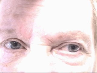
AAOx4, CN II-XII intact, gait not tested. A high-frequency, high-amplitude tremor is noted in the bilateral upper extremities at rest and with activity. Tremor appears associated with patient’s distress and pain.

Labs/Studies:

  • CBC: 9.4/12.8/38.1/311
  • BMP: 135/3.9/100/27/14/0.60/113
  • CRP: 0.18
  • ESRW: 20
  • Uric Acid: 2.6
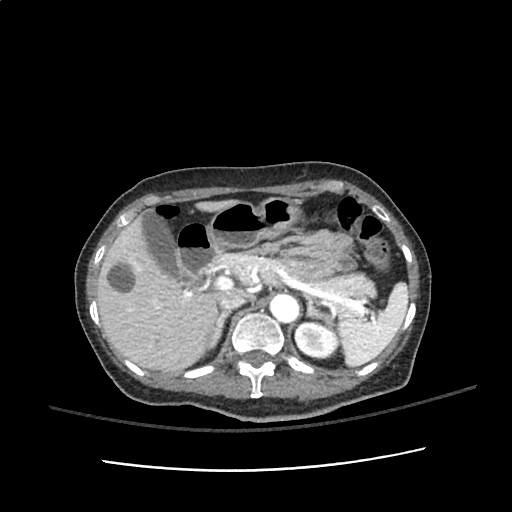
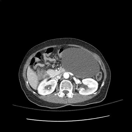
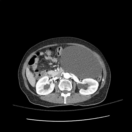
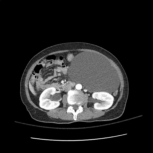
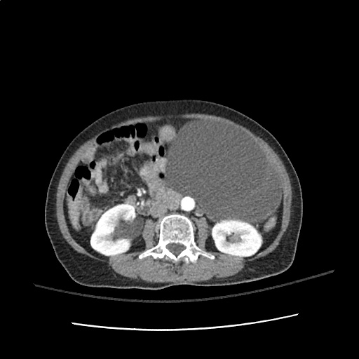
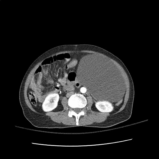
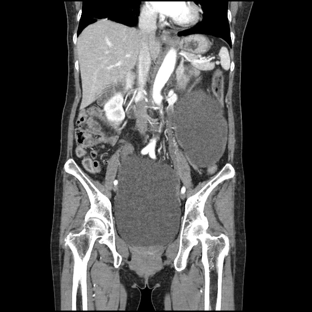
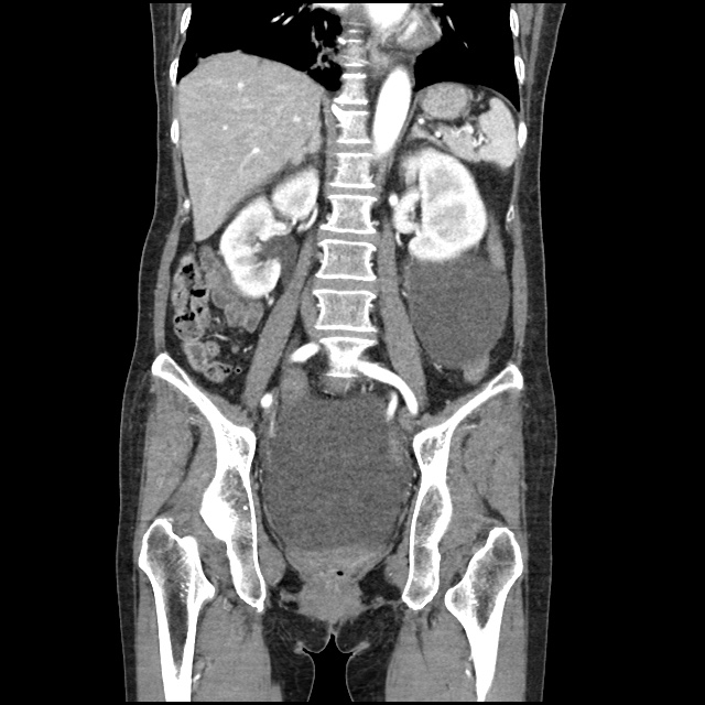
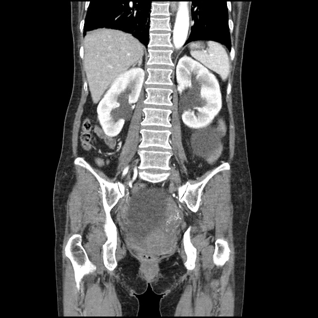
  • XR Left Tibia/Fibula: There is a metallic fixation device noted in the tibia.  There is no evidence of loosening of the metallic components.  The bones appear to be in gross anatomic alignment.

Assessment/Plan:

47F w/hx arthritis, gout, recent tib/fib fx s/p ORIF presenting with worsening polyarticular arthralgia unresponsive to OTC medications admitted for evaluation and pain management.

# Polyarticular arthralgia: Multiple joints affected, however patient notes most significant in bilateral knees and recently operated left leg. Polyarticular involvement, symmetric distribution and predominant involvement of large joints is suggestive of osteoarthritis. Also, given patient’s significant distress and multiple points of tenderness, potential extra-articular cause such as complex regional pain syndrome. Unlikely inflammatory arthritis (infectious, crystal arthropathy, rheumatic disease) given distribution and no significant erythema, warmth or effusion which could be aspirated. Obtained imaging of LLE to evaluate recent surgical repair of tib/fib fracture, with no evidence of loosening of fixation components or misalignment. Patient’s symptoms improved with morphine 5mg i.v. x2 in ED. Will admit for continued evaluation and pain management, consider addition of gabapentin for potential neuropathic pain associated with recent surgery.

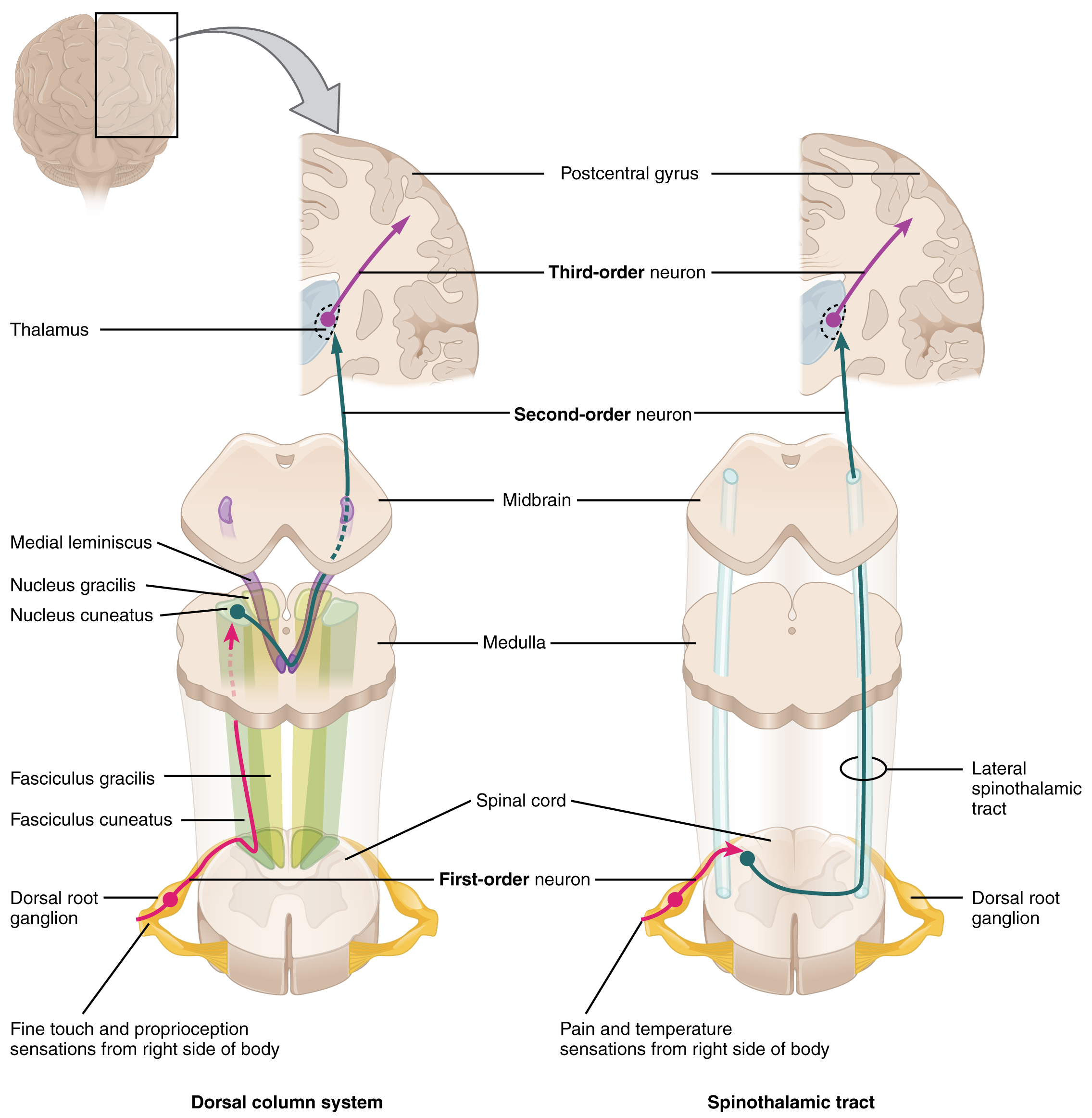
# Tremor: High-frequency, high-amplitude rest and action tremor suggestive of essential tremor or exaggerated physiological tremor associated with pain and emotional distress. No other neurological symptoms (HA, weakness) and non-focal neurological exam (aside from anesthesia localized around recent surgical incisions in LLE). Will continue monitoring, no need for imaging at this time.

# Gout: Patient with history of gout, however, doubt that current arthralgia associated with acute gouty flare. Will continue monitoring exam and consider ortho consultation for joint aspiration.

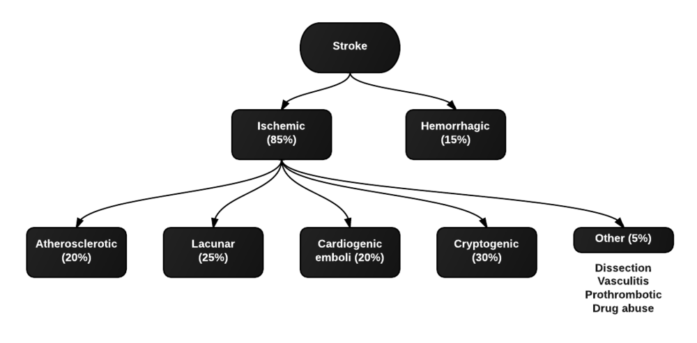
Differential Diagnosis of Monoarticular Arthralgia: 1,2

Differential Diagnosis of Monoarticular Arthralgia

Differential Diagnosis of Polyarticular Arthralgia 1,3

Differential Diagnosis of Polyarticular Arthralgia

Use of Serum Uric Acid in Acute Gout 4

In this patient, serum uric acid levels were checked. However, a normal serum urate (SU) does not exclude an acute gout flare. Studies have shown 12-43% of patients with acute gout flares have normal SU. An elevated SU (>8.0mg/dL) may support the diagnosis of a gout flare but is not itself diagnostic.

Approach to Joint Pain: 3

Approach to Joint Pain

Certain key historical elements can help narrow the differential diagnosis.

  1. Inflammation:
    1. Definition: Presence of erythema, warmth, swelling, pain with passive ROM, morning stiffness suggests inflammatory cause (arthritis)
    2. Common causes: Infection, gout, RA, SLE
  2. Chronology:
    1. Definition: Acute <6wks, chronic > 6wks
  3. Distribution:
    1. Definition: Location (large/small), symmetry
    2. Common causes:
      1. OA: DIP + PIP, spares wrists, elbows, ankles
      2. RA: PIP + MCP
      3. Spondylarthropathies (large joints)
      4. Symmetric involvement: RA, SLE
      5. Asymmetric involvement: Psoriatic arthritis, reactive arthritis, gout
  4. Course:
    1. Definition: Intermittent (gout), migrating (GC, Lyme, SLE)
  5. Demographics:
    1. Female <50: RA, SLE
    2. Male > 40: gout (♂ 20yr after puberty, ♀ 20yr after menopause)
  6. Extra-articular manifestations:
    1. Malar rash, oral ulcers: SLE
    2. Proximal muscle weakness: polymyositis
    3. Psoriatic skin/nail lesions: psoriatic arthritis
    4. Oral ulcers, vesicopustules on palms/soles, recent diarrheal illness: reactive arthritis

Differential Diagnosis of Tremor: 5,6

Differential Diagnosis of Joint Pain

References:

  1. Cush JJ, Lipsky PE. Chapter 331. Approach to Articular and Musculoskeletal Disorders. In: Longo DL, Fauci AS, Kasper DL, Hauser SL, Jameson JL, Loscalzo J, eds. Harrison’s Principles of Internal Medicine. 18th ed. New York: McGraw-Hill; 2012. http://www.accessmedicine.com/content.aspx?aID=9139045. Accessed August 27, 2013.
  2. Siva, C., Velazquez, C., Mody, A., & Brasington, R. (2003). Diagnosing acute monoarthritis in adults: a practical approach for the family physician. American family physician, 68(1), 83–90.
  3. Mies Richie, A., & Francis, M. L. (2003). Diagnostic approach to polyarticular joint pain. American family physician, 68(6), 1151–1160.
  4. Schlesinger, N., Norquist, J. M., & Watson, D. J. (2009). Serum urate during acute gout. The Journal of rheumatology, 36(6), 1287–1289. doi:10.3899/jrheum.080938
  5. Hess, C. W., & Pullman, S. L. (2012). Tremor: clinical phenomenology and assessment techniques. Tremor and other hyperkinetic movements (New York, N.Y.).
  6. Crawford, P., & Zimmerman, E. E. (2011). Differentiation and diagnosis of tremor. American family physician, 83(6), 697–702.

Syncope

ID:

A 50 year-old male with a reported two-year history of infrequent spells, presenting with two spells in the past two days.

HPI:

The patient’s spells began two years ago, he recounts that he was watching television when he lost consciousness and a friend noted he started shaking; he does not recall the event, and awoke in the hospital. The next spell occurred one year later, though the patient is unable to recall much about this episode. The patient remained spell-free until yesterday when he was on a bus, lost consciousness and awoke in a hospital. He notes that he had bit his tongue and lost control of his bladder. He was discharged hours later with a prescription for an AED which he was unable to fill. This morning, the patient had another spell while in the bathroom. His roommate heard him fall, found him on the ground, and noted that his mouth was moving but did not see any other movements.

The patient’s episodes are all associated with loss of consciousness and are followed by 5-10 minutes of disorientation after which he recovers fully. The episodes are sometimes preceded by a feeling of “euphoria”, though this feeling sometimes occurs without subsequent LOC.

The patient denies any associated palpitations, dizziness/LH, chest pain or muscle pain.  He has not had any recent fevers/chills, dysuria, cough, headache, changes in vision, numbness/tingling, weakness, difficulty speaking or swallowing or weight loss. He also denies any history of head trauma.

Physical Examination:

  • VS: Stable and WNL
  • General: Well-appearing, pleasant, and in NAD.
  • HEENT: NC/AT. MMM. Small lesion on tongue.
  • Lungs: CTAB.
  • CV: RRR with occasional ectopic beats, no M/R/G.
  • Abdomen: S/NT/ND. Bowel sounds present.
  • Neurological exam: AAOx4, CN II-XII intact, motor/sensation/reflexes/coordination/gait WNL

Imaging/Studies:

  • EKG: Occasional PAC/PVC
  • CT Brain: Unremarkable except for mild age-related cerebral atrophy

Assessment & Plan:

50 year-old male with a history of HTN and a reported two-year history of infrequent spells presenting with two spells in the past two days. The description of the patient’s episodes could be consistent with seizures. Aspects supporting this notion include loss of consciousness and period of confusion following each episode. One of the recent episodes was also associated with tongue-biting and loss of bladder control. Additionally, some episodes are associated with a sensation of euphoria rising from the abdomen to the head which could be indicative of an aura. Characteristics that suggest other causes include the absence of noted convulsions and non-stereotyped nature of each episode which could be due to the patient’s poor recollection of these events and absence of reliable witnesses. In the case of true seizures, the possible etiologies in this patient include a mass, metabolic abnormalities, substance use, or concomitant infection exacerbating an existing propensity for seizure activity. Other, non-seizure causes warranting evaluation include cardiogenic syncope particularly given the evidence of ectopic beats on examination and electrocardiogram.

Differential Diagnosis of Syncope

First, is it syncope? History is very important for distinguishing syncope from other causes (seizure, dizziness, vertigo, presyncope). Ask about precipitating events, prodromal symptoms, post-ictal confusion. Common causes of syncope and their associated symptoms are detailed in the figure below.

References:

  1. Kapoor, W. N. (2000). Syncope. The New England journal of medicine, 343(25), 1856–1862. doi:10.1056/NEJM200012213432507

Delirium

ID:

A 70 year-old female with a PMH of HTN, DM, hyperlipidemia and stage I breast cancer s/p lumpectomy with sentinel LN biopsy several years ago presented for elective surgery complicated by post-operative bleeding. She is now 4 days post-op and was found to be confused, somnolent and occasionally agitated.

HPI:

The patient could not be interviewed.

PE:

  • VS: Stable and within normal limits
  • General: unremarkable except for crackles in bilateral lung bases
  • MSE: only arouses to sternal rub and becomes agitated, moving all four extremities spontaneously and symmetrically.
  • Reflexes: corneal and gag reflexes present, suppresses eye movements with head turn, deep tendon reflexes 3+ throughout UE/LE bilaterally.

Assessment:

70 year-old woman with a history of HTN, DM, hyperlipidemia and breast cancer presents with worsening confusion, somnolence and occasional agitation four days after surgery. The combination of significantly altered consciousness and absence of focal neurological findings, all in the setting of a complicated surgical course suggest delirium.

Differential Diagnosis of Altered Mental Status:

Levels of consciousness

There are different levels of consciousness, they are named in the diagram below but are better described by the characteristics observed.

Initial assessment

Differential Diagnosis for Altered Mental Status

References:

  1. Inouye, S. K. (2006). Delirium in Older Persons. The New England journal of medicine, 354(11), 1157–1165. doi:10.1056/NEJMra052321
  2. Blueprints neurology. Philadelphia: Wolters Kluwer Health/Lippincott William & Wilkins, 2009.
  3. Tindall SC. Level of Consciousness. In: Walker HK, Hall WD, Hurst JW, editors. Clinical Methods: The History, Physical, and Laboratory Examinations. 3rd edition. Boston: Butterworths; 1990. Chapter 57. Available from: http://www.ncbi.nlm.nih.gov/books/NBK380/

Headache, vertigo and weakness

ID:

30 year-old male with no significant PMH presenting with right-sided “weakness”, vertigo and headache for three days.

HPI:

The patient was in his normal state of good health until three days prior to admission when he was out shopping and felt a headache and right-sided weakness. The headache came on suddenly, felt “sharp”, 7/10 in severity and radiated from the posterior occiput to his forehead on the right side. The headache was associated with vertigo and nausea/vomiting. At the same time, the patient noticed that he was no longer able to walk, describing “weakness” on the right side. He also began feeling dizzy, describing the sensation as “room spinning”. He denied changes in vision, hearing, difficulty speaking or swallowing (though he has had persistent hiccups for the past few days). He also denied CP, palpitations, SOB. He did seek immediate medical attention at an OSH but is unable to recall what was done and he was discharged from the ER on the same day. He presents 3 days later with persistent symptoms.

PE:

  • Mental status: normal
  • CN II-XII: intact with the exception of decreased sensation to sharp touch on right face and decreased gag reflex
  • Motor: normal bulk/tone, strength 5/5 in UE/LE bilaterally
  • Sensory: decreased sensation to pain and temperature on left body
  • Gait: wide-based, unable to tandem, heel, toe walk. Walking in place, he turns to the right.

Assessment:

30 year-old male with no PMH presenting with 3 days of HA, vertigo, hiccups, right-sided ataxia, and alternating decreased pain and temperature sensation on ipsilateral face and contralateral hemibody. These symptoms are suggestive of a brainstem lesion, localizing to the medulla. Hiccups suggest involvement of nucleus ambiguus (CN IX, X, XII). Alternating decreased pain/temperature sensation suggests involvement of the spinal tract of CN V, and interruption of fibers of the descending spinothalamic tract. These findings point further to a lesion in the right lateral medulla, likely vascular given the rapid onset of symptoms. Associated findings of right-sided ataxia suggests involvement of the superior cerebellar peduncle (restiform body) in the posterior lateral medulla. An MRI brain showed the lesion shown in Fig-1, and a CTA head/neck the following day showed dissection of the right vertebral artery.

A System for Cerebrovascular Disease:

Types of strokes

Clinical characteristics of strokes

Strokes are characterized by the sudden onset of focal neurological deficits. These are typically unilateral and consciousness is generally maintained.

  • dysphasia
  • dysarthria
  • weakness
  • ataxia
  • sensory loss
  • neglect
  • hemianopsia

If some of these typical features are not present (ex. gradual onset, significantly impaired consciousness, seizures early), consider alternative diagnoses (ex. hypoglycemia, subdural hematoma, mass, postictal paresis, migraine).

Common causes of stroke

The most common causes are atherosclerosis (leading to thromboembolism or local occlusion) and cardioembolism. If the patient does not have risk factors, consider alternatives:

  • contralateral ptosis/miosis: carotid artery dissection affecting sympathetic fibers
  • fever + murmur: infective endocarditis
  • HA + ↑ESR: giant-cell arteritis

References:

  1. Agabegi, S. (2013). Step-up to medicine. Philadelphia: Wolters Kluwer/Lippincott Williams & Wilkins.
  2. van der Worp, H. B., & van Gijn, J. (2007). Acute Ischemic Stroke. The New England journal of medicine, 357(6), 572–579. doi:10.1056/NEJMcp072057