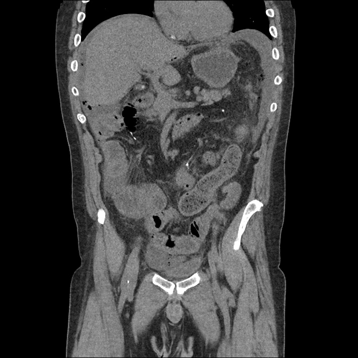
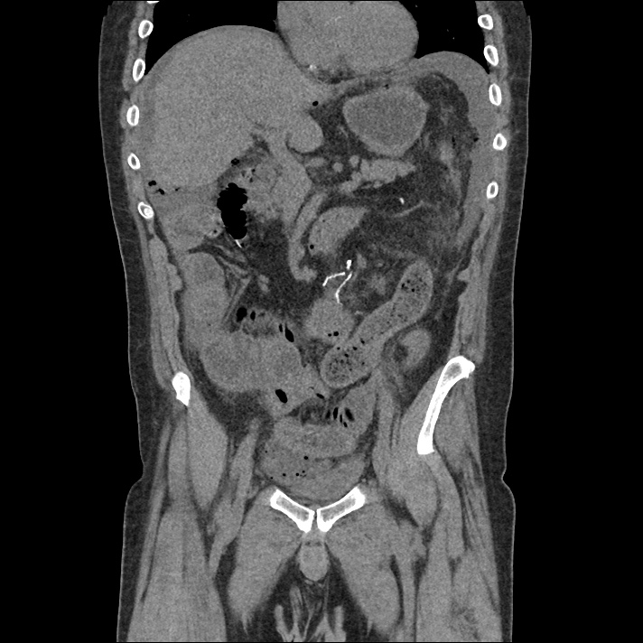
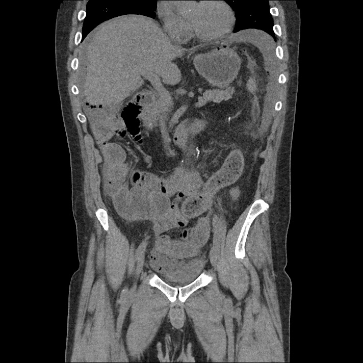
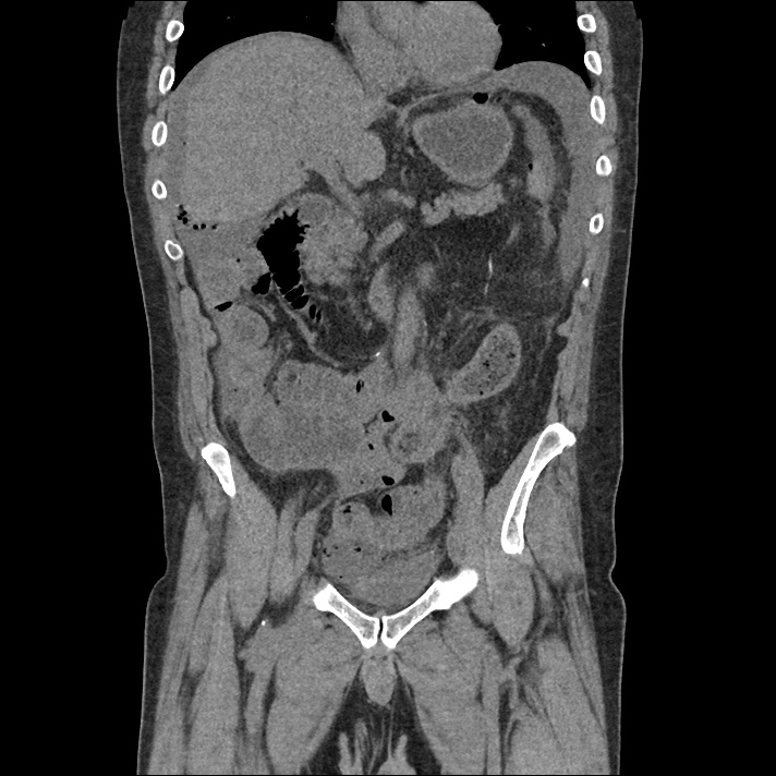
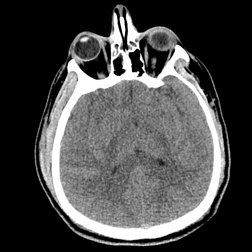
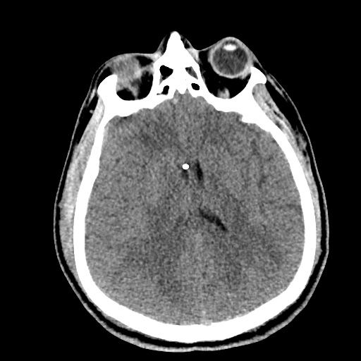
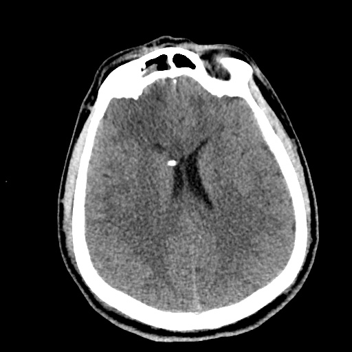
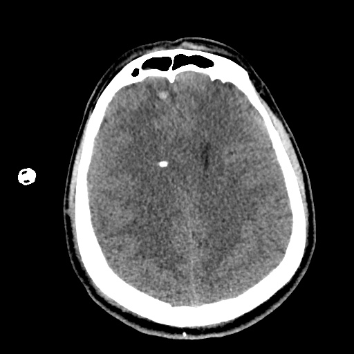
Hyponatremia

HPI:

A 62 year-old male with a history of hepatitis C cirrhosis complicated by hepatocellular carcinoma s/p radiofrequency ablation presenting after referral from hepatology clinic for hyponatremia. One week ago, the patient developed abdominal distension and shortness of breath that resolved after large-volume paracentesis and was started on furosemide 40mg p.o. daily and aldactone 100mg p.o. daily. After initiating diuretics, the patient noted worsening lower extremity edema, and increased thirst/fluid intake.

He reports two days of fatigue and intermittent confusion supported by family members who reported slowed speech. He otherwise denies abdominal pain, distension, nausea/vomiting, diarrhea/constipation, chest pain or shortness of breath. In the ED, the patient received 1L NS bolus.

PMH:

  • Hepatitis C cirrhosis c/b HCC s/p RFA
  • Rheumatoid arthritis

PSH:

  • None

Family History:

  • Non-contributory.

Social History:

  • Lives with partner, denies current or prior t/e/d abuse
  • HepC contracted from blood transfusions

Medications:

  • Furosemide 40mg p.o. daily
  • Spironolactone 100mg p.o. daily
  • Rifaximin 550mg p.o. b.i.d.

Allergies:

  • NKDA

Physical Exam:

VS: T 98.2 HR 80 RR 14 BP 95/70 O2 98% RA
Gen: Elderly male in no acute distress, alert and answering questions appropriately.
HEENT: NC/AT, PERRL, EOMI, faint scleral icterus, MMM.
CV: RRR, normal S1/S2, no murmurs. JVP 8cm.
Lungs: Faint basilar crackles on bilateral lung bases.
Abd: Normoactive bowel sounds, mildly distended but non-tender, without rebound/guarding.
Ext: 2+ pitting edema in lower extremities to knees bilaterally. 2+ peripheral pulses, warm and well perfused.
Neuro: AAOx3. CN II-XII intact. No asterixis. Normal gait. Normal FTN/RAM.

Labs/Studies:

  • BMP (admission): 112/5.6/88/22/28/1.1/97
  • BMP (+10h): 118/5.4/93/23/26/1.0/133
  • sOsm: 264
  • Urine: Na <20, K 26, Osm 453
  • BNP: 40
  • AST/ALT/AP/TB/Alb: 74/57/91/2.4/2.2

Assessment/Plan:

62M with HepC cirrhosis, with e/o decompensation (new-onset ascites) and hyponatremia.

  1. #Hyponatremia: Sodium 114, likely chronic, patient currently asymptomatic without concerning findings on neurological exam. Clinical findings suggestive of hypervolemic hyponatremia 2/2 decompensated cirrhosis resulting in decreased effective arterial blood volume and volume retention. However, the recent initiation of diuretics, mild AKI and early response to isotonic fluids in the ED suggests possible hypovolemic component.
    • 1L fluid restriction
    • q.4.h. sodium check, goal increase of 8mEq per 24h
    • hold diuretics
  2. #Hyperkalemia: Potassium 5.6, asymptomatic, AKI vs. medication-induced (aldactone). Continue monitoring.
  3. #AKI: Elevated creatinine 1.1 from baseline 0.7. Likely pre-renal given recent initiation of diuretics. Consider hepatorenal syndrome given decompensated cirrhosis. Follow-up repeat creatinine after 1L NS bolus in ED.
  4. #Hepatitis C: decompensated with new-onset ascites. No e/o encephalopathy, continue home rifaximin.

Physiology of Hyponatremia: 1,2,3,4

Physiology of Hyponatremia

Differential Diagnosis of Hyponatremia: 5

Differential Diagnosis of Hyponatremia

Evaluation of Hyponatremia: 2

  1. Identification of onset (acute vs. chronic)
  2. Presence of symptoms (HA, nausea, confusion, seizures)
  3. Assessment of volume status (edema, JVD, skin turgor, postural BP)
  4. Medical history (cardiac, liver, renal disease), drug history

References:

  1. Freda BJ, Davidson MB, Hall PM. Evaluation of hyponatremia: a little physiology goes a long way. Cleve Clin J Med. 2004;71(8):639–650.
  2. Biswas M, Davies JS. Hyponatraemia in clinical practice. Postgrad Med J. 2007;83(980):373–378. doi:10.1136/pgmj.2006.056515.
  3. Adrogué HJ, Madias NE. Hyponatremia. N. Engl. J. Med. 2000;342(21):1581–1589. doi:10.1056/NEJM200005253422107.
  4. Marx JA, Hockberger RS, Walls RM, Adams JG. Rosen’s emergency medicine: concepts and clinical practice. 2010;1.
  5. Milionis HJ, Liamis GL, Elisaf MS. The hyponatremic patient: a systematic approach to laboratory diagnosis. CMAJ. 2002;166(8):1056–1062.

Hypertensive Emergency

Brief HPI:

A 62 year-old female with a history of hypertension, diabetes and coronary artery disease is brought to the emergency department with altered mental status. The patient is confused and unable to provide history. Her family note that symptoms have been gradually worsening for the past one day and she had previously been in her usual state of good health. There was no history of recent illness, medication changes, recreational substance use, sick contacts, or travel.

On evaluation, vital signs were notable for hypertension (224/120mmHg, comparable on all extremities) though otherwise normal including afebrile core temperature – capillary glucose was 114mg/dL. On examination, the patient was awake and alert, making coordinated movements symmetrically in all four extremities without hyperreflexia or increased tone. Speech was unintelligible and the patient was unable to follow simple commands.

Labs/Imaging

Laboratory tests were notable for a serum creatinine of 1.2mg/dL (baseline unknown) but otherwise normal including CBC, troponin, TSH, and UA. ECG demonstrated left ventricular hypertrophy without acute ischemic changes. Imaging including chest radiograph and CT head non-contrast and CTA brain/neck were normal. Lumbar puncture was performed and CSF was normal.

Hospital Course

The patient was initiated on a continuous infusion of nicardipine for presumed hypertensive encephalopathy and admitted to the medical intensive care unit. An MRI was performed on hospital day 1 and demonstrated chronic microvascular ischemic changes. The patient’s mental status gradually improved over the course of her hospitalization and she was discharged home on hospital day 4.

An Algorithm for the Evaluation and Management of Hypertensive Emergencies

An Algorithm for the Evaluation and Management of Hypertensive Emergencies

References

General

  1. Lloyd-Jones DM, Morris PB, Ballantyne CM, et al. 2017 Focused Update of the 2016 ACC Expert Consensus Decision Pathway on the Role of Non-Statin Therapies for LDL-Cholesterol Lowering in the Management of Atherosclerotic Cardiovascular Disease Risk: A Report of the American College of Cardiology Task Force on Expert Consensus Decision Pathways. In: Vol 70. 2017:1785-1822. doi:10.1016/j.jacc.2017.07.745.
  2. Janke AT, McNaughton CD, Brody AM, Welch RD, Levy PD. Trends in the Incidence of Hypertensive Emergencies in US Emergency Departments From 2006 to 2013. J Am Heart Assoc. 2016;5(12). doi:10.1161/JAHA.116.004511.
  3. Rodriguez MA, Kumar SK, De Caro M. Hypertensive crisis. Cardiology in Review. 2010;18(2):102-107. doi:10.1097/CRD.0b013e3181c307b7.
  4. Katz JN, Gore JM, Amin A, et al. Practice patterns, outcomes, and end-organ dysfunction for patients with acute severe hypertension: the Studying the Treatment of Acute hyperTension (STAT) registry. Am Heart J. 2009;158(4):599–606.e1. doi:10.1016/j.ahj.2009.07.020.
  5. Elliott WJ. Clinical features in the management of selected hypertensive emergencies. Prog Cardiovasc Dis. 2006;48(5):316-325. doi:10.1016/j.pcad.2006.02.004.
  6. Aggarwal M, Khan IA. Hypertensive crisis: hypertensive emergencies and urgencies. Cardiol Clin. 2006;24(1):135-146. doi:10.1016/j.ccl.2005.09.002.
  7. Varon J, Marik PE. Clinical review: the management of hypertensive crises. Crit Care. 2003;7(5):374-384. doi:10.1186/cc2351.
  8. Shayne PH, Pitts SR. Severely increased blood pressure in the emergency department. YMEM. 2003;41(4):513-529. doi:10.1067/mem.2003.114.
  9. Vaughan CJ, Delanty N. Hypertensive emergencies. The Lancet. 2000;356(9227):411-417. doi:10.1016/S0140-6736(00)02539-3.

Ischemic Stroke

  1. Powers WJ, Rabinstein AA, Ackerson T, et al. 2018 Guidelines for the Early Management of Patients With Acute Ischemic Stroke: A Guideline for Healthcare Professionals From the American Heart Association/American Stroke Association. Stroke. 2018;49(3):e46-e110. doi:10.1161/STR.0000000000000158.

Hemorrhagic Stroke

  1. Hemphill JC, Greenberg SM, Anderson CS, et al. Guidelines for the Management of Spontaneous Intracerebral Hemorrhage: A Guideline for Healthcare Professionals From the American Heart Association/American Stroke Association. Stroke. 2015;46(7):2032-2060. doi:10.1161/STR.0000000000000069.

Subarachnoid Hemorrhage

  1. Connolly ES, Rabinstein AA, Carhuapoma JR, et al. Guidelines for the management of aneurysmal subarachnoid hemorrhage: a guideline for healthcare professionals from the American Heart Association/american Stroke Association. Stroke. 2012;43(6):1711-1737. doi:10.1161/STR.0b013e3182587839.

Renal

  1. Gillies MA, Kakar V, Parker RJ, Honoré PM, Ostermann M. Fenoldopam to prevent acute kidney injury after major surgery-a systematic review and meta-analysis. Crit Care. 2015;19(1):449. doi:10.1186/s13054-015-1166-4.
  2. Tumlin JA, Dunbar LM, Oparil S, et al. Fenoldopam, a dopamine agonist, for hypertensive emergency: a multicenter randomized trial. Fenoldopam Study Group. Academic Emergency Medicine. 2000;7(6):653-662.
  3. Shusterman NH, Elliott WJ, White WB. Fenoldopam, but not nitroprusside, improves renal function in severely hypertensive patients with impaired renal function. Am J Med. 1993;95(2):161-168.

Aortic Disease

  1. Hiratzka LF, Bakris GL, Beckman JA, et al. 2010 ACCF/AHA/AATS/ACR/ASA/SCA/SCAI/SIR/STS/SVM guidelines for the diagnosis and management of patients with Thoracic Aortic Disease: a report of the American College of Cardiology Foundation/American Heart Association Task Force on Practice Guidelines, American Association for Thoracic Surgery, American College of Radiology, American Stroke Association, Society of Cardiovascular Anesthesiologists, Society for Cardiovascular Angiography and Interventions, Society of Interventional Radiology, Society of Thoracic Surgeons, and Society for Vascular Medicine. Circulation. 2010;121(13):e266-e369. doi:10.1161/CIR.0b013e3181d4739e.

Pregnancy

  1. Townsend R, O’Brien P, Khalil A. Current best practice in the management of hypertensive disorders in pregnancy. Integr Blood Press Control. 2016;9:79-94. doi:10.2147/IBPC.S77344.
  2. Al-Safi Z, Imudia AN, Filetti LC, Hobson DT, Bahado-Singh RO, Awonuga AO. Delayed Postpartum Preeclampsia and Eclampsia. Obstet Gynecol. 2011;118(5):1102-1107. doi:10.1097/AOG.0b013e318231934c.
  3. Hypertension in pregnancy: diagnosis and management. National Institute for Health and Care Excellence. https://www.nice.org.uk/guidance/cg107. Published August 1, 2010. Accessed May 20, 2019.

Principles of Neonatal Resuscitation

The following resource for neonatal resuscitation and neonatal critical care was developed with the guidance of Dr. Agrawal (Neonatology) while on rotation at the White Memorial Medical Center Neonatal Intensive Care Unit.

Endotracheal Tube Size1-3

Simplified Formula
Estimated gestational age in weeks ÷ 10 = round to nearest half-size uncuffed tube

NRP Recommendation

Gestation age (weeks) Weight (kg) ETT Size (ID, mm) Depth (cm from lip)
<28 <1.0 2.5 6-7
28-34 1.0-2.0 3.0 7-8
34-38 2.0-3.0 3.5 8-9
>38 >3.0 3.5-4.0 9-10

Laryngoscope Blade Size

Age Blade
Preterm 0
Term 1

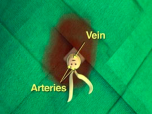
Umbilical Vein Catheter Placement4

ED Indications
Unstable neonate
Contraindications
Omphalocele
Gastroschisis
Necrotizing enterocolitis
Depth
4-5cm or until blood return (for emergent placement)

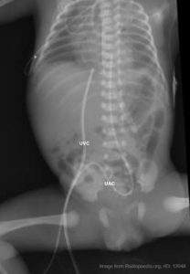
Umbilical artery/vein catheter position on plain radiograph.

Umbilical catheter size

Weight (kg) Size (F)
<1.5 3.5
1.5-3.5 5
>3.5 8

Umbilical catheter positioning on plain radiographs

Umbilical venous catheter position can be verified with a plain radiograph. Positioning within the umbilical vein can be confirmed by tracing a cephalad trajectory from the insertion point at the umbilicus. An umbilical artery catheter will first pass caudally into the internal iliac artery before travelling cephalad into a common iliac artery and the abdominal aorta.

Medications5

Medication Dose
Epinephrine 0.1mL/kg (1:10,000) IV, 0.01mg/kg
Volume Expansion 10mL/kg (normal saline, blood)
Naloxone 0.1-0.2mg/kg
Dopamine 5-20mcg/kg/min IV infusion

Neonatal Physiology and Transition to Extrauterine Life6

An important principle in neonatal resuscitation is supporting the appropriate transition from intra- to extra-uterine life which is dependent on several key anatomic and physiologic changes occurring in an optimal environment.

Anatomy7

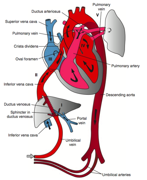
Fetal Circulation
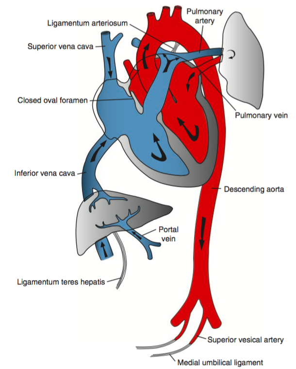
Neonatal Circulation

Fetal Circulation

In the fetal circulatory system, oxygenated blood is delivered via the umbilical vein, entering the inferior vena cava via the ductus venosus. The majority of this oxygenated blood passes through the right atrium and into the left atrium through the foramen ovale to enter the systemic circulation.

Meanwhile, high pulmonary pulmonary vascular resistance (due to hypoxic vasoconstriction in fluid-filled alveoli) means that most of the deoxygenated right ventricular output is routed through the ductus arteriosus and enters into the systemic circulation – mixing with oxygenated blood distal to the highest priority end-organs (brain and heart), to be reoxygenated at the placenta.

Post-transition Circulation

The transition to extra-uterine life involves several key steps detailed below and is supported by appropriate ventilation, oxygenation and temperature regulation.

  1. Alveolar Fluid Clearance
    Catecholamine and hormone changes (predominantly corticosteroids) during the process of labor induce changes in enzymatic expression that result in the resorption of alveolar fluid into the interstitial space. At the time of delivery, negative intra-thoracic pressure from inspiration further promotes the resorption of alveolar fluid. Mechanical thoracic compression from delivery may also contribute.
  2. Respiration and Breathing
    Disconnection from the placenta ceases the transfer of placenta-derived factors including prostaglandins. The withdrawal of tonic inhibition of central respiratory drive from prostaglandins with cord clamping stimulates rhythmic breathing. The infant’s initial breaths and resultant lung expansion promotes alveolar expansion and stimulates surfactant production – this decreases alveolar surface tension, increases lung compliance and further facilitates breathing.
  3. Circulatory Changes
    At delivery, clamping the umbilical cord removes a large bed of low-resistance circulation, increasing systemic vascular resistance and systemic blood pressure. At the same time, lung expansion and alveolar aeration decreases pulmonary vascular resistance and pulmonary arterial pressures. At the ductus arteriosus, increased systemic vascular resistance combined with decreased pulmonary vascular resistance decreases shunting and contributes to closure. Similarly, as left atrial pressure approaches and exceeds right atrial pressure, right-to-left flow across the foramen ovale ceases. Collectively, these changes serve to effectively separate the left- and right-sided circulations.

NRP Resuscitation Algorithm5,8

Neonatal Resuscitation Algorithm

References

  1. Luten R, Kahn N, Wears R, Kissoon N. Predicting Endotracheal Tube Size by Length in Newborns. J Emerg Med. 2007;32(4):343-347. doi:10.1016/j.jemermed.2007.02.035.
  2. Peterson J, Johnson N, Deakins K, Wilson-Costello D, Jelovsek JE, Chatburn R. Accuracy of the 7-8-9 Rule for endotracheal tube placement in the neonate. J Perinatol. 2006;26(6):333-336. doi:10.1038/sj.jp.7211503.
  3. Kempley ST, Moreiras JW, Petrone FL. Endotracheal tube length for neonatal intubation. Resuscitation. 2008;77(3):369-373. doi:10.1016/j.resuscitation.2008.02.002.
  4. Anderson J, Leonard D, Braner DAV, Lai S, Tegtmeyer K. Videos in Clinical Medicine. Umbilical Vascular Catheterization. Vol 359. 2008:e18. doi:10.1056/NEJMvcm0800666.
  5. Association AAOPAAH. Textbook of Neonatal Resuscitation. 2016.
  6. Caraciolo J Fernandes MD. Physiologic transition from intrauterine to extrauterine life. UpToDate.
  7. Sadler TW. Langman’s Medical Embryology. Lippincott Williams & Wilkins; 2011.
  8. Perlman JM, Wyllie J, Kattwinkel J, et al. Part 7: Neonatal Resuscitation: 2015 International Consensus on Cardiopulmonary Resuscitation and Emergency Cardiovascular Care Science With Treatment Recommendations. In: Vol 132. American Heart Association, Inc.; 2015:S204-S241. doi:10.1161/CIR.0000000000000276.

Hypotension

Brief H&P:

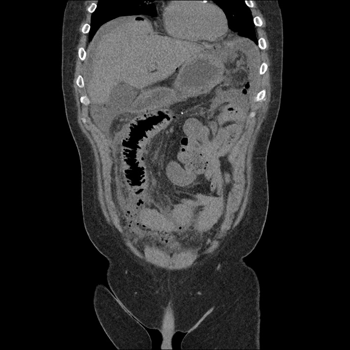
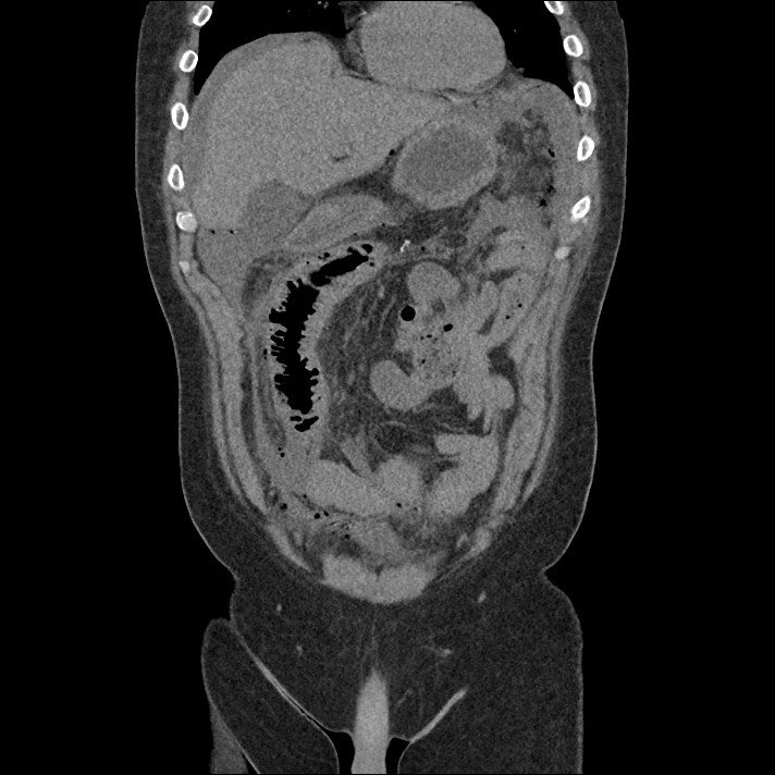
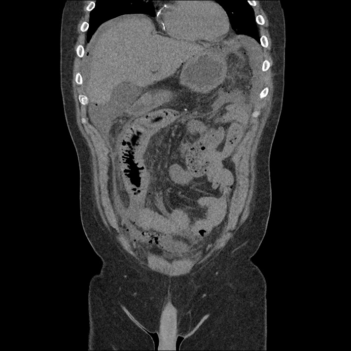
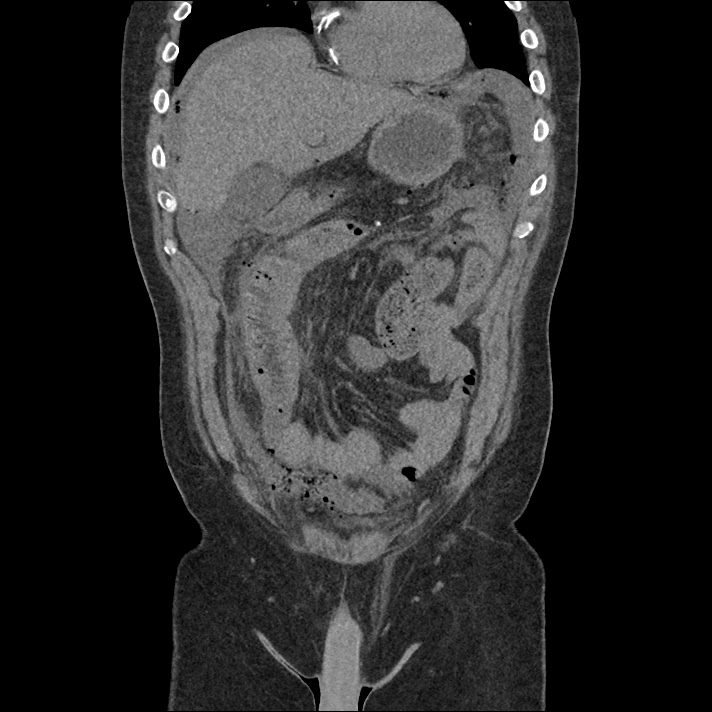
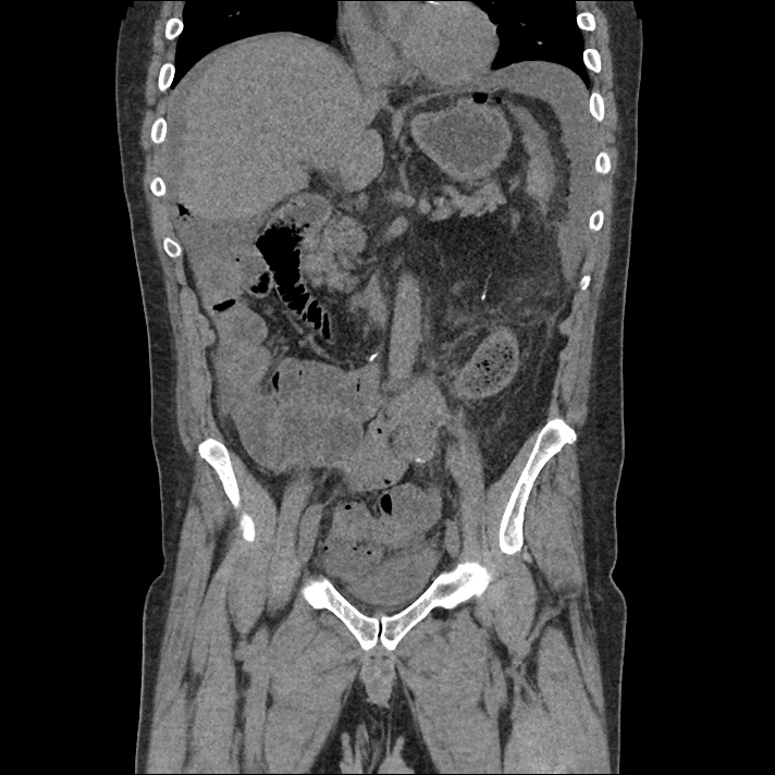
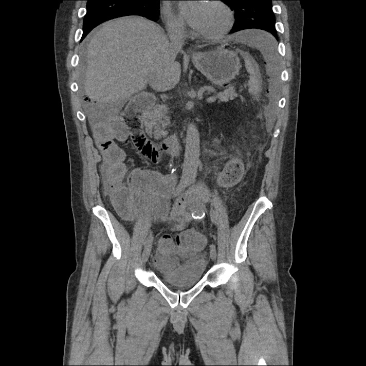
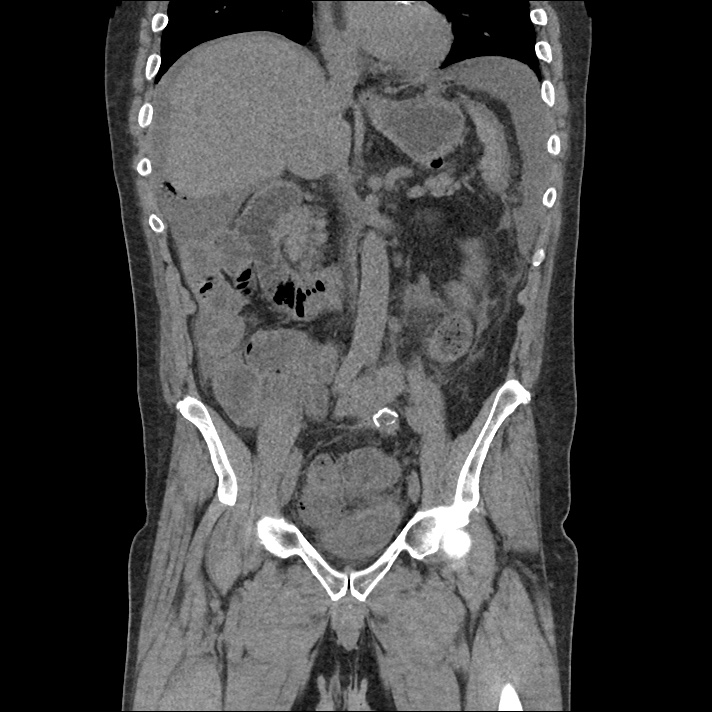
A 50 year-old male with a history of colonic mucinous adenocarcinoma on chemotherapy presented with a chief complaint of “vomiting”. He was unwilling to provide further history, repeating that he had vomited blood prior to presentation. His initial vital signs were notable for tachycardia. Physical examination showed some dried vomitus, brown in color, at the nares and lips; left upper quadrant abdominal tenderness to palpation; and guaiac-positive stool. Point-of-care hemoglobin was 3g/dL below the most recent measure two months prior. As his evaluation progressed, he developed hypotension and was transfused two units of uncrossmatched blood with adequate blood pressure response – he was started empirically on broad-spectrum antibiotics for an intra-abdominal source. Notable laboratory findings included a normal hemoglobin/hematocrit, acute kidney injury, and elevated anion gap metabolic acidosis presumably attributable to serum lactate of 10.7mmol/L. Computed tomography of the abdomen and pelvis demonstrated pneumoperitoneum with complex ascites concerning for bowel perforation. The patient deteriorated, was intubated, started on vasopressors and admitted to the surgical intensive care unit. The initial operative report noted extensive adhesions and perforated small bowel with feculent peritonitis. He has since undergone multiple further abdominal surgeries and remains critically ill.

Imaging

IM-0001-0032
IM-0001-0033
IM-0001-0034
IM-0001-0035
IM-0001-0036
IM-0001-0037
IM-0001-0038
IM-0001-0039
IM-0001-0040
IM-0001-0041
IM-0001-0042
IM-0001-0043
IM-0001-0044
IM-0001-0045
IM-0001-0046
IM-0001-0047
IM-0001-0048
IM-0001-0049
IM-0001-0050

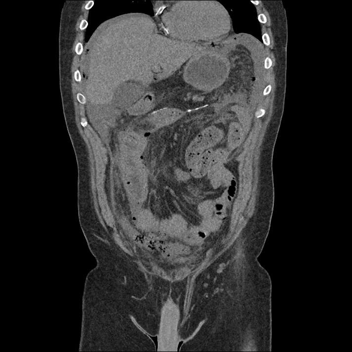
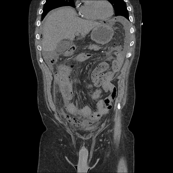
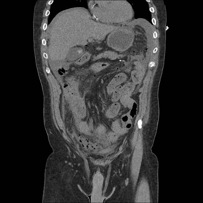
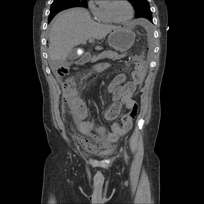
CT Abdomen/Pelvis

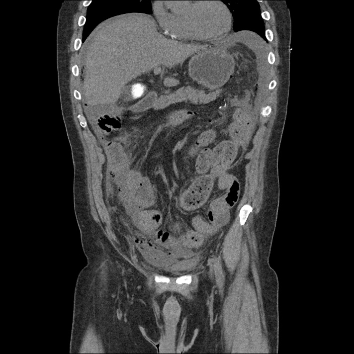
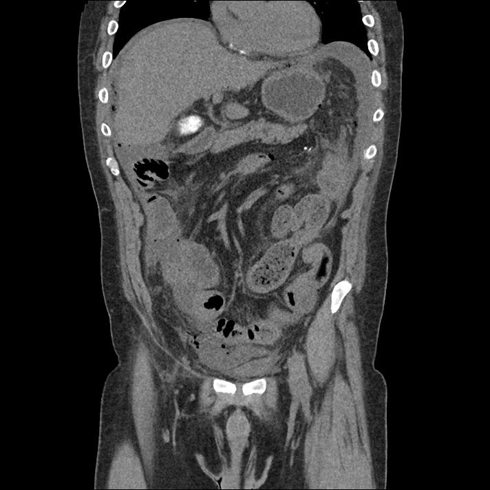
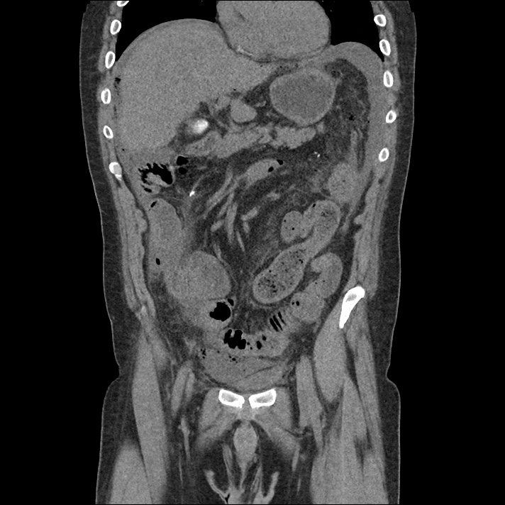
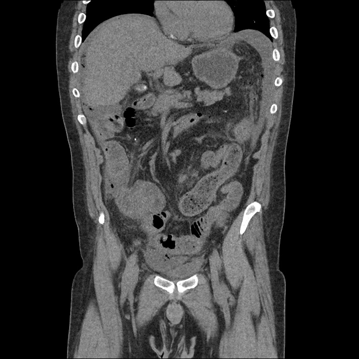
Free air is seen diffusely in the non-dependent portions of the abdomen: in the anterior abdomen and pelvis, inferior to the diaphragm, and in the perisplenic region. There is complex free fluid in the abdomen.

Algorithm for the Evaluation of Hypotension1

This process for the evaluation of hypotension in the emergency department was developed by Dr. Ravi Morchi. In the case above, a systematic approach to the evaluation of hypotension using ultrasonography and appropriately detailed physical examination may have expedited the patient’s care. The expertly-designed algorithm traverses the cardiovascular system, halting at evaluable checkpoints that may contribute to hypotension.

  1. The process begins with the cardiac conduction system to identify malignant dysrhythmias (bradycardia, or non-sinus tachycardia >170bpm), which, in unstable patients are managed with electricity.
  2. The next step assesses intravascular volume with physical examination or bedside ultrasonography of the inferior vena cava. Decreased right atrial pressure (whether due to hypovolemia, hemorrhage, or a distributive process) is evidenced by a small and collapsible IVC. If hemorrhage is suspected, further ultrasonography with FAST and evaluation of the abdominal aorta may identify intra- or retroperitoneal bleeding.
  3. If a normal or elevated right atrial pressure is identified, evaluate for dissociation between the RAP and left ventricular end-diastolic volume. This is typically caused by a pre- or intra-pulmonary obstructive process such as tension pneumothorax, cardiac tamponade, massive pulmonary embolism, pulmonary hypertension, or elevated intra-thoracic pressures secondary to air-trapping. Thoracic ultrasonography can identify pneumothorax, pericardial effusion, or signs of elevated right ventricular systolic pressures (RV:LV, septal flattening).
  4. Assuming adequate intra-vascular volume is arriving at the left ventricle, rapid echocardiography can be used to provide a gross estimate of cardiac contractility and point to a cardiogenic process. If there is no obvious pump failure, auscultation may reveal murmurs that would suggest systolic output is refluxing to lower-resistance routes (ex. mitral insufficiency, aortic insufficiency, or ventricular septal defect).
  5. Finally, if the heart rate is suitable, volume deficits are not grossly at fault, no obstructive process is suspected, and cardiac contractility is adequate and directed appropriately through the vascular tree, the cause may be distributive. Physical examination may reveal dilated capillary beds and low systemic vascular resistance.

Algorithm for the Evaluation of Hypotension

Guided Lecture

EM Ed
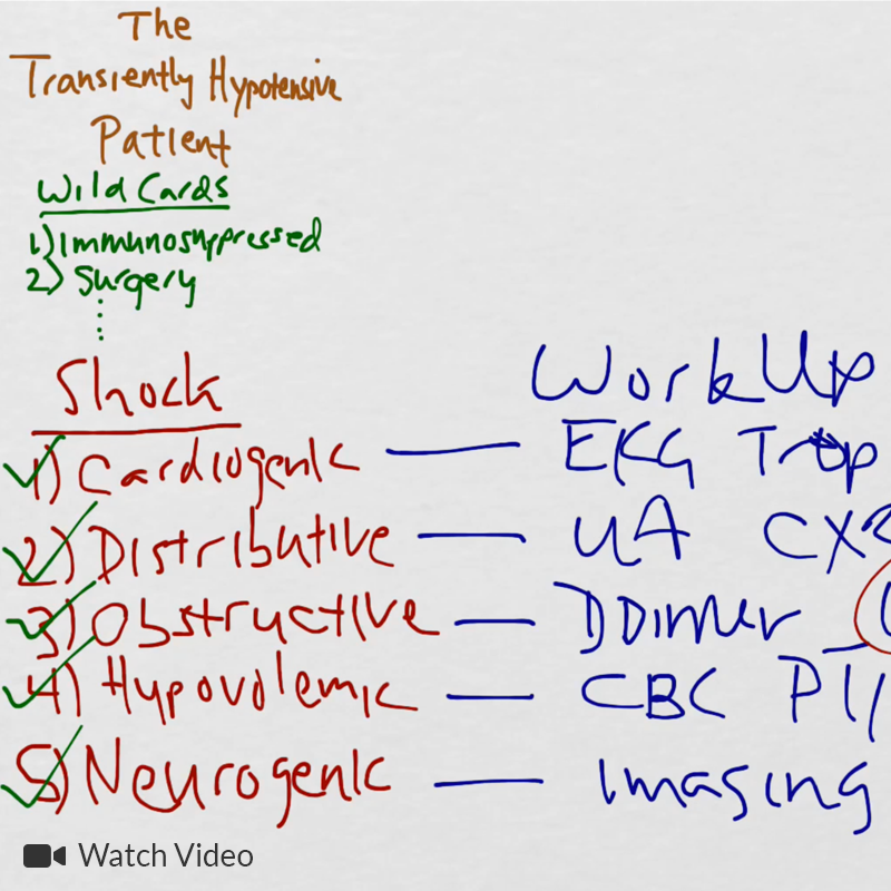
Watch “The Transiently Hypotensive Patient: Who Cares?” from EM Ed. In this lecture Dr. Basrai reviews the diagnostic pathway for a patient who presents with transient hypotension.

References

  1. Morchi R. Diagnosis Deconstructed: Solving Hypotension in 30 Seconds. Emergency Medicine News. 2015.

Severe Traumatic Brain Injury

HPI:

34 year-old male brought in by ambulance s/p assault. Field GCS reportedly 7, in trauma bay assessed as E2-V4-M6. Witnessed seizure in CT scanner, resolved with lorazepam. Intubated for airway protection, underwent external ventricular drain placement and transferred to surgical ICU.

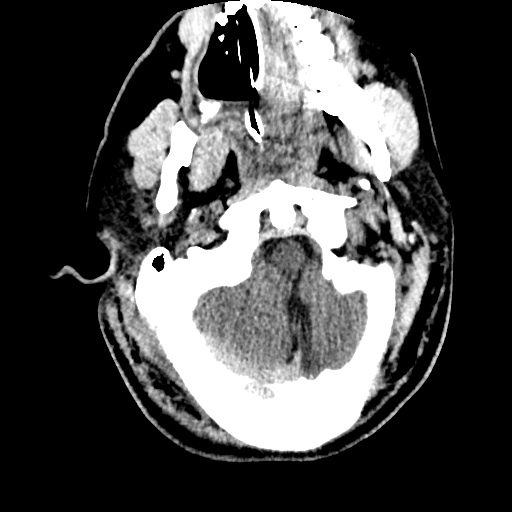
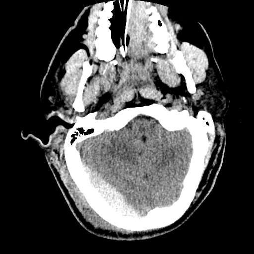
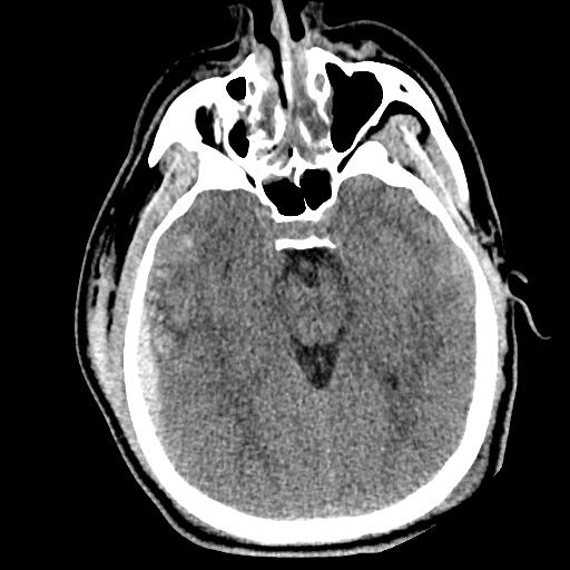
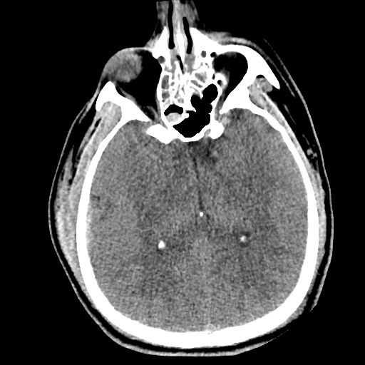
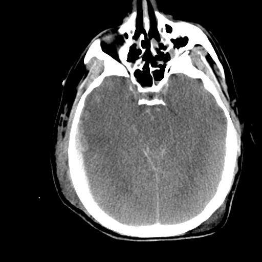
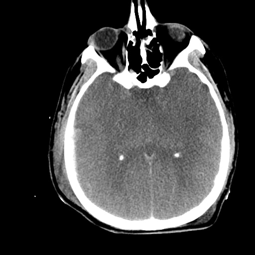
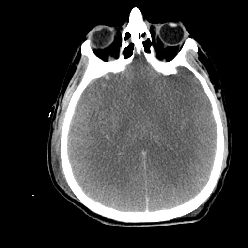
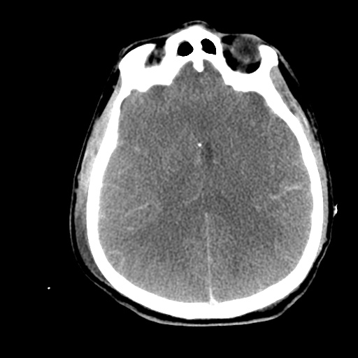
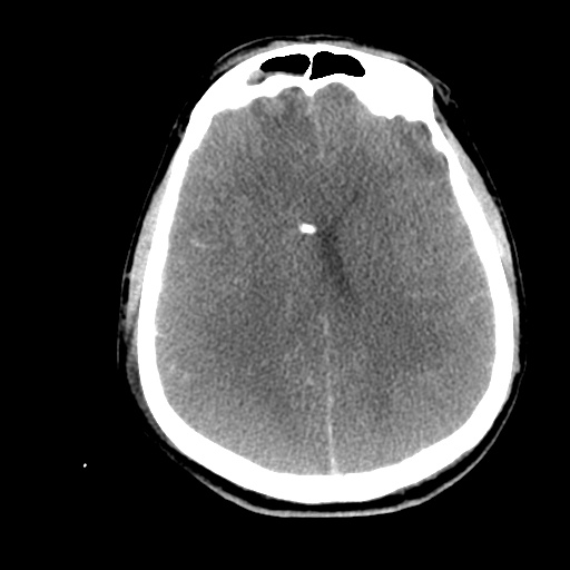
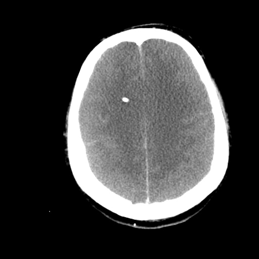
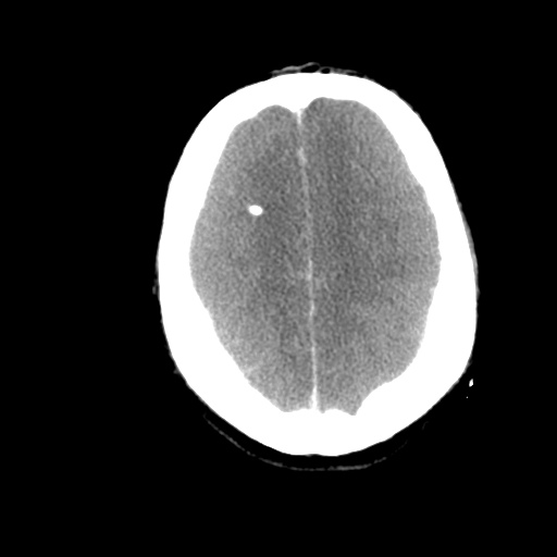
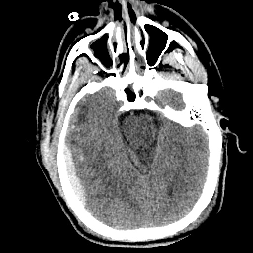
Initial imaging revealed bifrontal subdural hematomas and right temporal hemorrhagic contusion with generalized edema. Repeat imaging one hour later showed interval development of large extra-axial hemorrhage overlying the right occipital and parietal lobes (2.2cm), representing subdural or epidural hematoma.

The patient’s ICU course was complicated by continued seizures and refractory elevation in intracranial pressure. A pentobarbital infusion was started and titrated to adequate burst suppression and hyperosmolar therapy with both mannitol and hypertonic saline continued. Additional imaging revealed stable hemorrhage but continued diffuse cerebral edema evidenced by sulcal effacement.

On hospital day 5, examination revealed bilateral fixed and dilated pupils. Imaging revealed effacement of basilar cisterns, pre-pontine cistern, and cisterna magna suggestive of impending/ongoing transtentorial and tonsillar herniation. Pentobarbital was weaned and conventional cerebral angiography as well as cerebral perfusion studies were consistent with brain death.

Images

tbi1_01
tbi1_02
tbi1_03
tbi1_04
tbi1_05
tbi1_06
tbi1_07
tbi1_08
tbi1_09
tbi1_10
tbi1_11
tbi1_12

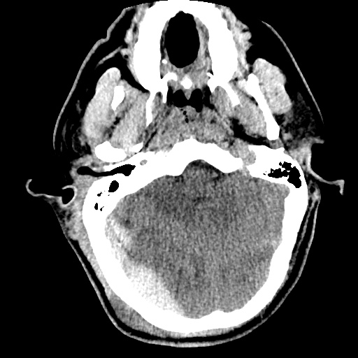
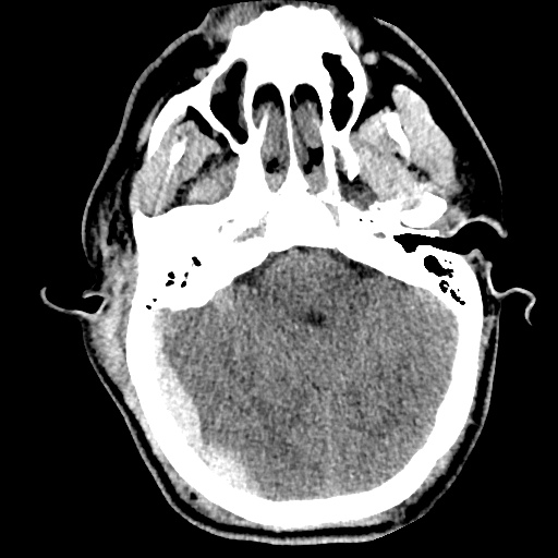
CT head without contrast one hour after presentation

  • Large extra-axial posterior hemorrhages. Hemorrhagic contusions in the right frontal and temporal lobes.
  • The cerebral sulci appear effaced – findings suggest diffuse cerebral edema.
  • S/p EVD using a right frontal approach.
tbi2_01
tbi2_02
tbi2_03
tbi2_04
tbi2_05
tbi2_06
tbi2_07
tbi2_08
tbi2_09
tbi2_11
tbi2_12

CT head without contrast on hospital day 5

  • Interval evidence of global hypoxic/ischemic injury to the brain.
  • Interval apparent effacement of the basilar cisterns, pre-pontine cistern, and cisterna magna suggesting impending/ongoing downward transtentorial herniation and tonsillar herniation.
  • Stable supra/infratentorial subdural/epidural hematoma.

Algorithm for the Management of Severe Traumatic Brain Injury1,2

Algorithm for the Management of Severe Traumatic Brain Injury

References

  1. Brain Trauma Foundation, American Association of Neurological Surgeons, Congress of Neurological Surgeons, Joint Section on Neurotrauma and Critical Care, AANS/CNS, Carney, N. A., & Ghajar, J. (2007). Guidelines for the management of severe traumatic brain injury. Introduction. Journal of neurotrauma, 24 Suppl 1, S1–2. doi:10.1089/neu.2007.9997
  2. Stocchetti, N., & Maas, A. I. R. (2014). Traumatic intracranial hypertension. The New England journal of medicine, 370(22), 2121–2130. doi:10.1056/NEJMra1208708
  3. WikEM: Severe traumatic brain injury

Electrolyte Abnormalities

Routine laboratory studies are common in the intensive care unit; abnormalities are even more common. Typically these studies include a chemistry panel (Chem 10). The differential diagnoses of the most frequent and clinically relevant electrolyte abnormalities are detailed below.

Differential Diagnosis and Evaluation of Hyponatremia

Differential Diagnosis and Evaluation of Hyponatremia

Differential Diagnosis and Evaluation of Hypernatremia

Differential Diagnosis and Evaluation of Hypernatremia

Differential Diagnosis and Evaluation of Hypokalemia

Differential Diagnosis and Evaluation of Hypokalemia

Differential Diagnosis of Hyperkalemia

Differential Diagnosis of Hyperkalemia

Differential Diagnosis of Hypo and Hypercalcemia

Differential Diagnosis of Hypo and Hypercalcemia

Differential Diagnosis of Hypo and Hypermagnesemia

Differential diagnosis of hypo and hypermagnesemia

Differential Diagnosis of Hypo and Hyperphosphatemia

Differential diagnosis of hypo and hyperphosphatemia

References:

  1. Marino, P. (2014). Marino’s the ICU book. Philadelphia: Wolters Kluwer Health/Lippincott Williams & Wilkins.
  2. Fulop, M. (1998). Algorithms for diagnosing some electrolyte disorders. American Journal of Emergency Medicine, 16(1), 76–84.
  3. WikEM: Hypokalemia
  4. WikEM: Hyponatremia

Acute Respiratory Distress Syndrome

HPI:

25M with a history of mild asthma transferred from an outside hospital after 10 days in the intensive care unit for continued management of ARDS.

The patient was well until one week prior to admission when he developed intermittent subjective fevers and general malaise associated with a non-productive cough and nausea/vomiting. He presented to the emergency department of an outside hospital with difficulty breathing and was noted to have respiratory distress and was subsequently admitted. Initial CT at the outside hospital revealed pneumomediastinum but no evidence of pulmonary embolism. Results from the outside hospital reveal a wide array of bacterial/fungal cultures and viral serologies including bronchoscopy but no obvious infectious source. The patient was treated with broad spectrum antibiotics for several days but his condition worsened requiring intubation, mechanical ventilation and transfer to the ICU. Further imaging was suggestive of ARDS, and the patient was transferred for additional management.

The patient was well until one week prior to admission when he reported development of malaise and fatigue. On the day of hospitalization, the patient presented to primary care doctor with complaint of cough and shortness of breath and was found to be in respiratory distress and was admitted. The patient received IV antibiotics (cefepime, vancomycin) and was intubated when respiratory distress worsened. Found to have evidence of ARDS on CXR.

PMH:

  • Mild asthma, not requiring medication
  • MRSA skin abscess

PSH:

  • None

FH:

  • Non-contributory.

SHx:

  • Lives with family and works at a local supermarket. Rare alcohol use and no prior tobacco or drug use.
  • No recent travel or sick contacts.

Meds:

  • ciprofloxacin 400mg i.v. q12h
  • linezolid 600mg i.v. q12h
  • meropenem 1g i.v. q8h
  • heparin 5000units s.q. q8h
  • cisatracurium 1.48mcg/kg/min
  • fentanyl 125mcg/hr
  • midazolam 10mg/hr
  • propofol 20mcg/kg/min

Allergies:

NKDA

Physical Exam:

VS: T 97.8 HR 120 RR 35 BP 123/68 O2 96%
Vent: PRVC, VT 320, RR 35, PEEP 6, FiO2 95%
Gen: Young male, thin-appearing, intubated and sedated and not responding to verbal commands
HEENT: PERRL, unable to assess EOM, ET tube in place
CV: RRR, normal S1/S2, no murmurs
Lungs: Coarse breath sounds bilaterally
Abd: Normoactive bowel sounds, soft, non-distended, no hepatosplenomegaly
Ext: No clubbing, cyanosis, edema
Neuro: Unable to assess

Labs/Studies:

  • CBC: 25.06/7.0/21.3/426
  • BMP: 132/3.3/88/32/18/1.1/103
  • ABG: 7.30/97/76/18
  • Blood/sputum/urine cultures: Negative
  • Aspergillus, crypto, cocci: Negative
  • EBV, HIV, influenza, RSV: Negative

Imaging


CT Chest: Evidence of pneumomediastinum and pneumopericardium. Bilateral pulmonary infiltrates, but no pulmonary embolism.

Assessment/Plan:

25M with ARDS transferred from outside hospital for further management.
# ARDS: Severe (P/F ratio <100). Etiology unclear, thorough infectious workup without obvious source. Consider autoimmune or allergic cause.

  • Ventilator: PRVC lung-protective ventilation
  • Consider NMB for dyssynchrony despite sedation
  • Monitor strict I/O, maintain net negative fluid balance.

# Sepsis: Leukocytosis, tachycardia. Continue broad-spectrum antibiotics and monitor cultures.
# Acidosis: Largely respiratory, place dialysis catheter if acute need arises.
# Pneumomediastinum: Possible Boerhaave syndrome given reports of nausea/vomiting.

Pathophysiology of Acute Respiratory Distress Syndrome (ARDS):1

ARDS represents a stereotyped response to multiple insults. It is characterized by damaged capillary endothelium and alveolar epithelium resulting in increased permeability and the accumulation of fluid in the alveolar space. This causes diffuse alveolar damage and triggers the release of various cytokines (TNF, IL-1, IL-6) which recruit and activate neutrophils causing oxidative cell damage.

Definition of ARDS (Berlin):2,3

Timing Acute in onset (<1 week)
Chest imaging Bilateral opacities
Origin of pulmonary edema Not explained by heart failure or fluid overload (assessed with echocardiography)
Oxygenation (PaO2/FiO2)
  1. Mild: 200-300
  2. Moderate: 100-200
  3. Severe: <100

Causes of ARDS:2,4

Causes of ARDS


An Introduction to Mechanical Ventilation:5,6,7

This is a simplification of the general principles underlying the most common ventilator modes. For more detail, see the articles cited in the references.
An Introduction to Mechanical Ventilation

Breath Sequences:

Continuous Mandatory Ventilation (CMV)

Continuous Mandatory Ventilation (CMV)

All breaths controlled by ventilator, no triggered breaths.

Assist-Control Ventilation (AC)

Assist-Control Ventilation (AC)

Every patient-triggered breath is fully supported, a backup rate is set. In the absence of patient-triggered breaths, AC acts like CMV.

Synchronized Intermittent Mandatory Ventilation (SIMV)

Synchronized Intermittent Mandatory Ventilation (SIMV)

Preset minimum mandatory breaths are “synchronized” to patient’s efforts. The patient is allowed to breathe spontaneously between supported breaths.

Pressure Support (PS)

Pressure Support (PS)

All breaths are triggered by the patient and each is supported by preset pressure.

Continuous Positive Airway Pressure (CPAP)

Continuous Positive Airway Pressure (CPAP)

Spontaneous breathing at elevated baseline pressure.

Control Variables:

Volume Control (VC)
Volume Control (VC)

Volume Control (VC)

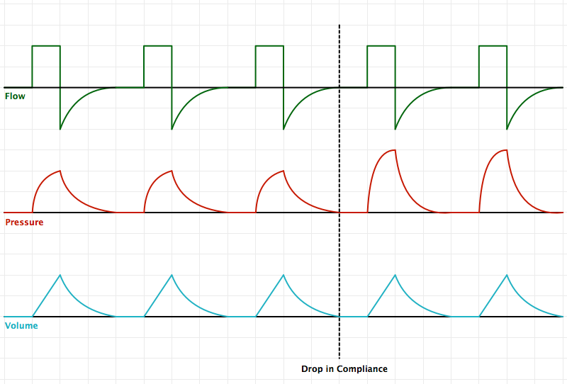
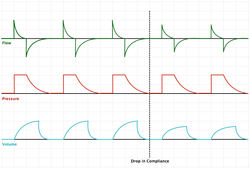
Volume is set, pressure is variable. With a drop in compliance, the preset minimum volume is maintained with an increase in pressure.

Pressure Control (PC)

Pressure Control (PC)

Pressure is set, volume is variable. With a drop in compliance, a smaller volume is delivered to maintain pressures at the preset limit.

Pressure-Regulated Volume Control (PRVC)

Pressure-Regulated Volume Control (PRVC)

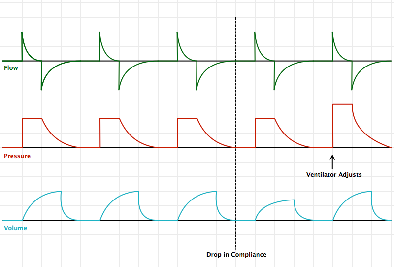
Pressure is targeted with a set minimum volume. The ventilator makes breath-to-breath adjustments of pressure to maintain minimum volumes. Breath mechanics are therefore comparable to pressure-control as a defined pressure is delivered based on prior breath’s respiratory mechanics (note pressure and flow tracings for PRVC/PC vs. VC)

References:

  1. Pierrakos C, Karanikolas M, Scolletta S, Karamouzos V, Velissaris D. Acute respiratory distress syndrome: pathophysiology and therapeutic options. J Clin Med Res. 2012;4(1):7–16. doi:10.4021/jocmr761w.
  2. Fanelli V, Vlachou A, Ghannadian S, Simonetti U, Slutsky AS, Zhang H. Acute respiratory distress syndrome: new definition, current and future therapeutic options. J Thorac Dis. 2013;5(3):326–334. doi:10.3978/j.issn.2072-1439.2013.04.05.
  3. ARDS Definition Task Force, Ranieri VM, Rubenfeld GD, et al. Acute respiratory distress syndrome: the Berlin Definition. In: Vol 307. 2012:2526–2533. doi:10.1001/jama.2012.5669.
  4. Ware LB, Matthay MA. The acute respiratory distress syndrome. N. Engl. J. Med. 2000;342(18):1334–1349. doi:10.1056/NEJM200005043421806.
  5. Deng, J. (10/20/13). Principles of Mechanical Ventilation. Medical Intensive Care Unit Lecture. Los Angeles, CA.
  6. Singer BD, Corbridge TC. Basic invasive mechanical ventilation. South. Med. J. 2009;102(12):1238–1245. doi:10.1097/SMJ.0b013e3181bfac4f.
  7. Hamed HMF, Ibrahim HG, Khater YH, Aziz ES. Ventilation and ventilators in the ICU: What every intensivist must know. Current Anaesthesia & Critical Care. 2006;17(1-2):77–83. doi:10.1016/j.cacc.2006.07.008.