Synovial Fluid

Brief HPI:

A 38 year-old female with a history of obesity and obstructive sleep apnea presents with right knee pain. She cannot identify a clear precipitant for her symptoms which she first noted 2 weeks ago. Her pain is worsened with ambulation and while previously tolerable, has grown more severe despite over-the-counter analgesics over the past two days. She denies fevers, intravenous drug use, recent travel or instrumentation.

On evaluation, vital signs are normal. Physical examination demonstrates a moderate-sized right knee effusion with overlying warmth though no edema. There is minimal pain with range of motion, no pain with heel percussion, and she is ambulatory independently with a mildly antalgic gait. Clinical suspicion for septic arthritis was low. A diagnostic arthrocentesis was performed without complication. Synovial fluid was less-viscous than normal with slight debris. Laboratory analysis revealed 14,230 white blood cells with 85% neutrophils and no crystals visualized. The patient was discharged with supportive care and outpatient follow-up – cultures were ultimately negative.

An Algorithm for the Analysis of Synovial Fluid

An Algorithm for the Analysis of Synovial Fluid

References

  1. Margaretten ME, Kohlwes J, Moore D, Bent S. Does this adult patient have septic arthritis? JAMA. 2007;297(13):1478-1488. doi:10.1001/jama.297.13.1478.
  2. Brannan SR, Jerrard DA. Synovial fluid analysis. J Emerg Med. 2006;30(3):331-339. doi:10.1016/j.jemermed.2005.05.029.
  3. Couderc M, Pereira B, Mathieu S, et al. Predictive value of the usual clinical signs and laboratory tests in the diagnosis of septic arthritis. CJEM. 2015;17(4):403-410. doi:10.1017/cem.2014.56.
  4. MD HJC, MD LAB, MD ML. Septic Arthritis. Hospital Medicine Clinics. 2014;3(4):494-503. doi:10.1016/j.ehmc.2014.06.009.

Cervical Spine Injuries

Brief H&P

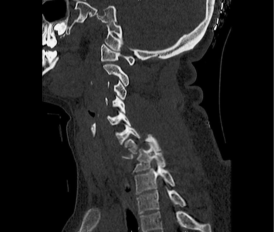
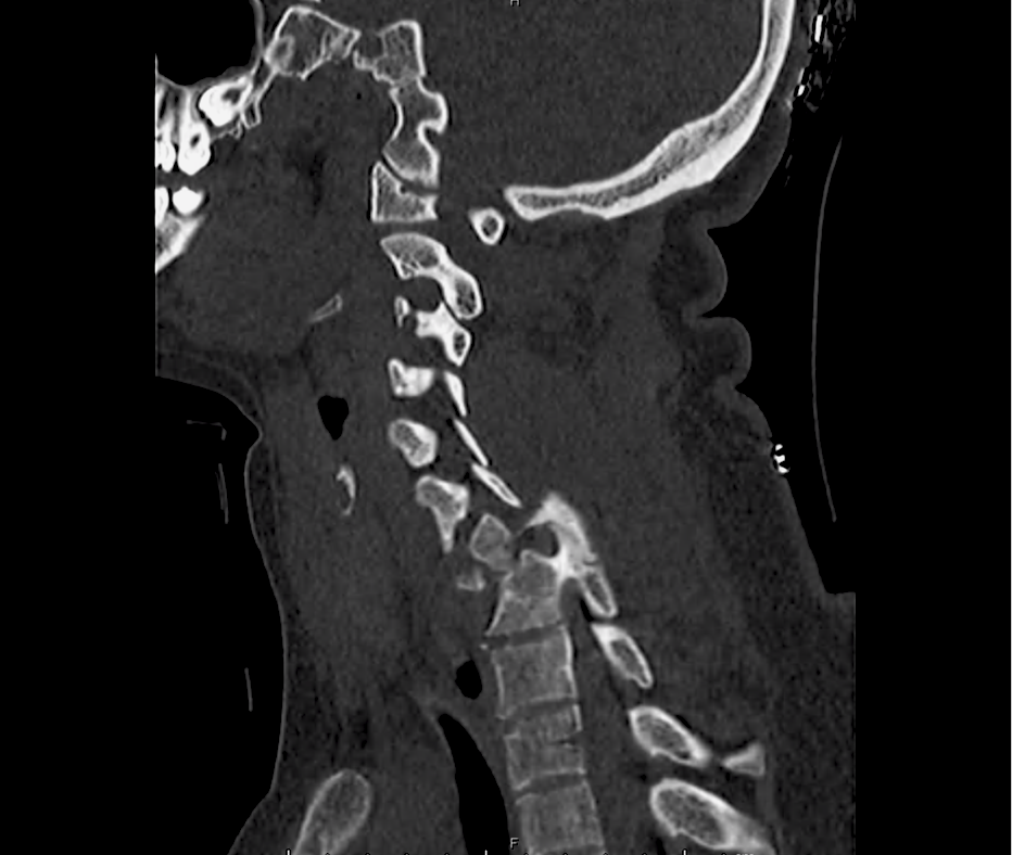
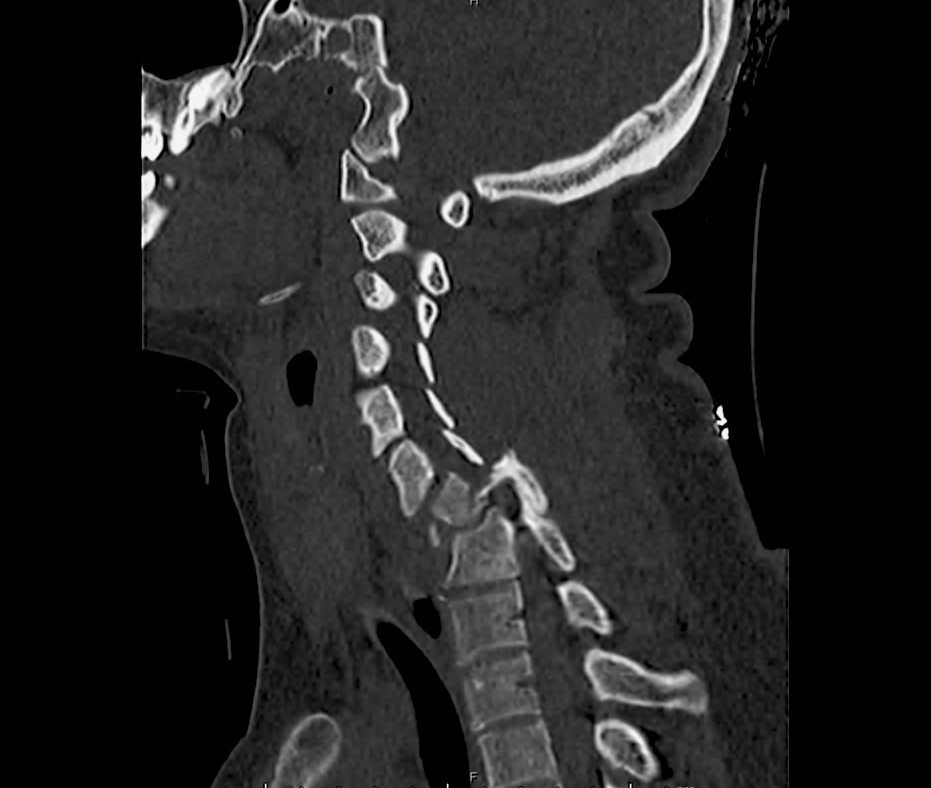
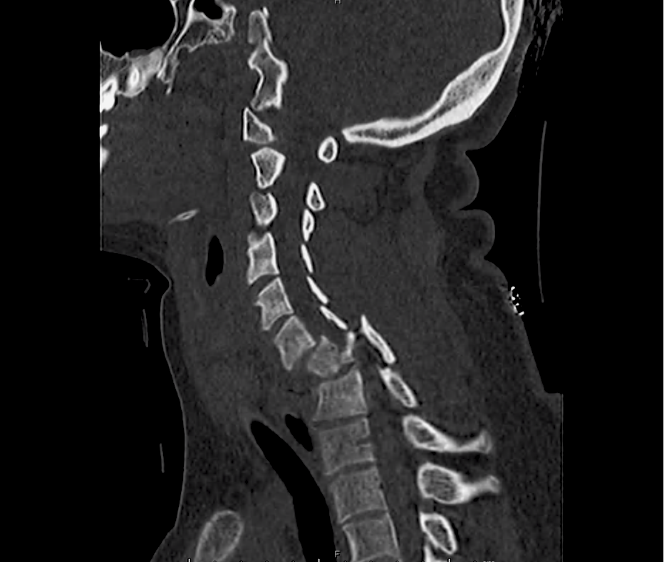
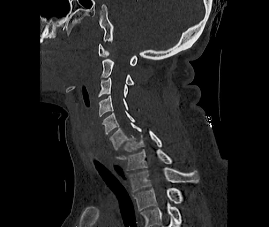
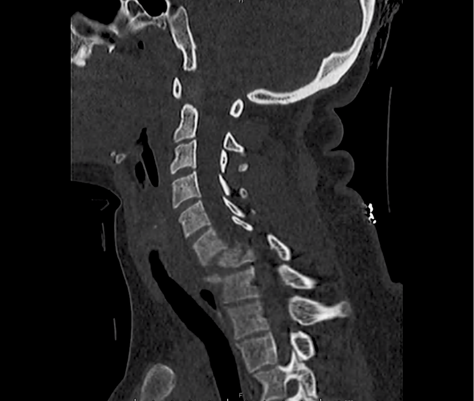
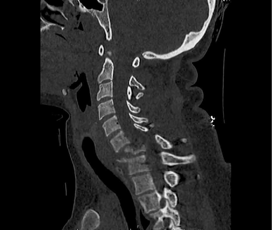
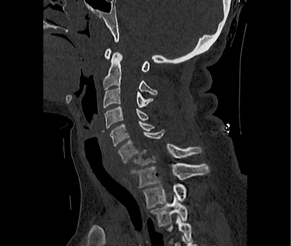
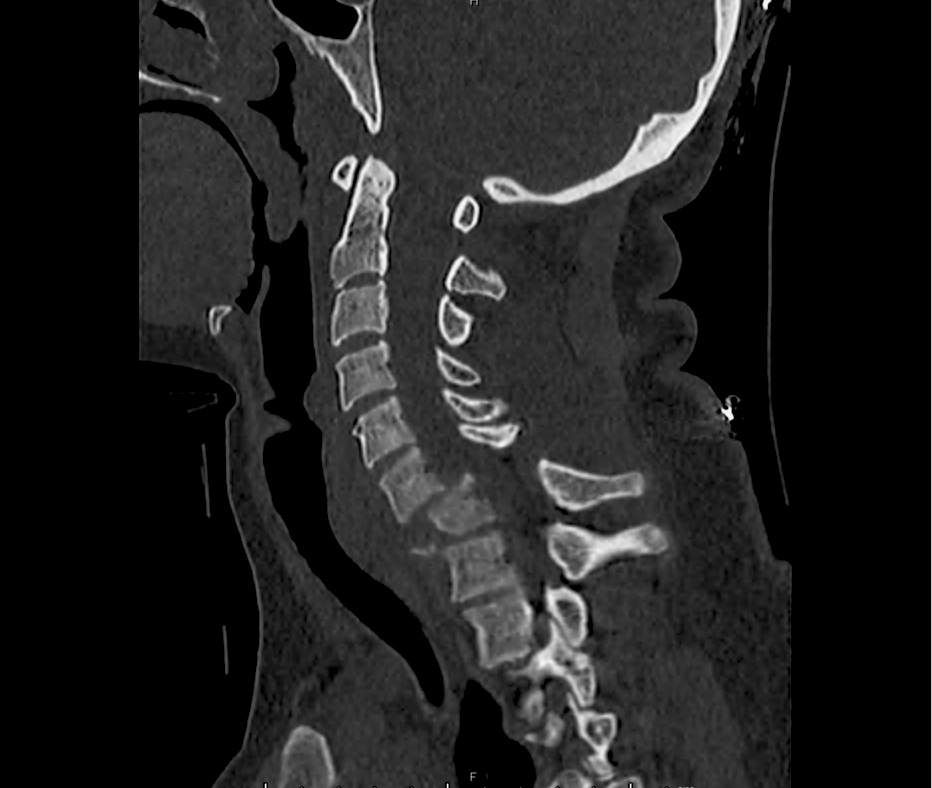
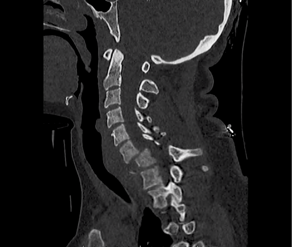
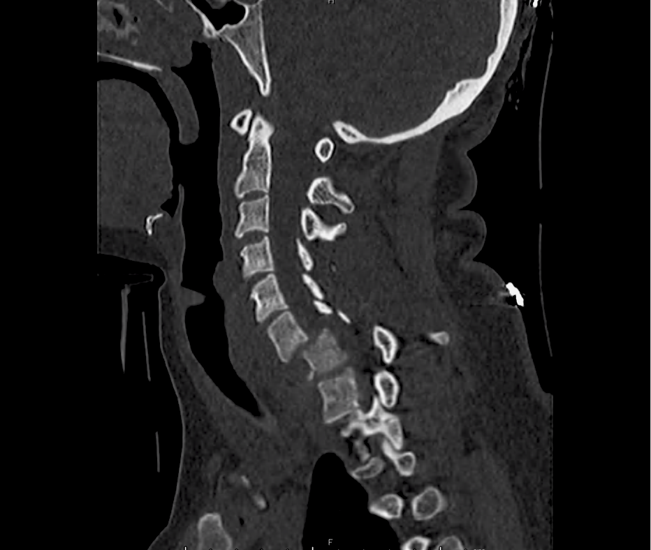
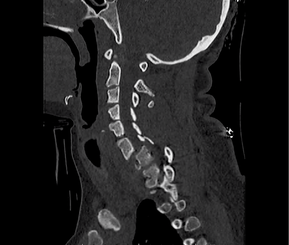
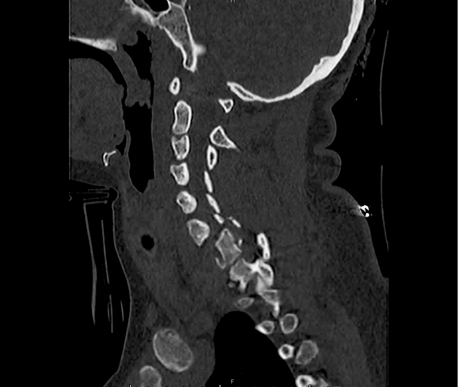
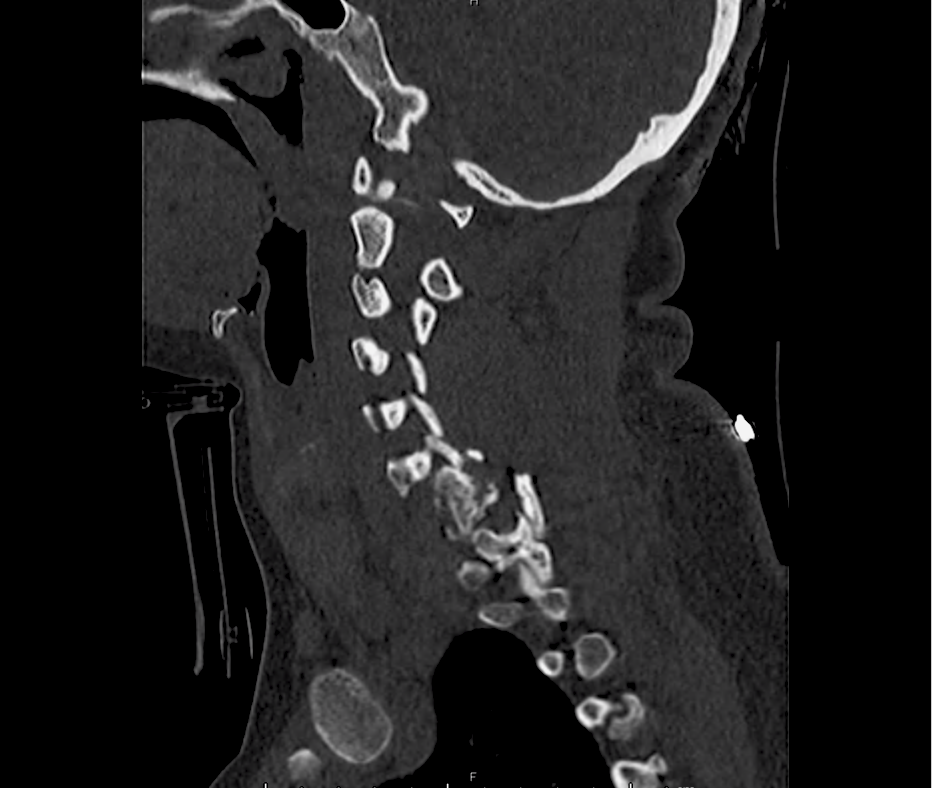
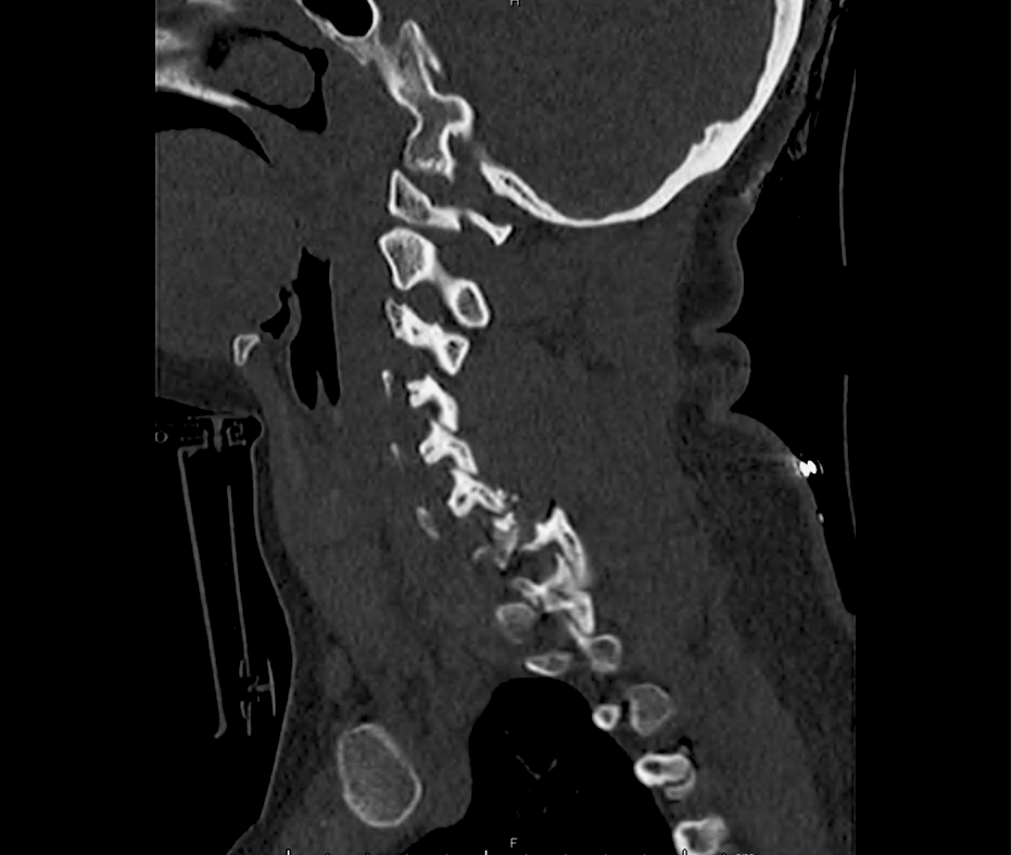
A young patient with no past medical history is brought in by ambulance after a high-speed motor vehicle accident. Trauma survey demonstrates absent motor/sensation in bilateral lower extremities with sensory level at T3-T4. Computed tomography of the cervical spine was obtained and is shown below.

Imaging

c-spine_01
c-spine_02
c-spine_03
c-spine_04
c-spine_05
c-spine_06
c-spine_07
c-spine_08
c-spine_09
c-spine_10
c-spine_11
c-spine_12
c-spine_13
c-spine_14
c-spine_15

CT C-Spine

Fracture-dislocation at C6-C7 and C7-T1 with comminuted burst fracture to C7 and locked facet joint with resultant anterior migration of C6 over C7, unstable cervical spine fracture.

Anatomy

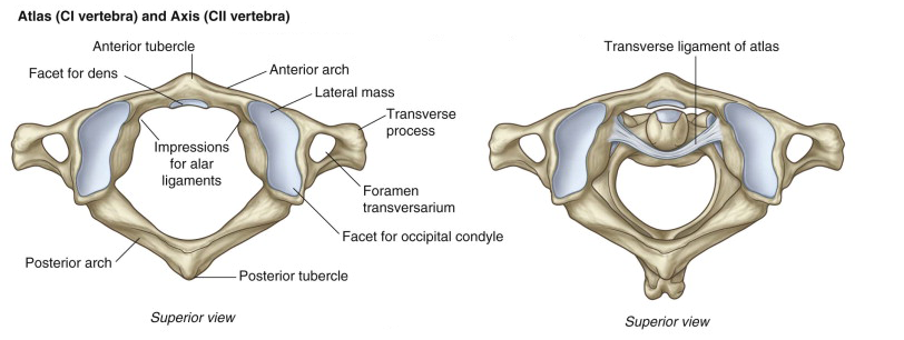
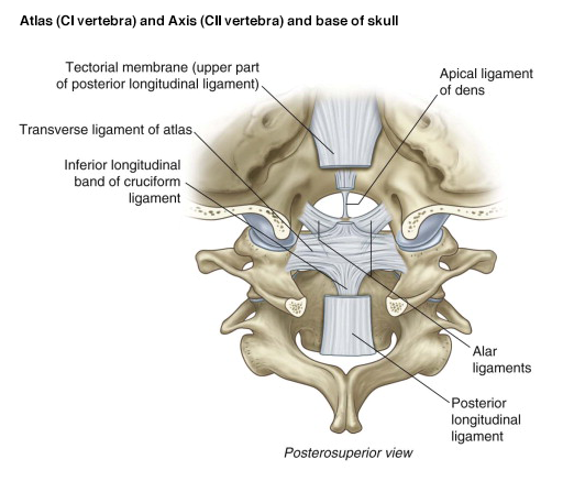
Atlas and Axis
Axis (C2 vertebra)
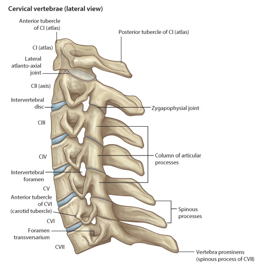
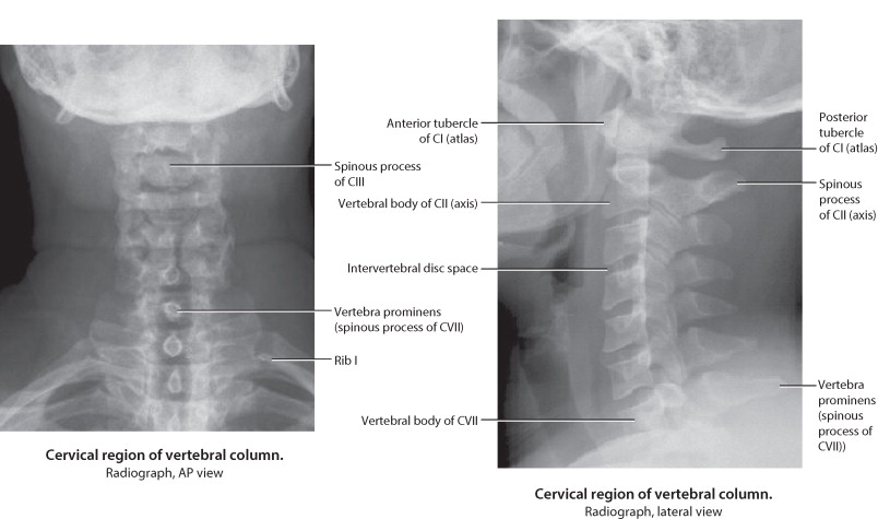
C-spine Lateral View
C-spine Radiographs
Skull base and C1/C2
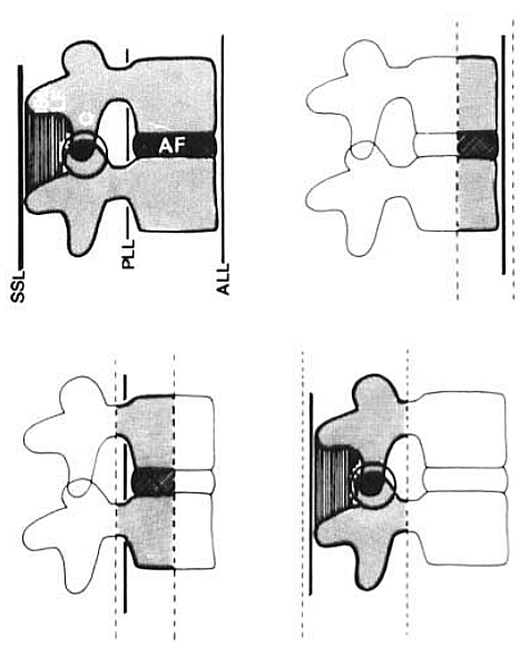
Vertebral Columns

Flexion

C1/C2

Wedge fracture

  • Stretch on strong nuchal ligament transmits force to vertebral body.
  • Stability: Generally stable unless >50% compression or multiple contiguous.

Flexion-teardrop fracture

  • Severe flexion force, avulsion of fragment of anterior/inferior portion of vertebral body.
  • Stability: Unstable, involves anterior/posterior ligamentous disruptions.

Clay shoveler’s fracture

  • Oblique fracture of spinous process of lower cervical spine.
  • Stability: Stable

Subluxation

  • Pure ligamentous injury without associated fracture.
  • Imaging: Widening of interspinous and intervertebral spaces on lateral.
  • Stability: Potentially unstable.

Bilateral facet dislocation

  • Anterior displacement of spine above level of injury caused by dislocation of upper inferior facet from lower superior facet.
  • Imaging: Anterior displacement greater than ½ AP diameter of vertebral body.
  • Stability: Unstable

Odontoid process fracture

  • Head trauma with shear force directed at odontoid.
  • Sub-classification: Type I (above transverse ligament), type II (odontoid base), type III (extension to body of C2)
  • Stability: Types II, III unstable.

Flexion/Rotation

Rotary atlantoaxial dislocation

  • Imaging: Open-mouth odontoid, asymmetric lateral masses of C1.
  • Stability: Unstable

Unilateral facet dislocation

  • Flexion and rotation centered around single facet results in contralateral facet dislocation.
  • Imaging: AP radiograph shows spinous processes above dislocation displaced from midline, lateral radiograph shows anterior displacement of lower vertebra (less than ½ AP diameter of vertebral body).

Extension

Posterior neural arch fracture (C1)

  • Forced extension causes compressive force on posterior elements of C1 between occiput and C2.
  • Stability: Unstable

Hangman’s fracture (spondylolysis C2)

  • Abrupt deceleration causes fracture of bilateral pedicles of C2, potentially with associated subluxation. Rarely associated with SCI due to large diameter of neural canal at C2.
  • Imaging: May be associated with retropharyngeal space edema.
  • Stability: Unstable

Extension-teardrop fracture

  • Abrupt extension (ex. diving) results in stretch along anterior longitudinal ligament with avulsion of anterior/inferior fragment of vertebral body (usually C5-C7).
  • Imaging: May be radiographically similar to flexion-teardrop fracture.
  • Complications: Central cord syndrome
  • Stability: Unstable in extension

Vertical compression

Burst fracture

  • Force applied from above or below causes transmission of force to intervertebral disc and vertebral body.
  • Imaging: Comminuted vertebral body, >40% compression of anterior vertebral body.
  • Complications: Fracture fragments may impinge on spinal cord.
  • Stability: Stable

Jefferson fracture (C1)

  • Vertical force transmitted from occipital condyles to superior articular facets of atlas, resulting in fractures of anterior and posterior arches.
  • Imaging: Widening of predental space. Open-mouth odontoid view may reveal bilateral offset distance of >7mm between lateral masses of C1/C2.
  • Stability: Unstable

Cervical Spine Imaging Decision Rule (Canadian)

Algorithm for the Evaluation of Cervical Spine Trauma (Canadian)

References:

  1. MD RK, MD BED, CAQ-SM KHM, MD WF. Emergency Department Evaluation and Treatment of Cervical Spine Injuries. Emergency Medicine Clinics of NA. 2015;33(2):241-282. doi:10.1016/j.emc.2014.12.002.
  2. Denis F. Spinal instability as defined by the three-column spine concept in acute spinal trauma. Clin Orthop Relat Res. 1984;(189):65-76.
  3. Munera F, Rivas LA, Nunez DB, Quencer RM. Imaging evaluation of adult spinal injuries: emphasis on multidetector CT in cervical spine trauma. Radiology. 2012;263(3):645-660. doi:10.1148/radiol.12110526.

Back Pain

Causes of Back Pain

Causes of Back Pain

Key Historical Findings

Onset
Acute onset with associated activity suggests mechanical process
Acute onset without trigger, particularly if severe pain may suggest vascular process
Progressive onset without trigger suggests non-mechanical process (i.e. malignancy)
Aggravating/Alleviating Factors
Worsening with cough/valsalva suggests herniated disk
Relief with flexion associated with spinal stenosis
Location/Radiation
Radicular pain typically extends below knee, associated with nerve root involvement
Radiation to/from chest or abdomen suggests visceral source
Flank location suggests retroperitoneal source
History/Associated Symptoms
Fever
Medications (particularly anti-coagulants)
Hematuria
Malignancy
IVDA
Vascular disease

Key Physical Findings

  • Abnormal vital signs

    • Fever: abscess, osteomyelitis, discitis
    • Hypertension: dissection
    • Shock: AAA
  • Localize point of greatest tenderness
  • Examine abdomen for pulsatile mass
  • Perform thorough neurological examination including rectal tone and perianal sensation
  • Positive straight leg raise associated with sciatic nerve root irritation and is sensitive (but not specific) for disk disease.

References

  1. Mahoney, B. (2013). Back Pain. In Rosen’s Emergency Medicine – Concepts and Clinical Practice (8th ed., Vol. 1, pp. 278-284). Elsevier Health Sciences.
  2. WikEM: Lower back pain