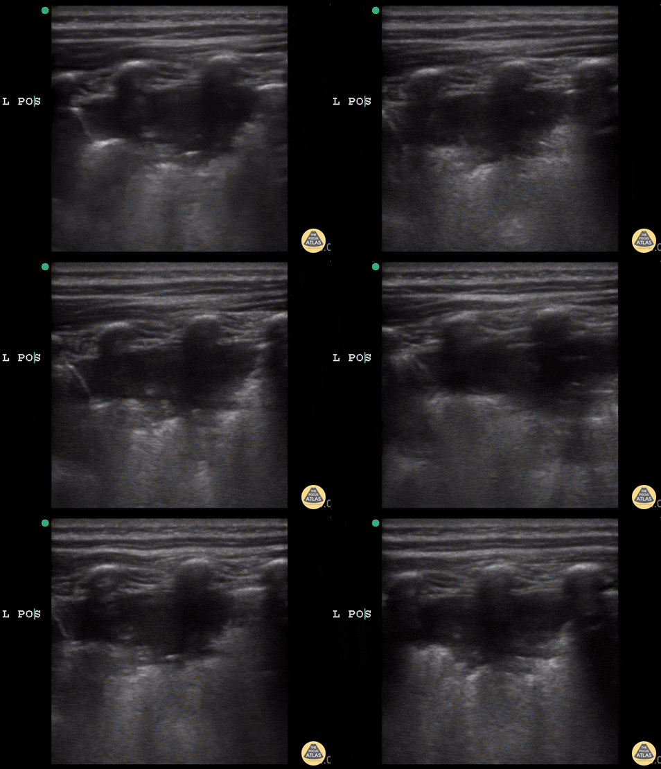
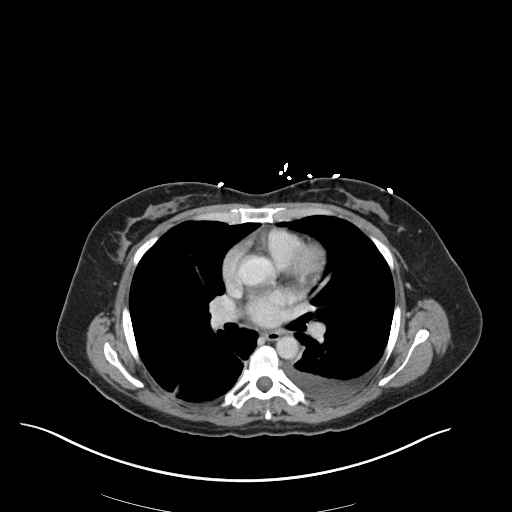
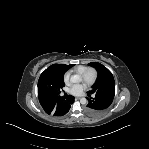
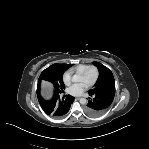
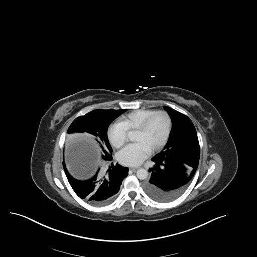
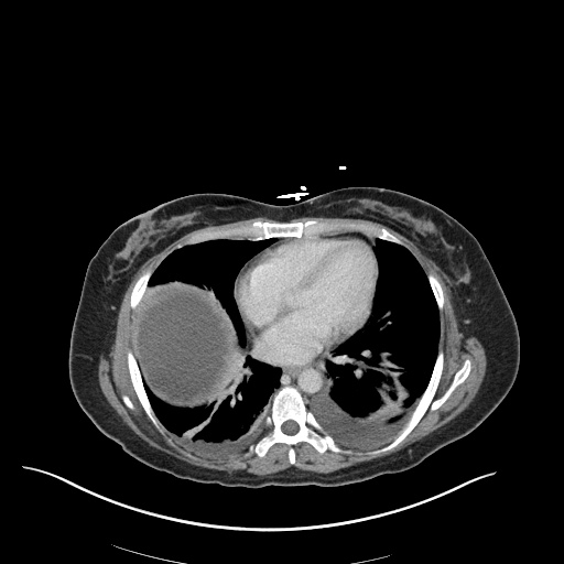
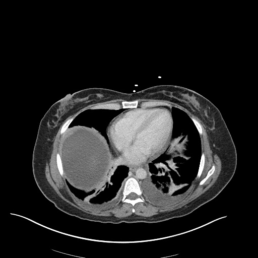
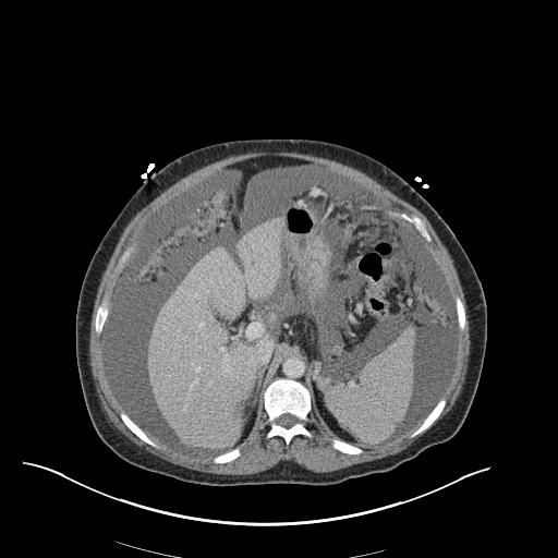
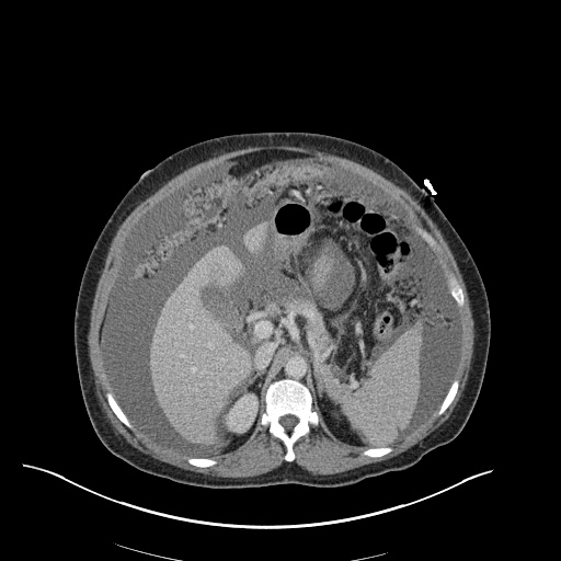
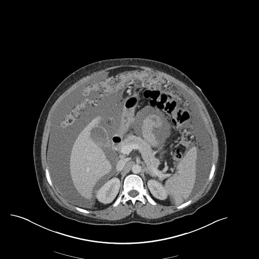
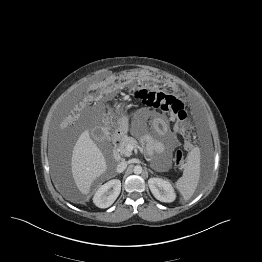
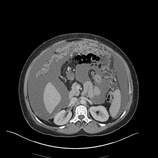
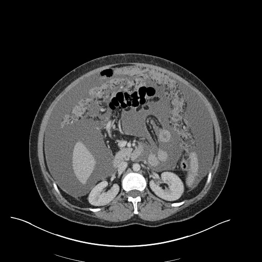
Wheezing and Stridor

Brief HPI:

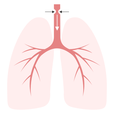
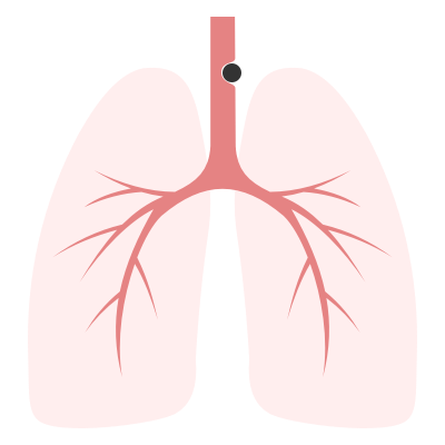
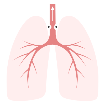
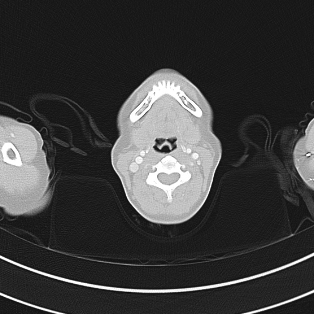
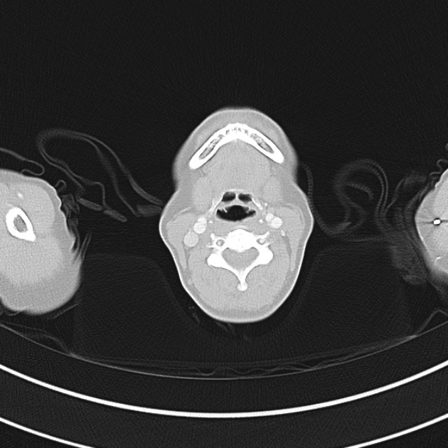
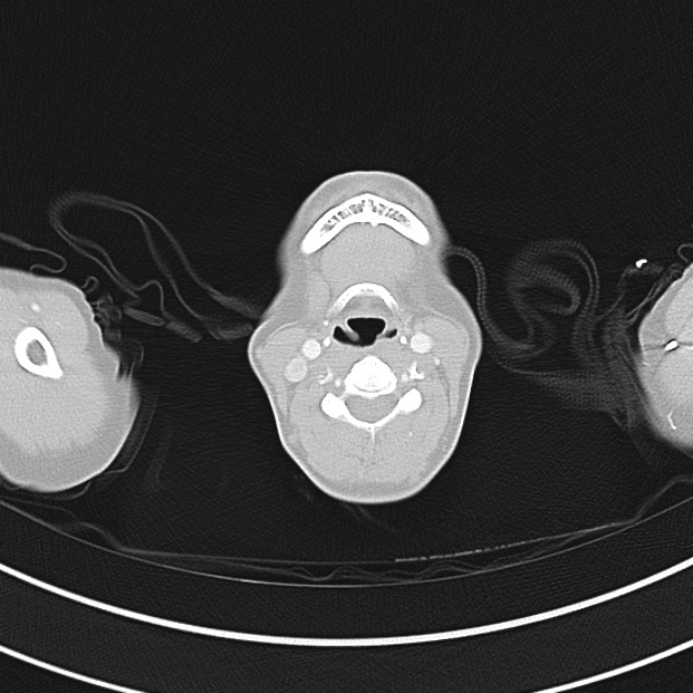
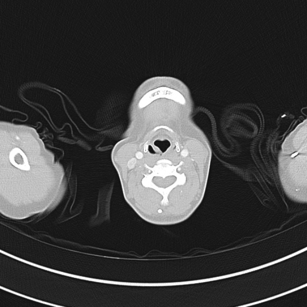
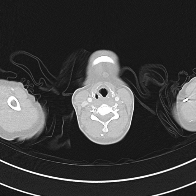
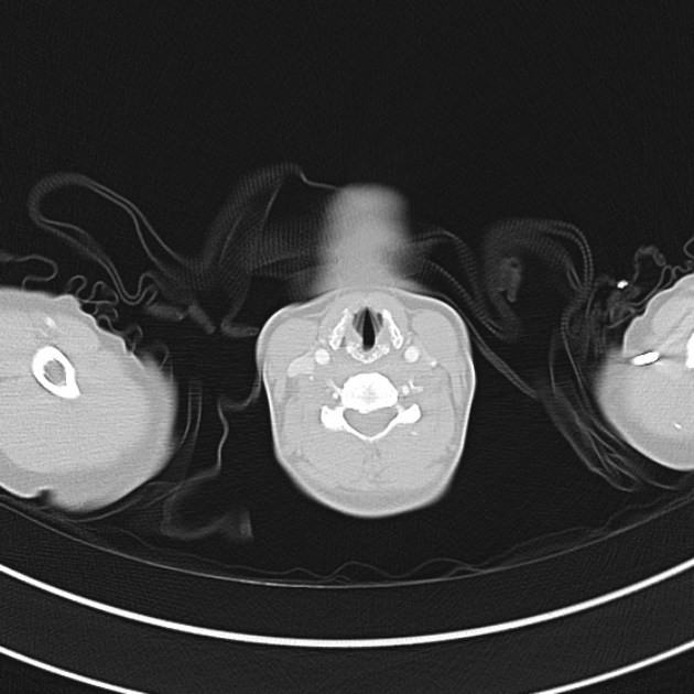
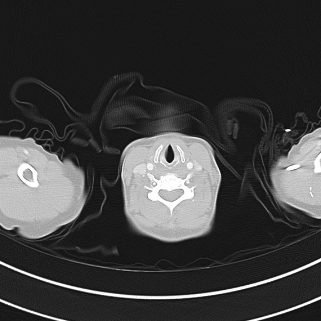
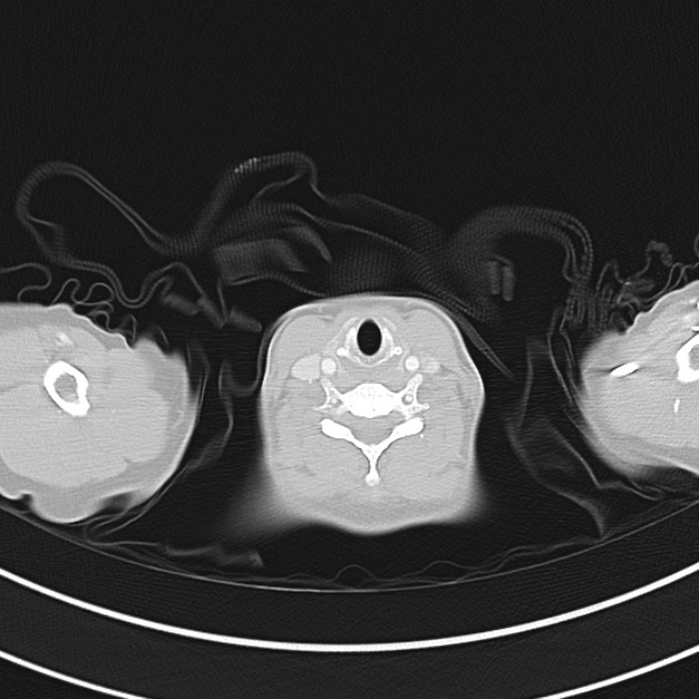
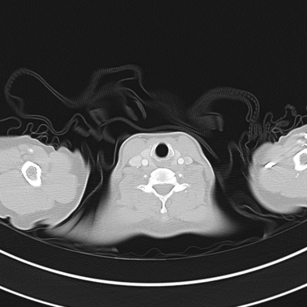
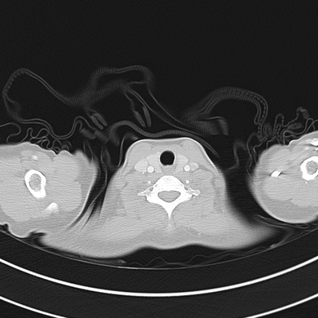
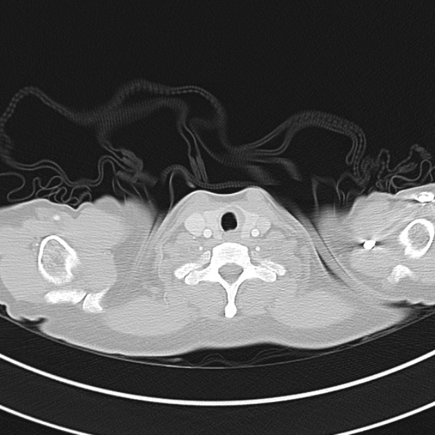
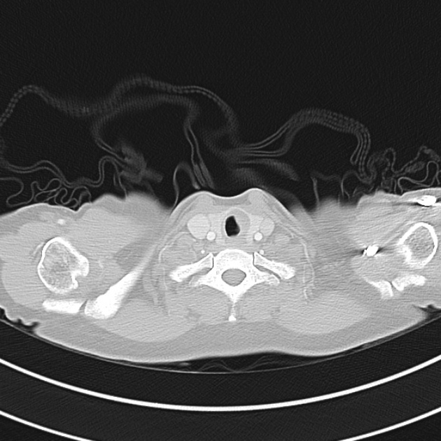
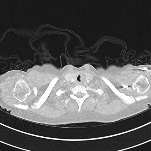
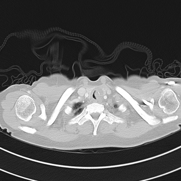
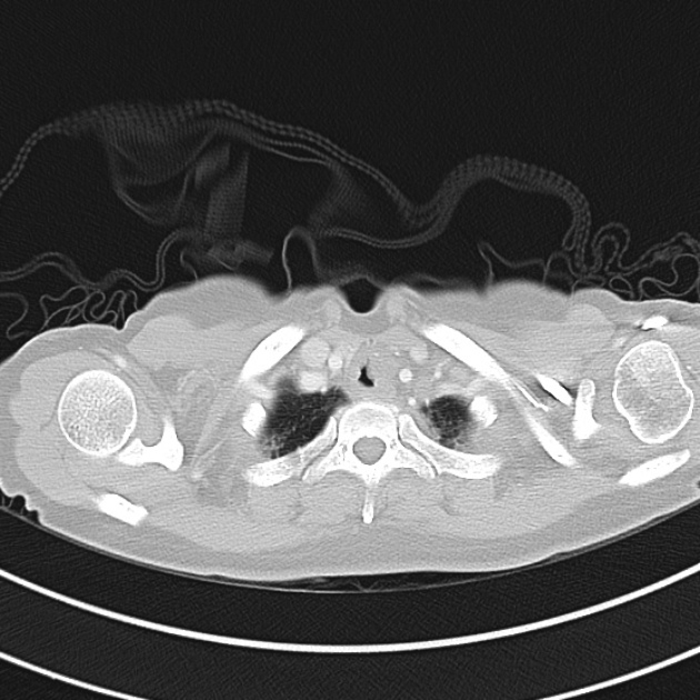
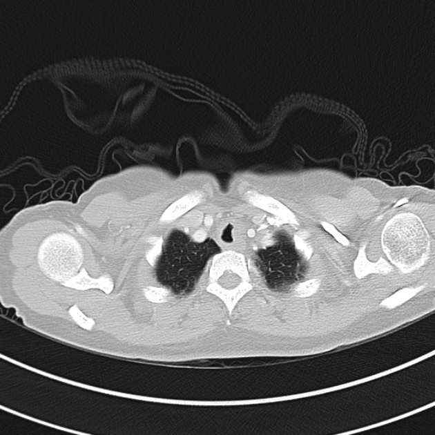
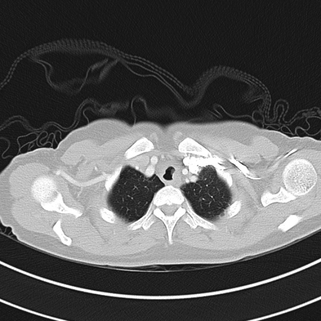
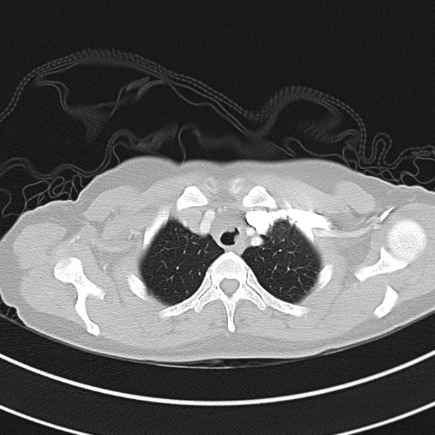
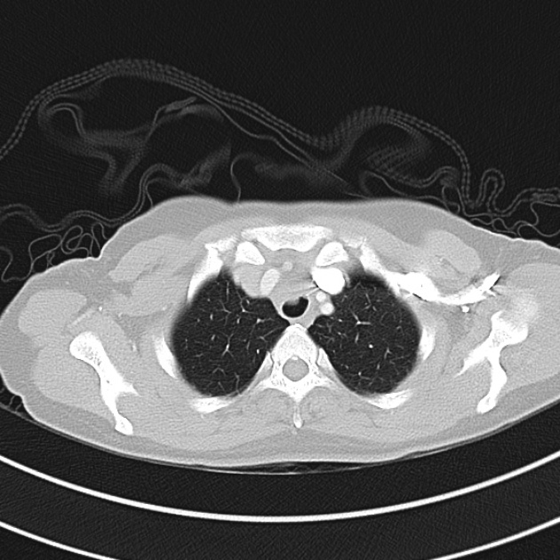
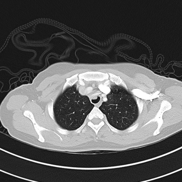
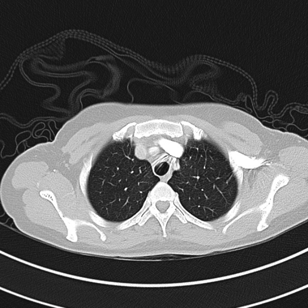
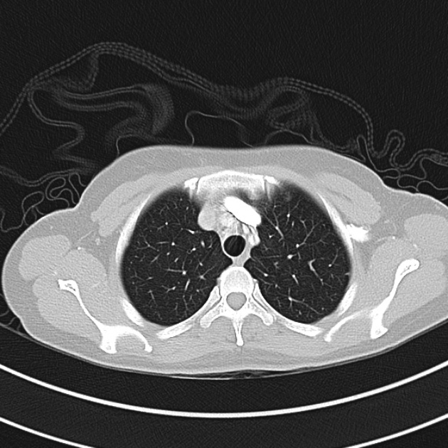
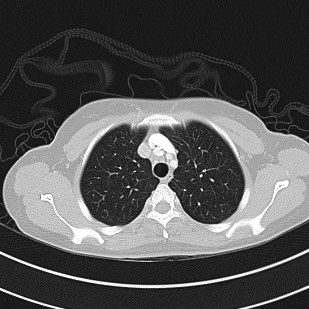
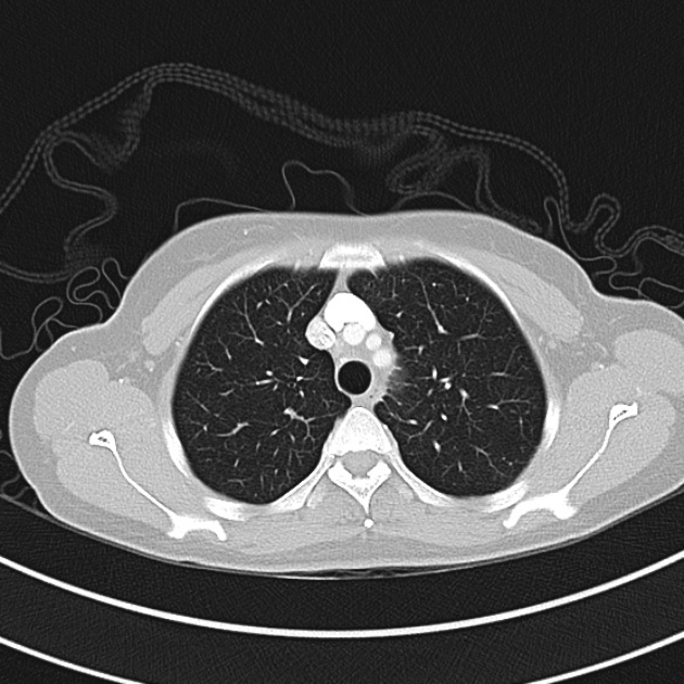
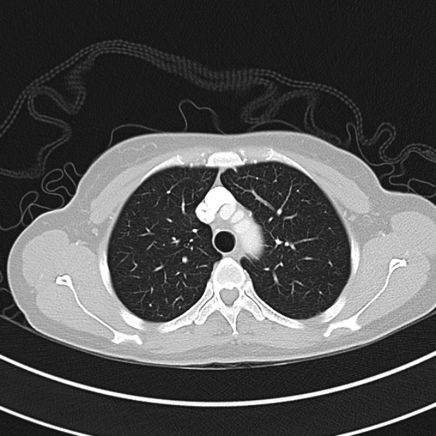
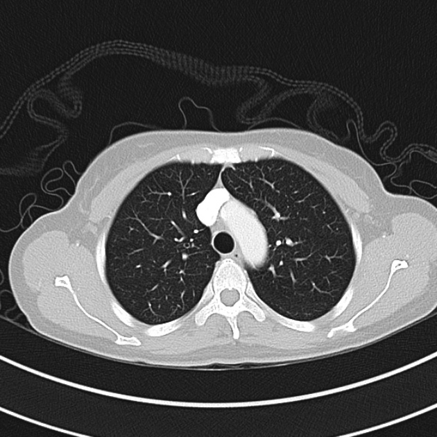
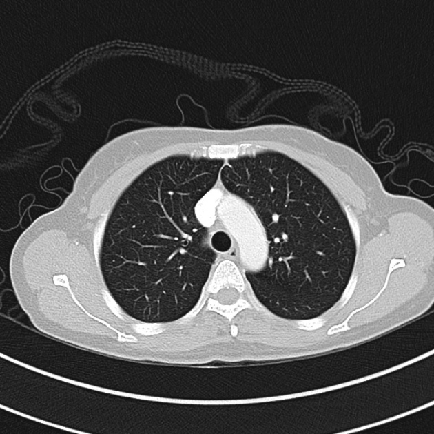
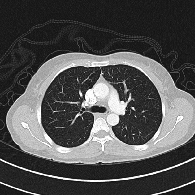
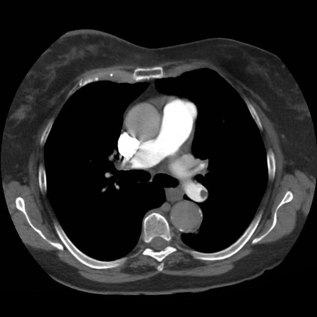
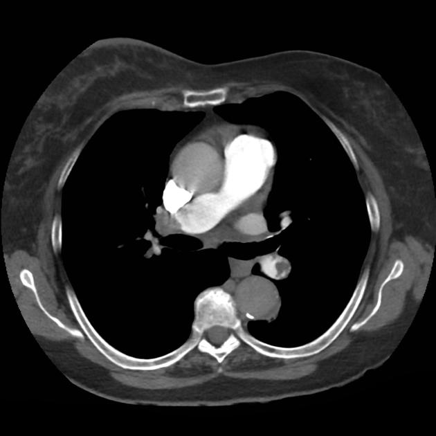
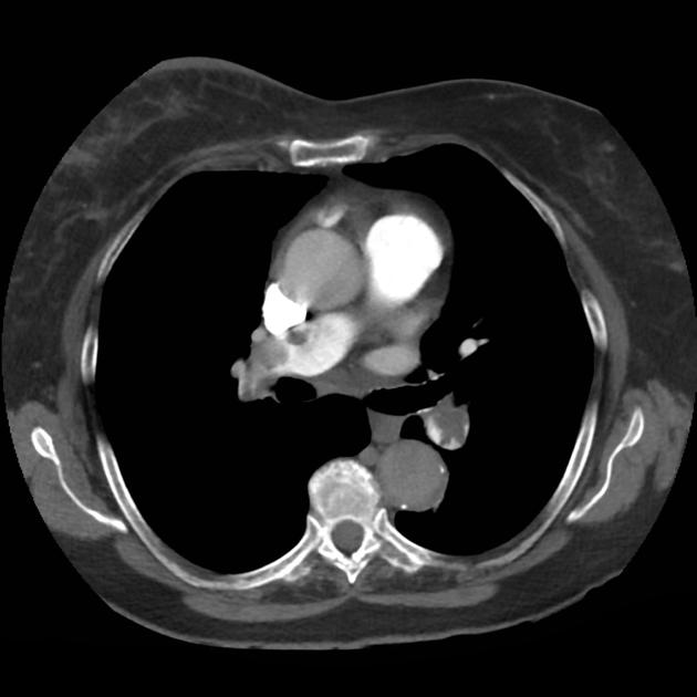
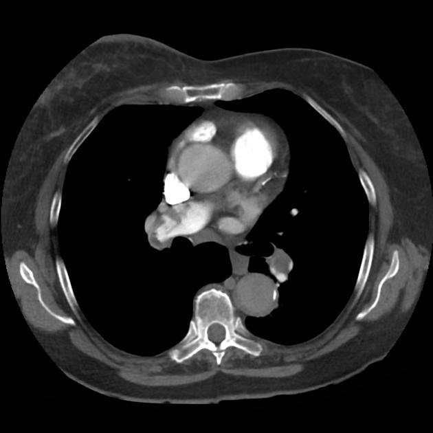
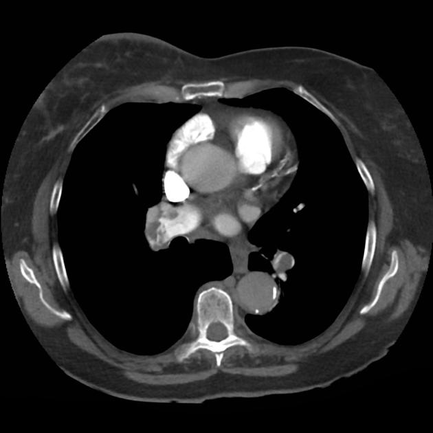
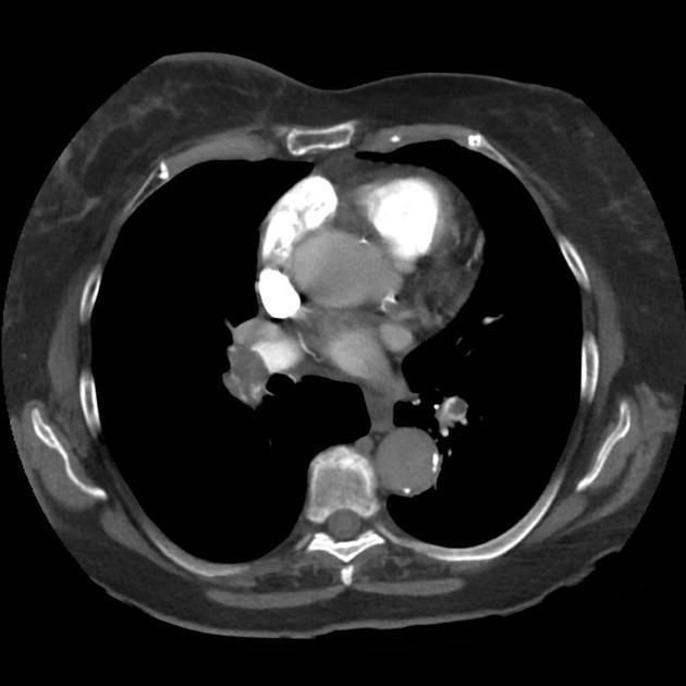
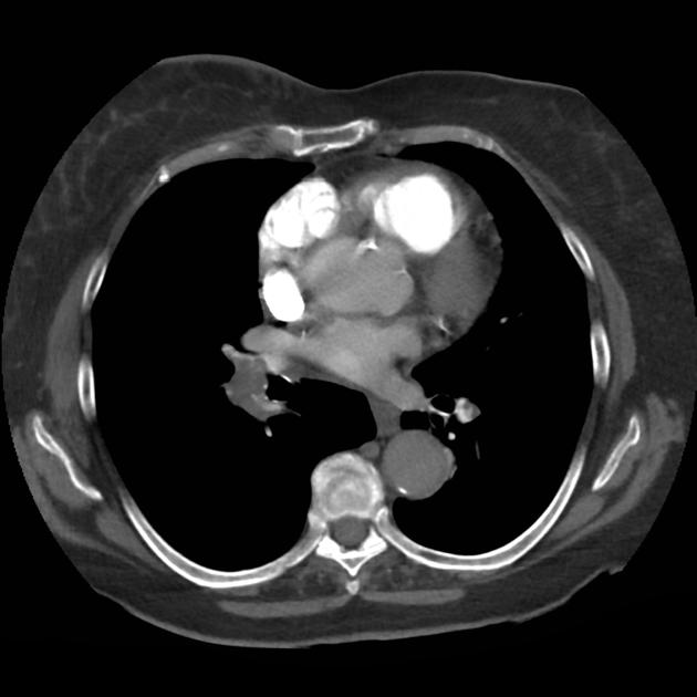
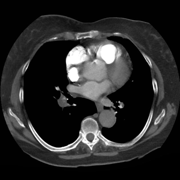
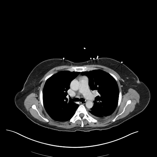
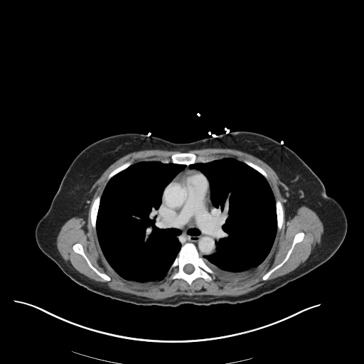
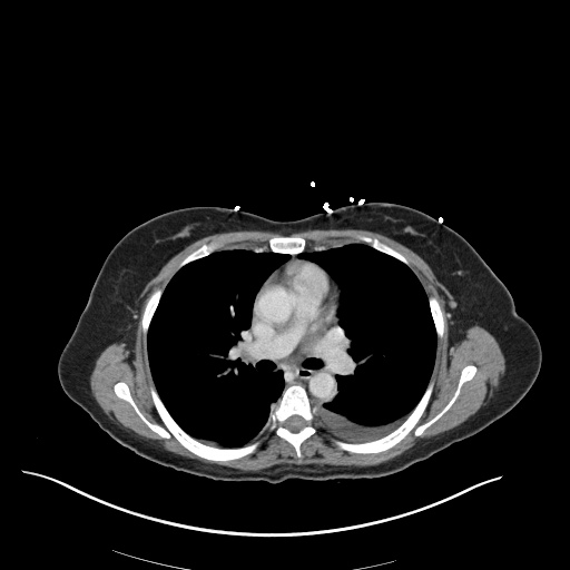
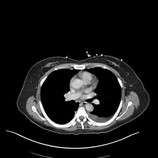
A 66 year-old male with a history of hypertension and COPD presents with shortness of breath. He states that his symptoms are unimproved with home nebulizer treatments and denies fever, cough or new sputum production. On examination, he has stridor appreciated during inspiratory and expiratory phases.

01_stridor
02_stridor
03_stridor
04_stridor
05_stridor
06_stridor
07_stridor
08_stridor
09_stridor
10_stridor
11_stridor
12_stridor
13_stridor
14_stridor
15_stridor
16_stridor
17_stridor
18_stridor
19_stridor
20_stridor
21_stridor
22_stridor
23_stridor
24_stridor
25_stridor
26_stridor
27_stridor
28_stridor
29_stridor
30_stridor
31_stridor
32_stridor

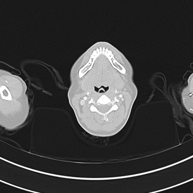
CT Chest:

1.9cm soft tissue thickening of the left tracheal wall at the level of the inferior thyroid gland. Luminal narrowing to 4 mm at this level.
Case courtesy of Dr Ian Bickle from Radiopaedia.org: 47677

Sound Characteristics

Stridor

An inspiratory, expiratory, or continuous monophonic sound that is loudest over the central airways.

Wheezing

A musical, high-pitched sound – more commonly expiratory. Requires sufficient airflow to induce airway oscillations.

Respiratory Phase

Inspiratory

Supraglottic: negative intratracheal pressure during inspiration causes airway collapse.

Biphasic

Glottic/Subglottic: fixed obstruction not impacted by changes in luminal/thoracic pressure.

Expiratory

Intrathoracic: increased pleural pressure compresses the narrowed airway.

An Algorithm for the Diagnosis of Wheezing and Stridor

An Algorithm for the Diagnosis of Wheezing and Stridor

Special thanks to Dr. Denna Zebda, Assistant Professor, Department of Otorhinolaryngology – McGovern Medical School for her expertise and review of this algorithm.

References

  1. Sicari V, Zabbo CP. Stridor. [Updated 2021 Jul 10]. In: StatPearls [Internet]. Treasure Island (FL): StatPearls Publishing; 2022 Jan-. Available from: https://www.ncbi.nlm.nih.gov/books/NBK525995/
  2. Patel PH, Mirabile VS, Sharma S. Wheezing. [Updated 2021 May 12]. In: StatPearls [Internet]. Treasure Island (FL): StatPearls Publishing; 2022 Jan-. Available from: https://www.ncbi.nlm.nih.gov/books/NBK482454/
  3. Bohadana A, Izbicki G, Kraman SS. Fundamentals of lung auscultation. N Engl J Med. 2014;370(21):2053.
  4. Orient JM, Sapira JD. Sapira’s Art & Science of Bedside Diagnosis. 4th ed. Wolters Kluwer Health/Lippincott Williams & Wilkins; 2010.

Hypoxia

Brief H&P:

A 67 year-old male with a history of hypertension and diabetes presents to the emergency department after a syncopal episode. He had been completing his normal morning routine when he developed a sensation of lightheadedness and awoke on the ground of his kitchen. He denies associated chest pain, palpitations, diaphoresis, or recent illness. He has no known sick contacts nor exposures to individuals undergoing evaluation for COVID-19.

On arrival in the emergency department, the patient was noted to be hypoxic with pulse oximetry measuring 74%. He was placed on supplemental oxygen via non-rebreather with improvement of oxygen saturation to 94%. Examination demonstrated diminished alertness (requiring constant stimulation for responses) and generalized motor weakness. Cardiac and pulmonary examinations were unremarkable with the exception of tachypnea and no extremity edema was appreciated.

A chest radiograph was obtained which demonstrated platelike atelectasis. An arterial blood gas was obtained with PaO2 of 72mmHg suggesting a prominent A-a gradient. CT pulmonary angiography was obtained:

01_pe
02_pe
03_pe
04_pe
05_pe
06_pe
07_pe
08_pe
09_pe
10_pe
11_pe
12_pe

CT Pulmonary Angiography:

Bilateral pulmonary emboli. Case courtesy of Associate Prof Frank Gaillard, Radiopaedia.org, rID: 19636

Upon return, the patient’s mental status worsened associated with hypotension and he was intubated for airway protection and received systemic thrombolysis. He was subsequently taken for emergent endovascular treatment of massive pulmonary embolus.

An Algorithm for the Differential Diagnosis of Hypoxemia & Hypoxia1-7

Hypoxemia is defined as low PaO2 while hypoxia is insufficient global or local tissue oxygen content.

An Algorithm for the Differential Diagnosis of Hypoxemia and Hypoxia

References

  1. Stapczynski J. Respiratory Distress. In: Tintinalli JE, Ma O, Yealy DM, Meckler GD, Stapczynski J, Cline DM, Thomas SH. eds. Tintinalli’s Emergency Medicine: A Comprehensive Study Guide, 9e New York, NY: McGraw-Hill; . http://accessmedicine.mhmedical.com/content.aspx?bookid=2353&sectionid=219642010. Accessed April 12, 2020.
  2. Gas Transport & pH. In: Barrett KE, Barman SM, Brooks HL, Yuan JJ. eds. Ganong’s Review of Medical Physiology, 26e New York, NY: McGraw-Hill; . http://accessmedicine.mhmedical.com/content.aspx?bookid=2525&sectionid=204297654. Accessed April 12, 2020.
  3. Loscalzo J. Hypoxia and Cyanosis. In: Jameson J, Fauci AS, Kasper DL, Hauser SL, Longo DL, Loscalzo J. eds. Harrison’s Principles of Internal Medicine, 20e New York, NY: McGraw-Hill; . http://accessmedicine.mhmedical.com/content.aspx?bookid=2129&sectionid=192012521. Accessed April 12, 2020.
  4. West NE, Lechtzin N. Chapter 93. Hypoxia. In: McKean SC, Ross JJ, Dressler DD, Brotman DJ, Ginsberg JS. eds. Principles and Practice of Hospital Medicine New York, NY: McGraw-Hill; 2012. http://accessmedicine.mhmedical.com/content.aspx?bookid=496&sectionid=41304065. Accessed April 12, 2020.
  5. Pulmonary Physiology. In: Kibble JD, Halsey CR. eds. Medical Physiology: The Big Picture New York, NY: McGraw-Hill; 2014. http://accessmedicine.mhmedical.com/content.aspx?bookid=1291&sectionid=75576764. Accessed April 12, 2020.
  6. Petersson, J., Glenny, R. (2014). Gas exchange and ventilation–perfusion relationships in the lung European Respiratory Journal 44(4), 1023-1041. https://dx.doi.org/10.1183/09031936.00037014
  7. Morchi, R. (2011). Diagnosis Deconstructed: The Case of the Patient with No Chief Complaint Emergency Medicine News XXXIII(3)
    Rodríguez-Roisin, R., Roca, J. (2005). Mechanisms of hypoxemia Intensive Care Medicine 31(8), 1017-1019. https://dx.doi.org/10.1007/s00134-005-2678-1

COVID-19

Brief HPI:

A 38 year-old male with a history of hypertension presents to the emergency department with fever, cough and shortness of breath. He notes 4 days of symptoms which have been gradually worsening despite over-the-counter treatments. He denies recent travel or sick contacts. While he attempted to remain isolated – his symptoms grew intolerable.

On arrival in the emergency department, vital signs were notable for tachycardia and hypoxia (SpO2 85%, improving to 92% on 4L by nasal cannula). Physical examination demonstrated tachypnea and accessory muscle use but clear lung fields, and no extremity edema nor jugular venous distension. A chest radiograph revealed patchy airspace opacities. A presumptive diagnosis of COVID-19 pneumonia was made.

While awaiting hospitalization, the patient’s hypoxia worsened though he remained otherwise alert and oriented. He was placed on 15L via non-rebreather and instructed regarding self-prone positioning. He was admitted to the intensive care unit.

An Algorithm for the Management of COVID-19 Hypoxic Respiratory Failure1-6

An algorithm for the management of COVID-19 respiratory failure

References

  1. Whittle, J., Pavlov, I., Sacchetti, A., Atwood, C., Rosenberg, M. (2020). Respiratory Support for Adult Patients with COVID‐19 Journal of the American College of Emergency Physicians Open https://dx.doi.org/10.1002/emp2.12071
  2. Hui, D., Chow, B., Chu, L., Ng, S., Lee, N., Gin, T., Chan, M. (2012). Exhaled Air Dispersion during Coughing with and without Wearing a Surgical or N95 Mask PLoS ONE  7(12), e50845. https://dx.doi.org/10.1371/journal.pone.0050845
  3. Hui, D., Chow, B., Lo, T., Ng, S., Ko, F., Gin, T., Chan, M. (2015). Exhaled Air Dispersion During Noninvasive Ventilation via Helmets and a Total Facemask Chest  147(5), 1336-1343. https://dx.doi.org/10.1378/chest.14-1934
  4. Hui, D., Chow, B., Lo, T., Tsang, O., Ko, F., Ng, S., Gin, T., Chan, M. (2019). Exhaled air dispersion during high-flow nasal cannula therapy versus CPAP via different masks European Respiratory Journal  53(4), 1802339. https://dx.doi.org/10.1183/13993003.02339-2018
  5. Sun, Q., Qiu, H., Huang, M., Yang, Y. (2020). Lower mortality of COVID-19 by early recognition and intervention: experience from Jiangsu Province Annals of Intensive Care  10(1), 33. https://dx.doi.org/10.1186/s13613-020-00650-2
  6. Roca, O., Caralt, B., Messika, J., Samper, M., Sztrymf, B., Hernández, G., García-de-Acilu, M., Frat, J., Masclans, J., Ricard, J. (2018). An Index Combining Respiratory Rate and Oxygenation to Predict Outcome of Nasal High-Flow Therapy American Journal of Respiratory and Critical Care Medicine  199(11), 1368-1376. https://dx.doi.org/10.1164/rccm.201803-0589oc

Pleural Fluid

Brief HPI:

A 43 year-old female with no reported medical history presents with shortness of breath. She notes 2 months of gradually worsening symptoms associated with unproductive cough and intermittent subjective fevers. Symptoms are worsened with activity and when laying flat. She has no history of similar symptoms in the past.

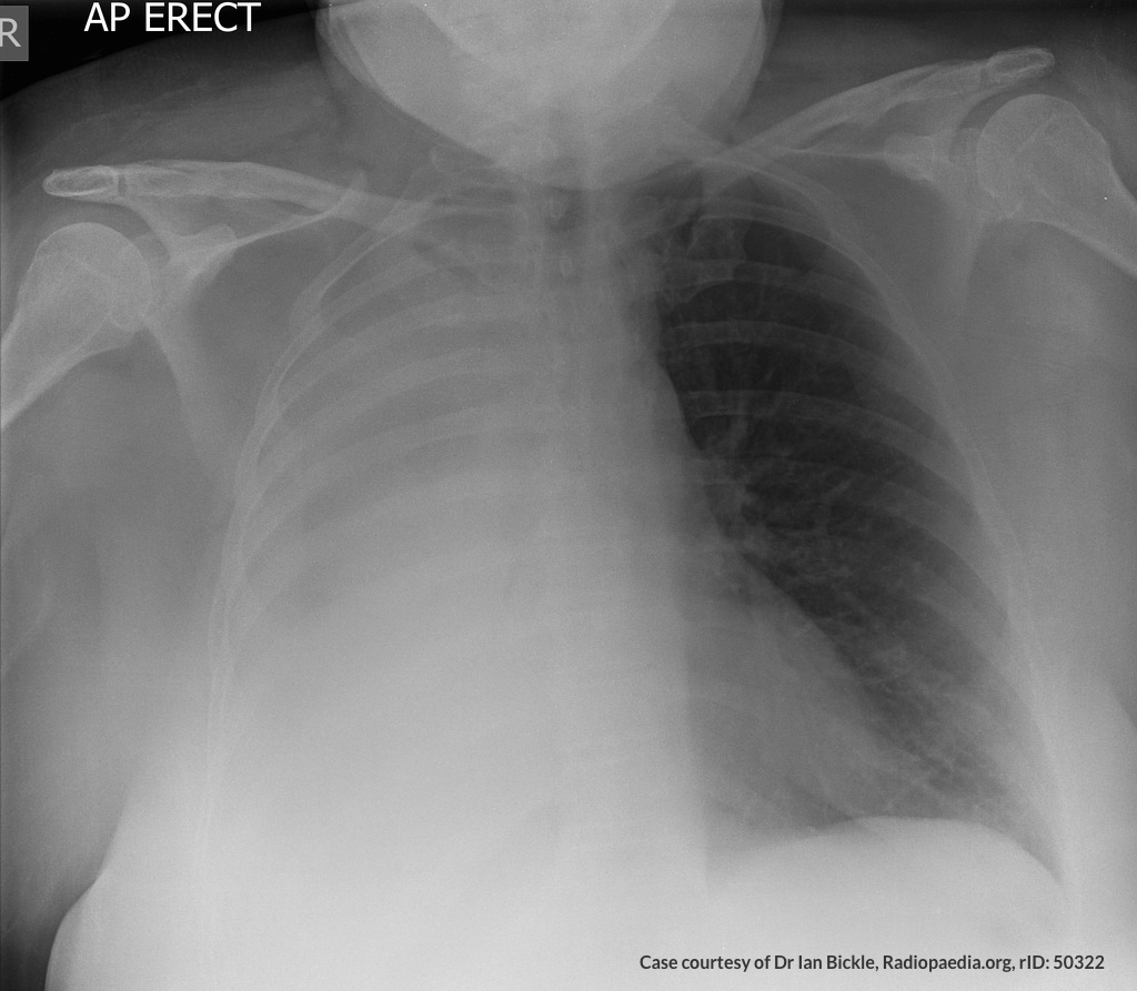
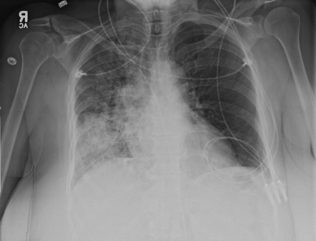
Vital signs are notable for tachycardia, tachypnea and hypoxia. Examination demonstrates absent breath sounds in the entire right lung field. A plain chest radiograph is obtained and shown below. The patient was placed on non-invasive positive pressure with minimal improvement and an emergent therapeutic thoracentesis was performed. Pleural fluid was exudative and a large volume was submitted for cytology.

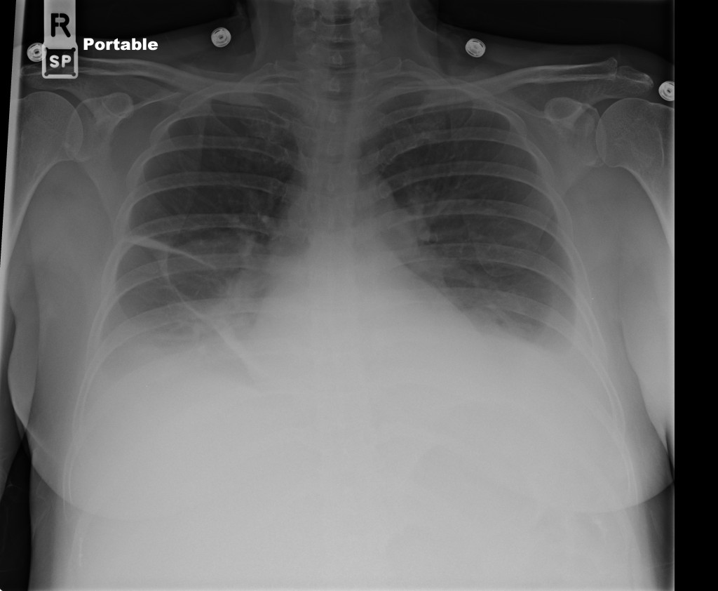
Whiteout right lung field Whiteout right lung field

An Algorithm for the Analysis of Pleural Fluid

An Algorithm for the Analysis of Pleural Fluid

References

  1. Light RW, Girard WM, Jenkinson SG, George RB. Parapneumonic effusions. Am J Med. 1980;69(4):507-512.
  2. Heffner JE, Brown LK, Barbieri CA. Diagnostic value of tests that discriminate between exudative and transudative pleural effusions. Primary Study Investigators. Chest. 1997;111(4):970-980. doi:10.1378/chest.111.4.970.
  3. Romero S, Martinez A, Hernandez L, et al. Light’s criteria revisited: consistency and comparison with new proposed alternative criteria for separating pleural transudates from exudates. Respiration. 2000;67(1):18-23. doi:10.1159/000029457.
  4. Light RW. Clinical practice. Pleural effusion. N Engl J Med. 2002;346(25):1971-1977. doi:10.1056/NEJMcp010731.
  5. Sahn SA, Huggins JT, San Jose E, Alvarez-Dobano JM, Valdes L. The Art of Pleural Fluid Analysis. Clinical Pulmonary Medicine. 2013;20(2):77-96. doi:10.1097/CPM.0b013e318285ba37.
  6. Light RW. The Light criteria: the beginning and why they are useful 40 years later. Clinics in Chest Medicine. 2013;34(1):21-26. doi:10.1016/j.ccm.2012.11.006.
  7. Aggarwal AN, Agarwal R, Sehgal IS, Dhooria S, Behera D. Meta-analysis of Indian studies evaluating adenosine deaminase for diagnosing tuberculous pleural effusion. Int J Tuberc Lung Dis. 2016;20(10):1386-1391. doi:10.5588/ijtld.16.0298.

Ultrasound in Dyspnea

Brief H&P:

A 68 year-old male with a history of hypertension, diabetes, hyperlipidemia, chronic obstructive pulmonary disease and congestive heart failure (CHF) with depressed ejection fraction presents via ambulance with a chief complaint of shortness of breath. EMS reports that the patient was tachypneic and saturating 80% on ambient air on their arrival. En route, he received nebulized albuterol, nitroglycerin and was started on non-invasive positive pressure ventilation (NI-PPV).

On arrival, he remains uncomfortable-appearing with a respiratory rate of 35 breaths/min and accessory muscle use. His heart rate is 136bpm, blood pressure is 118/85mmHg, and he is saturating 95% on an FiO2 of 100%. Attempts to obtain a history are limited due to difficulty comprehending his responses with the PPV mask on, and prompt desaturation with it off. Lung auscultation is similarly challenging due to ambient and transmitted sounds, although basilar crackles and diffuse expiratory wheezing are appreciated. Cardiovascular examination reveals a rapid and irregularly irregular rhythm. Assessment of jugular venous distension is limited due to the patient’s body habitus and the presence of mask straps around the patient’s neck. Lower extremities demonstrate 2+ pitting edema, symmetric bilaterally. Intravenous access is established and laboratory tests are sent. The ECG technician and portable chest x-ray are called.

The case presentation above demonstrates a common emergency department scenario: a critically-ill patient with undifferentiated dyspnea. Specifically, the scenario reveals a situation where the physical examination is either obfuscated by technical challenges or otherwise indeterminate. The patient is at risk for deterioration and targeted intervention is mandatory. If a COPD exacerbation is assumed, additional nebulized breathing treatments are indicated – a potentially costly jolt of beta agonists if the patient’s atrial fibrillation and rapid ventricular response are the consequence of decompensated systolic heart failure. Take the route of decompensated CHF and prompt afterload reduction with diuresis would be next – if incorrect, not only would the primary cause go untreated, but his tenuously-maintained blood pressure may suffer.

Algorithm for the Use of Ultrasound in the Evaluation of Dyspnea

Algorithm for the Use of Ultrasound in the Evaluation of Dyspnea

1. Lung Ultrasound

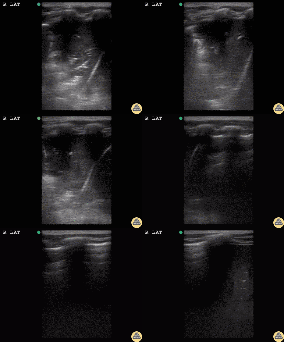
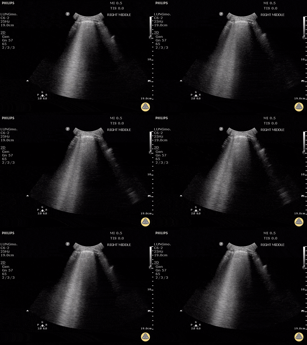
An approach incorporating point-of-care ultrasonography may be useful. First, a thoracic ultrasound is performed where certain causative etiologies might be identified immediately – for example absent lung sliding suggesting pneumothorax, or signs of generalized or subpleural consolidation.

The POCUS Atlas
The ultrasound images and videos used in this post come from The POCUS Atlas, a collaborative collection focusing on rare, exotic and perfectly captured ultrasound images.
The POCUS Atlas

Pneumothorax

Hepatization

Shred Sign

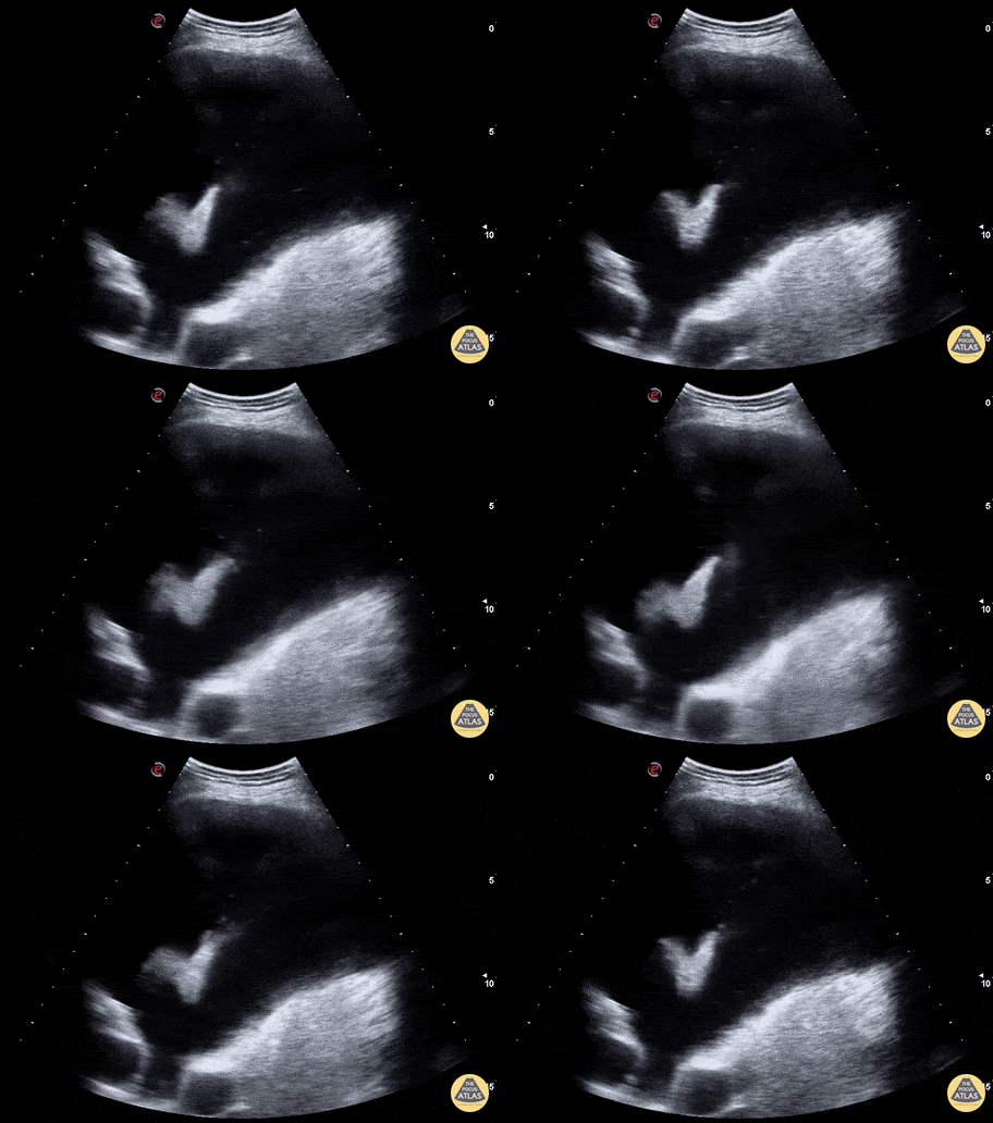
Pleural Effusion

2. Cardiac Ultrasound

Other findings on lung ultrasound may point to causes that are not primarily pulmonary. For example, if diffuse B-lines are encountered a focused cardiac ultrasound can be performed to grossly evaluate ejection fraction and estimate right atrial pressure.

B-Lines

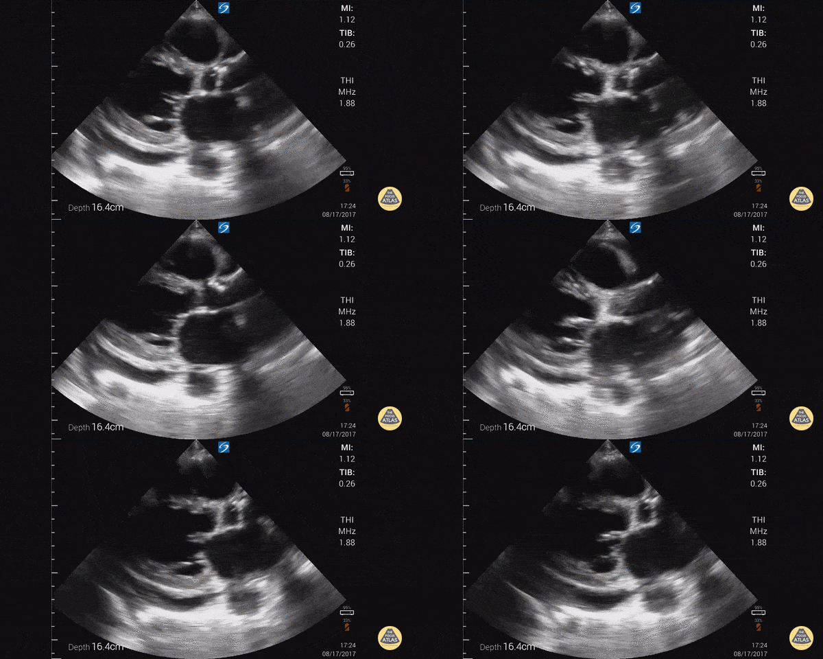
Depressed EF

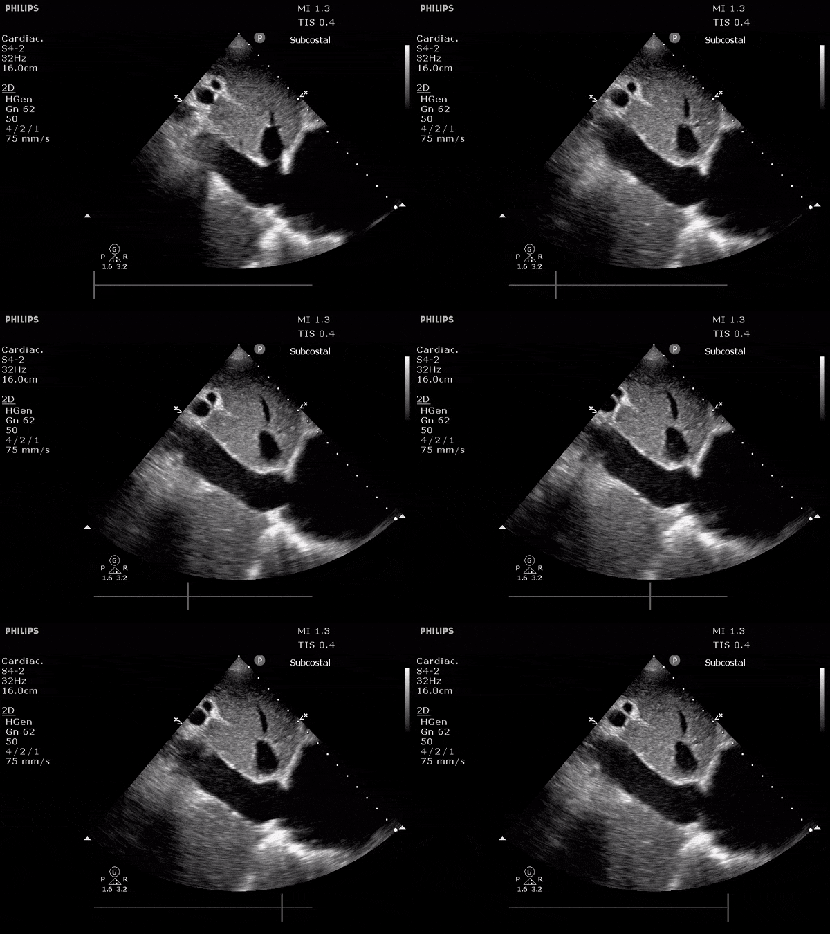
Dilated IVC

3. Venous Ultrasound

Finally, if the lung ultrasound is largely unremarkable (A-lines), a sequence of ultrasonographic findings including right ventricular dilation and the presence of a deep venous thrombosis would point to pulmonary embolism as the diagnosis.

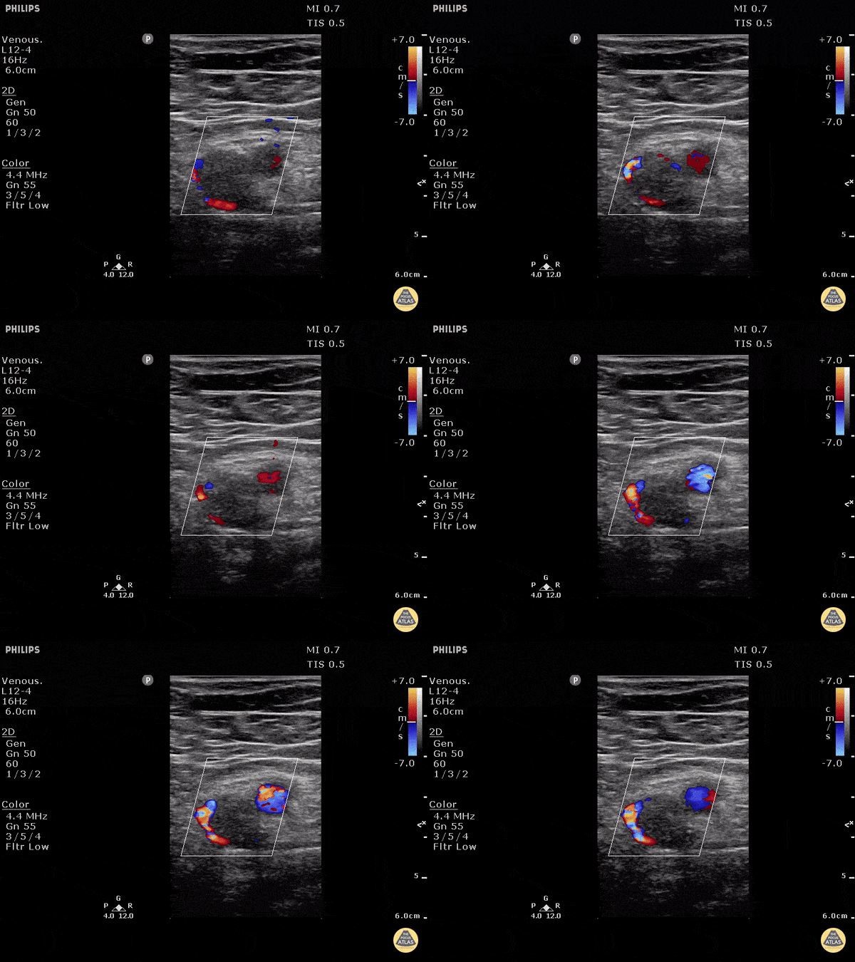
DVT

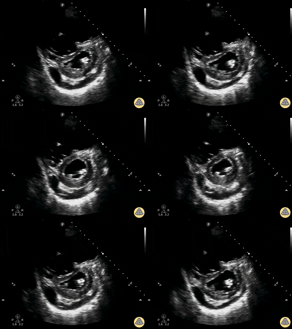
RV Dilation

All illustrations are available for free, licensed (along with all content on this site) under Creative Commons Attribution-ShareAlike 4.0 International Public License.

Downloads Page License

References

  1. Lichtenstein DA, Mezière GA, Lagoueyte J-F, Biderman P, Goldstein I, Gepner A. A-lines and B-lines: lung ultrasound as a bedside tool for predicting pulmonary artery occlusion pressure in the critically ill. Chest. 2009;136(4):1014-1020. doi:10.1378/chest.09-0001.
  2. Copetti R, Soldati G, Copetti P. Chest sonography: a useful tool to differentiate acute cardiogenic pulmonary edema from acute respiratory distress syndrome. Cardiovasc Ultrasound. 2008;6(1):16. doi:10.1186/1476-7120-6-16.
  3. Gallard E, Redonnet J-P, Bourcier J-E, et al. Diagnostic performance of cardiopulmonary ultrasound performed by the emergency physician in the management of acute dyspnea. Am J Emerg Med. 2015;33(3):352-358. doi:10.1016/j.ajem.2014.12.003.
  4. Lichtenstein DA. Lung ultrasound in the critically ill. Ann Intensive Care. 2014;4(1):1. doi:10.1186/2110-5820-4-1.
  5. Zanobetti M, Scorpiniti M, Gigli C, et al. Point-of-Care Ultrasonography for Evaluation of Acute Dyspnea in the ED. Chest. 2017;151(6):1295-1301. doi:10.1016/j.chest.2017.02.003.
  6. Lichtenstein DA, Mezière GA. Relevance of lung ultrasound in the diagnosis of acute respiratory failure: the BLUE protocol. Chest. 2008;134(1):117-125. doi:10.1378/chest.07-2800.
  7. Images from The POCUS Atlas
  8. Special thanks to Dr. Timothy Jang, Director Emergency Ultrasound Program, Director Emergency Ultrasound Fellowship, Associate Professor of Clinical Emergency Medicine, Department of Emergency Medicine at Harbor-UCLA

Dyspnea

Causes of Dyspnea

Causes of Dyspnea

Findings in Selected Causes of Dyspnea

Condition History Symptoms Findings Evaluation
Anaphylaxis Exposure to allergen Abrupt onset, facial swelling Stridor, wheezing, hives  
PE Immobilization, malignancy, prior DVT/PE, surgery, OCP Abrupt onset, pleuritic chest pain Tachycardia, hypoxia ECG (RV strain)
CT PA, D-dimer
LE US (DVT)
Pneumonia Exposure, tobacco use Fever, productive cough Focal rales CXR
CBC
Blood/respiratory cultures
Pneumothorax Trauma, thin male Abrupt onset, chest pain Decreased BS, subQ emphysema, JVD and tracheal deviation if tension CXR
US
Fluid overload Dietary indiscretion, medication non-adherence Orthopnea, PND JVD, S3/S4, peripheral edema CXR
US
ECG
BNP
COPD/Asthma Tobacco use, personal/family history Progressive Retractions, accessory muscle use, wheezing CXR
US (distinguish from fluid overload)
Malignancy Tobacco use, weight loss Hemoptysis   CXR
CT Chest

References

  1. Braithwaite, S., & Perina, D. (2013). Dyspnea. In Rosen’s Emergency Medicine – Concepts and Clinical Practice (8th ed., Vol. 1, pp. 206-213). Elsevier Health Sciences.

Pleural Effusion

HPI:

64F with a history of CAD, MI, CHF, CLL and rheumatoid arthritis who presented to the emergency department after transfer from a rehabilitation facility for respiratory distress. The patient reported several days of progressive shortness of breath with dyspnea on exertion. She also noted some associated orthopnea and lower extremity edema. The patient was recently hospitalized for similar symptoms and was diagnosed with CHF at the time. At the rehab facility, the patient became hypoxemic and hypertensive, reporting shortness of breath and chest pain prior to presentation.

Hospital Course:

The patient was initially managed with BiPAP and nitroglycerin continuous infusion, but was then stable on supplemental O2 via nasal cannula and was transitioned to long-acting nitrates and anti-hypertensives. The patient’s hypoxemic respiratory failure was initially attributed to acute exacerbation of left-ventricular heart failure, and the patient was managed with spot diuresis. However, there was no symptomatic improvement and the patient became hypernatremic so diuresis was held as alternative diagnoses were explored.

A transthoracic echocardiogram showed preserved LVEF (50-55%), but some diastolic dysfunction and elevated PAP/RAP. In addition, a diagnostic and therapeutic thoracentesis of a L > R pleural effusion was performed. Pleural fluid studies were suggestive of a transudative process, though with some abnormal characteristics (including lymphocyte predominance, as well as presence of signet cells).

Rheumatology and pulmonology services were consulted for input and recommendations for further evaluation were appreciated. Per rheumatology, the patient’s diagnosis of rheumatoid arthritis may not be consistent with her presentation or prior serologic studies. Her pleural fluid analysis was also not consistent with rheumatoid disease. According to pulmonary consult, the patient’s hypoxemia remains most consistent with left ventricular dysfunction though primary pulmonary processes cannot be excluded (and would warrant further evaluation with imaging and pulmonary function testing).

PMH:

  • CHF
  • CAD
  • CVA
  • Myocardial Infarction
  • HTN
  • Hypothyroidism
  • CLL
  • Anemia

PSH:

  • TAH-BSO

FH:

  • No family history of autoimmune disease.
  • Mother: DM

SHx:

  • Denies tobacco/EtOH/drug use
  • Lives at home, at SNF since discharge

Meds:

  • Furosemide 20mg p.o. daily
  • Gabapentin 300mg p.o. t.i.d.
  • Hydralazine 50mg p.o. t.i.d.
  • Hydrochlorothiazide 25mg p.o. b.i.d.
  • Hydroxychloroquine 200mg p.o. daily
  • Levothyroxine 25mcg p.o. daily
  • Minoxidil 2.5mg p.o. b.i.d.
  • Pantoprazole 40mg p.o. daily
  • Prednisone 15mg p.o. daily

Allergies:

  • Shellfish
  • Physical Exam:

    VS: T 37.2 HR 84 RR 15 BP 147/75 O2 97% 4LNC
    Gen: Elderly female, alert and oriented to self and place, responding appropriately to questions.
    HEENT: Mucous membranes moist, sclera anicteric, no cervical lymphadenopathy.
    CV: Regular rate and rhythm, normal S1/S2, no additional heart sounds. III/VI mid-systolic murmur heard best at LLSB with diastolic component, no radiation appreciated. Non-displaced PMI. JVP measured to 14cm.
    Lungs: Decreased breath sounds in left lung field to inferior 2/3 with crackles above, on right crackles to inferior 1/2 of lung fields posteriorly. Dullness to percussion of inferior left lung field posteriorly.
    Abdomen: Soft, non-tender, non-distended, no hepatosplenomegaly, no appreciable fluid wave.
    Ext: Bilateral lower extremities with 2+ pitting edema to knees, some hyperpigmentation to right lower extremity.

    Labs/Studies:

    • CBC: 11.2/10.2/32.7/179
    • BMP: 141/3.9/103/30/15/0.85/107
    • INR: 0.9
    • BNP: 1857
    • UA: WBC 4, RBC 29 , +Bacteria, UCr 90, UPr 14
    • Rheum: CCP <16, ANCA neg, RF 936
    • Pleural Fluid: LDH 98 (serum 237), Protein 2.8 (serum 6.0), Glucose 107
    • Cytology: Reactive mesothelial cells, histiocytes, lymphocytes, signet cells

    Imaging

    CXR
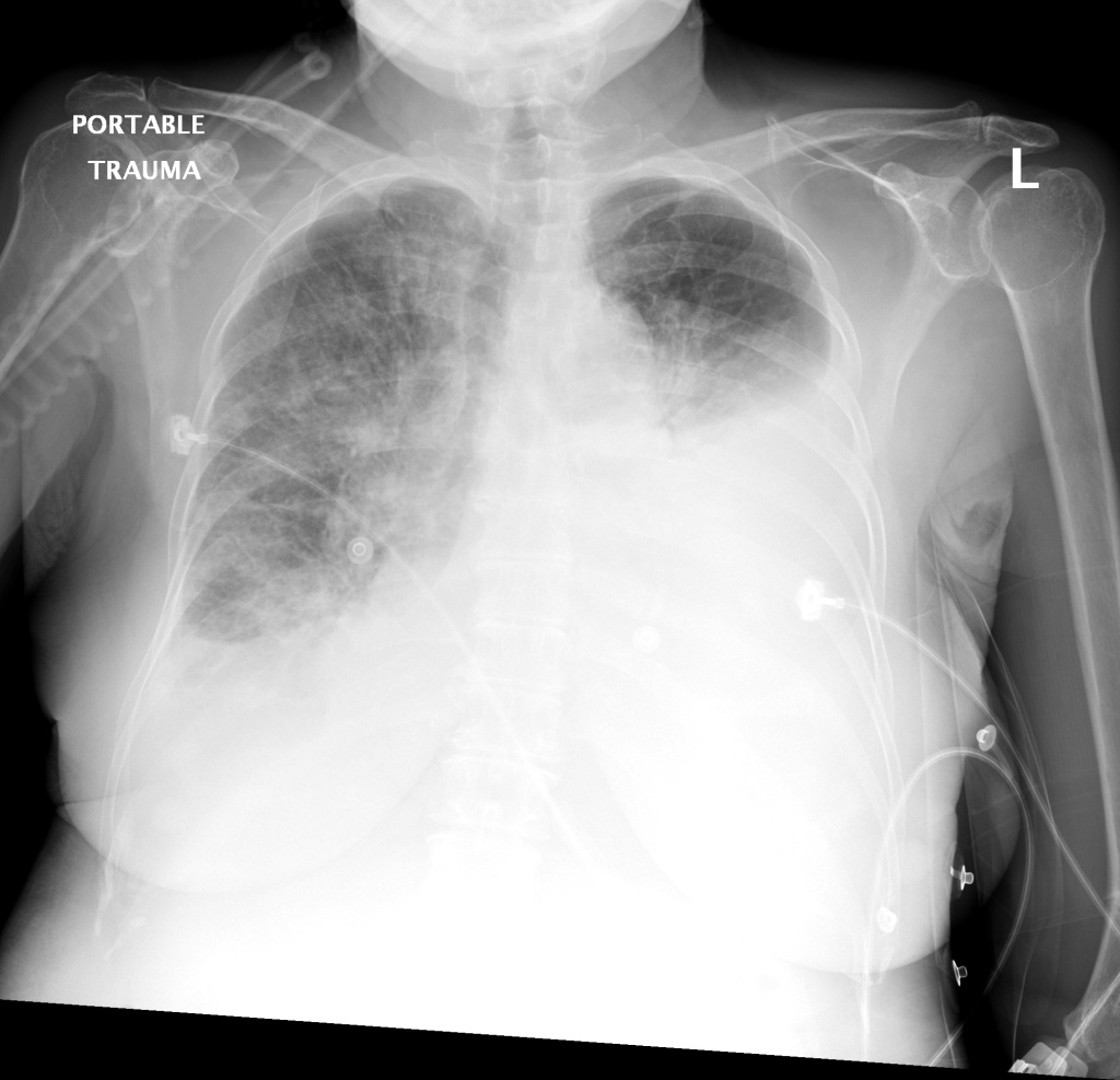
    CXR: Pleural Effusion

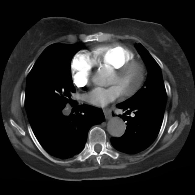
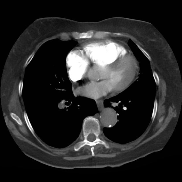
    There is a large left pleural effusion obscuring the lower half of the left hemi thorax. The cardiac silhouette is also obscured. There is pulmonary venous vascular congestion. There is also a right pleural effusion with fluid tracking into the minor fissure. Pulmonary interstitial edema is also noted.

    CT Chest (High-Resolution):

    • Bilateral, left greater than right, pleural effusions with adjacent atelectasis and collapse versus consolidation of the left lower lobe.
    • Prominent main pulmonary artery measuring 3.3 cm in diameter, which can be seen with pulmonary arterial hypertension.

    TTE:

    • LVEF is 50-55%.
    • Impaired left ventricular relaxation, which is associated with grade I/IV or mild diastolic dysfunction.
    • Moderate aortic stenosis with mild regurgitation (AVA 1.4 cm3, mean gradient 14mmHg, peak velocity 2.4 m/s).
    • Severe pulmonary hypertension (est PASP 52-62mmHg).
    • The inferior vena cava appeared dilated and decreased <50% with respiration (RAP 10-20 mmHg).
    • Minimal pericardial effusion without echocardiographic evidence of tamponade.

    Assessment/Plan:

    64F with history of CAD (prior MI), CHF, hypertension, CLL, hypothyroidism presented from a SNF with progressive shortness of breath, orthopnea and LE swelling, found to have bilateral (L>R) pleural effusion now s/p thoracentesis with transudative fluid.

    #Acute hypoxic respiratory failure: Large pleural effusions, s/p thoracentesis with pleural fluid suggestive of transudative process. Most likely secondary to left ventricular diastolic dysfunction. Improved after thoracentesis and diuresis. High-resolution CT chest performed without evidence of autoimmune-related pulmonary fibrosis or ILD (though persistent pleural effusions, pulmonary vascular congestion).

    #Pleural fluid signet cells: Identified on cytology, potentially related to history of untreated CLL or alternative primary malignancy.

    #Left ventricular diastolic dysfunction, decompensated: Associated with pleural effusions and hypoxemic respiratory failure. Management with diuresis.

    #Pulmonary Hypertension: Severe, noted on transthoracic echocardiography, may be secondary to hypoxemia associated with pleural effusions, consider repeat imaging once euvolemic or right-heart catheterization.

    #Microscopic Hematuria: No evidence of infection, no symptoms suggestive of nephrolithiasis. No casts identified or significant proteinuria. Plan for renal ultrasound.

    #Rheumatoid Arthritis: History of rheumatoid arthritis, on prednisone and hydroxychloroquine. Imaging without evidence of inflammatory arthropathy, RF elevated but CCP negative. Per rheumatology, the patient’s symptoms are not consistent with RA, continuing home medications while evaluation is ongoing. Pleural effusions unlikely associated with RA as transudative, and without monocyte predominance or low glucose.

    Case 2: Malignant Pleural Effusion

    malig_01
    malig_02
    malig_03
    malig_04
    malig_05
    malig_06
    malig_07
    malig_08
    malig_09
    malig_10
    malig_11
    malig_12
    malig_13
    malig_14
    Within the lungs there are ground-glass opacities bilaterally, and a left pleural effusion with adjacent consolidation vs compressive atelectasis.
    • Protein: 2.6 (serum: 4.9)
    • LDH: 1275 (serum: 219)
    • Cytology: Malignant cells

    Case 3: Traumatic Thoracentesis

    trauma_01
    trauma_02
    trauma_03
    trauma_04
    trauma_05
    trauma_06
    trauma_07
    trauma_08
    trauma_09
    trauma_10
    trauma_11
    trauma_12
    trauma_13
    trauma_14
    trauma_15
    trauma_16
    trauma_17
    trauma_18
    trauma_19
    Moderate right pleural effusion, some fluid in non-dependent portions suggestive of loculation. Diffuse nodules and opacification in right lung with compressive atelectasis. Left pleural effusion with high density material at the posterior costophrenic angle. Left chest tube.
    • Protein: 2.7 (serum 6.4)
    • LDH: 344 (serum 236)
    • Cell count: 100,000 RBC

    Case 4: Pneumonia

    pna_01
    pna_02
    pna_03
    pna_04
    pna_05
    pna_06
    pna_07
    pna_08
    pna_09
    pna_10
    pna_11
    pna_12
    pna_13
    pna_14
    pna_15
    Loculated right pleural effusion with foci of atelectasis and consolidative changes concerning for pneumonia. Minimal left-sided pleural effusion with consolidative changes. Enlarged mediastinal lymph nodes, possibly reactive.
    • Protein: 4.3 (serum 6.7)
    • LDH: 377 (serum 108)
    • pH: 7.46
    • Glucose: 153
    • Neutrophils: 84%

    Etiology of Pleural Effusions: 1

    Etiology Frequency (%)
    CHF 35
    Pneumonia 22
    Malignancy 15
    Pulmonary Embolism 11

    Clinical Features in the Diagnosis of Pleural Effusions and Identifying Etiology: 1,2

    Pleural effusions can be easily identified on chest radiography, physical examination findings include dullness to percussion, decreased tactile fremitus and decreased (or absent) breath sounds.

    • Hemoptysis: Malignancy, PE, TB
    • Weight Loss: Malignancy, TB
    • Ascites: Cirrhosis, ovarian cancer
    • Unilateral Leg Swelling: PE
    • Bilateral Leg Swelling: CHF, cirrhosis, nephrotic syndrome
    • Jugular Venous Distension: CHF

    Differential Diagnosis of Pleural Effusions: 1,2,3,4

    Differential Diagnosis of Pleural Effusions

    References:

    1. Light, R. W. (2002). Clinical practice. Pleural effusion. The New England journal of medicine, 346(25), 1971–1977. doi:10.1056/NEJMcp010731
    2. McGrath, E. E., & Anderson, P. B. (2011). Diagnosis of pleural effusion: a systematic approach. American journal of critical care : an official publication, American Association of Critical-Care Nurses, 20(2), 119–27– quiz 128. doi:10.4037/ajcc2011685
    3. Thomsen, T. W., DeLaPena, J., & Setnik, G. S. (2006). Videos in clinical medicine. Thoracentesis. The New England journal of medicine (Vol. 355, p. e16). doi:10.1056/NEJMvcm053812
    4. Wilcox, M. E., Chong, C. A. K. Y., Stanbrook, M. B., Tricco, A. C., Wong, C., & Straus, S. E. (2014). Does this patient have an exudative pleural effusion? The Rational Clinical Examination systematic review. JAMA : the journal of the American Medical Association, 311(23), 2422–2431. doi:10.1001/jama.2014.5552
    5. WikEM: Pleural effusion
    Pulmonary Embolism in Hospitalized Patients

    Brief Progress Note

    Notified by nursing of abnormal vital signs, SpO2 91%. Briefly, this patient is a 52 year-old G1P1 with no prior medical history who is post-operative day three status post total abdominal hysterectomy, bilateral salpingoophorectomy as well as tumor debulking and staging for suspected primary ovarian adenocarcinoma based on peritoneal fluid cyctology.

    On evaluation, the patient denied shortness of breath, chest pain, pleuritic chest pain, cough/hemoptysis, or calf/thigh pain. She states that she had been ambulating around the ward prior to having her vital signs assessed.

    Physical Exam:

    VS: T 98.9 HR 94 RR 18 BP 117/72 O2 91% RA
    Gen: No acute distress, speaking in full sentences.
    HEENT: No jugular venous distension.
    CV: RRR, normal S1/S2, no prominent P2, no additional heart sounds.
    Lungs: Decreased breath sounds at inferior 1/3 posterior lung fields bilaterally, faint crackles above, no wheezing. Dullness to percussion in inferior lung fields.
    Ext: Warm, well-perfused. Sequential compression devices on bilateral lower extremities, removed revealing trace pitting edema symmetric bilaterally, no tenderness to palpation of posterior leg, no pain with passive dorsiflexion.

    Medications:

    • Lovenox 40mg s.q. daily
    • Norco 5-325mg p.o. q.4.h. p.r.n. pain
    • Morphine 2mg i.v. q.3.h. p.r.n. breakthrough pain
    • Colace 100mg p.o. b.i.d. p.r.n. constipation
    • Zofran 4mg i.v. q.6.h. p.r.n. nausea/vomiting

    Imaging:

    CXR

    Bilateral pleural effusions with pleural fluid tracking along the minor fissure.

    CT Chest

    IM-0001-0001
    IM-0001-0003
    IM-0001-0005
    IM-0001-0007
    IM-0001-0009
    IM-0001-0011
    IM-0001-0013
    IM-0001-0015
    IM-0001-0017
    IM-0001-0019
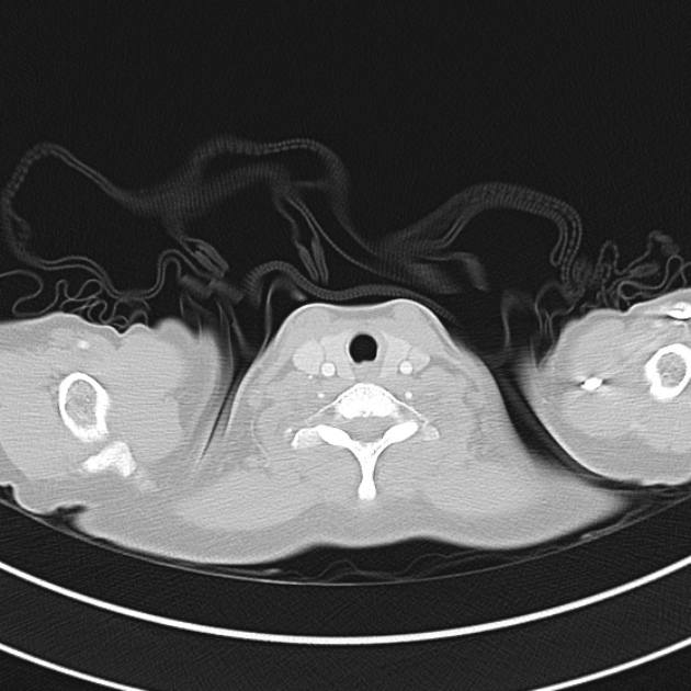
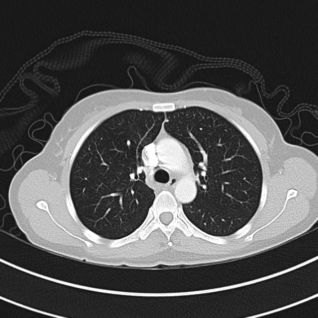
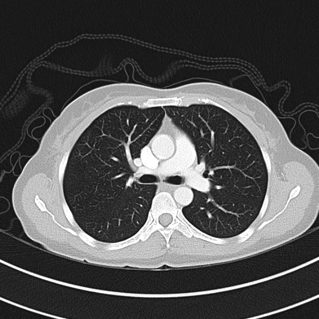
    No evidence of pulmonary embolism, study performed 5 days prior to onset of symptoms.

    CT Abdomen/Pelvis

    IM-0001-0051
    IM-0001-0055
    IM-0001-0059
    IM-0001-0063
    IM-0001-0067
    IM-0001-0071
    IM-0001-0075
    IM-0001-0079
    IM-0001-0083
    IM-0001-0087
    IM-0001-0091
    IM-0001-0095
    Significant peritoneal carcinomatosis, ascites delineates peritoneal from retroperitoneal spaces.

    Assessment and Plan:

    52yo G1P1 with likely primary ovarian adenocarcinoma with extensive peritoneal involvement, complicated by malignant ascites and pleural effusions with hypoxemia. The primary concern in this post-operative patient with a history of malignancy is venous thromboembolism, particularly pulmonary embolism. Aside from hypoxemia, this patient had no symptoms suggestive of pulmonary embolism (denied dyspnea, chest pain, cough, or lower extremity pain). Her examination had some signs infrequently associated with pulmonary embolism which were otherwise adequately explained by known bilateral pleural effusions (including decreased breath sounds, rales, and tachypnea), she was not tachycardic, had no evidence of jugular venous distension, nor a prominent P2. In addition, she was receiving appropriate VTE prophylaxis (both pharmacological and mechanical). The application of the Modified Wells clinical decision rule suggests low likelihood of pulmonary embolism (with 2.5 points assigned for recent surgery and history of malignancy). The patient had a recent CT chest without evidence of pulmonary embolism, and given that an elevated D-dimer was virtually assured which would have necessitated repeat imaging (and the associated risks of radiation exposure and contrast injury), the episode was ascribed to ventilation/perfusion mismatch secondary to large pleural effusions. Hypoxemia resolved after several minutes at rest and no further testing was performed.
    Signs and Symptoms of Pulmonary Embolism1

    Signs Symptoms
    Tachypnea Dyspnea (rest/exertion)
    Tachycardia Pleurtic chest pain
    Rales Cough
    Decreased breath sounds Orthopnea
    Prominent P2 Calf/thigh pain or swelling
    JVD Wheezing

    Modified Wells Criteria1,2

    Feature Points
    Clinical symptoms of DVT (leg swelling, tenderness to palpation) 3.0
    Pulmonary embolism most likely diagnosis 3.0
    Tachycardia (HR >100) 1.5
    Immobilization >3d, surgery in prior 4 weeks 1.5
    Prior DVT/PE 1.5
    Hemoptysis 1.0
    Malignancy 1.0

    >4.0 Likely
    ≤4.0 Unlikely

    Algorithm for Evaluation of Suspected Pulmonary Embolism2

    Utility of D-Dimer:2
    Of limited utility in patients with high suspicion for pulmonary embolism
    Decreased specificity: malignancy, hospitalized patients, pregnancy, elderly
    Efficacy of DVT prophylaxis:3
    LMWH prevents approximately ½ of VTE events (including PE, symptomatic and asymptomatic DVT)

    References:

    • Kruip, M. J. H. A., Söhne, M., Nijkeuter, M., Kwakkel-Van Erp, H. M., Tick, L. W., Halkes, S. J. M., Prins, M. H., et al. (2006). A simple diagnostic strategy in hospitalized patients with clinically suspected pulmonary embolism. Journal of internal medicine, 260(5), 459–466. doi:10.1111/j.1365-2796.2006.01709.x
    • Tapson, V. F. (2008). Acute pulmonary embolism. The New England journal of medicine, 358(10), 1037–1052. doi:10.1056/NEJMra072753
    • Själander, A., Jansson, J.-H., Bergqvist, D., Eriksson, H., Carlberg, B., & Svensson, P. (2008). Efficacy and safety of anticoagulant prophylaxis to prevent venous thromboembolism in acutely ill medical inpatients: a meta-analysis. Journal of internal medicine, 263(1), 52–60. doi:10.1111/j.1365-2796.2007.01878.x

    Acute Respiratory Distress Syndrome

    HPI:

    25M with a history of mild asthma transferred from an outside hospital after 10 days in the intensive care unit for continued management of ARDS.

    The patient was well until one week prior to admission when he developed intermittent subjective fevers and general malaise associated with a non-productive cough and nausea/vomiting. He presented to the emergency department of an outside hospital with difficulty breathing and was noted to have respiratory distress and was subsequently admitted. Initial CT at the outside hospital revealed pneumomediastinum but no evidence of pulmonary embolism. Results from the outside hospital reveal a wide array of bacterial/fungal cultures and viral serologies including bronchoscopy but no obvious infectious source. The patient was treated with broad spectrum antibiotics for several days but his condition worsened requiring intubation, mechanical ventilation and transfer to the ICU. Further imaging was suggestive of ARDS, and the patient was transferred for additional management.
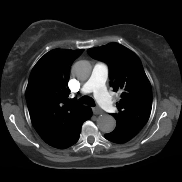
    The patient was well until one week prior to admission when he reported development of malaise and fatigue. On the day of hospitalization, the patient presented to primary care doctor with complaint of cough and shortness of breath and was found to be in respiratory distress and was admitted. The patient received IV antibiotics (cefepime, vancomycin) and was intubated when respiratory distress worsened. Found to have evidence of ARDS on CXR.
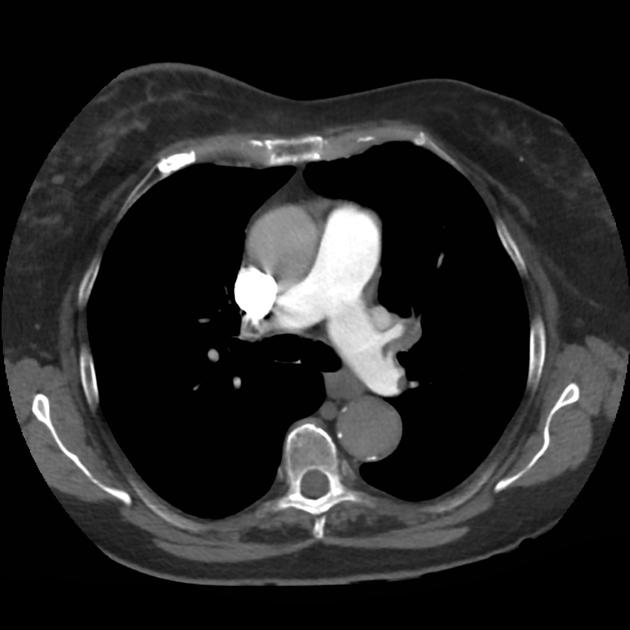
    PMH:

    • Mild asthma, not requiring medication
    • MRSA skin abscess

    PSH:

    • None

    FH:

    • Non-contributory.

    SHx:

    • Lives with family and works at a local supermarket. Rare alcohol use and no prior tobacco or drug use.
    • No recent travel or sick contacts.

    Meds:

    • ciprofloxacin 400mg i.v. q12h
    • linezolid 600mg i.v. q12h
    • meropenem 1g i.v. q8h
    • heparin 5000units s.q. q8h
    • cisatracurium 1.48mcg/kg/min
    • fentanyl 125mcg/hr
    • midazolam 10mg/hr
    • propofol 20mcg/kg/min

    Allergies:

    NKDA

    Physical Exam:

    VS: T 97.8 HR 120 RR 35 BP 123/68 O2 96%
    Vent: PRVC, VT 320, RR 35, PEEP 6, FiO2 95%
    Gen: Young male, thin-appearing, intubated and sedated and not responding to verbal commands
    HEENT: PERRL, unable to assess EOM, ET tube in place
    CV: RRR, normal S1/S2, no murmurs
    Lungs: Coarse breath sounds bilaterally
    Abd: Normoactive bowel sounds, soft, non-distended, no hepatosplenomegaly
    Ext: No clubbing, cyanosis, edema
    Neuro: Unable to assess

    Labs/Studies:

    • CBC: 25.06/7.0/21.3/426
    • BMP: 132/3.3/88/32/18/1.1/103
    • ABG: 7.30/97/76/18
    • Blood/sputum/urine cultures: Negative
    • Aspergillus, crypto, cocci: Negative
    • EBV, HIV, influenza, RSV: Negative

    Imaging


    CT Chest: Evidence of pneumomediastinum and pneumopericardium. Bilateral pulmonary infiltrates, but no pulmonary embolism.

    Assessment/Plan:

    25M with ARDS transferred from outside hospital for further management.
    # ARDS: Severe (P/F ratio <100). Etiology unclear, thorough infectious workup without obvious source. Consider autoimmune or allergic cause.

    • Ventilator: PRVC lung-protective ventilation
    • Consider NMB for dyssynchrony despite sedation
    • Monitor strict I/O, maintain net negative fluid balance.

    # Sepsis: Leukocytosis, tachycardia. Continue broad-spectrum antibiotics and monitor cultures.
    # Acidosis: Largely respiratory, place dialysis catheter if acute need arises.
    # Pneumomediastinum: Possible Boerhaave syndrome given reports of nausea/vomiting.

    Pathophysiology of Acute Respiratory Distress Syndrome (ARDS):1

    ARDS represents a stereotyped response to multiple insults. It is characterized by damaged capillary endothelium and alveolar epithelium resulting in increased permeability and the accumulation of fluid in the alveolar space. This causes diffuse alveolar damage and triggers the release of various cytokines (TNF, IL-1, IL-6) which recruit and activate neutrophils causing oxidative cell damage.

    Definition of ARDS (Berlin):2,3

    Timing Acute in onset (<1 week)
    Chest imaging Bilateral opacities
    Origin of pulmonary edema Not explained by heart failure or fluid overload (assessed with echocardiography)
    Oxygenation (PaO2/FiO2)
    1. Mild: 200-300
    2. Moderate: 100-200
    3. Severe: <100

    Causes of ARDS:2,4

    Causes of ARDS


    An Introduction to Mechanical Ventilation:5,6,7

    This is a simplification of the general principles underlying the most common ventilator modes. For more detail, see the articles cited in the references.
    An Introduction to Mechanical Ventilation

    Breath Sequences:

    Continuous Mandatory Ventilation (CMV)

    Continuous Mandatory Ventilation (CMV)

    All breaths controlled by ventilator, no triggered breaths.

    Assist-Control Ventilation (AC)

    Assist-Control Ventilation (AC)

    Every patient-triggered breath is fully supported, a backup rate is set. In the absence of patient-triggered breaths, AC acts like CMV.

    Synchronized Intermittent Mandatory Ventilation (SIMV)

    Synchronized Intermittent Mandatory Ventilation (SIMV)

    Preset minimum mandatory breaths are “synchronized” to patient’s efforts. The patient is allowed to breathe spontaneously between supported breaths.

    Pressure Support (PS)

    Pressure Support (PS)

    All breaths are triggered by the patient and each is supported by preset pressure.

    Continuous Positive Airway Pressure (CPAP)

    Continuous Positive Airway Pressure (CPAP)

    Spontaneous breathing at elevated baseline pressure.

    Control Variables:

    Volume Control (VC)
    Volume Control (VC)

    Volume Control (VC)

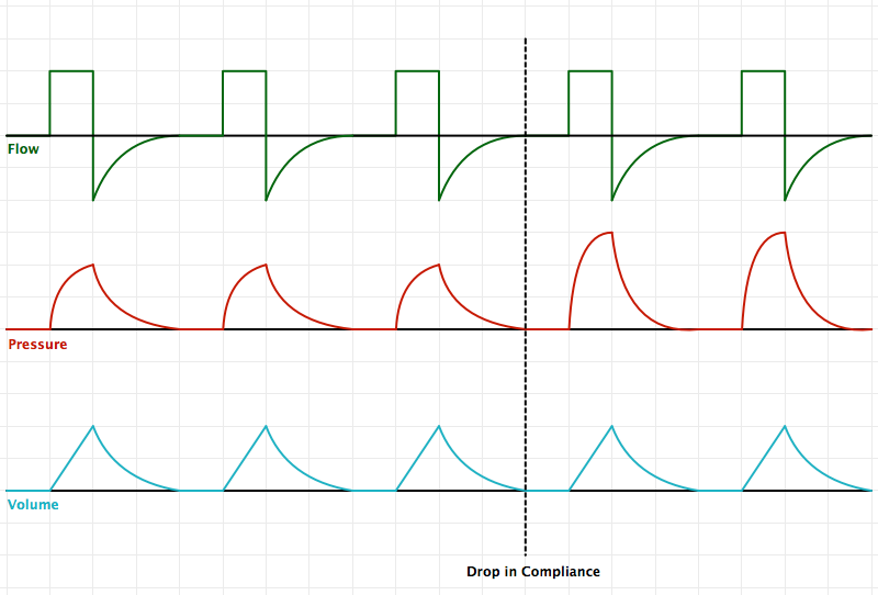
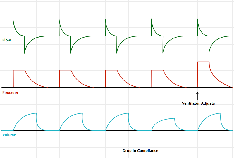
    Volume is set, pressure is variable. With a drop in compliance, the preset minimum volume is maintained with an increase in pressure.

    Pressure Control (PC)
    Pressure Control (PC)

    Pressure Control (PC)

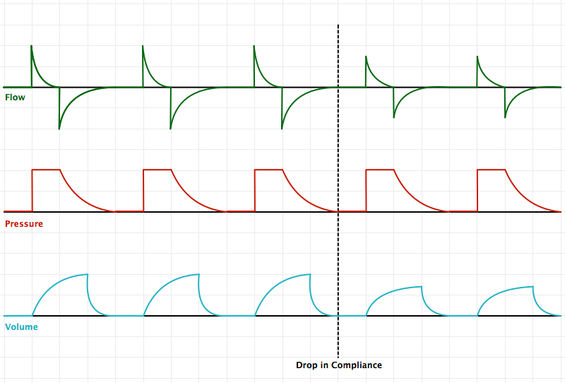
    Pressure is set, volume is variable. With a drop in compliance, a smaller volume is delivered to maintain pressures at the preset limit.

    Pressure-Regulated Volume Control (PRVC)
    Pressure-Regulated Volume Control (PRVC)

    Pressure-Regulated Volume Control (PRVC)

    Pressure is targeted with a set minimum volume. The ventilator makes breath-to-breath adjustments of pressure to maintain minimum volumes. Breath mechanics are therefore comparable to pressure-control as a defined pressure is delivered based on prior breath’s respiratory mechanics (note pressure and flow tracings for PRVC/PC vs. VC)

    References:

    1. Pierrakos C, Karanikolas M, Scolletta S, Karamouzos V, Velissaris D. Acute respiratory distress syndrome: pathophysiology and therapeutic options. J Clin Med Res. 2012;4(1):7–16. doi:10.4021/jocmr761w.
    2. Fanelli V, Vlachou A, Ghannadian S, Simonetti U, Slutsky AS, Zhang H. Acute respiratory distress syndrome: new definition, current and future therapeutic options. J Thorac Dis. 2013;5(3):326–334. doi:10.3978/j.issn.2072-1439.2013.04.05.
    3. ARDS Definition Task Force, Ranieri VM, Rubenfeld GD, et al. Acute respiratory distress syndrome: the Berlin Definition. In: Vol 307. 2012:2526–2533. doi:10.1001/jama.2012.5669.
    4. Ware LB, Matthay MA. The acute respiratory distress syndrome. N. Engl. J. Med. 2000;342(18):1334–1349. doi:10.1056/NEJM200005043421806.
    5. Deng, J. (10/20/13). Principles of Mechanical Ventilation. Medical Intensive Care Unit Lecture. Los Angeles, CA.
    6. Singer BD, Corbridge TC. Basic invasive mechanical ventilation. South. Med. J. 2009;102(12):1238–1245. doi:10.1097/SMJ.0b013e3181bfac4f.
    7. Hamed HMF, Ibrahim HG, Khater YH, Aziz ES. Ventilation and ventilators in the ICU: What every intensivist must know. Current Anaesthesia & Critical Care. 2006;17(1-2):77–83. doi:10.1016/j.cacc.2006.07.008.

    Hemoptysis

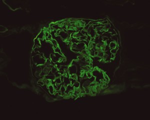
    Source: Mulpuru, S., Touchie, C., Karpinski, J., & Humphrey-Murto, S. (2010). Coexistent Wegener“s granulomatosis and Goodpasture”s disease. The Journal of rheumatology, 37(8), 1786–1787. doi:10.3899/jrheum.091404

    Linear IgG deposits consistent with anti-GBM disease.

    CC:

    “bad cough”

    HPI:

    61yo African American female w/hx of HTN presenting with 1mo of persistent cough productive of green-yellow sputum, noticed streaks of blood for the past 5 days. She came to the ED today because she has been feeling increasingly fatigued. She reports subjective fevers at the onset of symptoms which has resolved. She denies shortness of breath, chest pain, chills, night sweats. She sought medical care for this problem 2wk ago and was treated with amoxicillin and a cough suppressant. She recalls a coworker was ill one month ago. She is US-born, had a negative PPD in the past and has no known exposures to tuberculosis.

    Of note, the patient reports her urine had a foamy appearance and has been darker in color beginning 3 weeks ago, but this had resolved. She denied dysuria, or frank hematuria.

    PMH:

    • HTN
    • Asthma – last required medications >30yrs ago

     PSH:

    • None

    FH:

    • Non-contributory

     SHx:

    • No t/e/d
    • Works as librarian

    Meds:

    • benazepril
    • amlodipine
    • amoxicillin
    • promethazine

    Allergies:

    • NKDA

    Physical Exam:

    VS: T 99.4 HR 97 BP 132/60 RR 20 O2 92%
    Gen: Well-appearing, pleasant, speaking in complete sentences
    HEENT: PERRL, MMM no lesions, no cervical lymphadenopathy
    CV: RRR, normal S1/S2, no murmur appreciated
    Lungs: Crackles in posterior: right middle/inferior and left inferior fields, no wheezing, no dullness to percussion
    Abd: +BS, soft, non-tender, no CVAT
    Ext: Warm, well-perfused, 2+ peripheral pulses, 1+ pitting edema to knee
    Skin: No lesions on exposed skin
    Neuro: AAO

    Labs:

    • CBC: 12.3/6.7/19.8/52.3 (S: 94, B: 1, L: 4, M: 1, MCV: 92.3); baseline Hb/Hct (1/11/2012) 13.4
    • BMP: 136/3.6/101/25/46/3.43/126; baseline creatinine (1/11/2012) 1.18
    • UA: brown, trace LE, – nitrites, 2+ protein, 81 RBC

    Imaging:

    CXR PA

    • Right mid-lung zone consolidation is present, suggests pneumonia if acute.
    • Mild asymmetric right parenchymal increased density is seen diffusely as well.

    Assessment/Plan:

    65AAF w/hx HTN presents with persistent productive cough, recently with hemoptysis.

    # Cough: Symptoms and physical findings of abnormal breath sounds (crackles, though no strict consolidation) concerning for community-acquired pneumonia. Addition of hemoptysis raises concern for TB, particularly when taking into consideration the duration of cough and presence of constitutional symptoms. CBC shows leukocytosis with left shift, CXR with right mid/lower lob infiltrates consistent with pneumonia. Recommend admission and isolation to rule out TB, start empiric therapy for community acquired pneumonia with ceftriaxone, azithromycin. Obtain induced sputum samples for culture, AFB smear and culture.

    # Abnormal urine: Patient describes changes in urine suggestive of proteinuria and hematuria. Acuity of onset and apparent spontaneous resolution suggests a chronic kidney injury 2/2 hypertension is unlikely. Absence of dysuria, or tenderness (suprapubic, costovertebral) suggests complicated UTI unlikely. Urinalysis notable for 2+ protein and significant RBC’s, possible nephritic syndrome. In the setting of hemoptysis, this raises concern for anti-GBM disease vs. vasculitis.

    # Anemia: Normocytic anemia. No evidence of acute, life-threatening hemorrhage as patient is currently hemodynamically stable. Possible sites of blood loss include alveoli, glomeruli. Given that patient sought care today for worsening fatigue, will monitor hemoglobin closely and consider transfusion. Obtain iron studies.

    # HTN: BP stable, hold home medications.

    Interval History:

    The patient was admitted for management of community-acquired pneumonia and isolation to rule out TB. Empiric therapy with CTX + azithromycin was continued. On HOD1, the patient was transfused two units of PRBC’s. On HOD2, the patient underwent CT chest/abdomen/pelvis due to worsening respiratory status despite antimicrobial therapy. On HOD3, the patient went into atrial fibrillation with RVR which was converted to sinus rhythm with metoprolol 5mg IV x3. On HOD5, nephrology consult recommended starting steroid therapy, plasmapheresis and obtaining a renal biopsy, however the biopsy was delayed due to worsening respiratory status.

    Interval Labs:

    • Iron studies: Fe 8, TIBC 203, Ferritin 468, haptoglobin 333, retics 2.7
    • Inflammatory markers: ESR 120, CRP 34
    • Micro: BCx NGTD, RCx moderate Candida, sputum AFB smear negative x3
    • LFT: AST 34, ALT 29, ALP 52, protein 6.3, albumin 2.4, T.bil 0.8, D.bil 0.2
    • Quant-gold: negative
    • Anti-GBM 1.2 (nl <1.0)
    • p-ANCA: positive 1:640, [ELISA pending]
    • ANA: positive 1:320, speckled
    • HIV: negative

    Interval Imaging:

    CT Chest

    • Diffuse right lung, tree and bud opacities, ground-glass opacities and areas of confluence with scattered air bronchograms. Less severe similar pattern in the left lung as well particular at the base.
    • Right paratracheal, subcarinal and perihilar LAD.
    • Findings concerning for primary TB in the right clinical setting. DDx nonspecific bacterial PNA and fungal PNA.

    CT Abdomen/Pelvis

    • Mild nonspecific R > L perinephric stranding.

    Interval Assessment/Plan:

    # Acute respiratory failure: Unlikely simple CA-PNA given worsening status while on appropriate antibiotic therapy. Active tuberculosis possible given history of chronic productive cough with hemoptysis, constitutional symptoms and imaging findings. IGRA’s of limited utility in diagnosis of active disease, further, while three negative sputum AFB smears decreases the likelihood of TB, additional testing with NAAT and culture is required. Another possibility is a vasculitic process given concomitant hematuria and acute renal failure, with respiratory symptoms now 2/2 alveolar hemorrhage. This was evaluated with ANCA assay which was positive for p-ANCA with high titer. This is often suggestive of primary vasculitis (in this case likely microscopic polyangiitis vs. Churg-Strauss), however ELISA for target antigen is of particular importance as p-ANCA with specificity for antigens other than MPO can be associated with another condition on the differential: Goodpasture’s syndrome. This patient was found to have elevated anti-GBM antibodies which are highly suggestive of Goodpasture’s syndrome, and can be associated with ANCA-positivity (often suggesting a poorer prognosis with decreased likelihood of recovery of renal function).1

    # Acute kidney injury: The patient had significant elevation of serum creatinine compared to last-recorded baseline. She also described darkening and foamy appearance of urine 3 weeks prior to admission, suggestive of proteinuria/hematuria of relatively acute onset. This was supported by urinalysis findings of protein and RBC’s (with casts). Given presence of anti-GBM antibodies, high specificity of such findings, and correlation with glomerulonephritis with evidence of pulmonary alveolar hemorrhage, this appears to be the most likely cause at this time. Definitive diagnosis with renal biopsy to be obtained following stabilization of respiratory status. Patient will be started on plasmapheresis and immunosuppressive therapy (corticosteroids, cyclophosphamide).

    # Normocytic Anemia: Likely combination of acute blood loss (2/2 hematuria, pulmonary alveolar hemorrhage) and chronic disease. Normocytic anemia with some reticulocytosis suggestive of acute blood loss, however iron studies with low Fe, TIBC and elevated ferritin suggest chronic disease as an associated factor.

    Differential Diagnosis of Hemoptysis: 2, 3

    A System for Hemoptysis

    A System for the Diagnosis of Tuberculosis: 4, 5

    A System for the Diagnosis of Tuberculosis


    A System for Vasculitides: 8, 9

    A System for Vasculitidies


    Vasculitis Mimics: 9

    Vasculitis Mimics


    Interpretation of antineutrophil cytoplasmic autoantibodies (ANCA): 10

    Pattern Target Associated vasculitis Other diseases
    C-ANCA PR3
    • Granulomatosis with polangiitis (Wegener’s)
    • Eosinophilic granulomatosis with polyangiitis (Churg-Strauss)
    • Microscopic polyangiitis
    • Pauci-immune glomerulonephritis
    C-ANCA (atypical) BPIMPO
    • IBD
    • Cystic fibrosis


    P-ANCA MPO
    • Microscopic polyangiitis
    • Eosinophilic granulomatosis with polyangiitis (Churg-Strauss)
    • Pauci-immune glomerulonephritis
    Non-MPO
    • Autoimmune hepatitis
    • IBD, PSC
    • SLE, RA
    • Drugs
    • Infection (HIV, fungal)

    Differential Diagnosis of Anemias: 11

    A System for Anemias

    References:

    1. Levy, J. B., Hammad, T., Coulthart, A., Dougan, T., & Pusey, C. D. (2004). Clinical features and outcome of patients with both ANCA and anti-GBM antibodies. Kidney international, 66(4), 1535–1540. doi:10.1111/j.1523-1755.2004.00917.x
    2. Bidwell, J. L., & Pachner, R. W. (2005). Hemoptysis: diagnosis and management. American family physician, 72(7), 1253–1260.
    3. Hirshberg, B., Biran, I., Glazer, M., & Kramer, M. R. (1997). Hemoptysis: etiology, evaluation, and outcome in a tertiary referral hospital. Chest, 112(2), 440–444. doi:10.1378/chest.112.2.440
    4. Campbell, I. A., & Bah-Sow, O. (2006). Pulmonary tuberculosis: diagnosis and treatment. BMJ (Clinical research ed.), 332(7551), 1194–1197. doi:10.1136/bmj.332.7551.1194
    5. Zumla, A., Raviglione, M., Hafner, R., & Reyn, von, C. F. (2013). Tuberculosis. The New England journal of medicine, 368(8), 745–755. doi:10.1056/NEJMra1200894
    6. Diagnostic Standards and Classification of Tuberculosis in Adults and Children. American journal of respiratory and critical care medicine. doi:10.1164/ajrccm.161.4.16141
    7. Laraque, F., Griggs, A., Slopen, M., & Munsiff, S. S. (2009). Performance of nucleic acid amplification tests for diagnosis of tuberculosis in a large urban setting. Clinical infectious diseases : an official publication of the Infectious Diseases Society of America, 49(1), 46–54. doi:10.1086/599037
    8. Gross, W. L., Trabandt, A., & Reinhold-Keller, E. (2000). Diagnosis and evaluation of vasculitis. Rheumatology (Oxford, England), 39(3), 245–252.
    9. Suresh, E. (2006). Diagnostic approach to patients with suspected vasculitis. Postgraduate medical journal, 82(970), 483–488. doi:10.1136/pgmj.2005.042648
    10. Rus, V., & Handwerger, B. S. (2000). Clinical value of antineutrophil cytoplasmic antibodies. Current rheumatology reports, 2(5), 383–389.
    11. Goljan, E. (2011). Pathology. Philadelphia, PA: Mosby/Elsevier.

    Quick Case: Pleuritic Chest Pain

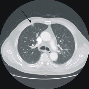
    Image from: Maeng, C. H., Chin, S. O., Yang, B. H., Kim, S.-Y., Youn, H.-J., Cho, K. S., Baek, S. K., et al. (2007). A case of organizing pneumonia associated with rituximab. Cancer research and treatment : official journal of Korean Cancer Association, 39(2), 88–91. doi:10.4143/crt.2007.39.2.88

    30yo male presenting with forearm cellulitis, also complaining of right-sided sharp chest pain worse with deep inspiration and some movements of the ipsilateral shoulder. Found to have multiple pulmonary nodules (suggestive of metastasis) with the largest being a subpleural nodule in the superior/anterior portion of the RUL (roughly the location of the patient’s pain).


    Differential Diagnosis of Pleuritic Chest Pain

    Causes of Pleuritic CP

    Location of Referred Pain

    Referred Pain