CSF Shunt Complications

Brief HPI:

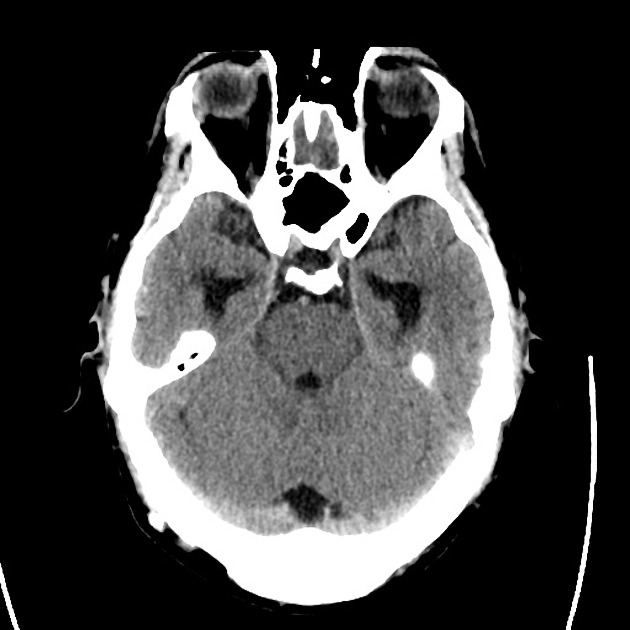
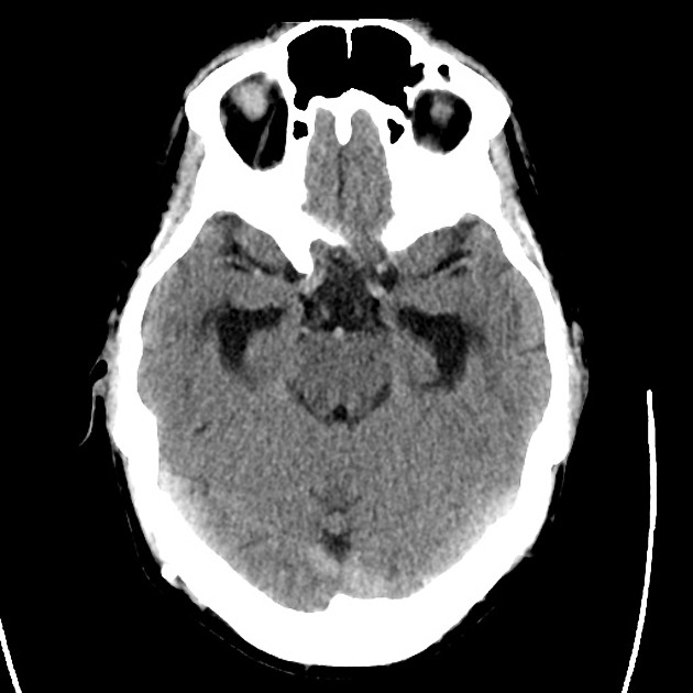
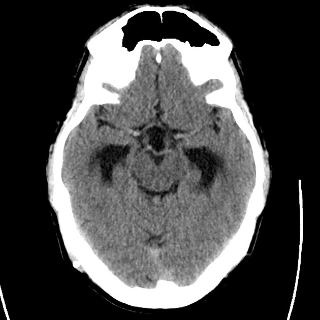
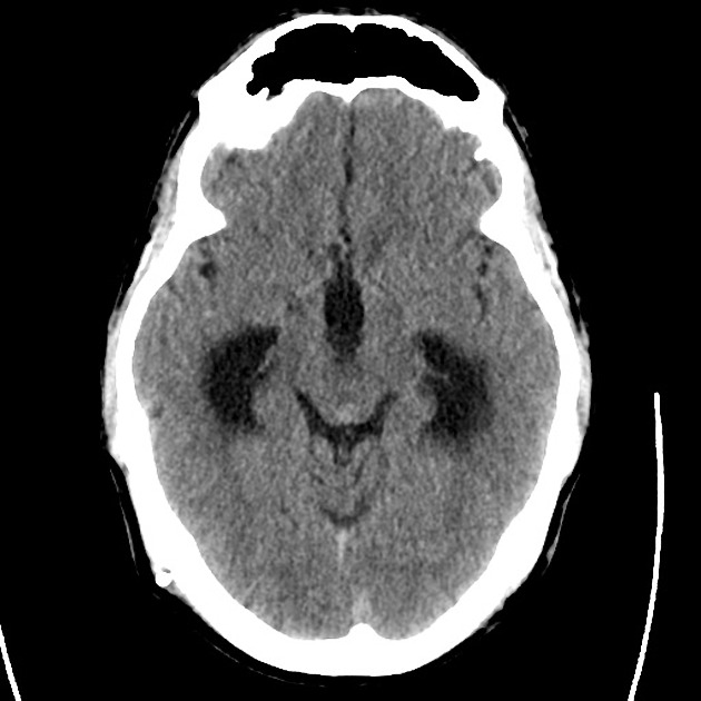
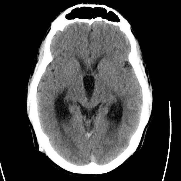
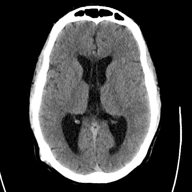
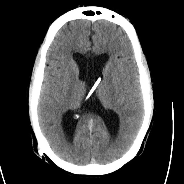
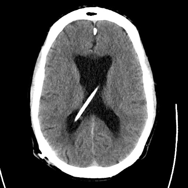
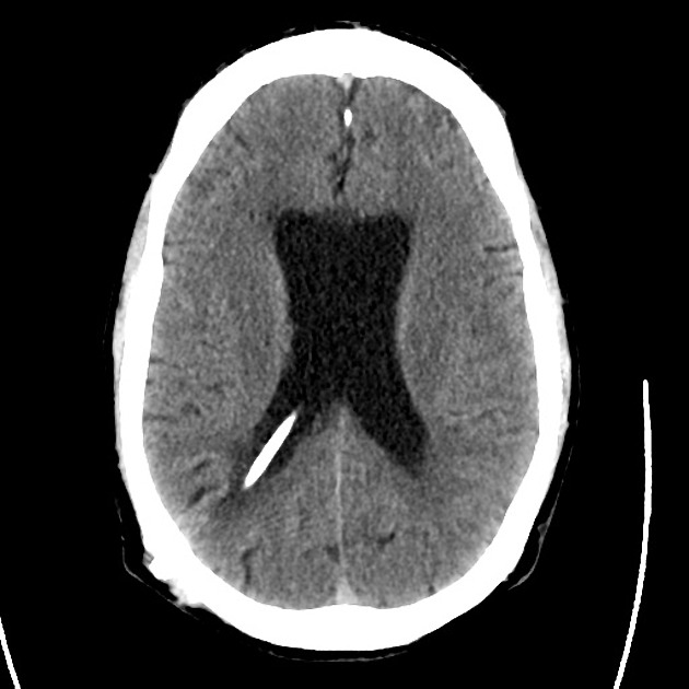
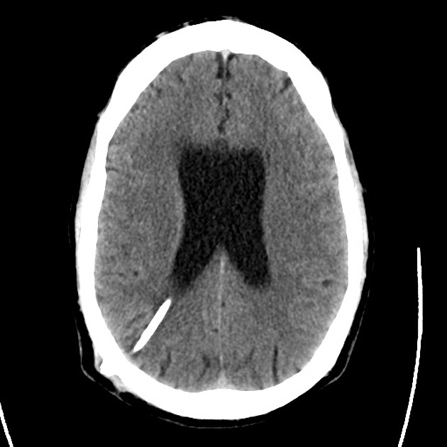
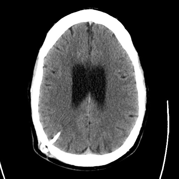
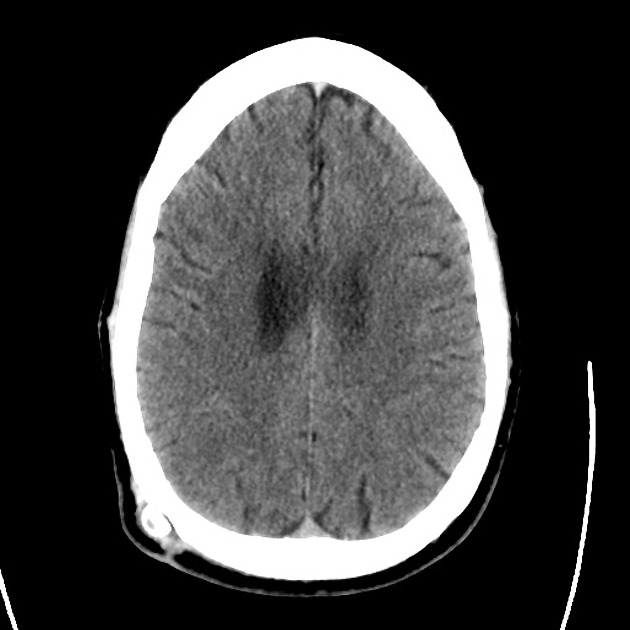
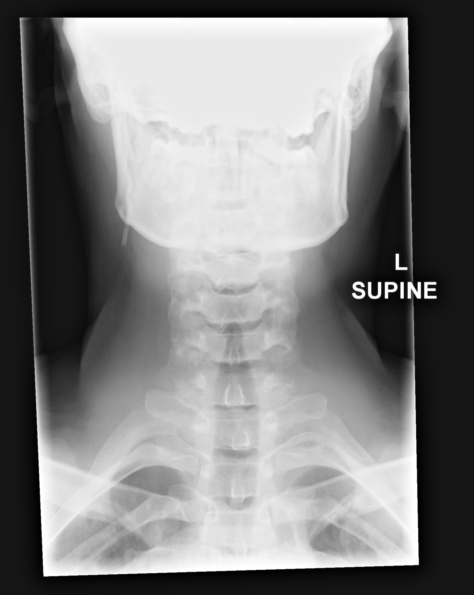
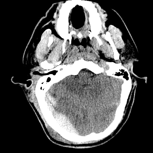
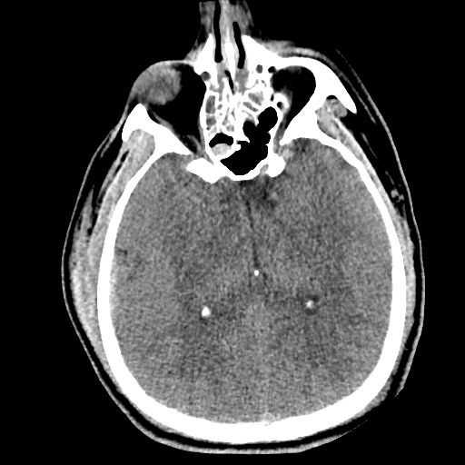
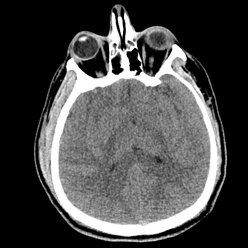
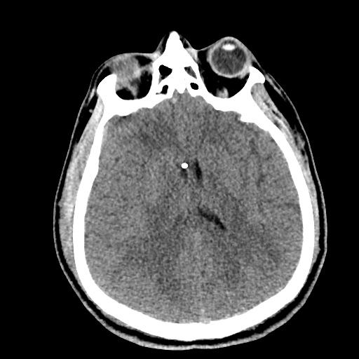
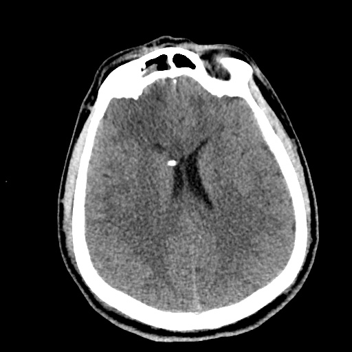
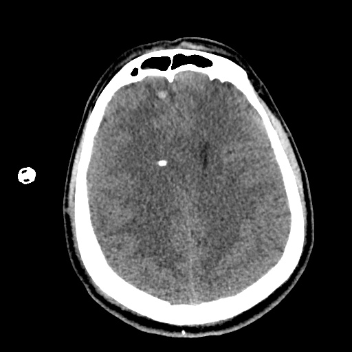
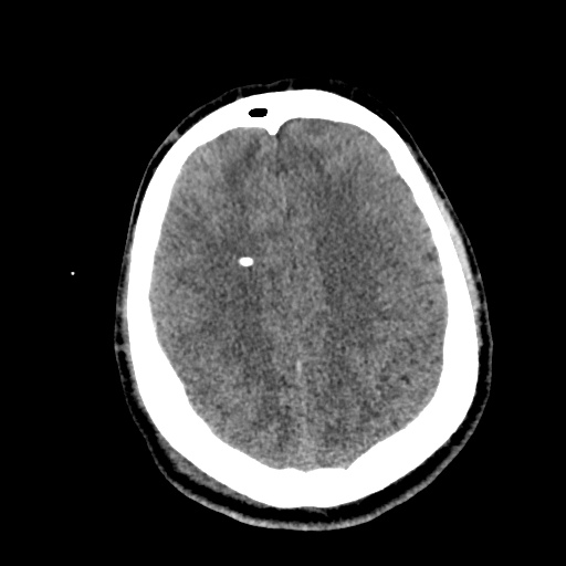
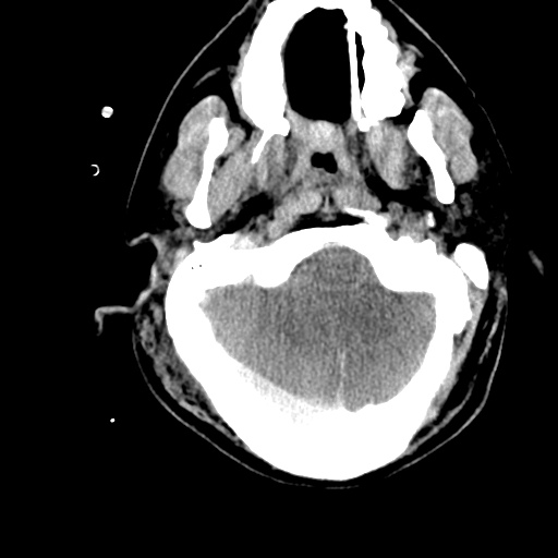
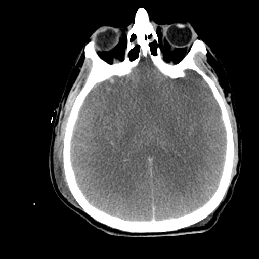
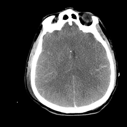
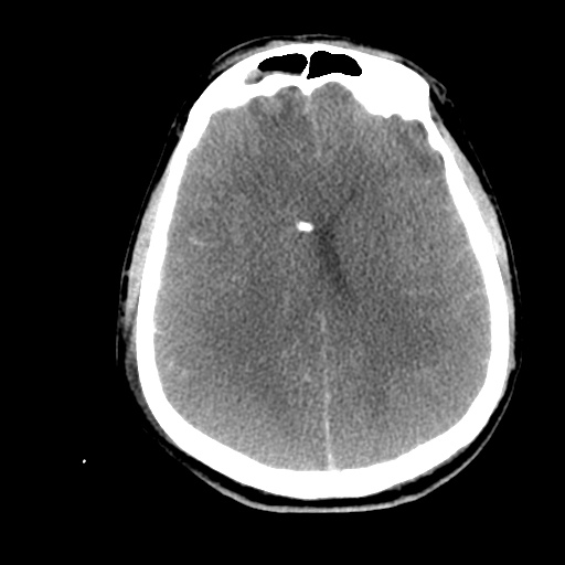
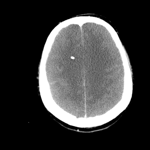
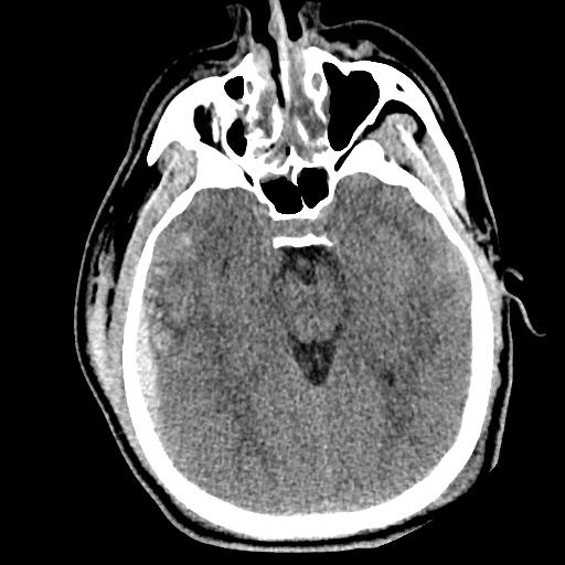
A 33 year-old female with a history of idiopathic intracranial hypertension and ventriculoperitoneal shunt placement presents with headache and confusion. She denies fever, trauma, neck pain or stiffness. She has not had symptoms like this since her shunt was placed 2 years ago. Imaging was obtained which showed ventriculomegaly and a fracture of the shunt at the level of the cervical spine. Neurosurgery was consulted and the patient was admitted for shunt repair.

00_ct_csf_shunt
01_ct_csf_shunt
02_ct_csf_shunt
03_ct_csf_shunt
04_ct_csf_shunt
05_ct_csf_shunt
06_ct_csf_shunt
07_ct_csf_shunt
08_ct_csf_shunt
09_ct_csf_shunt
10_ct_csf_shunt
11_ct_csf_shunt
12_ct_csf_shunt

CT Head

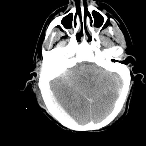
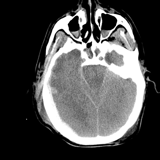
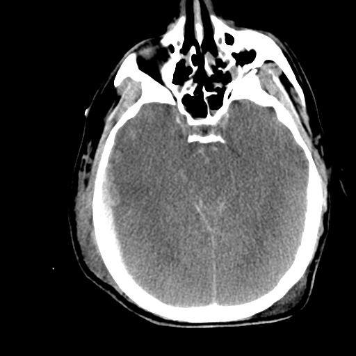
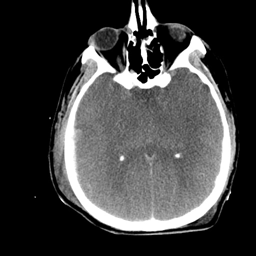
Ventriculomegaly with dilatation of the temporal horns in particular. Right parietal approach ventricular drain. Case courtesy of Dr. Henry Knipe, Radiopaedia.org, rID: 39615

XR Shunt Series

XR Shunt Series

Shunt tubing fractured at the level of the upper cervical spine

An Algorithm for CSF Shunt Complications

An Algorithm for CSF Shunt Complications

References:

  1. Madsen MA. Emergency department management of ventriculoperitoneal cerebrospinal fluid shunts. Ann Emerg Med. 1986;15(11):1330-1343.
  2. Ferras M, McCauley N, Stead T, Ganti L, Desai B. Ventriculoperitoneal shunts in the emergency department: a review. Cureus. 2020;12(2):e6857.
  3. Paff M, Alexandru-Abrams D, Muhonen M, Loudon W. Ventriculoperitoneal shunt complications: A review. Interdisciplinary Neurosurgery. 2018;13:66-70.
  4. Pitetti R. Emergency department evaluation of ventricular shunt malfunction: is the shunt series really necessary? Pediatr Emerg Care. 2007;23(3):137-141.
  5. Fowler JB, De Jesus O, Mesfin FB. Ventriculoperitoneal Shunt. [Updated 2021 Feb 7]. In: StatPearls [Internet]. Treasure Island (FL): StatPearls Publishing; 2021 Jan-. Available from: https://www.ncbi.nlm.nih.gov/books/NBK459351/
  6. Broggi M, Zattra CM, Schiariti M, et al. Diagnosis of ventriculoperitoneal shunt malfunction: a practical algorithm. World Neurosurg. 2020;137:e479-e486.

Spinal Epidural Abscess

Case Presentation

HPI:

34M with no PMH presenting with joint pain and rash. The patient was in his usual state of good health until 1 week prior to presentation, noting bilateral shoulder pain. Diagnosed with musculoskeletal process at outside hospital and discharged with analgesics. Presented with partner due to worsening pain involving multiple joints, a non-painful, non-pruritic rash on bilateral lower extremities, and apparent confusion/hallucinations. Social history was non-contributory, no recent procedures or instrumentation.

Objectively, vital signs were notable for tachycardia and elevated core temperature. The patient was ill-appearing, disoriented and unable to provide detailed history. Skin examination was notable for non-blanching petechial rash with areas of confluence most dense in anterior distal lower extremities, rarer proximally, and otherwise without palm/sole involvement. Mucous membranes were dry, neck was supple. There was tenderness to palpation and manipulation of bilateral shoulders. No back tenderness to palpation or percussion was identified. Neurological examination notable for disorientation, intact cranial nerve function, pain-limited weakness in bilateral upper extremities particularly shoulder abduction, and 4/5 hip flexion, knee flexion/extension in bilateral lower extremities.

Labs:

  • CBC: 34.0/11.8/35.7/216
  • Differential: 31 bands
  • INR: 1.94
  • BMP: 131/5.3/102/17/88/2.55/215
  • LFT: AST 93, ALT 57, AP 237, TB 2.9, DB 1.9, Alb 1.4
  • Lactate: 3.3
  • UA: 47WBC, 5RBC
  • Utox: Negative
  • ESR: 83, CRP: 11.9
  • HIV: Nonreactive

Radiology

  • CT head: Negative
  • CXR: Negative
  • XR Shoulder: Negative
  • CT Chest/Abdomen/Pelvis non-contrast: Mild bilateral hydrouereter/hyndronephrosis, L4-L5 grade 2 anterolisthesis.
IM-0001-0005
IM-0001-0006
IM-0001-0007
IM-0001-0008
IM-0001-0009
IM-0001-0010
IM-0001-0011
IM-0001-0012
IM-0001-0013
IM-0001-0014

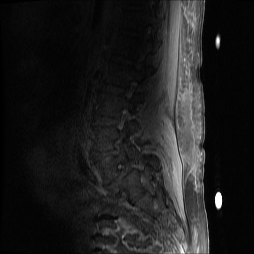
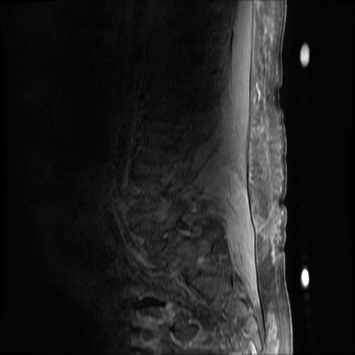
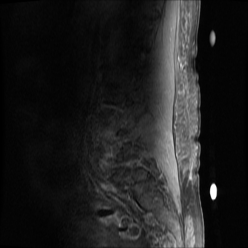
MRI Lumbar Spine w/contrast

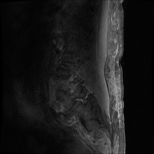
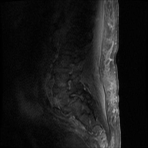
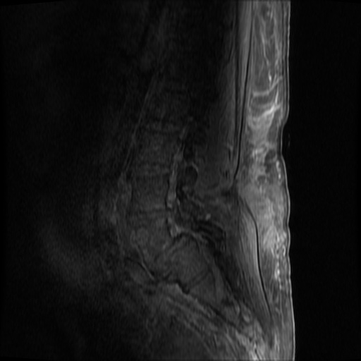
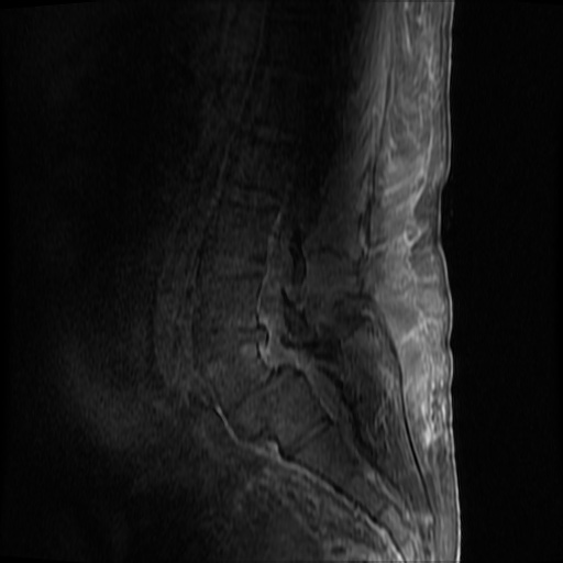
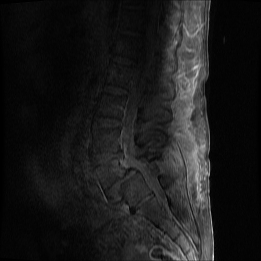
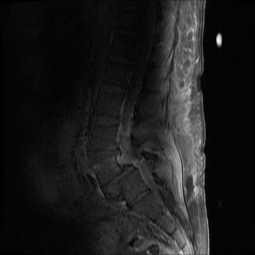
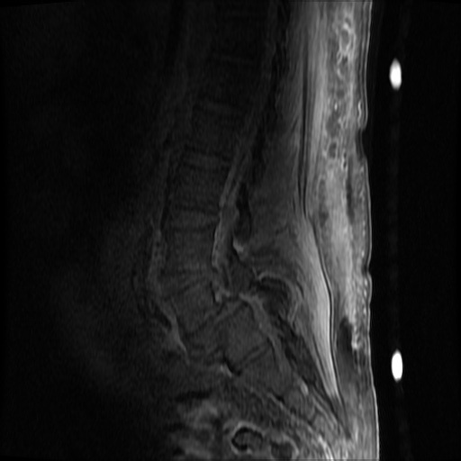
Diffuse epidural enhancement posterior to the L4 and L5 vertebral bodies compressing the thecal sac and resulting in moderate severe spinal canal stenosis. Rim enhancement of the 1.5 cm left paraspinal fluid that may be within the L4 tendon sheath or simply paraspinal abscess.

Assessment/Plan:

Severe sepsis with end-organ dysfunction, unclear source (urinary tract involvement unlikely to account for severity of illness). Covered empirically with broad-spectrum anti-microbials including CNS infection given component of encephalitis. Admitted to the intensive care unit.

Hospital Course:

On hospital day 1, the patient underwent non-contrast MRI of the entire neuraxis with findings concerning for L4-L5 and L5-S1 epidural and paraspinal infection resulting in moderate-severe spinal canal stenosis. Blood and urine cultures grew gram-positive cocci in clusters.

On hospital day 2, the patient became increasingly somnolent. Repeat examination by consulting neurology service was concerning for evidence of meningeal irritation. Cultures speciated as methicillin-sensitive staphylococcus aureus and oxacillin was added. MRI was repeated with gadolinium, findings concerning for L4 epidural vs. paraspinal abscess.

On hospital day 3, the patient’s mental status continued to worsen and he was intubated for airway protection. Neurosurgical intervention was deferred due to deteriorating clinical status. Shoulder synovial fluid aspirate culture positive for MSSA, orthopedic surgery consulted for washout/serial arthrocentesis. TTE performed without evidence of valvular vegetation.

On hospital day 4, additional warm joints were aspirated by orthopedic surgery including knee, bilateral ankles, and shoulder each of which ultimately grew MSSA.

On hospital day 6, the patient underwent OR washout of affected joints with intraoperative findings of purulent fluid. TEE performed without evidence of valvular vegetation. The following day, underwent fluoroscopically-guided lumbar puncture, CSF studies inconclusive. Rifampin added for high-grade bacteremia with multiple seeded sites.

The patient was extubated on hospital day 9 and transferred out of the intensive care unit. The following day, he became increasingly tachypneic with evidence of volume overload on examination and was intubated and returned to the intensive care unit. Sustained PEA arrest post-intubation with ROSC, possibly secondary to pneumothorax vs. hypoxia from extensive mucous plugging. Required increasing vasopressor support over the subsequent 12 hours, emergent CVVHD for worsening academia and hypervolemia. The patient sustained another arrest and ultimately expired.

The final impression was that of high-grade bacteremia from unclear source (vague history of proximate hand laceration/infection) with resultant seeding of epidural/paraspinal space, urinary tract, multiple joints, and likely CNS/meninges. Review of abdominal ultrasonography with evidence of cirrhosis, suggesting that some component of initial hepatic synthetic dysfunction may have been chronic and this may have increased the patient’s risk for disseminated infection and SEA. Neurosurgical intervention was not pursued due to unstable clinical status and as the patient’s neurological findings were not consistent with the location of the identified lesion.

Spinal Epidural Abscess (SEA)1

Risk factors:

  • Immunocompromise: diabetes, cirrhosis, CKD, HIV/AIDS
  • Anatomic: DJD, trauma, prior surgery
  • Introduction: IVDA, epidural anesthesia, tattoo

Organism:

  • S. aureus, 2/3
  • S. epidermidis (associated with device, instrumentation)
  • E. coli (urine spread)
  • P. aeruginosa (IVDA)
  • Rare: anaerobes, mycobacteria, fungi

Staging:

  1. Back pain at affected site
  2. Nerve root pain from affected level
  3. Weakness, sensory deficit, bladder/bowel dysfunction
  4. Paralysis

Clinical features:

  • Back pain (75%)
  • Fever (50%)
  • Neuro deficit (33%)

Diagnosis:

  • Labs: Leukocytosis, ESR/CRP, blood cultures
  • Imaging: MRI with gadolinium, 90% sensitivity
  • Clinical findings and laboratory studies are insensitive and non-specific, in one study, approximately ½ of patients had >2 visits.

Prevalence of abnormal physical findings 2

Finding Prevalence
Fever (T>38°C) 19-32%
Focal spinal TTP 52-62%
Diffuse spinal TTP 63-65%
Positive SLR 11-13%
Abnormal sensation 17-27%
Weakness 29-40%
Abnormal reflexes 8-17%
Abnormal rectal tone 5-10%
Saddle anesthesia 2%

Clinical Decision Guideline 3

Spinal Epidural Abscess Clinical Decision Guideline

Management:

  • Neurosurgical evacuation/fusion
  • Antibiotics (vancomycin, oxacillin, cefepime)
  • Neurosurgical intervention may not result in neurological recovery if symptoms present for > 24-36 hours and may be impractical in the setting of panspinal infection.

References:

  1. Darouiche RO. Spinal epidural abscess. N Engl J Med. 2006;355(19):2012–2020. doi:10.1056/NEJMra055111.
  2. Davis DP, Wold RM, Patel RJ, et al. The clinical presentation and impact of diagnostic delays on emergency department patients with spinal epidural abscess. J Emerg Med. 2004;26(3):285–291. doi:10.1016/j.jemermed.2003.11.013.
  3. Davis DP, Salazar A, Chan TC, Vilke GM. Prospective evaluation of a clinical decision guideline to diagnose spinal epidural abscess in patients who present to the emergency department with spine pain. J Neurosurg Spine. 2011;14(6):765–770. doi:10.3171/2011.1.SPINE1091.
  4. WikEM: Epidural abscess (spinal)
Seizure

Definition

Seizure
Pathologic neuronal activation leading to abnormal function
Epilepsy
Recurrent unprovoked seizures

Classification

  • Cause
    • Primary: Unprovoked
    • Secondary: Provoked, caused by trauma, illness, intoxication, metabolic disturbances, etc.
  • Effect on mentation
    • Generalized: involvement of both hemispheres with associated loss of consciousness (tonic-clonic, absence, atonic, myoclonic)
    • Focal: Involving single hemisphere with preserved level of consciousness
  • Status epilepticus
    • Witnessed convulsions lasting >5min
    • Recurrent seizure without recovery from postictal period

Causes of Seizures

Causes of Seizures

Management of Seizures

Management of Seizures

Medications for Treatment of Seizures

Medication Dose (adult) Dose (peds) Comment
1st Line
Lorazepam 4mg IV <13kg: 0.1mg/kg (max 2mg)
13-39kg: 2mg

>39kg: 4mg
Repeat in 10min
Midazolam 10mg IM 0.2mg/kg IM (max 5mg) Repeat in 10min
Midazolam 10mg buccal 0.5mg/kg buccal (max 5mg) Repeat in 10min
2nd Line
Fosphenytoin 20mg PE/kg IV    
Phenytoin 20mg/kg IV   May cause hypotension
3rd Line
Midazolam 0.05-2mg/kg/hr    
Propofol 1-2mg/kg bolus then 20-200mcg/kg/min    
Pentobarbital 5-15mg/kg bolus then 0.5-5mg/kg/hr    
Special Conditions
Glucose 50mL D50/W   Hypoglycemia
MgSO4 6g IV over 15min   Eclampsia (20wks gestation to 6wks post-partum)
Pyridoxine 0.5g/min until seizures stop, max 5g   INH ingestion
3% saline 100-200mL over 1-2h   Confirmed hyponatremia

History

Points suggestive of seizure over alternative process
Abrupt onset
Duration < 120s
LOC
Purposeless activity: automatisms, tonic-clonic
Provocation: fever in children, substance withdrawal
Postictal state
Retrograde amnesia
Incontinence, oral trauma (buccal maceration, tongue laceration)
Rapidly resolving lactic acidosis
Important historical points for patients with seizure history
Recent illness
Medications (adherence, changes, interactions)
Substance use
Ictogenic factors
Recent/remote head trauma
Developmental abnormalities
Substance use
Sleep deprivation
Pregnancy

Key Physical Examination Findings

  • Vital sign abnormalities persisting beyond immediate postictal state (may suggest drug/toxin exposure, CNS lesion)
  • Nuchal rigidity
  • Signs of IVDA
  • Sequela

    • Head trauma
    • Tongue laceration
    • Shoulder dislocation (posterior)
  • Neurological exam

    • Stroke
    • Elevated ICP
    • Failure to note improvement in postictal confusion (encephalopathy, subclinical seizures)

Labs

  • Glucose
  • BMP (Na, Ca, Mg)
  • AED levels
  • CBC (leukocytosis and bandemia common post-seizure)
  • CSF
  • B-hCG
  • LFT (hepatic dysfunction, alcoholic hepatitis)
  • Lactate (rapidly resolves on repeat)

Indications for Imaging

  • New seizures
  • History of trauma
  • History of malignancy
  • Immunocompromised
  • Headache
  • Anti-coagulation
  • Focal neurological exam
  • Persistent AMS

References

  1. McMullan, J., Davitt, A., & Pollack, C. (2013). Seizures. In Rosen’s Emergency Medicine – Concepts and Clinical Practice (8th ed., Vol. 1, pp. 156-161). Elsevier Health Sciences
  2. WikEM: Seizure

Weakness

View Algorithm
There is a ddxof algorithm for the evaluation of weakness. View it here.

Motor Neuron Signs

Upper Motor Neuron:
Spasticity
Hyperreflexia
Pronator drift
Babinski
Lower Motor Neuron:
Flaccidity
Hyporeflexia
Fasciculation
Atrophy

Causes of Weakness

Lesion Critical Emergent
Non-neurological Shock (VS, clinical assessment)
Hypoglycemia (POC glucose)
Electrolyte derangement (BMP)
Anemia (POC Hb, CBC)
MI (ECG, troponin)
CNS depression (Utox, EtOH)
 
Cortex Stroke Tumor
Abscess
Demyelination
Brainstem Stroke Demyelination
Spinal Cord Ischemia
Compression (disk, abscess, hematoma)
Demyelination (transverse myelitis)
Peripheral Acute demyelination (GBS) Compressive plexopathy
Muscle Rhabdomyolysis Inflammatory myositis

Weakness Syndromes

Unilateral weakness, ipsilateral face
Lesion: Contralateral cortex, internal capsule
Causes: Stroke (sudden onset), demyelination/mass (gradual onset)
Symptoms: Neglect, visual field cut, aphasia
Findings: UMN signs
Key features: Association with headache suggests hemorrhage or mass
Unilateral weakness, contralateral face
Lesion: Brainstem
Causes: Vertebrobasilar insufficiency, demyelination
Symptoms: Dysphagia, dysarthria, diplopia, vertigo, nausea/vomiting
Findings: CN involvement, cerebellar abnormalities
Unilateral weakness, no facial involvement
Lesion: Contralateral medial cerebral cortex, discrete internal capsule
Causes: Stroke
Rare Cause: Brown-Sequard if contralateral hemibody pain and temperature sensory disturbance
Unilateral weakness single limb (monoparesis/plegia)
Lesion: Spinal cord, peripheral nerve, NMJ
UMN signs: Brown-Sequard if contralateral pain and temperature sensory disturbance
LMN signs: Radiculopathy if associated sensory disturbance
Normal reflexes, normal sensation: Consider NMJ disorder
Bilateral weakness of lower extremities (paraparesis/plegia)
Lesion: Spinal cord, peripheral nerve
UMN signs: Anterior cord syndrome (compression, ischemia, demyelination) if contralateral pain and temperature sensory disturbance
Cauda equina: Loss of perianal sensation, loss of rectal tone, or urinary retention
GBS: If no signs of cauda equina and sensory disturbances paralleling ascending weakness (with hyporeflexia)
Bilateral weakness of upper extremities
Lesion: Central cord syndrome
Causes: Syringomyelia, hyperextension injury
Findings: Pain and temperature sensory disturbances in upper extremities (intact proprioception)
Bilateral weakness of all four extremities (quadriparesis/plegia)
Lesion: Cervical spinal cord
Findings: UMN signs below level of injury, strength/sensory testing identifies level
Bilateral weakness, proximal groups
Lesion: Muscle
Causes: Rhabdomyolysis, polymyositis, dermatomyositis, myopathies
Findings: Muscle tenderness to palpation, no UMN signs, no sensory disturbances
Facial weakness, upper and lower face
Lesion: CNVII
Causes: Bell’s palsy, mastoiditis, parotitis
Other CN involvement suggests brainstem lesion, multiple cranial neuropathies, or NMJ

Review of Spinal Cord Anatomy

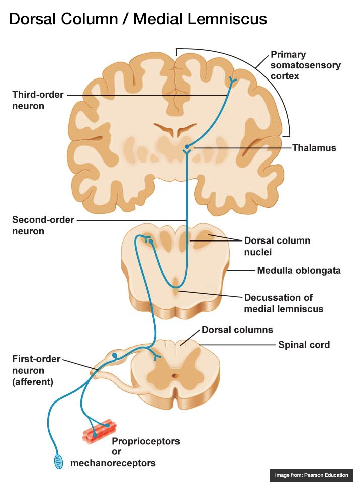
  • Dorsal Column – Medial Lemniscus (fine touch, proprioception)
    1. Afferent sensory fibers with cell body in DRG
    2. Ascend in ipsilateral posterior column
    3. Synapse in medulla, decussate, ascend in contralateral medial lemniscus
    4. Synapse in thalamus (VPL)
    5. Synapse in sensory strip of post-central gyrus
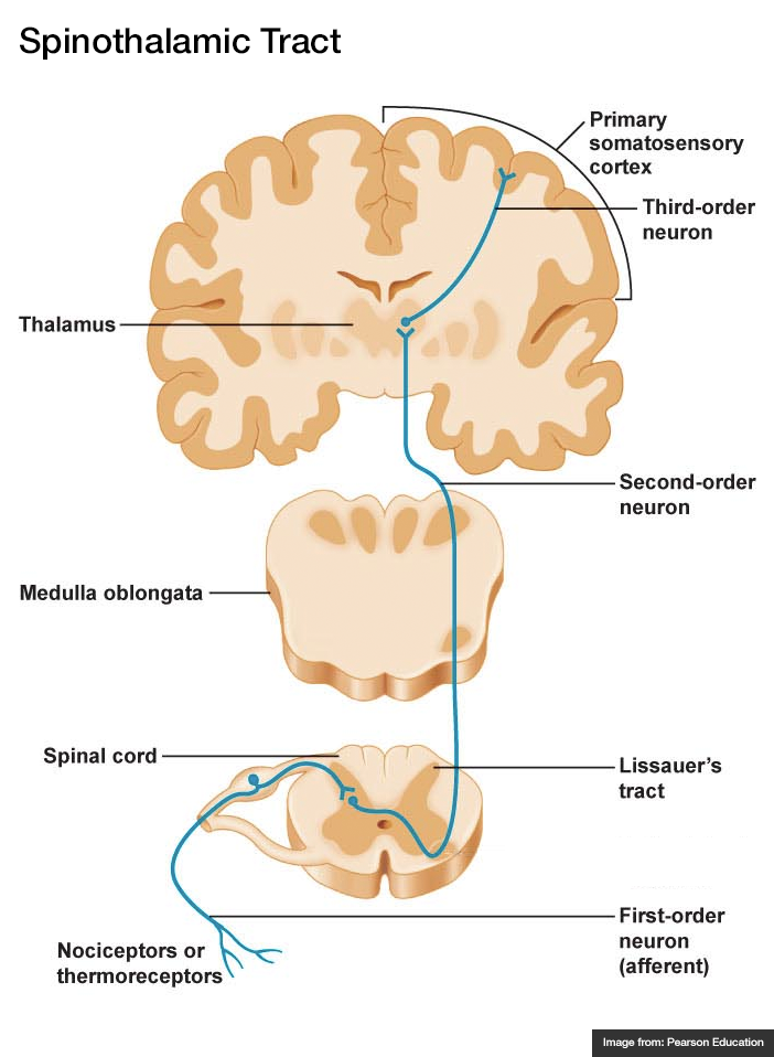
  • Spinothalamic Tract (pain, temperature)

    1. Afferent sensory fibers with cell body in DRG
    2. Ascends 1-2 levels
    3. Synapse in ipsilateral spinal cord, decussate, ascend in contralateral lateral spinothalamic tract
    4. Synapse in thalamus (VPL)
    5. Synapse in sensory strip of post-central gyrus
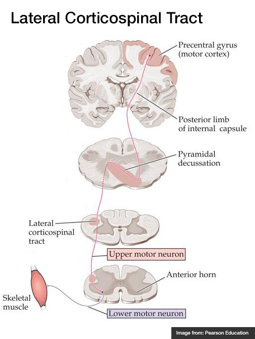
  • Lateral Corticospinal Tract (motor)

    1. Efferent cell body in motor strip of pre-central gyrus
    2. Descends through internal capsule
    3. Decussates in pyramid of medulla, descends in contralateral lateral corticospinal tract
    4. Synapse in anterior horn, lower motor neuron to muscle fiber
Spinal Cord Syndromes
Spinothalamic Tract
Dorsal Column / Medial Lemniscus
Lateral Corticospinal Tract

References

  1. Morchi, R. (2013). Weakness. In Rosen’s Emergency Medicine – Concepts and Clinical Practice (8th ed., Vol. 1, pp. 124-128). Elsevier Health Sciences.

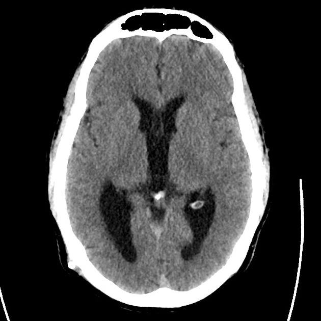
Severe Traumatic Brain Injury

HPI:

34 year-old male brought in by ambulance s/p assault. Field GCS reportedly 7, in trauma bay assessed as E2-V4-M6. Witnessed seizure in CT scanner, resolved with lorazepam. Intubated for airway protection, underwent external ventricular drain placement and transferred to surgical ICU.

Initial imaging revealed bifrontal subdural hematomas and right temporal hemorrhagic contusion with generalized edema. Repeat imaging one hour later showed interval development of large extra-axial hemorrhage overlying the right occipital and parietal lobes (2.2cm), representing subdural or epidural hematoma.

The patient’s ICU course was complicated by continued seizures and refractory elevation in intracranial pressure. A pentobarbital infusion was started and titrated to adequate burst suppression and hyperosmolar therapy with both mannitol and hypertonic saline continued. Additional imaging revealed stable hemorrhage but continued diffuse cerebral edema evidenced by sulcal effacement.

On hospital day 5, examination revealed bilateral fixed and dilated pupils. Imaging revealed effacement of basilar cisterns, pre-pontine cistern, and cisterna magna suggestive of impending/ongoing transtentorial and tonsillar herniation. Pentobarbital was weaned and conventional cerebral angiography as well as cerebral perfusion studies were consistent with brain death.

Images

tbi1_01
tbi1_02
tbi1_03
tbi1_04
tbi1_05
tbi1_06
tbi1_07
tbi1_08
tbi1_09
tbi1_10
tbi1_11
tbi1_12

CT head without contrast one hour after presentation

  • Large extra-axial posterior hemorrhages. Hemorrhagic contusions in the right frontal and temporal lobes.
  • The cerebral sulci appear effaced – findings suggest diffuse cerebral edema.
  • S/p EVD using a right frontal approach.
tbi2_01
tbi2_02
tbi2_03
tbi2_04
tbi2_05
tbi2_06
tbi2_07
tbi2_08
tbi2_09
tbi2_11
tbi2_12

CT head without contrast on hospital day 5

  • Interval evidence of global hypoxic/ischemic injury to the brain.
  • Interval apparent effacement of the basilar cisterns, pre-pontine cistern, and cisterna magna suggesting impending/ongoing downward transtentorial herniation and tonsillar herniation.
  • Stable supra/infratentorial subdural/epidural hematoma.

Algorithm for the Management of Severe Traumatic Brain Injury1,2

Algorithm for the Management of Severe Traumatic Brain Injury

References

  1. Brain Trauma Foundation, American Association of Neurological Surgeons, Congress of Neurological Surgeons, Joint Section on Neurotrauma and Critical Care, AANS/CNS, Carney, N. A., & Ghajar, J. (2007). Guidelines for the management of severe traumatic brain injury. Introduction. Journal of neurotrauma, 24 Suppl 1, S1–2. doi:10.1089/neu.2007.9997
  2. Stocchetti, N., & Maas, A. I. R. (2014). Traumatic intracranial hypertension. The New England journal of medicine, 370(22), 2121–2130. doi:10.1056/NEJMra1208708
  3. WikEM: Severe traumatic brain injury

Head Trauma: Radiographic Evolution

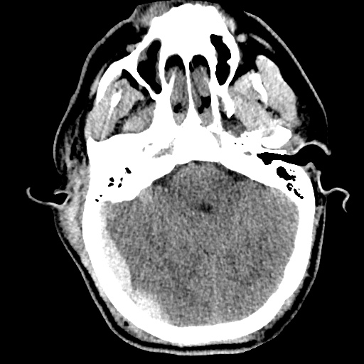
CT Head (Initial)
CT Head (Initial)
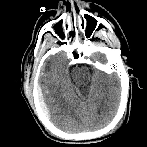
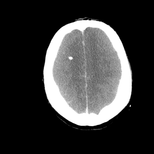
- Noncontrast axial images through the head demonstrate no evidence of skull fracture.
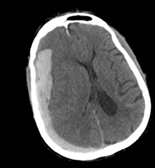
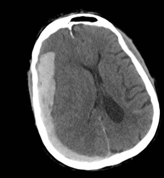
- Large lentiform-shaped mixed density extra-axial acute epidural hematoma in the right parietal occipital
- Associated subdural hematoma tracking along right convexity toward the right temporal lobe.
- There is no evidence of midline shift.

CT Head (+8h)
CT Head (+8h)

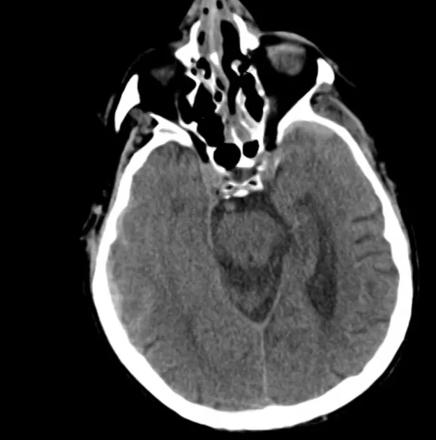
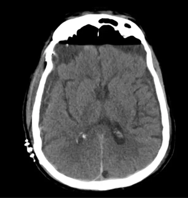
- Significant interval increase in the size of the right hemispheric subdural hematoma
- There is now midline shift from right to left at the level of the septum pellucidum measuring 10 mm, partial effacement of the right lateral ventricle and subfalcial herniation.
- Scattered subarachnoid blood is redemonstrated.
- Comminuted fractures of the nasal bone are present and there is overlying and associated periorbital soft tissue swelling.

CT Head (+16h, s/p SDH evacuation)

CT Head (+16h, s/p SDH evacuation)

- Interval gross total evacuation of right hemispheric subdural hematoma.
- Moderate anterior bifrontal subdural and right epidural air is present.
- Small scattered subarachnoid and intraventricular blood is redemonstrated.

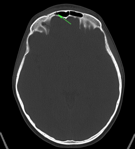
Skull Fracture

Frontal bone fractureID:

14 year-old female, previously healthy, brought in by ambulance s/p auto vs. pedestrian.

HPI:

Incident unwitnessed, paramedics report no LOC with GCS 15 at scene. GCS 10 upon arrival to ED, with 2min GTC seizure. Patient intubated for airway protection and CT head showed non-displaced frontal bone fracture and small frontal SAH. Patient self-extubated, returned to baseline mental status and was transferred to PICU.

PE:

  • VS: 128/76mmHg, 120bpm, 22 R/min, 100% RA, 37.6°C
  • General: Alert and responsive young female with multiple bandages on extremities
  • HEENT: Right frontal hematoma, no bony defect palpated, multiple facial abrasions, no otorrhea, no rhinorrhea, TM clear b/l, no other ecchymosis.
  • CV: RRR, normal S1/S2, no M/R/G
  • Lungs: CTAB
  • Abdomen: +BS, soft, NT/ND, no rebound/guarding, no flank ecchymoses
  • Neuro: AAOx3, CN II-XII intact, sensation/motor/reflexes symmetric and intact.
  • Extremities: Well-perfused with good pulses, no focal bony tenderness, no joint effusions, multiple abrasions on extensor surfaces of all four extremities.

Assessment & Plan:

14yo female, previously healthy, s/p auto vs. peds followed by GTC seizure and CT head showing small SAH and non-displaced frontal bone skull fracture. No evidence of basilar skull fracture on examination or imaging. Seizure likely 2/2 irritation from SAH. Patient was followed closely in PICU with q1h neuro checks with low threshold for repeat CT if change in mental status or more seizures occurred. The patient was eventually transferred to the general ward and was discharged with neurology follow-up and Keppra for seizure prophylaxis for 6mo.

Types of Skull Fractures:

A system for skull fractures