Anticoagulant Reversal in Intracranial Hemorrhage

Brief HPI:

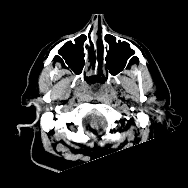
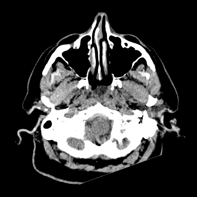
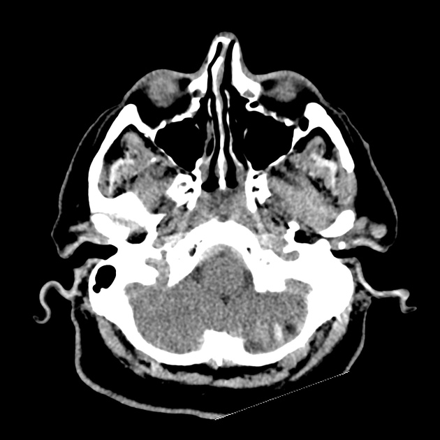
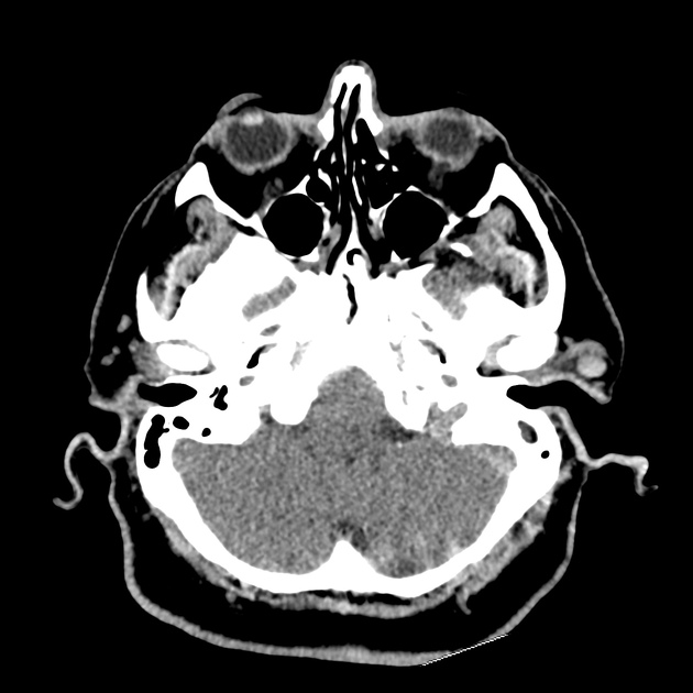
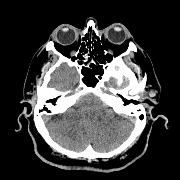
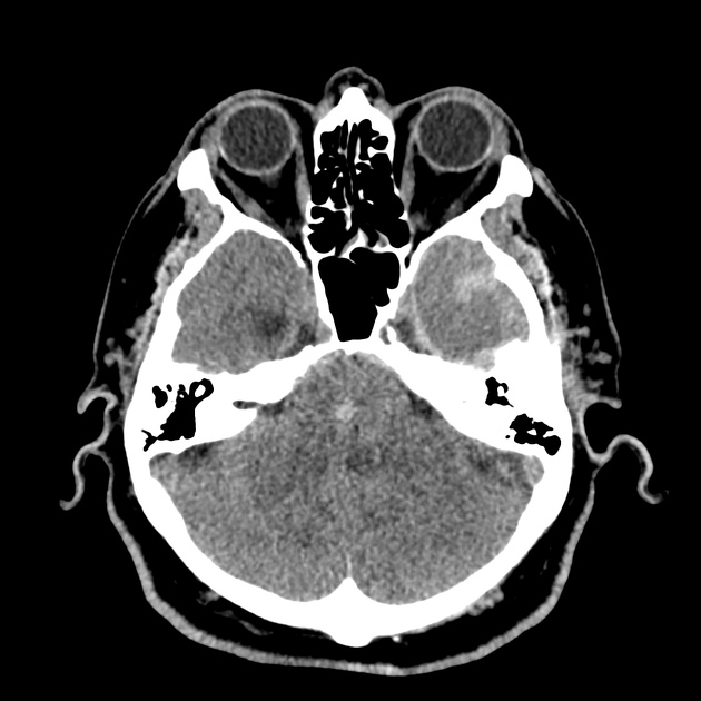
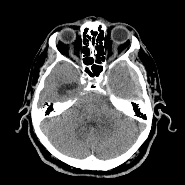
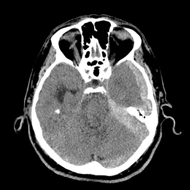
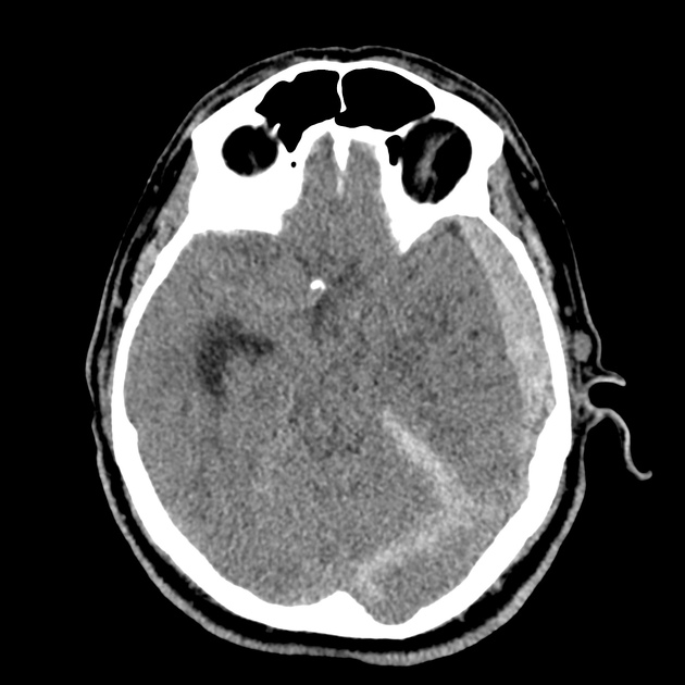
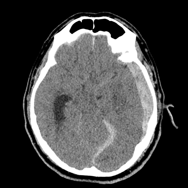
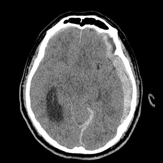
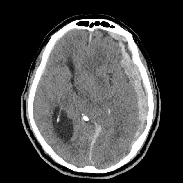
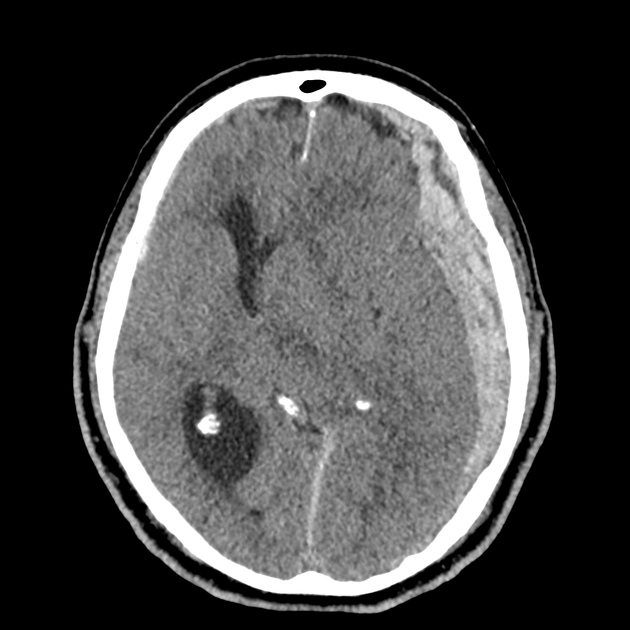
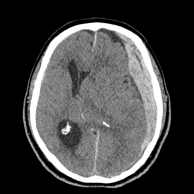
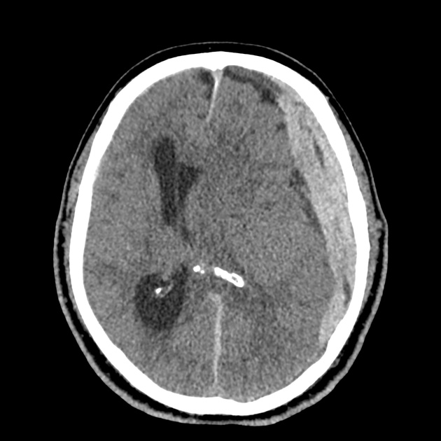
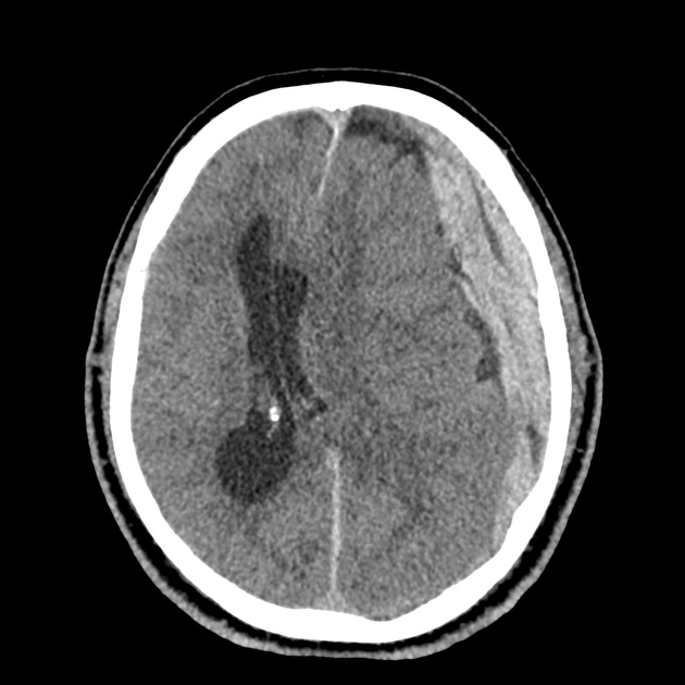
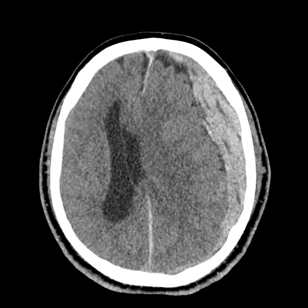
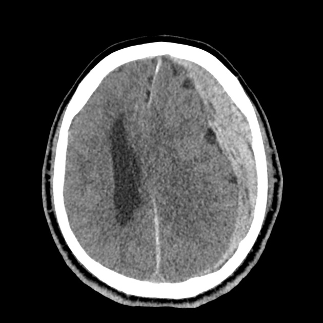
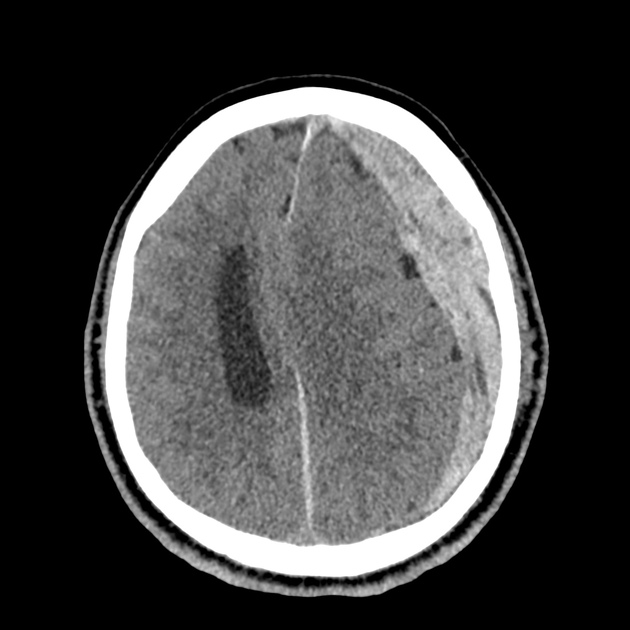
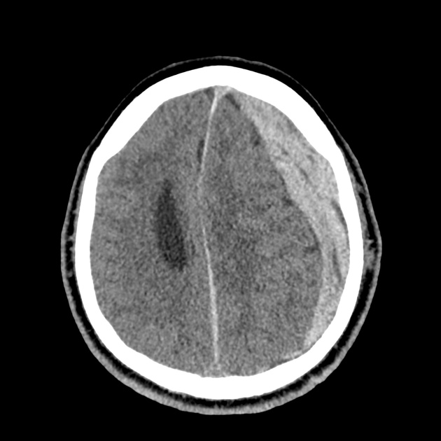
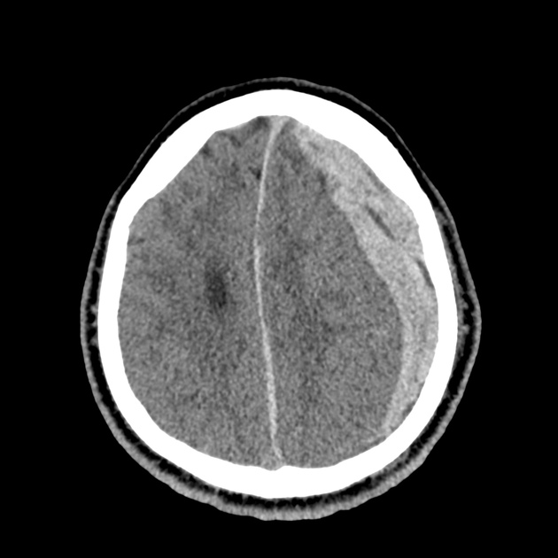
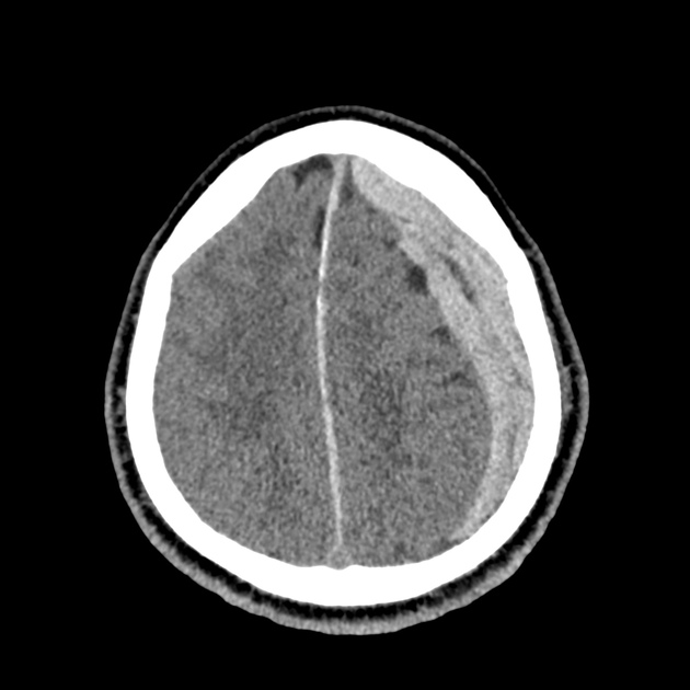
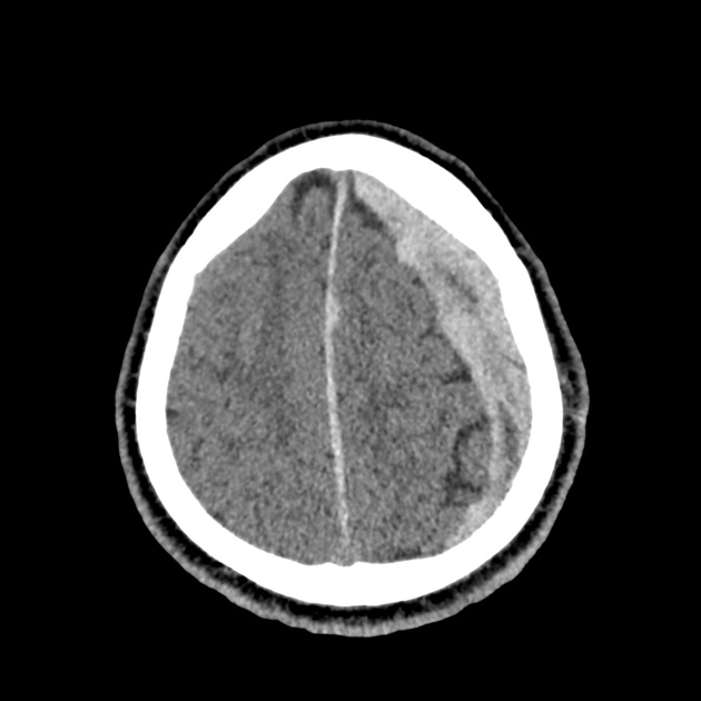
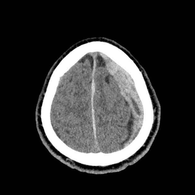
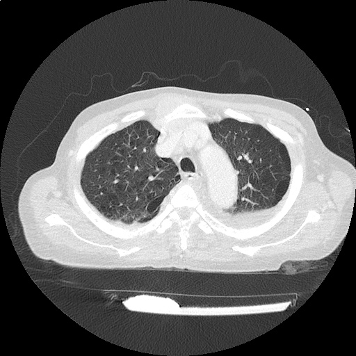
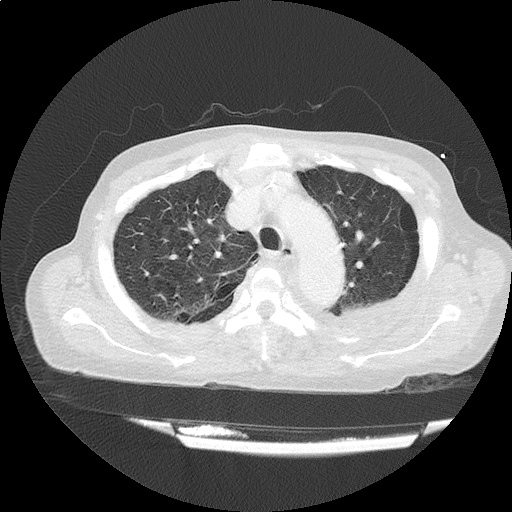
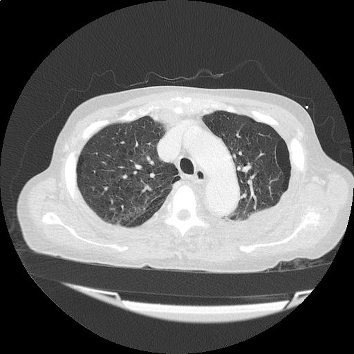
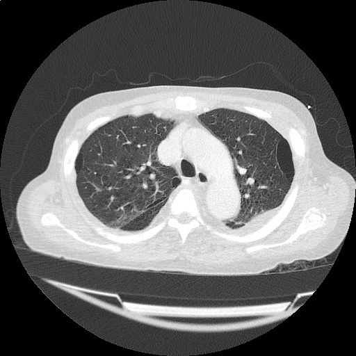
A 65-year-old male with a past medical history of hypertension, diabetes mellitus, and atrial fibrillation presents after a mechanical fall with a posterior scalp hematoma and altered mental status. The patient’s family reports that the patient is taking apixaban with his last dose 4 hours prior to arrival. Physical examination reveals a GCS of 13, blood pressure of 175/99, and asymmetric pupils. The patient is taken to CT where head imaging reveals left sided subdural hematoma with midline shift and developing uncal herniation.

00_subdural
02_subdural
04_subdural
06_subdural
08_subdural
10_subdural
12_subdural
14_subdural
16_subdural
18_subdural
20_subdural
22_subdural
24_subdural
26_subdural
28_subdural
30_subdural
32_subdural
34_subdural
36_subdural
38_subdural
40_subdural
42_subdural
44_subdural
46_subdural

CT Head:

Left cerebral convexity acute subdural hematoma producing substantial mass effect with midline shift and left uncal herniation.
Case courtesy of Dr Andrew Dixon, Radiopaedia.org. From the case rID: 32395

A nicardipine infusion is initiated and the head of the bed is elevated. Andexanet Alfa is not available, therefore an infusion of 4-Factor PCC is initiated. The patient is taken emergently to the operating room by neurosurgery for craniotomy and hematoma evacuation.

An Algorithm for the Reversal of Anticoagulation for Intracranial Hemorrhage 1-4

An Algorithm for Anticoagulant Reversal in Intracranial Hemorrhage


All Agents

For all agents, discontinue anticoagulation. Patients may require blood pressure control including anti-hypertensive infusions (goal SBP <140). Avoid reversal for intracranial hemorrhage associated with cerebral venous thrombosis. Use cautiously in patients with concomitant life-threatening ischemia, thrombosis, or severe DIC.

Vitamin K Antagonists (ex. warfarin)

Initial Dose

A fixed dose of 4F-PCC 1500 to 2000 units can be given as an initial dose with repeat dosing based on INR measurement 15 minutes after completion of infusion. Follow local institution guidelines if available.

Monitoring and Repeat Dosing

  • Vitamin K: if INR ≥1.4 at 12 hours 5
  • 4F-PCC: May consider repeat PCC dosing based on INR, though with increased DIC and thrombotic risk, it is recommended to correct further with FFP if INR remains ≥1.4 6

Direct Factor Xa Inhibitors (ex. rivaroxaban, apixaban)

Activated charcoal may be effective for up to six hours for apixaban 7 and eight hours for rivaroxaban 8.

*Andexanet alfa Regimens 9,10

  • Low-dose: rivaroxaban <10mg, apixaban <5mg, edoxaban <30mg or 8 or more hours since last dose
  • High-dose: If greater than above thresholds, or dose/timing unknown

Pentasaccharides (ex. fondaparinux)

Use high-dose Andexanet alfa regimen 12

Direct Thrombin inhibitors (ex. dabigatran)

Monitoring and Repeat Dosing

If ongoing significant bleeding after treatment, consider redosing idarucizumab and/or hemodialysis.

Alternative Regimens

If idarucizumab is not available, aPCC (50-80 units/kg) , 4F-PCC or 3F-PCC (50 units/kg) can be used in order of preference.

Unfractionated Heparin

Dosing

Determination of units of heparin is based on estimated active agent (half-life 1-2 hours)

  • Protamine sulfate 1mg/100 units IV, maximum dose 50mg
  • Alternatively, can give fixed dose of 25-50mg

Monitoring and Repeat Dosing

If aPTT is persistently elevated, repeat 0.5 mg/100 units

Low-Molecular Weight Heparin 13

Reversal is not indicated if more than 3-5 half-lives have passed since administration:

  • Enoxaparin mean half-life: 4-5 hours
  • Dalteparin mean half-life: 2.8 hours
  • Nadroparin mean half-life: 3.7 hours

If bleeding persists, or renal insufficiency, repeat dose .5 mg/1 mg enoxaparin or .5 mg/100 anti-Xa units.

This algorithm was developed by Dr. Taylor Martin. Taylor is an emergency medicine resident at McGovern Medical School at UTHealth Houston.

References

Guidelines & Reviews

  1. Greenberg SM, Ziai WC, Cordonnier C, et al. 2022 guideline for the management of patients with spontaneous intracerebral hemorrhage: a guideline from the american heart association/american stroke association. Stroke. Published online May 17, 2022:101161STR0000000000000407.
  2. Tomaselli GF, Mahaffey KW, Cuker A, et al. 2020 acc expert consensus decision pathway on management of bleeding in patients on oral anticoagulants: a report of the american college of cardiology solution set oversight committee. J Am Coll Cardiol. 2020;76(5):594-622.
  3. Frontera JA, Lewin JJ, Rabinstein AA, et al. Guideline for reversal of antithrombotics in intracranial hemorrhage: a statement for healthcare professionals from the neurocritical care society and society of critical care medicine. Neurocrit Care. 2016;24(1):6-46.
  4.  Freeman, W. David, Weitz, Jeffrey. “Reversal of anticoagulation in intracranial hemorrhage.” UpToDate. (2022) https://www.uptodate.com/contents/reversal-of-anticoagulation-in-intracranial-hemorrhage?search=anticoagulation%20reversal (Accessed on May 26, 2022)

Vitamin K Antagonists

  1. Ansell J, Hirsh J, Hylek E, Jacobson A, Crowther M, Palareti G. Pharmacology and management of the vitamin k antagonists: american college of chest physicians evidence-based clinical practice guidelines(8th edition). Chest. 2008;133(6 Suppl):160S-198S.
  2. Pabinger I, Brenner B, Kalina U, et al. Prothrombin complex concentrate (Beriplex p/n) for emergency anticoagulation reversal: a prospective multinational clinical trial. J Thromb Haemost. 2008;6(4):622-631.

Direct Factor Xa Inhibitors

  1. http://packageinserts.bms.com/pi/pi_eliquis.pdf
  2. https://www.bayer.com/sites/default/files/2020-11/xarelto-pm-en.pdf
  3. Demchuk AM, Yue P, Zotova E, et al. Hemostatic efficacy and anti-fxa (Factor xa) reversal with andexanet alfa in intracranial hemorrhage: annexa-4 substudy. Stroke. 2021;52(6):2096-2105.
  4. Cohen AT, Lewis M, Connor A, et al. Thirty-day mortality with andexanet alfa compared with prothrombin complex concentrate therapy for life-threatening direct oral anticoagulant-related bleeding. J Am Coll Emerg Physicians Open. 2022;3(2):e12655.
  5. Scaglione F. New oral anticoagulants: comparative pharmacology with vitamin K antagonists. Clin Pharmacokinet. 2013;52(2):69-82.

Pentasaccharides (ex. fondaparinux)

  1. Lu G, DeGuzman FR, Hollenbach SJ, et al. A specific antidote for reversal of anticoagulation by direct and indirect inhibitors of coagulation factor Xa. Nat Med. 2013;19(4):446-451.

Low-Molecular Weight Heparin

  1. Fareed J, Hoppensteadt D, Walenga J, et al. Pharmacodynamic and pharmacokinetic properties of enoxaparin : implications for clinical practice. Clin Pharmacokinet. 2003;42(12):1043-1057.

Transfusion Reactions

Brief HPI:

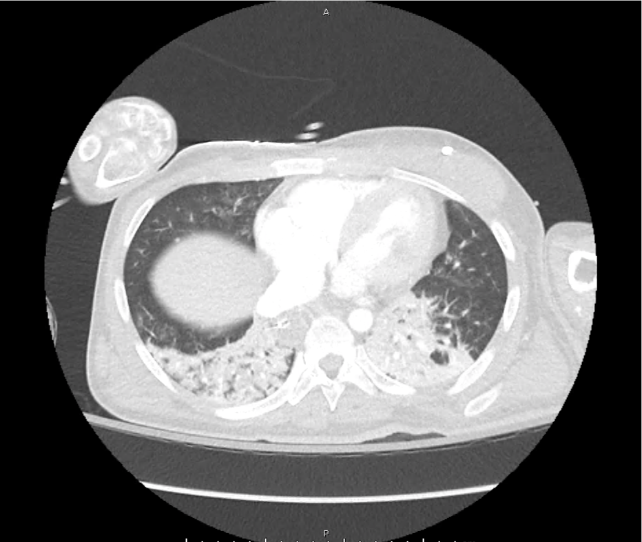
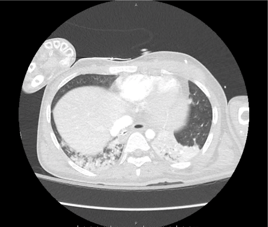
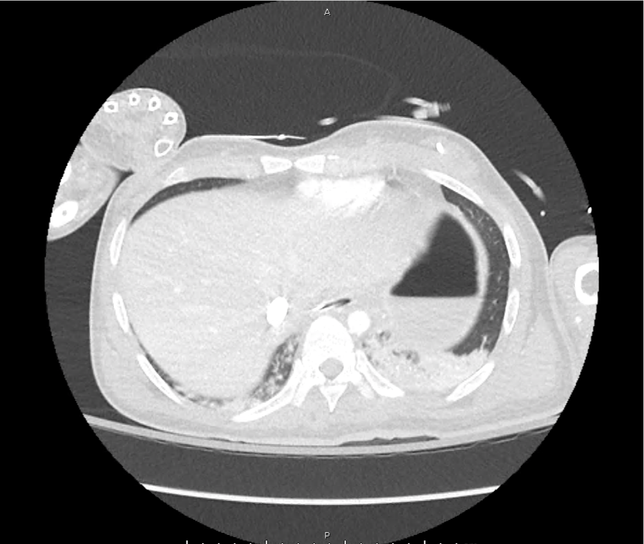
A 28 year-old female with a history of systemic lupus erythematosus and end-stage renal disease without access to scheduled hemodialysis presents to the emergency department with 1 week of worsening dyspnea, fatigue and leg swelling. Her symptoms are reminiscent of prior episodes resolving with hemodialysis. On evaluation, vital signs are normal and laboratory tests demonstrate microcytic anemia (Hb 5.9g/dL) but no hyperkalemia. A plain chest radiograph is normal and the patient ambulates without hypoxia.

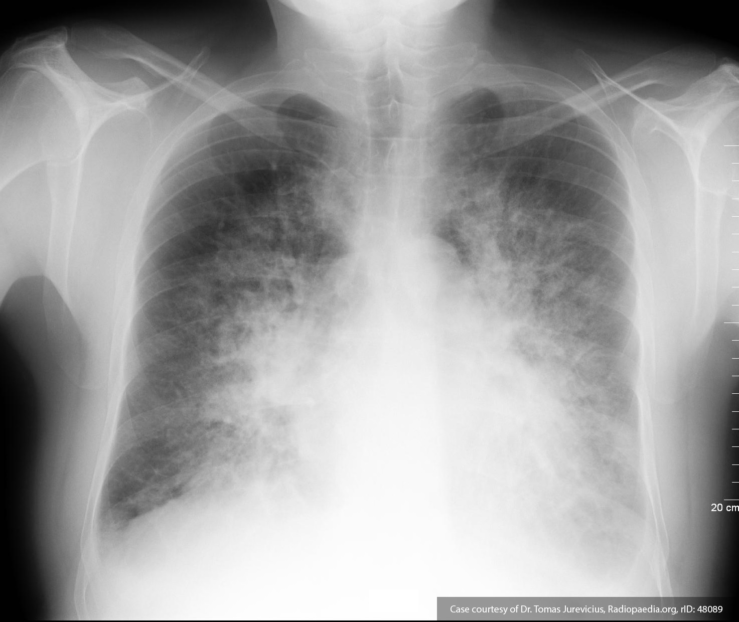
The patient was deemed to not meet any requirements for emergent hemodialysis. One unit of packed red blood cells was ordered for transfusion for symptomatic anemia. During transfusion, the patient developed worsening dyspnea and was found to be hypertensive and hypoxic. A chest radiograph was obtained and is shown below.

Chest x-ray with pulmonary edema

Pulmonary vascular congestion and bilateral pleural effusions.


The transfusion was discontinued, the patient was placed on non-invasive positive pressure ventilation, and emergent hemodialysis was initiated with subsequent resolution of presumed transfusion associated circulatory overload.

Algorithm for the Evaluation and Management of Transfusion Reactions

Algorithm for the Evaluation and Management of Transfusion Reactions

This algorithm was developed by Dr. Eric Madden, chief resident in emergency medicine at McGovern Med EM.

References

  1. Carson JL, Triulzi DJ, Ness PM. Indications for and Adverse Effects of Red-Cell Transfusion. N Engl J Med. 2017;377(13):1261-1272. doi:10.1056/NEJMra1612789.
  2. Delaney M, Wendel S, Bercovitz RS, et al. Transfusion reactions: prevention, diagnosis, and treatment. Lancet. 2016;388(10061):2825-2836. doi:10.1016/S0140-6736(15)01313-6.
  3. Goel R, Tobian AAR, Shaz BH. Noninfectious transfusion-associated adverse events and their mitigation strategies. Blood. 2019;133(17):1831-1839. doi:10.1182/blood-2018-10-833988.
  4. Osterman JL, Arora S. Blood product transfusions and reactions. Emerg Med Clin North Am. 2014;32(3):727-738. doi:10.1016/j.emc.2014.04.012.
  5. Silvergleid AJ. Approach to the patient with a suspected acute transfusion reaction. Post TW, ed. UpToDate. Waltham, MA: UpToDate Inc. https://www.uptodate.com (Accessed on September 01, 2019.)
  6. Suddock JT, Crookston KP. Transfusion Reactions. January 2019.

Pleural Fluid

Brief HPI:

A 43 year-old female with no reported medical history presents with shortness of breath. She notes 2 months of gradually worsening symptoms associated with unproductive cough and intermittent subjective fevers. Symptoms are worsened with activity and when laying flat. She has no history of similar symptoms in the past.

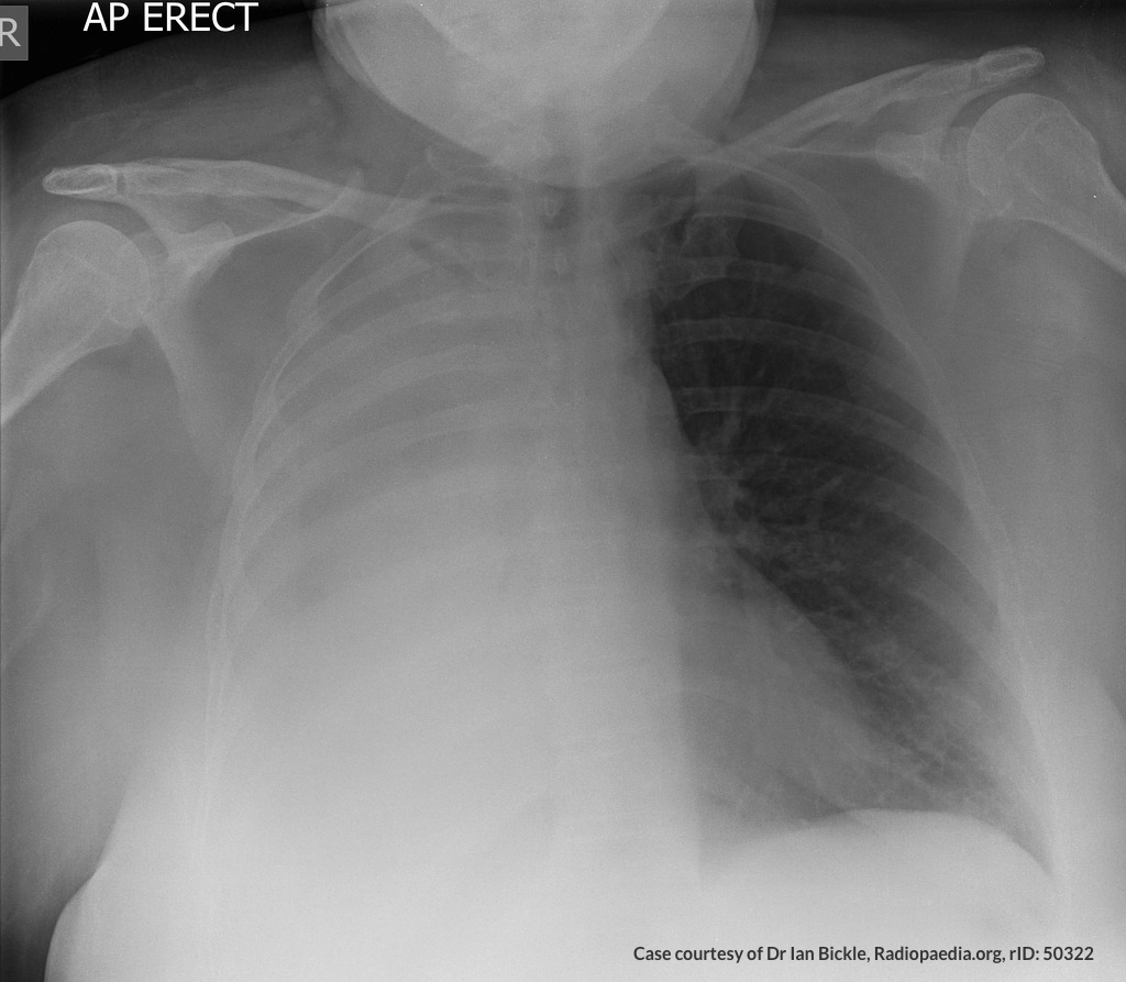
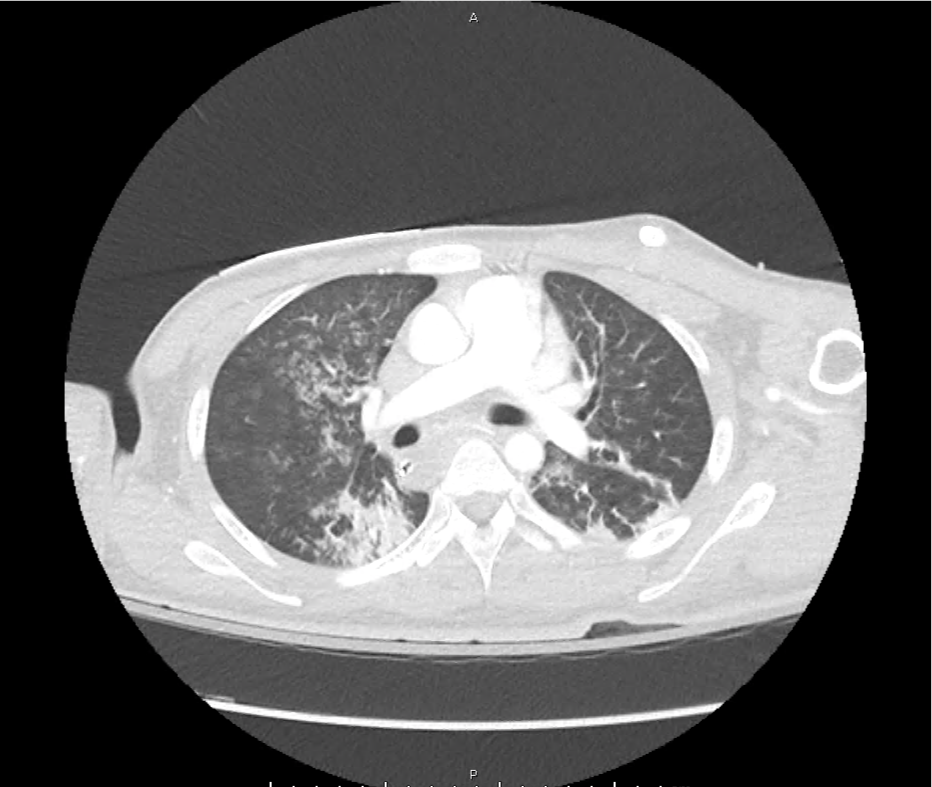
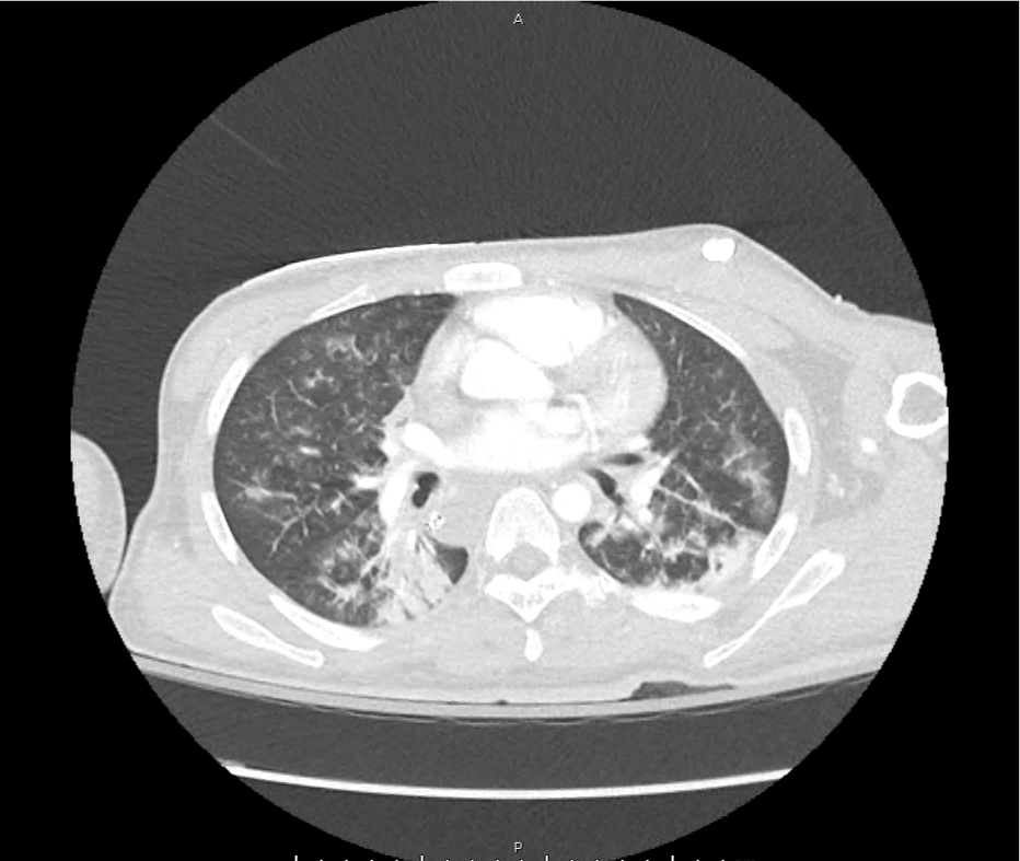
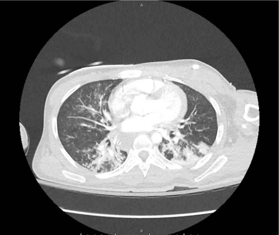
Vital signs are notable for tachycardia, tachypnea and hypoxia. Examination demonstrates absent breath sounds in the entire right lung field. A plain chest radiograph is obtained and shown below. The patient was placed on non-invasive positive pressure with minimal improvement and an emergent therapeutic thoracentesis was performed. Pleural fluid was exudative and a large volume was submitted for cytology.

Whiteout right lung field Whiteout right lung field

An Algorithm for the Analysis of Pleural Fluid

An Algorithm for the Analysis of Pleural Fluid

References

  1. Light RW, Girard WM, Jenkinson SG, George RB. Parapneumonic effusions. Am J Med. 1980;69(4):507-512.
  2. Heffner JE, Brown LK, Barbieri CA. Diagnostic value of tests that discriminate between exudative and transudative pleural effusions. Primary Study Investigators. Chest. 1997;111(4):970-980. doi:10.1378/chest.111.4.970.
  3. Romero S, Martinez A, Hernandez L, et al. Light’s criteria revisited: consistency and comparison with new proposed alternative criteria for separating pleural transudates from exudates. Respiration. 2000;67(1):18-23. doi:10.1159/000029457.
  4. Light RW. Clinical practice. Pleural effusion. N Engl J Med. 2002;346(25):1971-1977. doi:10.1056/NEJMcp010731.
  5. Sahn SA, Huggins JT, San Jose E, Alvarez-Dobano JM, Valdes L. The Art of Pleural Fluid Analysis. Clinical Pulmonary Medicine. 2013;20(2):77-96. doi:10.1097/CPM.0b013e318285ba37.
  6. Light RW. The Light criteria: the beginning and why they are useful 40 years later. Clinics in Chest Medicine. 2013;34(1):21-26. doi:10.1016/j.ccm.2012.11.006.
  7. Aggarwal AN, Agarwal R, Sehgal IS, Dhooria S, Behera D. Meta-analysis of Indian studies evaluating adenosine deaminase for diagnosing tuberculous pleural effusion. Int J Tuberc Lung Dis. 2016;20(10):1386-1391. doi:10.5588/ijtld.16.0298.

Leukemoid Reaction

Brief HPI:

An approximately 80-year-old male with unknown medical history is brought to the emergency department from a skilled nursing facility after unwitnessed arrest – EMS providers established return of spontaneous circulation after chest compressions and epinephrine. On arrival, the patient was hypotensive (MAP 40mmHg) and hypoxic (SpO2 85%) with mask ventilation. The patient was intubated, resuscitated with intravenous fluids and started on vasopressors. Imaging demonstrated lung consolidation consistent with multifocal pneumonia versus aspiration. Laboratory studies were obtained:

  • CBC: WBC: 49.2 (N: 64%, Bands: 20%)
  • ABG: pH: 7.07, pCO2: 73mmHg
  • Lactate: 9.1mmol/L
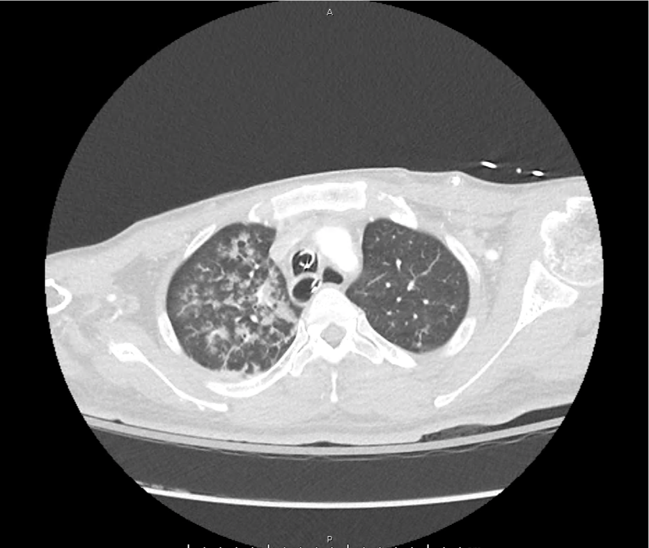
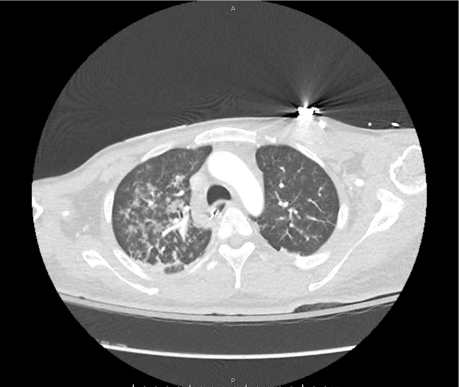
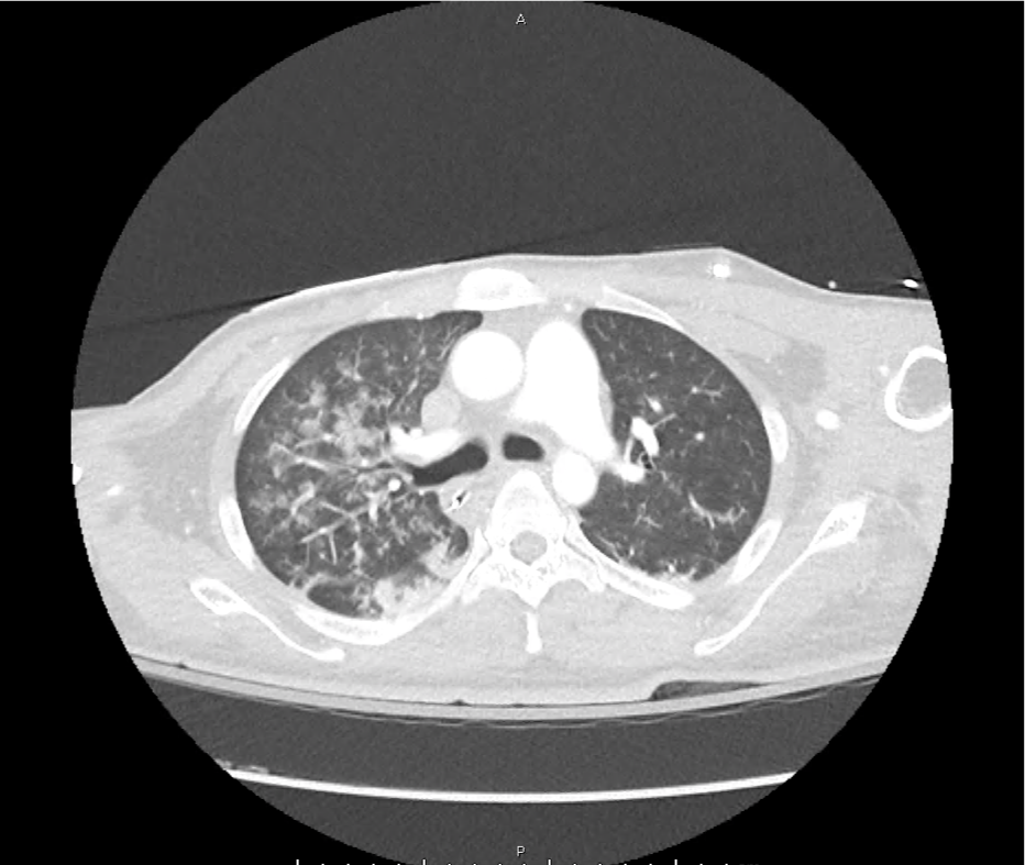
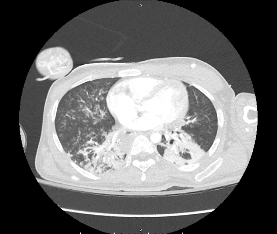
leukemoid_ct_01
leukemoid_ct_02
leukemoid_ct_03
leukemoid_ct_04
leukemoid_ct_05
leukemoid_ct_06
leukemoid_ct_07
leukemoid_ct_08
leukemoid_ct_09
leukemoid_ct_10
leukemoid_ct_11
leukemoid_ct_12

CT Pulmonary Angiography

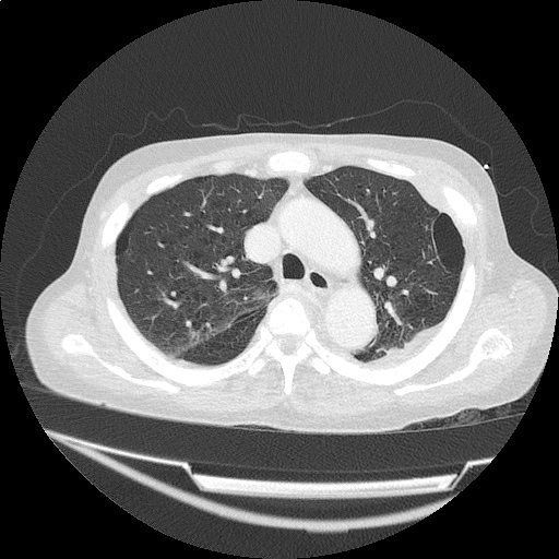
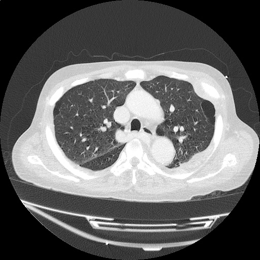
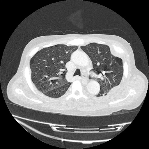
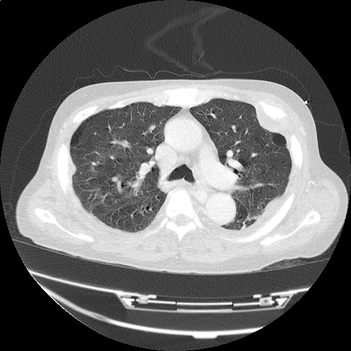
Peribronchial opacities and patchy consolidation in the lungs which may represent multifocal pneumonia and/or aspiration in the appropriate clinical setting.
Mildly dilated main pulmonary artery suggestive of pulmonary arterial hypertension.

ED Course:

The patient was admitted to the medical intensive care unit for cardiopulmonary arrest presumed secondary to hypoxia and septic shock from healthcare-associated pneumonia or aspiration. The markedly elevated white blood cell count was attributed to a combination of infection and tissue ischemia from transient global hypoperfusion.


Definition: 1

  • Markedly elevated leukocyte (particularly neutrophil) count without hematologic malignancy
  • Cutoff is variable, 25-50k

Review of Available Literature

Retrospective review of 135 patients with WBC >25k 2
48% infection
15% malignancy
9% hemorrhage
12% glucocorticoid or granulocyte colony stimulating therapy
Retrospective review of 173 patients with WBC >30k 3
48% infection (7% C. difficile)
28% tissue ischemia
7% obstetric process (vaginal or cesarean delivery)
5% malignancy
Observational study of 54 patients with WBC >25k 4
Consecutive patients presenting to the emergency department
Compared to age-matched controls with moderate leukocytosis (12-24k)
Patients with leukemoid reaction were more likely to have an infection, be hospitalized and die.

Differential Diagnosis of Leukemoid Reaction 1,5-8

Differential Diagnosis of Leukemoid Reaction

References

  1. Sakka V, Tsiodras S, Giamarellos-Bourboulis EJ, Giamarellou H. An update on the etiology and diagnostic evaluation of a leukemoid reaction. Eur J Intern Med. 2006;17(6):394-398. doi:10.1016/j.ejim.2006.04.004.
  2. Reding MT, Hibbs JR, Morrison VA, Swaim WR, Filice GA. Diagnosis and outcome of 100 consecutive patients with extreme granulocytic leukocytosis. Am J Med. 1998;104(1):12-16.
  3. Potasman I, Grupper M. Leukemoid reaction: spectrum and prognosis of 173 adult patients. Clin Infect Dis. 2013;57(11):e177-e181. doi:10.1093/cid/cit562.
  4. Lawrence YR, Raveh D, Rudensky B, Munter G. Extreme leukocytosis in the emergency department. QJM. 2007;100(4):217-223. doi:10.1093/qjmed/hcm006.
  5. Marinella MA, Burdette SD, Bedimo R, Markert RJ. Leukemoid reactions complicating colitis due to Clostridium difficile. South Med J. 2004;97(10):959-963. doi:10.1097/01.SMJ.0000054537.20978.D4.
  6. Okun DB, Tanaka KR. Profound leukemoid reaction in cytomegalovirus mononucleosis. JAMA. 1978;240(17):1888-1889.
  7. Halkes CJM, Dijstelbloem HM, Eelkman Rooda SJ, Kramer MHH. Extreme leucocytosis: not always leukaemia. Neth J Med. 2007;65(7):248-251.
  8. Granger JM, Kontoyiannis DP. Etiology and outcome of extreme leukocytosis in 758 nonhematologic cancer patients: a retrospective, single-institution study. Cancer. 2009;115(17):3919-3923. doi:10.1002/cncr.24480.

Thrombocytopenia

Brief HPI:

A middle-aged female with no known medical history is brought to the emergency department with altered mental status. Her family notes worsening confusion over the past 2-3 days associated with vomiting and yellow discoloration of skin and eyes.

Initial vital signs were normal, though with borderline hypotension (99/64mmHg). Examination demonstrated an alert, but lethargic patient with jaundice and scleral icterus, no skin lesions were appreciated. Laboratory studies were obtained:

CBC

  • WBC: 21.3 (N: 83%, Bands: 11%)
  • Hb: 5.5
  • Plt: 6k
  • Marked schistocytes

Coagulation Panel

  • INR: 1.26
  • PTT: Normal
  • Fibrinogen: Normal
  • FDP: Normal
  • D-dimer: >9,000 (normal 250)
  • Haptoglobin: Undetectable
  • LDH: 1493

CMP

  • Creatinine: 1.1
  • AST/ALT: Normal
  • TB: 4.3, DB: 0.8

Imaging:

CT Head: No acute intracranial process.

ttp_ct_01
ttp_ct_02
ttp_ct_03
ttp_ct_04
ttp_ct_05
ttp_ct_06
ttp_ct_07
ttp_ct_08
ttp_ct_09
ttp_ct_10
ttp_ct_11
ttp_ct_12
ttp_ct_13
ttp_ct_14
ttp_ct_15
ttp_ct_16
ttp_ct_17
ttp_ct_18
ttp_ct_19

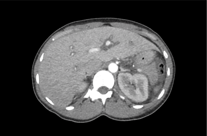
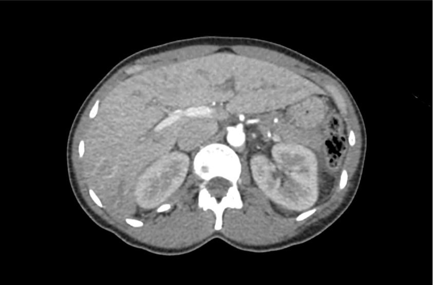
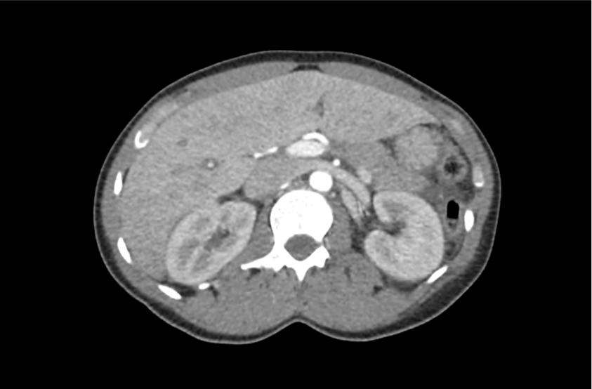
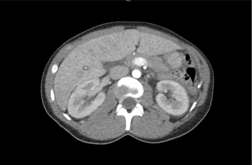
CT Abdomen/Pelvis with Contrast

Moderate free intra-abdominal fluid, heterogeneous liver with periportal edema, dense right middle lobe consolidation.

ED Course:

The patient developed worsening respiratory failure with hypoxia and tachypnea requiring endotracheal intubation. Thrombotic thrombocytopenic purpura was suspected and while awaiting emergent plasma exchange transfusion, the patient arrested and resuscitation efforts were unsuccessful.

The patient’s ADAMTS13 activity level was <3%. Autopsy demonstrated consolidation of the right middle lobe with possible lymphoproliferative mass, and lung petechial hemorrhages from microvascular thrombi.

Differential Diagnosis of Thrombocytopenia 1-7

Differential Diagnosis of Thrombocytopenia

Algorithm for the Evaluation of Thrombocytopenia 8

Algorithm for the Evaluation of Thrombocytopenia

Definition 9

  • Mild: <150k
  • Moderate: 100-150k
  • Severe: <50k
    • 10-30k: bleeding with minimal trauma
    • <10k: increased risk spontaneous bleeding

History 9,10

  • Prior platelet count
  • Family history bleeding disorders
  • Medications
    • Heparin
    • Quinine, quinidine
    • Rifampin
    • Trimethoprim-sulfamethoxazole
    • Vancomycin
  • Alcohol use
  • Travel-related infections

Physical Examination 9,10

  • Splenomegaly (liver disease)
  • Lymphadenopathy (infection, malignancy)

Workup 10,11

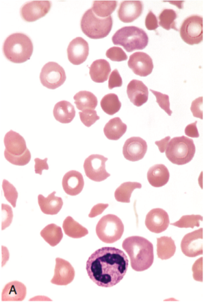
Schistocytes

Red blood cell fragments (schistocytes) 11

  • hCG
  • Repeat CBC
    • Detect spurious measure
    • Neutrophil-predominant leukocytosis: bacterial infection
    • Immature leukocytes (blasts): leukemia, myelodysplasia
  • Peripheral smear
    • Schistocytes: microangiopathic process (DIC, TTP, HUS)
    • Atypical lymphocytes: viral infection
    • Intracellular parasites: malaria
    • Hypersegmented neutrophils: nutritional deficiency
  • Infectious features: HIV, HCV, EBV, H.pylori, blood cultures
  • Autoimmune features: ANA, APL-Ab
  • Suspected occult liver disease: LFT, PT/PTT/INR
  • Suspected thrombotic microangiopathy: PT/PTT/INR, haptoglobin, LDH, fibrinogen, FDP, d-dimer

Specific Conditions 2-6,9,12-20

Disease Cause Presentation Laboratory Findings Treatment
DIC Sepsis
Trauma
Burn
Malignancy
Bleeding
Multi-organ failure
Shock
INR
Fibrinogen
FDP
D-dimer
Directed at underlying cause
Transfusion thresholds for hemorrhage:
FFP for INR >1.5
Platelets if <50k
Cryoprecipitate of fibrinogen <100mg/dL
TTP Insufficient ADAMTS-13 activity Non-specific constitutional symptoms (ex. weakness)
Neuro: headache, AMS, focal neuro deficit
GI: abdominal pain, nausea/vomiting
LDH
Reticulocyte
Unconjugated bilirubin
Haptoglobin
Plasma exchange
HUS Shiga-toxin-producing bacteria, E. coli O157:H7 Bloody diarrhea, anuria, oliguria, and hypertension Aggressive supportive care
HELLP Spectrum of eclampsia Hypertension
Visual symptoms
Headache
RUQ abdominal pain
AST/ALT
Uric acid
Unconjugated bilirubin
LDH
Reticulocyte
Haptoglobin
Delivery, MgSO4
ITP Primary ITP

Secondary ITP
– Drug
– Autoimmune
– Infection
– Malignancy

Usually asymptomatic, may have petechiae or easy bruising Isolated thrombocytopenia Steroids
HIT Exposure to heparin or LMWH Thrombocytopenia or a 50 percent reduction in platelet count between 5-10d exposure
New thrombosis or skin necrosis
4 T’s score
Platelet factor 4 antibodies Withdraw heparin

References

  1. Greinacher A, Selleng S. How I evaluate and treat thrombocytopenia in the intensive care unit patient. Blood. 2016;128(26):3032-3042. doi:10.1182/blood-2016-09-693655.
  2. Joly BS, Coppo P, Veyradier A. Thrombotic thrombocytopenic purpura. Blood. 2017;129(21):2836-2846. doi:10.1182/blood-2016-10-709857.
  3. Leslie SD, Toy PT. Laboratory hemostatic abnormalities in massively transfused patients given red blood cells and crystalloid. Am J Clin Pathol. 1991;96(6):770-773.
  4. Neunert C, Lim W, Crowther M, et al. The American Society of Hematology 2011 evidence-based practice guideline for immune thrombocytopenia. Blood. 2011;117(16):4190-4207. doi:10.1182/blood-2010-08-302984.
  5. Kappler S, Ronan-Bentle S, Graham A. Thrombotic microangiopathies (TTP, HUS, HELLP). Emerg Med Clin North Am. 2014;32(3):649-671. doi:10.1016/j.emc.2014.04.008.
  6. Greinacher A. Heparin-Induced Thrombocytopenia. Solomon CG, ed. N Engl J Med. 2015;373(3):252-261. doi:10.1056/NEJMcp1411910.
  7. Reardon JE Jr., Marques MB. Evaluation of Thrombocytopenia. Lab Med. 2006;37(4):248-250. doi:10.1309/R7P79KERAJHPRHLT.
  8. Stasi R. How to approach thrombocytopenia. Hematology Am Soc Hematol Educ Program. 2012;2012:191-197. doi:10.1182/asheducation-2012.1.191.
  9. Gauer RL, Braun MM. Thrombocytopenia. Am Fam Physician. 2012;85(6):612-622.
  10. Abrams CS. 172 – Thrombocytopenia. Twenty Fifth Edition. Elsevier Inc.; 2016:1159–1167.e2. doi:10.1016/B978-1-4557-5017-7.00172-0.
  11. Wilson CS, Vergara-Lluri ME, Brynes RK. Chapter 11 – Evaluation of Anemia, Leukopenia, and Thrombocytopenia. Second Edition. Elsevier Inc.; 2017:195-234.e195. doi:10.1016/B978-0-323-29613-7.00011-9.
  12. Hui P, Cook DJ, Lim W, Fraser GA, Arnold DM. The frequency and clinical significance of thrombocytopenia complicating critical illness: a systematic review. Chest. 2011;139(2):271-278. doi:10.1378/chest.10-2243.
  13. Jokiranta TS. HUS and atypical HUS. Blood. 2017;129(21):2847-2856. doi:10.1182/blood-2016-11-709865.
  14. Neunert CE. Management of newly diagnosed immune thrombocytopenia: can we change outcomes? Hematology Am Soc Hematol Educ Program. 2017;2017(1):400-405. doi:10.1182/asheducation-2017.1.400.
  15. Lambert MP, Gernsheimer TB. Clinical updates in adult immune thrombocytopenia. Blood. 2017;129(21):2829-2835. doi:10.1182/blood-2017-03-754119.
  16. Arepally GM. Heparin-induced thrombocytopenia. Blood. 2017;129(21):2864-2872. doi:10.1182/blood-2016-11-709873.
  17. Aster RH, Bougie DW. Drug-induced immune thrombocytopenia. N Engl J Med. 2007;357(6):580-587. doi:10.1056/NEJMra066469.
  18. Boral BM, Williams DJ, Boral LI. Disseminated Intravascular Coagulation. Am J Clin Pathol. 2016;146(6):670-680. doi:10.1093/ajcp/aqw195.
  19. Scully M, Hunt BJ, Benjamin S, et al. Guidelines on the diagnosis and management of thrombotic thrombocytopenic purpura and other thrombotic microangiopathies. Br J Haematol. 2012;158(3):323-335. doi:10.1111/j.1365-2141.2012.09167.x.
  20. Levine RL, Hursting MJ, Drexler A, Lewis BE, Francis JL. Heparin-induced thrombocytopenia in the emergency department. Ann Emerg Med. 2004;44(5):511-515. doi:10.1016/j.annemergmed.2004.06.004.

Sickle Cell Crises

Brief H&P

A 27 year-old male with sickle cell disease (HbSC) on hydroxurea and with a history of 2-3 hospitalizations per year for vaso-occlusive pain crises manifested by arthralgias and back pain presents to the emergency department with 3 days of worsening joint pain affecting his entire body but predominantly his knees and lower back. He is familiar with this pain and attempted therapy at home with ibuprofen, then hydrocodone-acetaminophen, and finally hydromorphone without improvement and presented to the emergency department.

On review of systems, he denied chest pain, cough, or shortness of breath. He has some periumbilical abdominal pain but tolerated normal oral intake on the day of presentation without vomiting nor changes in bowel habits. He otherwise denied fevers, peripheral numbness/weakness, urinary or fecal incontinence or retention. He similarly denies trauma, weight loss, night sweats, or intravenous drug use.

Objectively, the patient’s vital signs were normal and he was well-appearing. Mucous membranes were moist and skin turgor was normal. There were no appreciable joint effusions, warmth, nor limitation to active/passive range of motion of any joints. His back had no midline tenderness to palpation or percussion, normal range of motion in all axes and extremity sensation and strength testing were normal. Abdominal and genitourinary examinations were normal. The patient had preserved perineal sensation to light touch and normal rectal tone – a core temperature was obtained which was also normal.

Peripheral access was established and a parenteral dose of hydromorphone equivalent to his home oral dose was administered (0.015mg/kg). Repeat dosing was required at 15 minutes due to persistent pain scale of 10. Diphenhydramine and acetaminophen were also administered, for potential opioid-sparing effects, recognizing the limited evidence to support these relatively benign adjuncts.

Laboratory studies were notable for anemia (though stable compared to baseline measures), appropriate reticulocyte count, no evidence of hemolysis and with normal electrolytes and renal function.

A thorough history and examination did not identify a critical precipitant for the patient’s symptoms which were presumed to be secondary to a vaso-occlusive pain crisis. On reassessment, the patient’s pain was improved and an oral dose of hydromorphone was administered with continued observation and serial reassessments for two hours thereafter. The patient’s hematologist was available for follow-up the subsequent morning and the patient was discharged home.

Pharmacokinetics of Commonly-Used Opiate Analgesics1-3

Medication Route Onset Peak Duration
Morphine IV 5-10min 20min 3-5h
IM 15-30min 30-60min
PO 30min 1h
Oxycodone PO 10-15min 30-60min 3-6h
Hydrocodone PO 10-20min 4-8h
Fentanyl IV <1min 2-5min 30-60min
Hydromorphone IV 5min 10-20min 3-4h
PO 15-30min 30-60min
Codeine PO 30-60min 60-90min 4-6h

Spectrum of Sickle Cell Trait and Disease4

Algorithm for the Evaluation and Management of Sickle Cell Crises4-10

Algorithm for the Management of Sickle Cell Crises

References:

  1. Lexicomp Online®, Adult Drug Information, Hudson, Ohio: Lexi-Comp, Inc.; November 4, 2017.
  2. Trescot AM, Datta S, Lee M, Hansen H. Opioid pharmacology. Pain Physician. 2008;11(2 Suppl):S133-S153.
  3. Vieweg WVR, Lipps WFC, Fernandez A. Opioids and methadone equivalents for clinicians. Prim Care Companion J Clin Psychiatry. 2005;7(3):86-88.
  4. Glassberg J. Evidence-based management of sickle cell disease in the emergency department. Emergency Medicine Practice. 2011;13(8):1–20–quiz20.
  5. Raam R, Mallemat H, Jhun P, Herbert M. Sickle Cell Crisis and You: A How-to Guide. Ann Emerg Med. 2016;67(6):787-790. doi:10.1016/j.annemergmed.2016.04.016.
  6. Piel FB, Steinberg MH, Rees DC. Sickle Cell Disease. N Engl J Med. 2017;376(16):1561-1573. doi:10.1056/NEJMra1510865.
  7. Lovett PB, Sule HP, Lopez BL. Sickle cell disease in the emergency department. Emerg Med Clin North Am. 2014;32(3):629-647. doi:10.1016/j.emc.2014.04.011.
  8. Yawn BP, John-Sowah J. Management of Sickle Cell Disease: Recommendations From the 2014 Expert Panel Report. Vol 92. 2015:1069-1076.
  9. Zempsky WT. Evaluation and Treatment of Sickle Cell Pain in the Emergency Department: Paths to a Better Future. Clinical Pediatric Emergency Medicine. 2010;11(4):265-273. doi:10.1016/j.cpem.2010.09.002.
  10. Aliyu ZY, Tumblin AR, Kato GJ. Current therapy of sickle cell disease. Haematologica. 2006;91(1):7-10.

Hematologic Emergencies

Sickle Cell Crises

  • Triggers: infection, acidosis, dehydration, cold-exposure, hypoxia, pregnancy
  • Presentation: exclude alternative more serious pathology prior to ascribing pain to vaso-occlusive crisis

Effects by Organ System

System Symptom
CNS Focal or generalized neurological symptoms, stroke, seizure
Pulmonary Acute chest syndrome (fever, chest pain, cough, hypoxia, pulmonary infiltrates), pulmonary embolism
GI Abdominal pain, nausea/vomiting
Renal Papillary necrosis
GU Priapism, testicular/ovarian ischemia
Muskuloskeletal Bone pain (back, proximal extremities), exclude osteomyelitis, avascular necrosis
ID Infection, functional asplenia (streptococcus, haemophilus)
OB Preterm labor, placental abruptions, SAB
Ophthalmology Acute retinal ischemia, hyphema (with intra-ocular hypertension)
Hematology
  • Sequestration crisis: acute anemia, often post-viral
  • Hemolytic crisis: acute anemia, reticulocytosis, hyperbilirubinemia
  • Megaloblastic crisis: folate deficiency
  • Aplastic crisis: inadequate reticulocytosis

Evaluation

  • CBC with reticulocyte count
    •  Hemoglobin: suggests sequestration or hemolytic crisis
    • Reticulocyte index: suggests aplastic or megaloblastic crisis
  • LDH/haptoglobin: evaluate for hemolysis
  • UA: evaluate for infection/infarction
  • CXR: evaluate for acute chest syndrome

Management

  • Rehydration (hypotonic fluids)
  • Analgesia
  • Supplemental oxygen if hypoxic
  • Exchange transfusion for priapism, neurologic symptoms, aplastic/sequestration/hemolytic crises

Transfusion Reactions

  • Epidemiology: overall 0.25%, 0.09% severe
  • Management: stop transfusion

Management by Presumed Etiology

Reaction Mechanism Signs/symptoms Management
Acute, Severe
Acute hemolysis Incompatibility Fevers, HR, BP, vomiting, back pain IVF, vasopressors if needed, furosemide
Anaphylaxis IgA-mediated 1min: flushing laryngospasm, bronchospasm, BP Epinephrine, steroids, diphenhydramine, IVF
Sepsis Bacterial contamination (Y. entercolitica), increased risk in platelet transfusion Fevers, BP IVF, vasopressors if needed, broad-spectrum antibiotics
TRALI (transfusion-related acute lung injury) Non-cardiogenic pulmonary edema, increased risk in FFP transfusion Hypoxia, respiratory distress, XR bilateral infiltrates Supplemental oxygen, PPV/ETT
TACO (transfusion-associated circulatory overload) Hypervolemia in patients with history of CHF Hypoxia, respiratory distress, heart failure Supplemental oxygen, PPV/ETT, furosemide
Acute, Minor
Simple febrile reaction Cytokine-mediated Isolated fever Acetaminophen
Minor allergic reaction Response to transfused plasma proteins Urticaria, pruritus, flushing Diphenhydramine
Delayed
Delayed hemolysis Minor RBC antigens 5-10d, low-grade hemolysis  
GVHD Immunocompromised host Fever, rash, N/V, transaminitis, pancytopenia  
Massive Transfusion
Massive transfusion Large-volume, refrigerated products Coagulopathy, hypothermia, hypocalcemia, hyperkalemia, lactic acidosis

Bleeding Disorders

Overview

  • Disorders of primary hemostasis
    • General: present with mucocutaneous, post-operative bleeding
    • vWD
    • Platelet disorders
      • Medication-induced: NSAID, valproate, B-lactam, SSRI
      • Systemic disease: hepatic, renal failure
    • ITP: antibody-mediated platelet destruction
  • Disorders of secondary hemostasis
    • General: present with bleeding into soft-tissue, joints
    • Hemophilia A (VIII)
    • Hemophilia B (IX)
  • Disorders of both primary and secondary hemostasis
    • DIC
    • Liver disease
    • Severe vWD
  • Evaluation
    • PT: VII, vitamin K
    • PTT: VIII, IX, XI, XIII, vWD, heparin
    • Increased PT/PTT: XI, V, vitamin K, heparin, DIC
    • CBC: degree of anemia, platelet count, differential (hematopoetic disorders)
  • Management
    • Thrombocytopenia
      • Prophylactic transfusion for avoidance of spontaneous hemorrhage for platelet count <10,000
      • Transfusion for active bleeding at platelet count <50,000
      • Dosing
        • Adults: one RDP increases platelet count by 7-10,000
        • Pediatrics: 5-10ml/kg
      • ITP
        • Transfuse platelets for active bleeding
        • High-dose steroids (prednisone 1mg/kg)
        • IVIG (1g/kg/d)
      • Uremia
        • Hemodialysis
        • DDAVP (0.3ug/kg IV)
      • vWD
        • DDAVP (0.3ug/kg IV)
        • Severe: VWF (Humate-P) 40-80IU/kg
        • Tranexamic acid
      • Hemophilia A
        • Minor: 20IU/kg
        • Major: 50IU/kg
      • Hemophilia B
        • Minor: 40IU/kg
        • Major: 100IU/kg

DIC/TTP/HUS

  • Disseminated Intravascular Coagulation
    • Etiology: severe systemic illness/injury
      • Trauma, burn, crush
      • Sepsis
      • Malignancy
      • Obstetric complication: abruption, amniotic fluid embolism
      • Hemolytic anemia
    • Exam: petechiae/purpura, hemorrhage (puncture site, GI, GU, pulmonary)
    • Labs:
      • PT/PTT
      • Fibrinogen
      • CBC: schistocytes, thrombocytopenia
      • FDP/D-Dimer
    • Management
      • Treat underlying illness
      • Transfuse (PRBC, FFP for INR > 2, cryoprecipitate for fibrinogen < 100)
      • Heparin if apparent embolic events
      • Consult hematology
  • TTP/HUS
    • Presentation
      • Thrombocytopenia
      • Altered mental status
      • Renal dysfunction
      • Fever
      • MAHA
    • TTP: more commonly associated with altered mental status
      • Etiology: drugs, pregnancy, infection (HIV)
      • Mechanism: ULvWF uncleaved by dysfunctional ADAMTS-13
    • HUS: more commonly associated with renal dysfunction
      • Mechanism: toxin from E. coli, Shigella
      • Timing: 1-2wks after diarrheal illness
    • Evaluation
      • CBC: anemia, schistocytes, thrombocytopenia
      • PT/PTT (normal)
      • BUN/Creatinine
      • LDH
    • Management
      • Platelets contraindicated except as stopgap measure in ICH (can worsen process)
      • Plasma exchange with FFP (replaces functional ADAMTS-13)
      • Steroids (prednisone 1mg/kg daily)
      • Hematology consultation

Complications of anti-thrombotic therapy

  • Agents
    • Anti-platelet
      • TXA: Aspirin
      • ADP: clopidogrel, ticagrelor, prasugrel
      • GPIIb/IIIa: abciximab, eptifibatide, tirofiban
    • Anti-coagulants
      • Anti-thrombin: heparin, LMWH (enoxaparin, dalteparin)
      • Vitamin K antagonist: warfarn (anti-II, VII, IX, X)
      • Direct thrombin inhibitor: bivalirudin, argatroban, dabigatran
      • Xa inhibitor: rivaroxaban, apixaban
    • Fibrinolytics
      • Alteplase, tenectaplase
  • Complications
    • HIT: platelet count decrease >50% at 5 days

Summary of Management

Agent Reversal
Aspirin, clopidogrel 5-10U platelets

DDAVP 0.3ug/kg

GPIIb/IIIa Abciximab: 5-10U platelets

Eptifibatide/tirofiban: none

Heparin Protamine 1mg/100mg heparin in last 2-3 hours
LMWH Enoxaparin: 1mg/1mg

Dalteparin: 1mg/100U

Warfarin See supratherapeutic INR algorithm
DTI Dabigatran: Praxbind, hemodialysis, consider Factor VIIa
Xa PCC
Fibrinolytics 10U cryoprecipitate, 2U FFP, consider platelets and aminocaproic acid (4-5g IV)

Oncologic Emergencies

Overview

  •  Complications
    • Airway obstruction
    • PNA
    • Pleural effusion
    • Pericardial effusion
    • VTE
    • SVC syndrome
      • Symptoms: dyspnea (airway edema), chest fullness, blurred vision, headache (increased ICP)
    • Massive hemoptysis
      • Management: ETT (large-bore for bronschoscopy), affected side down
  • Brain Metastases
    • Cancers: melanoma, lung, breast, colorectal
    • Management: dexamethasone 10mg IV load, elevated HOB, hypertonic saline or mannitol, prophylactic anti-eplipetics
  • Meningitis
    • Pathogens: Listeria (ampicillin), Cryptococcus (amphotericin)
    • Evaluation: CSF sampling with cytology (diagnose leptomeningeal metastases)

Metabolic Disturbances

  • Hypercalcemia
    • Cancers: MM, RCC, lymphoma, bone metastases (breast, lung, prostate)
    • Mechanism: metastatic destruction, PTH-RP, tumor calcitriol
    • Prognosis: 50% 30-day mortality
    • Symptoms
      • Chronic: anorexia, nausea/vomiting, constipation, fatigue, memory loss
      • Acute: CNS (lethargy, somnolence)
    • Findings
      • Calcium: >13.0mg/dL
      • ECG: QT shortening
    • Treatment
      • Mild: IVF
      • Severe: IVF, loop diuretics, bisophosphanate (pamidronate 90mg IV infused over 4 hours), consider calcitriol, consider hemodialysis if cannot tolerate fluids or unlikely to respond to diuretics
  • Hyponatremia
    • Cancers: lung (small-cell), pancreatic, ovarian, lymphoma, thymoma, CNS
    • Mechanism: SIADH
    • Symptoms: muscle twitching, seizure, coma
    • Management: fluid restriction, if seizing administer 3% hypertonic saline at 100cc/hr until resolution
  • Hypernatremia
    • Mechanism: decreased intake, increased GI losses from chemotherapy
    • Management: cautious fluid resuscitation
  • Tumor Lysis Syndrome (TLS)
    • Cancers: hematologic, rapid-growth solid tumors
    • Mechanism: release of intracellular contents (uric acid, K, PO4, Ca)
    • Timing: 1-4 days after therapy (chemo, radiation)
    • Diagnosis
      • Uric acid >8mg/dL
      • Potassium >6mEq/L
      • Calcium <7mg/dL
      • PO4 >4.5mg/dL
      • Acute kidney injury
    • Management
      • IVF, allopurinol, rasburicase, urinary alkalinization
      • Consider hemodialysis if volume overloaded

Localized Complications

  • Musculoskeletal Complications
    • Spinal cord compression
      • Cancers: prostate, breast, lung, RCC, non-Hodgkin lymphoma, MM (5-10% of all cancer patients)
      • Sites: thoracic (60%), lumbosacral (30%), cervical (10%)
      • Symptoms: pain (worse lying flat, cough/sneeze, heavy lifting)
      • Evaluation: MRI (se 93%, sp 97%)
      • Management: dexamethasone 10mg IV load, 4mg q6h, neurosurgical consultation, radiation oncology consultation
    • Pathologic fracture
      • Features: sudden onset, low-force mechanism
  • Therapy Complications
    • Neutropenic fever
      • Definition: ANC <500 or ANC <1000 with expected nadir <500 (nadir typically occurs 5-10d after chemotherapy) with Tmax >38.3°C or >38.0°C for >1h
      • Examination: subtle signs of infection, thorough examination is critical (skin, catheter, perineum)
      • Treatment: carbapenem monotherapy, vancomycin if indwelling catheter, oncology consultation for colony stimulating factors
    • Chemotherapy-induced vomiting
      • Management: ondansetron with dexamethasone, consider NK-1 antagonist (aprepitant)

Hematologic Malignancies

  • Acute leukemia
    • Signs/Symptoms: leukopenia (infection), anemia (weakness/fatigue), thrombocytopenia (bleeding)
    • Diagnosis: >5% blasts
  • Thrombocytopenia
    • Management
      • No bleeding, goal >10,000
      • Fever, coagulopathy, hyperleukoctosis, goal >20,000
      • One unit of platelets increases count by 5,000
  • Hyperleukocytosis
    • Definition: WBC > 50-100k
    • Complications: microvascular congestion (pulmonary, cerebral, coronary)
    • Symptoms
      • CNS: confusion, somnolence, coma
      • Pulmonary: dyspnea, respiratory alkalosis
    • Management: cytoreduction (induction chemotherapy, increased risk TLS)
  • Hyperviscosity
    • Cancer: macroglobulinemia, MM
    • Symptoms: epistaxis, purpura, GIB, neuro deficits
    • Diagnosis: serum viscosity > 1.4-1.8
    • Management: emergent plasmapheresis
  • Polycythemia
    • Diagnosis: Hb >17
    • Differential: dehydration, hypoxia, smoking, altitude
    • Symptoms: HA, vertigo, angina, claudication, pruritus (after showering)
    • Complications: thrombosis (stroke), bleeding
    • Management: emergent phlebotomy (500cc if otherwise healthy)
  • Thrombocytosis
    • Diagnosis: platelet >1,000,000
    • Symptoms: vasomotor (HA, lightheadedness, syncope, chest pain, paresthesias)
    • Management: low-dose aspirin

Anemia

HPI

70M with a history of dementia presenting with 3 days of fatigue. The patient was unable to provide detailed history, however family members reported worsening fatigue with the patient requiring assistance with ambulation for several days. The patient was referred from an outside clinic after point-of-care hemoglobin of 6.7. No reported history of anemia, and no history suggestive of obvious external bleeding.

Vital signs stable, tachycardia and tachypnea noted with minimal exertion but saturating well on ambient air and in no acute distress. Examination notable for conjunctival pallor without scleral icterus, systolic flow murmur, brown stool guaiac negative.

CBC with hemoglobin of 7.5 , MCV 80.3 , RDW 22.4 , no leukocytosis and normal platelets. Also noted was an alkaline phosphatase of 828 , normal total and direct bilirubin, and undetectable serum troponin. Chest x-ray showed a possible pleural-based mass.

The patient was transfused two units of PRBC’s and admitted for further evaluation. CT chest/abdomen/pelvis revealed sternal and rib-based pleural soft-tissue mass, prostate mass, pelvic and retroperitoneal lymphadenopathy as well as extensive bony metastatic disease consistent with primary prostate cancer with diffused metastasis. Serum PSA was 2,087 . Iron studies suggested anemia of chronic disease. Reticulocytes were not obtained but may have suggested inadequate production index given extensive bony metastases and possible associated myelosuppression. The patient was symptomatically improved after transfusion and discharged with outpatient follow-up for discussions regarding possible biopsy and treatment.

Images

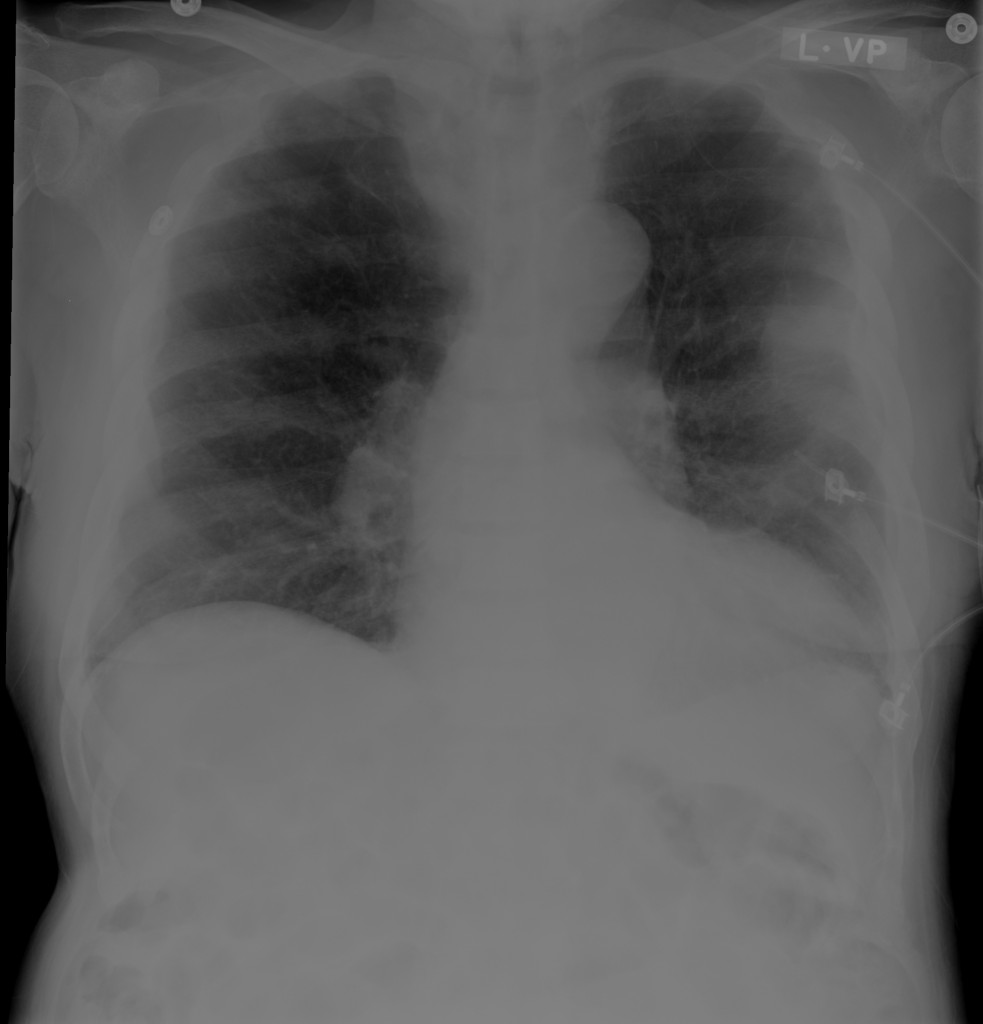
Chest x-ray with pleural-based mass

Areas of pleural thickening. Possible pleural based mass in left mid lung.

ct_chest_anemia_01
ct_chest_anemia_02
ct_chest_anemia_03
ct_chest_anemia_04
ct_chest_anemia_05
ct_chest_anemia_06
ct_chest_anemia_07
ct_chest_anemia_08
ct_chest_anemia_09
ct_chest_anemia_10
ct_chest_anemia_11
ct_chest_anemia_12
ct_chest_anemia_13

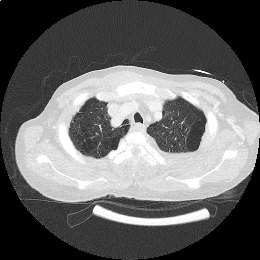
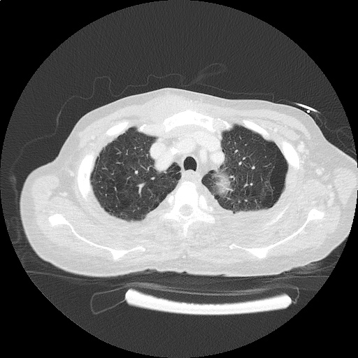
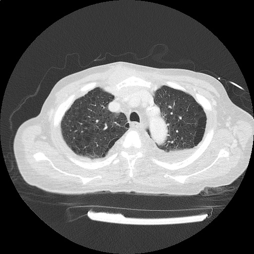
CT Chest: Lung Window

  • Rib-based pleural soft tissue masses.
  • Large 5.6 x 4.4cm anterior sternal soft-tissue mass.
ct_bone_anemia_01
ct_bone_anemia_02
ct_bone_anemia_03
ct_bone_anemia_04
ct_bone_anemia_05
ct_bone_anemia_06
ct_bone_anemia_07
ct_bone_anemia_08

CT Body: Bone Window

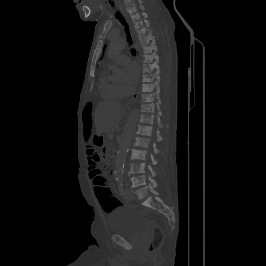
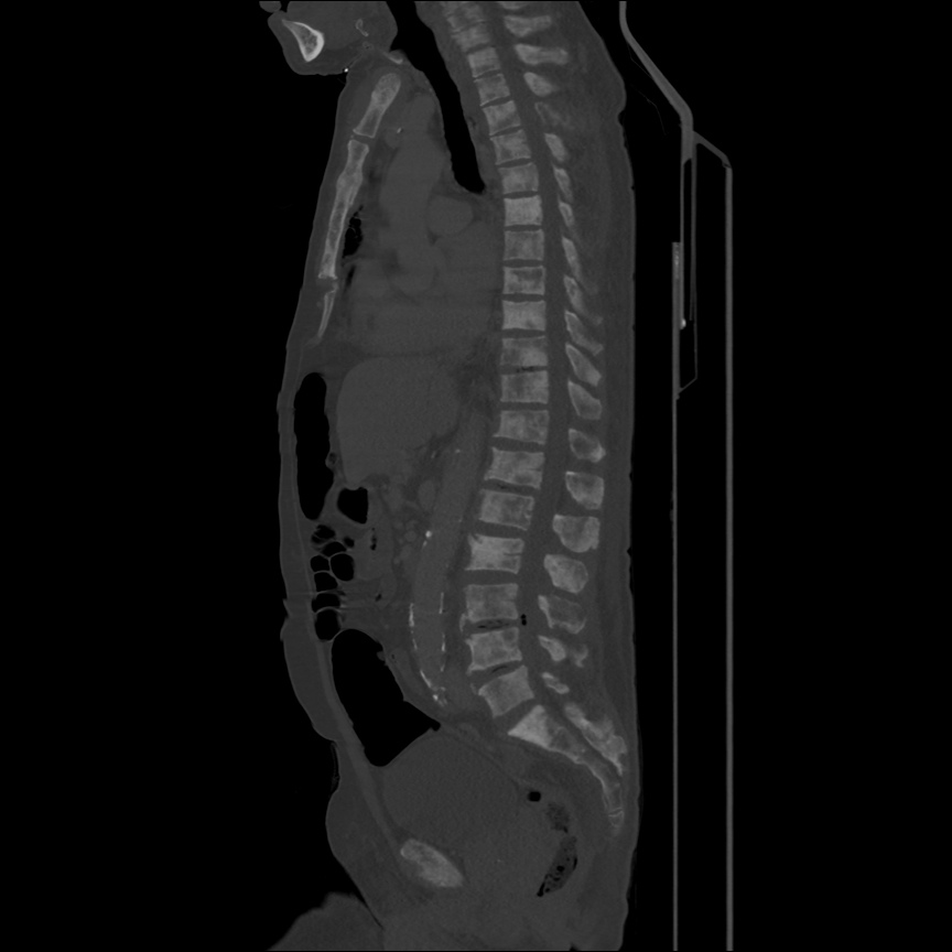
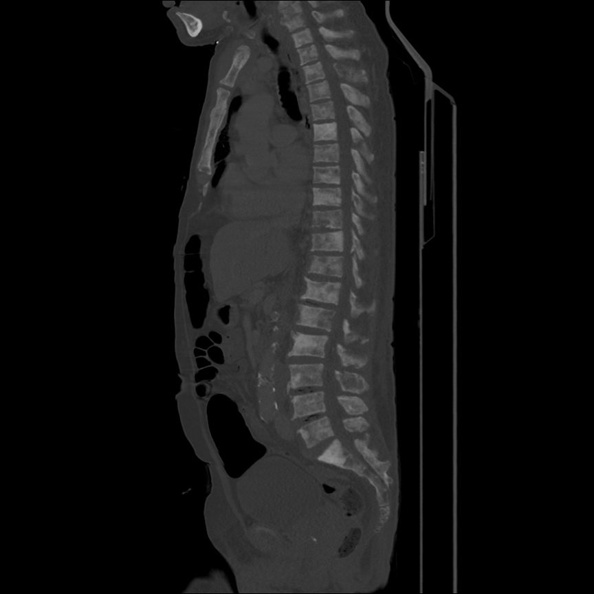
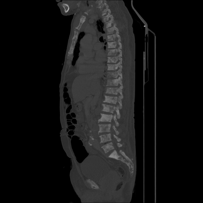
  • Extensive bony metastatic disease.
  • Prostate mass, large pelvic and retroperitoneal lymphadenopathy.
  • Consistent with primary prostate cancer with diffuse metastasis.

Algorithm for the Evaluation of Anemia 1,2

Algorithm for the Evaluation of Anemia

References:

  1. Zaiden R, Rana F. Evaluation of Anemia. BMJ Best Practice. Oct 2014. http://us.bestpractice.bmj.com/best-practice/monograph/93/overview.html. Last accessed 15 May 2015.
  2. Janz, T. G., Johnson, R. L., & Rubenstein, S. D. (2013). Anemia in the emergency department: evaluation and treatment. Emergency medicine practice, 15(11), 1–15– quiz 15–6.
  3. WiKEM: Anemia

Lymphadenopathy Applied: Lymphoma

HPI:

27 year-old female with no medical history presenting with neck swelling. She describes one month of progressive enlargement of a left-sided neck mass, and in the past two weeks has noted a new right-sided neck mass. This has been associated with worsening dysphagia to solids, describing a sensation of food lodging in the mid-chest and requiring liquids for passage – she attributes her recent 10lb weight loss to this. She also reports a non-productive cough for the past two weeks and generalized fatigue. She otherwise denies fevers, night sweats, chest pain, shortness of breath, nausea/vomiting, or changes in bowel/urinary habits. She has no known sick contacts or TB exposure risk factors. She has no medical history, no prior surgeries, does not take any medications and denies tobacco, alcohol or drug use.

Physical Exam:

VS: T 38.4 HR 98 RR 14 BP 108/68 O2 99% RA
Gen: Well-appearing young female, in no acute distress.
HEENT: PERRL, EOMI, MMM without lesions. There is a 2x3cm firm, non-tender, mobile left supraclavicular lymph node, as well as two 1x1cm firm, non-tender lymph nodes in the left and right anterior cervical chains.
CV: RRR, normal S1/S2, no murmurs. No JVD.
Lungs: Clear to auscultation bilaterally. There is a transition to bronchial breath sounds along the trachea inferior to the sternal angle with normal tracheal sounds superiorly.
Abd: Soft, non-tender without organomegaly.
Ext: Warm and well-perfused with normal peripheral pulses. No axillary or inguinal lymphadenopathy.
Neuro: Alert and oriented, responding appropriately to questions. PERRL, EOMI, facial sensation symmetric, facial muscles symmetric, hearing grossly normal, palate rises symmetrically, tongue movements normal without fasciculation, SCM/trapezius normal. Normal FTN, RAM. Gait intact. Peripheral sensation and motor grossly normal.

Imaging:

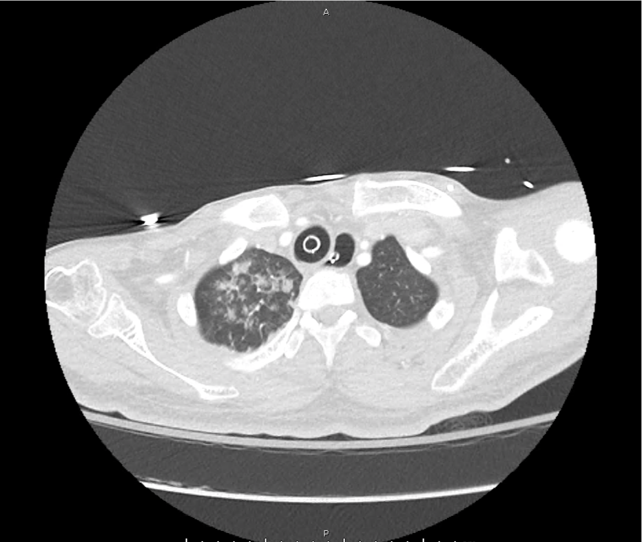
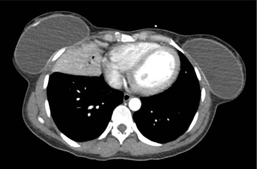
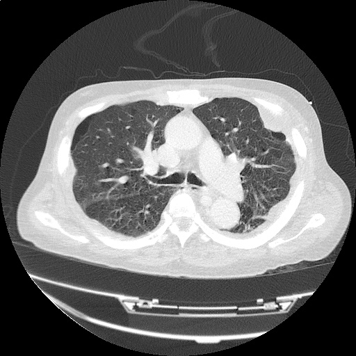
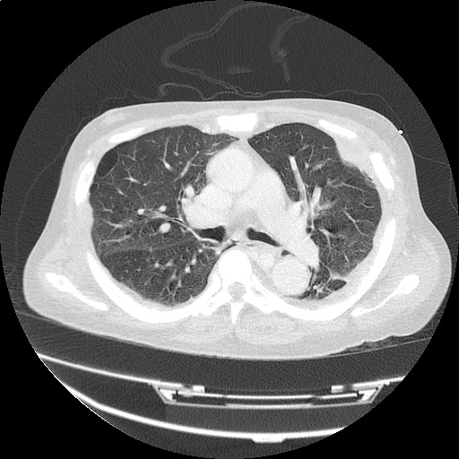
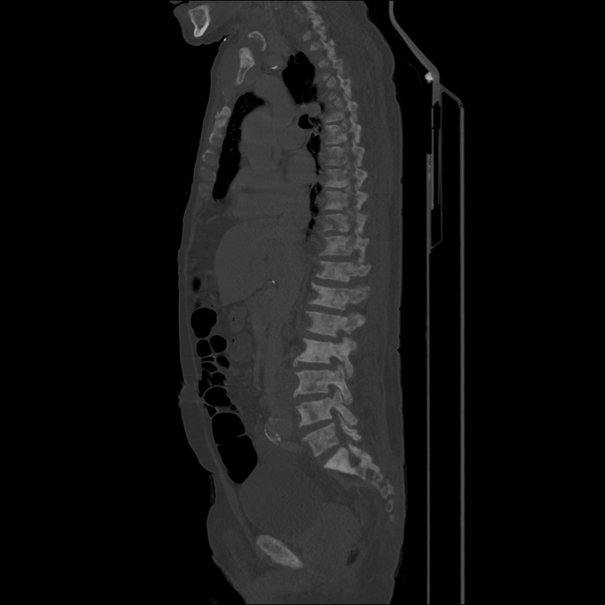
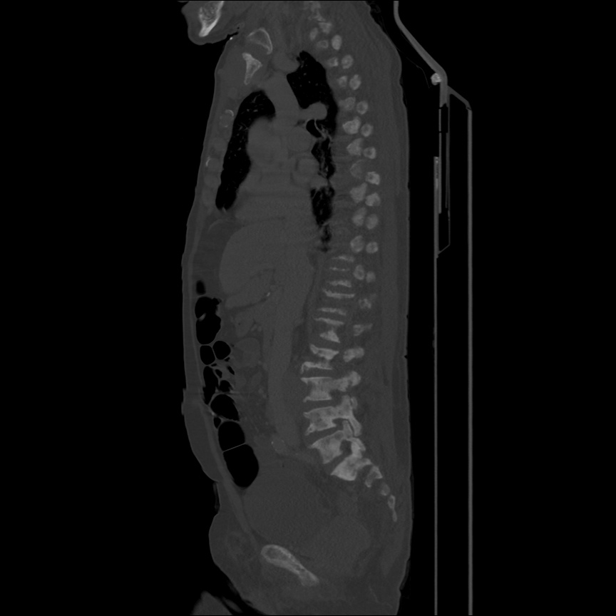
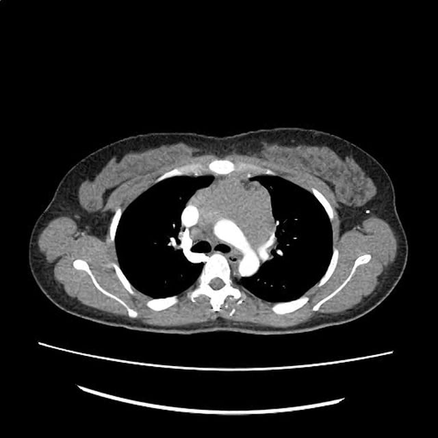
CT Chest - Axial

CT Chest - Axial

Anterior mediastinal mass with a wide differential - likely represents lymphoma or germ cell tumor. Less likely thymic or thyroid origin.

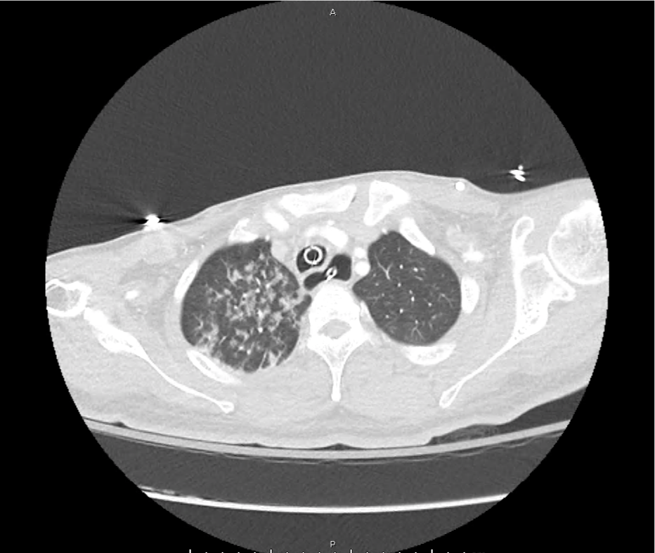
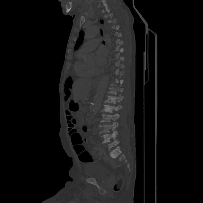
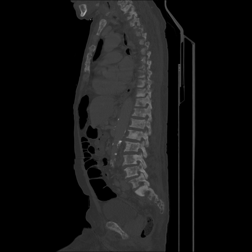
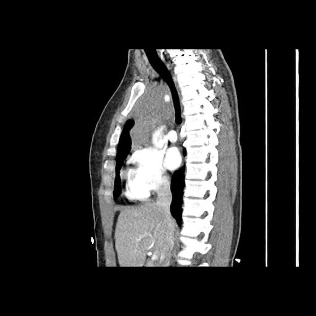
CT Chest - Sagittal

CT Chest - Sagittal

Anterior mediastinal mass with a wide differential - likely represents lymphoma or germ cell tumor. Less likely thymic or thyroid origin.

Assessment/Plan:

27F with no PMH presenting with progressive localized lymphadenopathy. Resultant dysphagia, cervical and supraclavicular distribution as well as abnormal tracheal sounds concerning for mediastinal involvement. The patient is currently stable without evidence of airway compromise. A CT of the chest was obtained to evaluate for thoracic malignancy, which showed a large anterior mediastinal mass concerning lymphoma or germ cell tumor. The location of the mass likely explains the patient’s dysphagia due to compression of the esophagus, as well as the abnormal pulmonary exam with compression potentially irritating the trachea and triggering her non-productive cough. The patient was admitted for further workup.

Lymphadenopathy Applied – Lymphoma

This case applies the differential diagnosis of lymphadenopathy. The most abnormal finding on examination was non-tender, left supraclavicular lymphadenopathy. The duration of symptoms and lack of tenderness is concerning for malignancy, and the left supraclavicular location suggests a thoracic or intra-abdominal source.

Lymphadenopathy Applied - Lymphoma