Types of Dizziness
Distinguishing Central vs. Peripheral Vertigo
| Characteristic | Peripheral | Central |
|---|---|---|
| Onset | Sudden | Gradual |
| Intensity | Severe | Mild |
| Duration | Minutes | Weeks |
| Timing | Intermittent | Continuous |
| Nystagmus | Horizontal | Vertical, bidirectional |
| Exacerbation with head movement | + | – |
| Auditory symptoms | + | – |
| Neurological findings | – | + |
Causes of Vertigo
Characteristics of common causes of vertigo
| Cause | Mechanism | Onset | Symptoms | Findings |
|---|---|---|---|---|
| Peripheral | ||||
| BPPV | Otolith | Brief, positional episodes | Nausea, vomiting, absent auditory symptoms. | Dix-Hallpike positive |
| Vestibular neuronitis | Viral, post-viral inflammation of vestibular portion of CNVIII | Acute and severe, subsiding over days. | Nausea, vomiting, absent auditory symptoms. | Head thrust abnormal |
| Meniere | Endolymphatic hydrops | Recurrent, lasting hours | Tinnitus, hearing loss. | SNHL |
| Central | ||||
| Vertebrobasilar insufficiency | Atherosclerosis (vascular risk factors) | Acute onset, recurrent episodes if TIA | Headache, gait impairment, diplopia, absent auditory symptoms. | Neurologic deficits |
| Cerebellar stroke | Atherosclerosis (vascular risk factors) | Acute and severe | Headache, dysphagia, gait impairment | Dysmetria, dysdiadochokinesia, ataxia, CN palsy |
| Brainstem stroke | Atherosclerosis (vascular risk factors), dissection | Acute and severe | Dysphagia, dysphonia, gait impairment, sensory disturbances | Loss of pain/temperature on ipsilateral face, contralateral body, palatal/pharyngeal paralysis |
| MS | Demyelination | Subacute onset | History of other, variable symptoms | INO |
History
- Onset, duration, timing, severity, exacerbating factors
- Vascular risk factors: age, male, HTN, CAD, DM, atrial fibrillation
- Vestibulotoxic medications: aminoglycosides, AED
Key Physical Examination Findings
- VS: Presence of hypotension suggests presyncope
- Head: Examine for evidence of trauma
- Neck: Auscultate for carotid bruit
- Ear: Effusion or perforation suggests peripheral process (possible perilymphatic fistula)
- Eye: Examine for pupillary defects (CNIII), papilledema, extraoccular muscles
- Neuro: Cerebellar testing
Positional Testing
- Dix-Hallpike
- Turn head 45°
- Upright sitting → supine (head overhanging bed)
- Positive: nystagmus + symptoms on one side
- Roll
- Supine
- Turn head 90°
- Positive: nystagmus + symptoms on both sides, more severe on affected
HINTS1
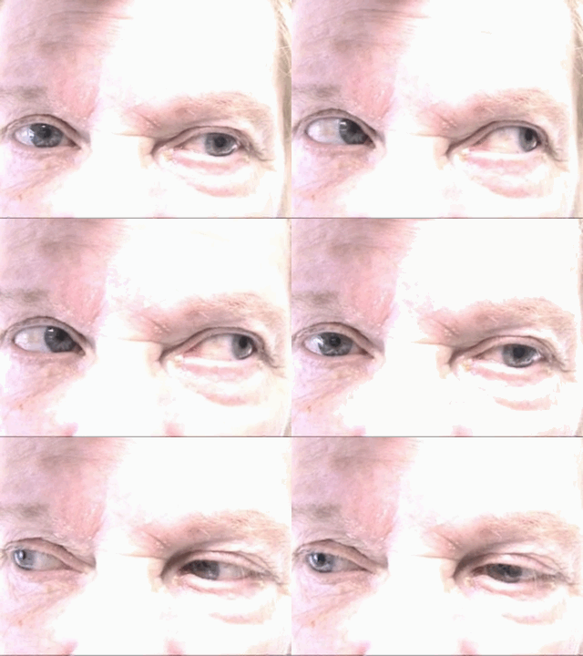
Normal head impulse, direction-changing nystagmus, or skew deviation suggests stroke.
- Head impulse
- Focus on examiner’s nose
- Rapidly turn head 10° in horizontal plan
- Presence of corrective saccade suggests defect of peripheral vestibular nerve
- Nystagmus
- Peripheral: Horizontal, unidirectional. Increases on gaze in direction of fast phase (decreases or resolves opposite)
- Central: Direction changing
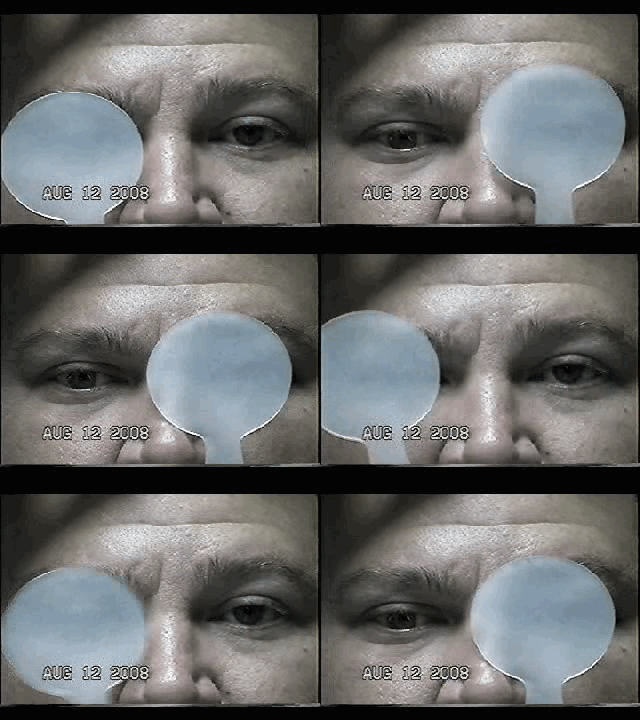
- Skew deviation
- Cross cover
- Presence of vertical disconjugate gaze suggests brainstem dysfunction
HINTS Gallery
Labs
- Glucose
- CBC/Chemistry
- ECG
Imaging
- Warranted if findings concerning for central process
- MRI preferred
Management
- Specific etiologies
- Vestibular neuronitis: steroids
- Meniere: dietary changes
- BPPV: canalith repositioning
- Symptomatic relief
- Promethazine (Phenergan) 12.5-25mg PO
- Ondansetron (Zofran) 4mg IV
- Lorazepam (Ativan) 1-2mg PO/IV
- Meclizine (Antivert) 25mg PO q6-8h PRN
References
- Kattah, J. C., Talkad, A. V., Wang, D. Z., Hsieh, Y.-H., & Newman-Toker, D. E. (2009). HINTS to diagnose stroke in the acute vestibular syndrome: three-step bedside oculomotor examination more sensitive than early MRI diffusion-weighted imaging. Stroke; a journal of cerebral circulation, 40(11), 3504–3510. doi:10.1161/STROKEAHA.109.551234
- Chang, A., & Olshaker, J. (2013). Dizziness and Vertigo. In Rosen’s Emergency Medicine – Concepts and Clinical Practice (8th ed., Vol. 1, pp. 162-169). Elsevier Health Sciences.


