History and Physical
38F with no medical history, presenting with double vision. The patient reported six weeks of intermittent diplopia for which she had presented to this hospital previously. She was briefly admitted for evaluation of possible cranial nerve IV palsy. Extensive imaging was unremarkable, without mass lesion, infarction, vascular malformation, or meningeal enhancement. She was discharged with outpatient follow-up including ophthalmology clinic and further imaging.
The patient represented due to persistent diplopia that is worse with right gaze. The diplopia is predominantly vertical, alleviated by head tilt. Now associated with three days of right ptosis as well as two weeks of progressive weakness and fatigue – most notable when climbing stairs.
Examination notable for right hypertropia increased on right or downward gaze suggestive of isolated inferior rectus weakness. Pupils were equal and reactive. There was marked fatigable ptosis with 2mm right palpebral fissure compared to 10mm on contralateral side. Symmetrical muscle weakness was noted, 4/5 neck flexion, elbow extension, wrist flexion/extension, shoulder abduction, hip flexion. Gait was wide-based. Application of ice for 5 minutes improved right palpebral fissure opening to >7mm.
Further evaluation included CXR and CT chest with intravenous contrast which did not identify a mediastinal mass. The patient’s respiratory status remained stable throughout hospitalization as assessed by measurements of forced vital capacity. On hospital day one, an edrophonium test was performed which was positive. The patient was started on pyridostigmine, completed a course of IVIG and was discharged with outpatient neurology follow-up.
Evaluation of Diplopia 1
History
- Onset/cadence
- Direction of gaze with worst diplopia
- Orientation (vertical/horizontal)
- Associated symptoms (headache, vertigo, dysarthria, eye pain)
Terms Describing Eye Position

Tropias are always present, phorias are identified by cross-cover testing (break fusion)
Algorithm for the Evaluation of Diplopia 2

Causes of Diplopia 3,4,5,6
| Finding | EOM | Causes | Features |
|---|---|---|---|
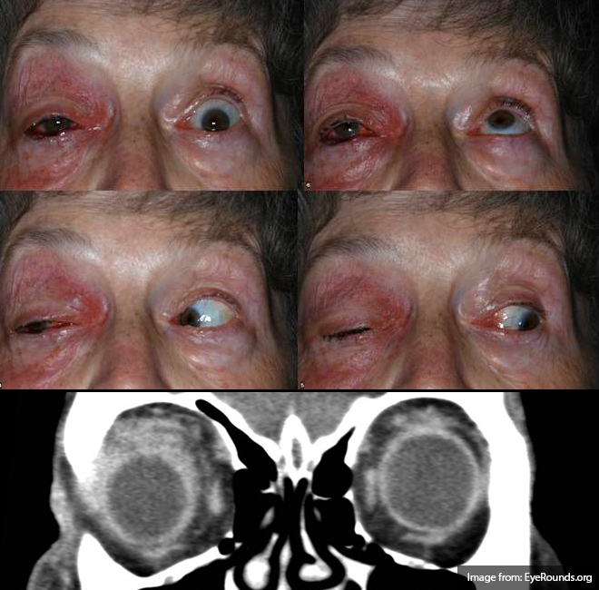
| Mechanical orbitopathy | Variable. Abrupt restriction of movement | Orbital cellulitis | Pain, erythema |
| Orbital pseudotumor | Autoimmune | ||
| Trauma | History | ||
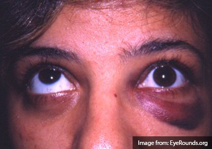
| Thyroid eye disease | Bilateral | ||
| Isolated CN III | Limited adduction/upgaze/downgaze | Microvascular ischemia | Pain, risk factors, pupil-sparing |
| Aneurysm | Pupil involvement | ||
| Demyelination | MRI | ||
| Isolated CN IV | Limited downgaze (hypertropia) | Trauma | May be mild |
| Microvascular ischemia | Less common than CN III | ||
| ICP | Fundoscopy, imaging | ||
| Demyelination | MRI | ||
| Isolated CN VI | Limited abduction
(esotropia) |
ICP | Fundoscopy, imaging |
| Demyelination | MRI | ||
| Microvascular ischemia | Less common than CN III | ||
| INO | Limited adduction
(exotropia) |
Demyelination | MRI |
| Stroke | Dysarthria, ataxia, facial weakness | ||
| Multiple CN involvement (III, IV, VI) | Variable | Cavernous sinus process | Retroorbital pain, conjunctival injection or chemosis |
| Brainstem deficits | Variable | Brainstem stroke | Weakness, dysmetria, tremor |
| Basilar artery occlusion | Vertigo, slurred speech | ||
| Wernicke | AMS, ataxia, nystagmus | ||
| Basilar meningitis | Fever, photophobia, meningismus | ||
| Miller-Fisher | Ataxia, areflexia | ||
| Neuromuscular process | Variable | Myasthenia gravis | Fatigability, ice test |
References
- Alves, M., Miranda, A., Narciso, M. R., Mieiro, L., & Fonseca, T. (2015). Diplopia: a diagnostic challenge with common and rare etiologies. The American journal of case reports, 16, 220–223. doi:10.12659/AJCR.893134
- Borooah, S., Wright, M., & Dhillon, B. (2011). Pocket Tutor Ophthalmology. JP Medical Limited. Retrieved from https://books.google.com/books?id=z\_CfWj8-ftoC
- Dinkin, M. (2014). Diagnostic approach to diplopia. Continuum (Minneapolis, Minn.), 20(4 Neuro-ophthalmology), 942–965. doi:10.1212/01.CON.0000453310.52390.58
- Rucker, J. C., & Tomsak, R. L. (2005). Binocular diplopia. A practical approach. The neurologist, 11(2), 98–110. doi:10.1097/01.nrl.0000156318.80903.b1
- Friedman, D. I. (2010). Pearls: diplopia. Seminars in neurology, 30(1), 54–65. doi:10.1055/s-0029-1244995
- Guluma, K. (2013). Diplopia. In Rosen’s Emergency Medicine – Concepts and Clinical Practice (8th ed., Vol. 1, pp. 176-183). Elsevier Health Sciences.
- WikEM: Diplopia
Pingback: Differential Diagnosis of Diplopia Applied