Diplopia Applied

Brief H&P:

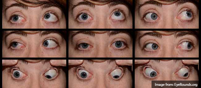
A young male with no past medical history presents to the emergency department after assault. He was punched multiple times in the face and has since noted double vision, worse with upward gaze. Examination revealed right peri-orbital edema with associated limitation to upward gaze.

Imaging:

entrapment_1
entrapment_2
entrapment_3
entrapment_4
entrapment_5
entrapment_6
entrapment_7
entrapment_8
entrapment_9

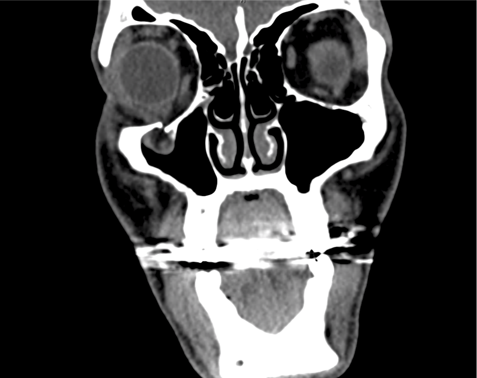
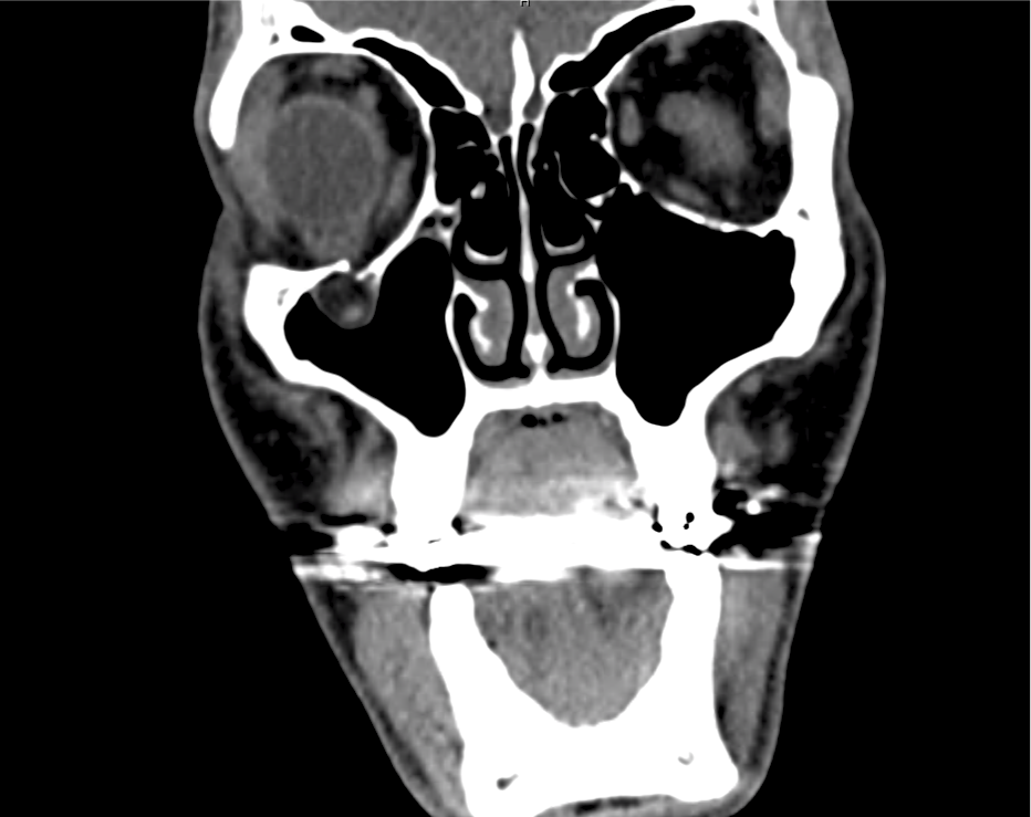
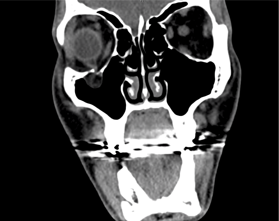
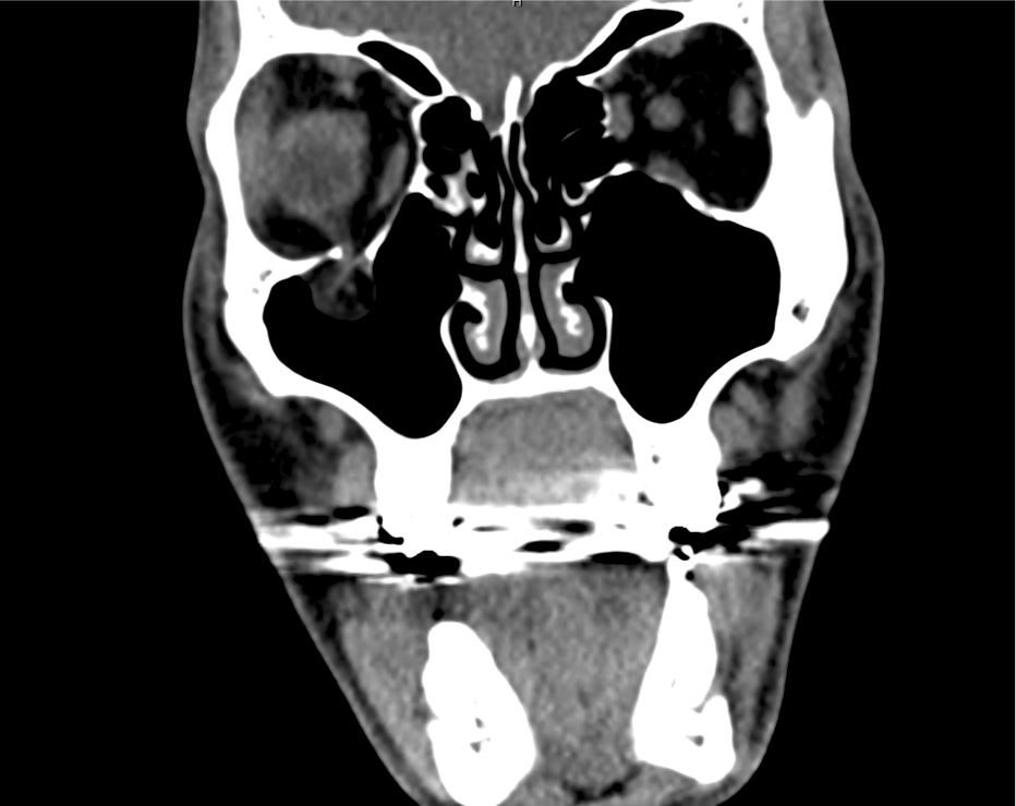
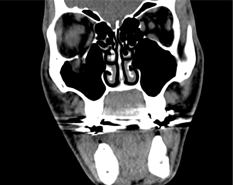
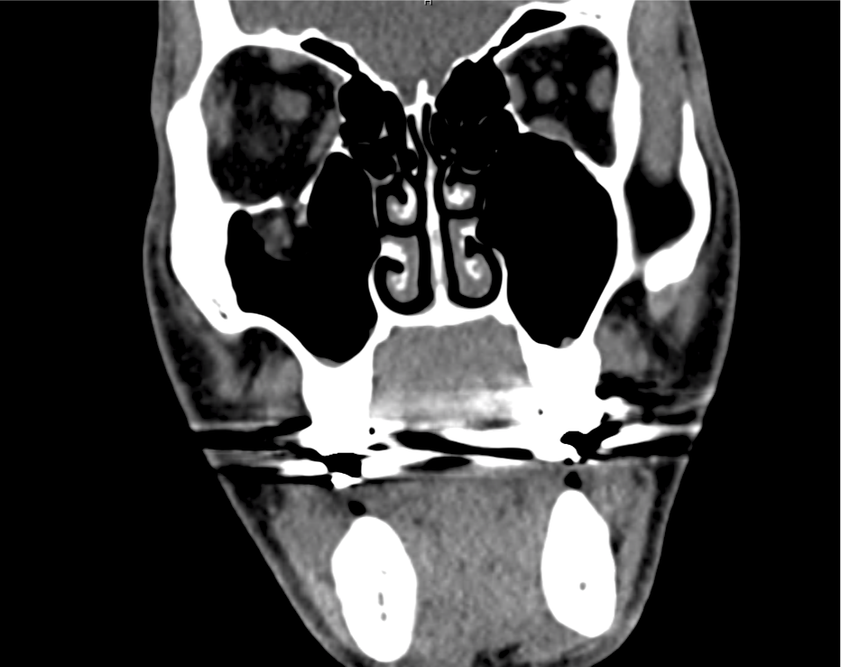
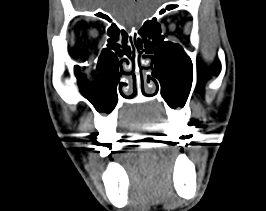
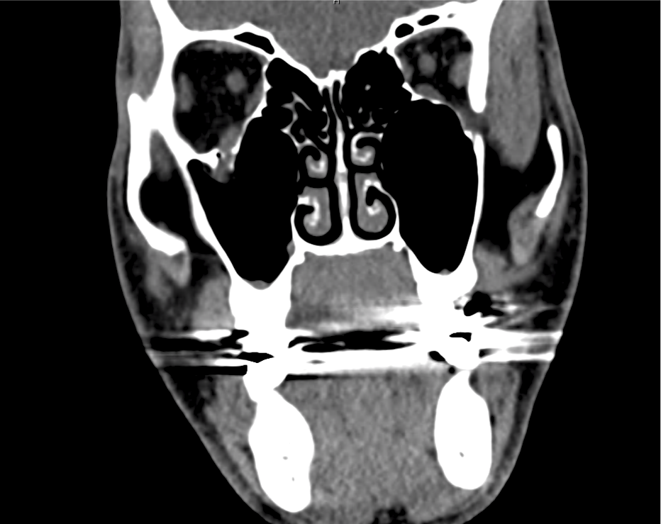
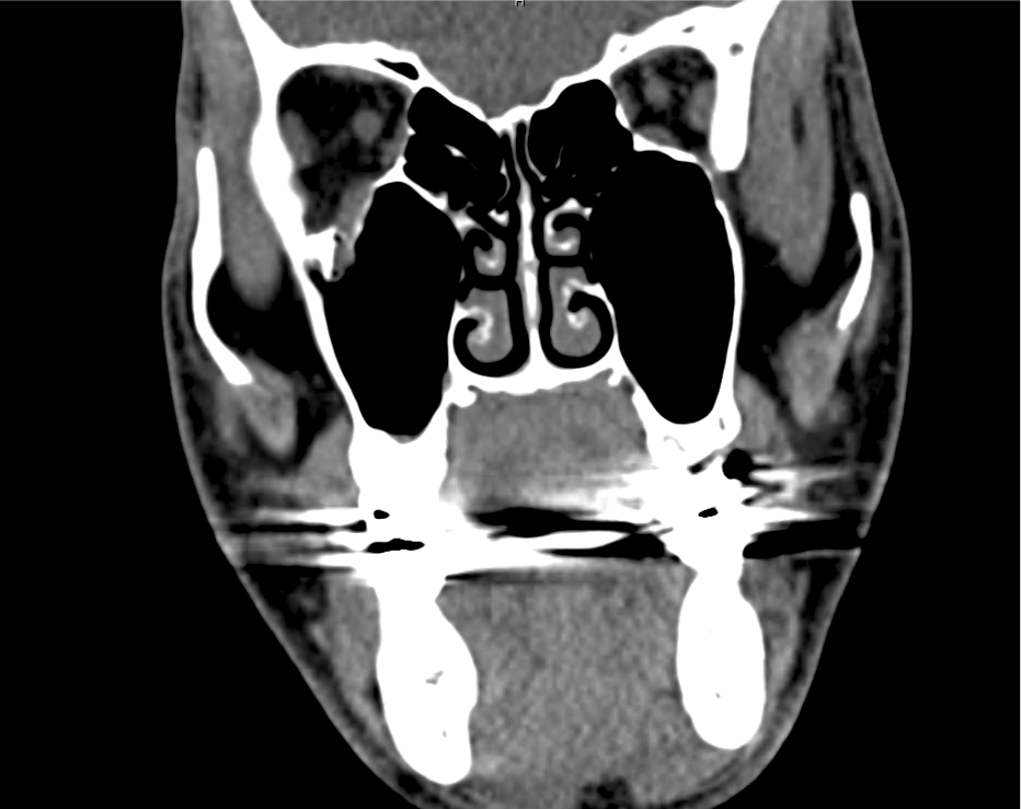
CT Maxillofacial Non-contrast

Inferior orbital wall fracture with herniation of the inferior rectus muscle.

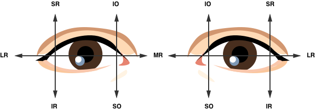
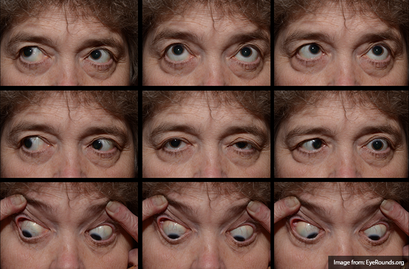
Extraocular Muscle Actions:

Extra-ocular movement actions.

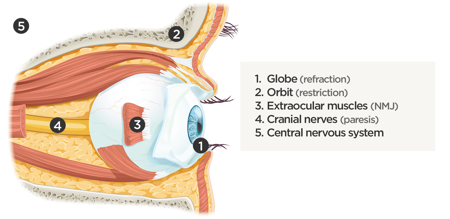
Affected Anatomic Sites in Diplopia:

Coordinated eye positioning is affected by voluntary movements (requiring cranial nerve control for conjugate eye movements), vergence (for depth adjustments), as well as reflexive adjustments for head movement (requiring vestibular input). As with any motor activity, neuromuscular control must be normal with unrestricted movement of the globe within the orbit.

Sites causing diplopia

Algorithm for the Evaluation of Diplopia:

Diplopia has been explored previously on ddxof. The earlier algorithm was focused on identifying the paretic nerve. This algorithm uses features of the history and physical examination to identify potential etiologic causes of diplopia.

Algorithm for the Evaluation of Diplopia

References:

  1. Rucker JC, Tomsak RL. Binocular diplopia. A practical approach. Neurologist. 2005;11(2):98-110. doi:10.1097/01.nrl.0000156318.80903.b1.
  2. Friedman DI. Pearls: diplopia. Semin Neurol. 2010;30(1):54-65. doi:10.1055/s-0029-1244995.
  3. Alves M, Miranda A, Narciso MR, Mieiro L, Fonseca T. Diplopia: a diagnostic challenge with common and rare etiologies. Am J Case Rep. 2015;16:220-223. doi:10.12659/AJCR.893134.
  4. Dinkin M. Diagnostic approach to diplopia. Continuum (Minneap Minn). 2014;20(4 Neuro-ophthalmology):942-965. doi:10.1212/01.CON.0000453310.52390.58.
  5. Marx J, Walls R, Hockberger R. Rosen’s Emergency Medicine – Concepts and Clinical Practice. 8 ed. Elsevier Health Sciences; 2013:176-183.
  6. Nazerian P, Vanni S, Tarocchi C, et al. Causes of diplopia in the emergency department: diagnostic accuracy of clinical assessment and of head computed tomography. Eur J Emerg Med. 2014;21(2):118-124. doi:10.1097/MEJ.0b013e3283636120.
  7. Low L, Shah W, MacEwen CJ. Double vision. BMJ. 2015;351:h5385. doi:10.1136/bmj.h5385.
  8. Danchaivijitr C, Kennard C. Diplopia and eye movement disorders. J Neurol Neurosurg Psychiatry. 2004;75 Suppl 4:iv24-iv31. doi:10.1136/jnnp.2004.053413.
  9. Huff JS, Austin EW. Neuro-Ophthalmology in Emergency Medicine. Emerg Med Clin North Am. 2016;34(4):967-986. doi:10.1016/j.emc.2016.06.016.

Neurosyphilis

Brief H&P

A young male with a history of HIV (untreated for the last year, with unknown CD4 count), and syphilis (reportedly treated with an intramuscular injection 1 year ago), presents with 4 months of a painful rash on the palms and soles and diplopia. Examination revealed the rash pictured below, ocular examination with minimal papilledema and anterior chamber inflammation.

Labs were unremarkable. CSF sampling was notable for 34 WBC’s with lymphocyte predominance (92%), and elevated protein (56mg/dL). The patient was admitted for syphilis with presumed neurosyphilis. Serum RPR titer was elevated at 1:64,  FTA-ABS and CSF VDRL were reactive. The patient was treated with intravenous penicillin and anti-retroviral therapy was reinitiated.

Epidemiology1

  • Transmission
    • Sexual contact (estimated transmission probability 60% per partner)
    • Trans-placental
  • Race/Sex
    •  African-American, Hispanic
    • Male > Female
    • Male (primary syphilis), female (secondary syphilis) – lesion visibility
    • Urban > rural

Natural History1

Stage Signs/Symptoms Incubation Period
Primary Chancre, reginal lymphadenopathy 3 weeks
Secondary Rash, fever, malaise, generalized lymphadenopathy, mucous membrane lesions, condyloma lata, headache, meningitis 2-12 weeks
Latent Asymptomatic Early (<1 year)

Late (>1 year)

Tertiary Cardiovascular:

Aortic aneurysm, aortic insufficiency, coronary artery ostial stenosis

<2 years
CNS:
Acute syphilitic meningitis: headache, confusion, meningeal irritation <2 years
Meningovascular: cranial nerve palsy 5-7 years
General paresis: headache, vertigo, personality changes, vascular event 5-7 years
Tabes dorsalis: dementia, ataxia, Argyl-Robertson, [arrow-down] proprioception 10-20 years
Gumma:

Local tissue destruction

1-46 years

Diagnosis1

  • Serologic
    • Non-treponemal (screening)
      • RPR, VDRL
      • Limitations:  sensitivity, false positive (age, pregnancy, drugs, malignancy, autoimmune, viral infections)
    • Treponemal (confirmatory)
      • FTA-ABS
    • Neurosyphilis
      • Indications for CSF sampling: neurologic/ophthalmologic symptoms, tertiary syphilis (aortitis, gumma, iritis), HIV coinfection with elevated RPR titer (> 1:32)
      • CSF: leukocytosis (predominantly lymphocytes),  protein
      • CSF VDRL reactive
      • Negative CSF FTA-ABS may rule out neurosyphilis

Syphilis in HIV-infected Individuals2

  • Primary: larger and more lesion, multiple ulcers
  • Secondary: genital ulcers more common, higher RPR/VDRL titers
  • Tertiary: possibly more rapid progression to neurosyphilis

References

  1. Singh AE, Romanowski B. Syphilis: review with emphasis on clinical, epidemiologic, and some biologic features. Clin Microbiol Rev. 1999;12(2):187-209.
  2. French P. Syphilis. BMJ. 2007;334(7585):143-147. doi:10.1136/bmj.39085.518148.BE.

Diplopia

History and Physical

38F with no medical history, presenting with double vision. The patient reported six weeks of intermittent diplopia for which she had presented to this hospital previously. She was briefly admitted for evaluation of possible cranial nerve IV palsy. Extensive imaging was unremarkable, without mass lesion, infarction, vascular malformation, or meningeal enhancement. She was discharged with outpatient follow-up including ophthalmology clinic and further imaging.

The patient represented due to persistent diplopia that is worse with right gaze. The diplopia is predominantly vertical, alleviated by head tilt. Now associated with three days of right ptosis as well as two weeks of progressive weakness and fatigue – most notable when climbing stairs.

Examination notable for right hypertropia increased on right or downward gaze suggestive of isolated inferior rectus weakness. Pupils were equal and reactive. There was marked fatigable ptosis with 2mm right palpebral fissure compared to 10mm on contralateral side. Symmetrical muscle weakness was noted, 4/5 neck flexion, elbow extension, wrist flexion/extension, shoulder abduction, hip flexion. Gait was wide-based. Application of ice for 5 minutes improved right palpebral fissure opening to >7mm.

Further evaluation included CXR and CT chest with intravenous contrast which did not identify a mediastinal mass. The patient’s respiratory status remained stable throughout hospitalization as assessed by measurements of forced vital capacity. On hospital day one, an edrophonium test was performed which was positive. The patient was started on pyridostigmine, completed a course of IVIG and was discharged with outpatient neurology follow-up.

Evaluation of Diplopia 1

History

  1. Onset/cadence
  2. Direction of gaze with worst diplopia
  3. Orientation (vertical/horizontal)
  4. Associated symptoms (headache, vertigo, dysarthria, eye pain)

Terms Describing Eye Position

Terms describing eye position

Tropias are always present, phorias are identified by cross-cover testing (break fusion)

Algorithm for the Evaluation of Diplopia 2

Algorithm for the Evaluation of Diplopia

Causes of Diplopia 3,4,5,6

Finding EOM Causes Features
Mechanical orbitopathy Variable. Abrupt restriction of movement Orbital cellulitis Pain, erythema
Orbital pseudotumor Autoimmune
Trauma History
Thyroid eye disease Bilateral
Isolated CN III Limited adduction/upgaze/downgaze Microvascular ischemia Pain, risk factors, pupil-sparing
Aneurysm Pupil involvement
Demyelination MRI
Isolated CN IV Limited downgaze (hypertropia) Trauma May be mild
Microvascular ischemia Less common than CN III
ICP Fundoscopy, imaging
Demyelination MRI
Isolated CN VI Limited abduction

(esotropia)

ICP Fundoscopy, imaging
Demyelination MRI
Microvascular ischemia Less common than CN III
INO Limited adduction

(exotropia)

Demyelination MRI
Stroke Dysarthria, ataxia, facial weakness
Multiple CN involvement (III, IV, VI) Variable Cavernous sinus process Retroorbital pain, conjunctival injection or chemosis
Brainstem deficits Variable Brainstem stroke Weakness, dysmetria, tremor
Basilar artery occlusion Vertigo, slurred speech
Wernicke AMS, ataxia, nystagmus
Basilar meningitis Fever, photophobia, meningismus
Miller-Fisher Ataxia, areflexia
Neuromuscular process Variable Myasthenia gravis Fatigability, ice test

References

  1. Alves, M., Miranda, A., Narciso, M. R., Mieiro, L., & Fonseca, T. (2015). Diplopia: a diagnostic challenge with common and rare etiologies. The American journal of case reports, 16, 220–223. doi:10.12659/AJCR.893134
  2. Borooah, S., Wright, M., & Dhillon, B. (2011). Pocket Tutor Ophthalmology. JP Medical Limited. Retrieved from https://books.google.com/books?id=z\_CfWj8-ftoC
  3. Dinkin, M. (2014). Diagnostic approach to diplopia. Continuum (Minneapolis, Minn.), 20(4 Neuro-ophthalmology), 942–965. doi:10.1212/01.CON.0000453310.52390.58
  4. Rucker, J. C., & Tomsak, R. L. (2005). Binocular diplopia. A practical approach. The neurologist, 11(2), 98–110. doi:10.1097/01.nrl.0000156318.80903.b1
  5. Friedman, D. I. (2010). Pearls: diplopia. Seminars in neurology, 30(1), 54–65. doi:10.1055/s-0029-1244995
  6. Guluma, K. (2013). Diplopia. In Rosen’s Emergency Medicine – Concepts and Clinical Practice (8th ed., Vol. 1, pp. 176-183). Elsevier Health Sciences.
  7. WikEM: Diplopia