Hyponatremia

HPI:

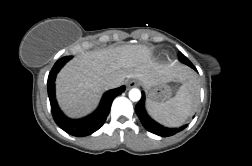
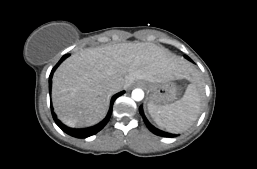
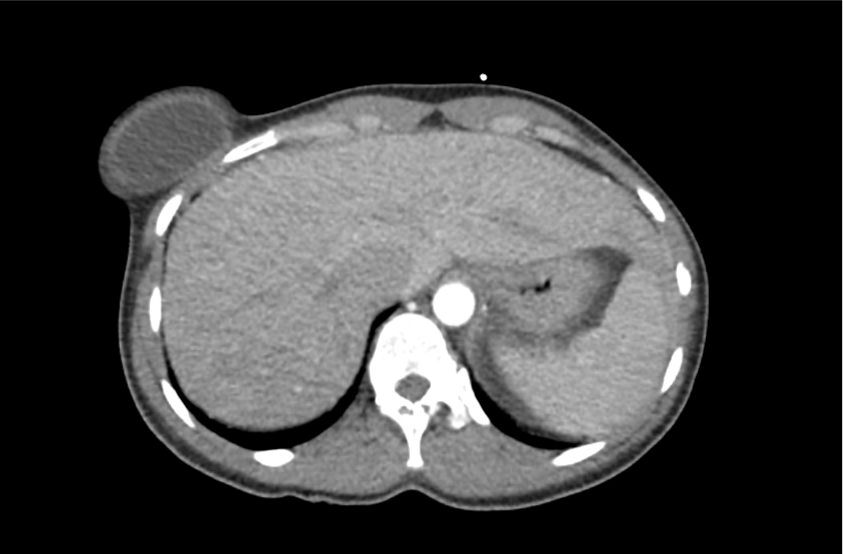
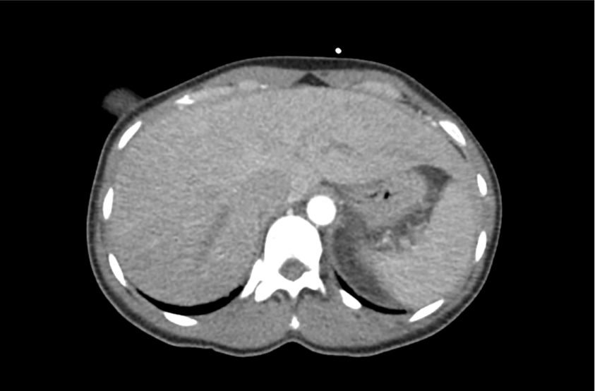
A 62 year-old male with a history of hepatitis C cirrhosis complicated by hepatocellular carcinoma s/p radiofrequency ablation presenting after referral from hepatology clinic for hyponatremia. One week ago, the patient developed abdominal distension and shortness of breath that resolved after large-volume paracentesis and was started on furosemide 40mg p.o. daily and aldactone 100mg p.o. daily. After initiating diuretics, the patient noted worsening lower extremity edema, and increased thirst/fluid intake.

He reports two days of fatigue and intermittent confusion supported by family members who reported slowed speech. He otherwise denies abdominal pain, distension, nausea/vomiting, diarrhea/constipation, chest pain or shortness of breath. In the ED, the patient received 1L NS bolus.

PMH:

  • Hepatitis C cirrhosis c/b HCC s/p RFA
  • Rheumatoid arthritis

PSH:

  • None

Family History:

  • Non-contributory.

Social History:

  • Lives with partner, denies current or prior t/e/d abuse
  • HepC contracted from blood transfusions

Medications:

  • Furosemide 40mg p.o. daily
  • Spironolactone 100mg p.o. daily
  • Rifaximin 550mg p.o. b.i.d.

Allergies:

  • NKDA

Physical Exam:

VS: T 98.2 HR 80 RR 14 BP 95/70 O2 98% RA
Gen: Elderly male in no acute distress, alert and answering questions appropriately.
HEENT: NC/AT, PERRL, EOMI, faint scleral icterus, MMM.
CV: RRR, normal S1/S2, no murmurs. JVP 8cm.
Lungs: Faint basilar crackles on bilateral lung bases.
Abd: Normoactive bowel sounds, mildly distended but non-tender, without rebound/guarding.
Ext: 2+ pitting edema in lower extremities to knees bilaterally. 2+ peripheral pulses, warm and well perfused.
Neuro: AAOx3. CN II-XII intact. No asterixis. Normal gait. Normal FTN/RAM.
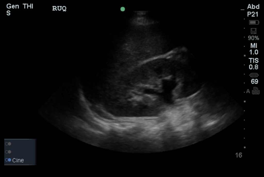
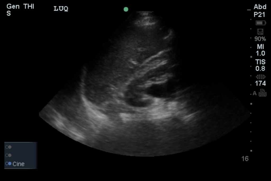
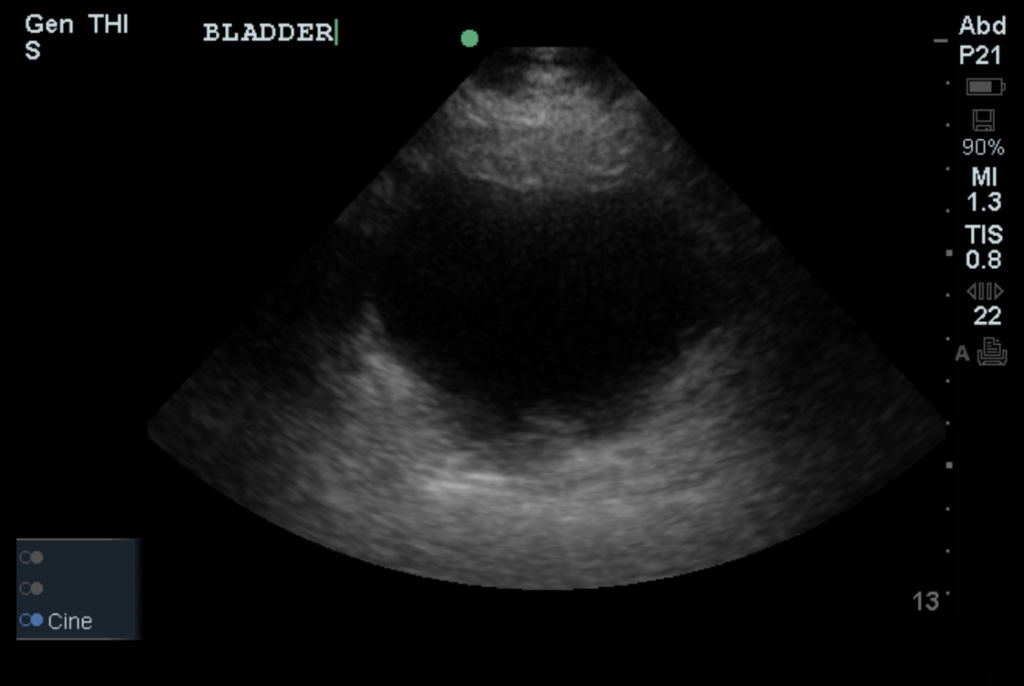
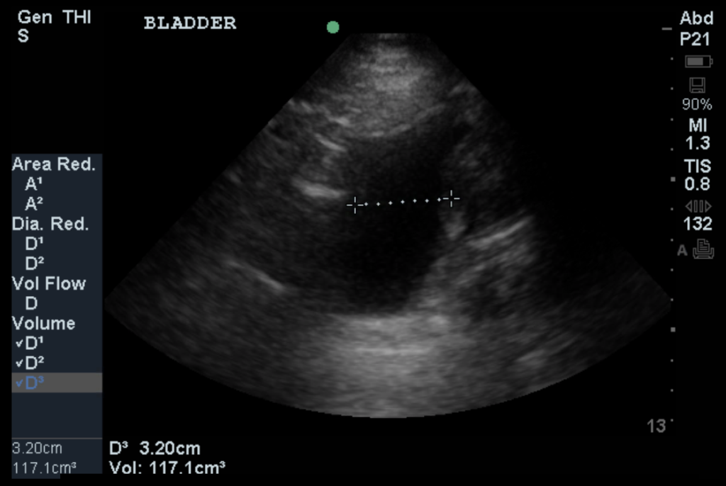
Labs/Studies:

  • BMP (admission): 112/5.6/88/22/28/1.1/97
  • BMP (+10h): 118/5.4/93/23/26/1.0/133
  • sOsm: 264
  • Urine: Na <20, K 26, Osm 453
  • BNP: 40
  • AST/ALT/AP/TB/Alb: 74/57/91/2.4/2.2

Assessment/Plan:

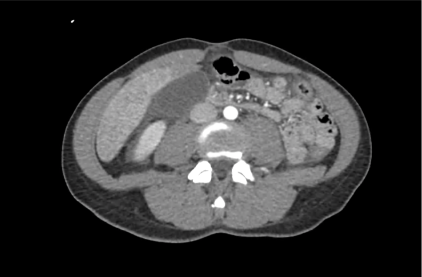
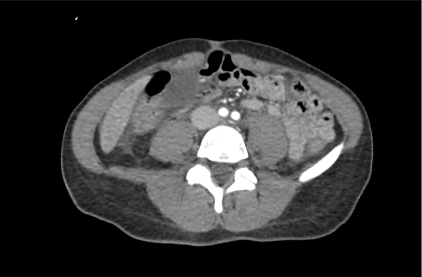
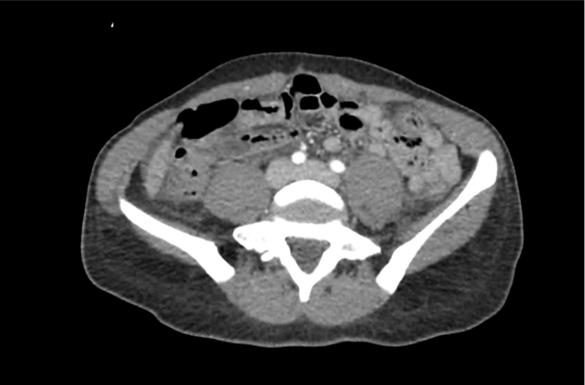
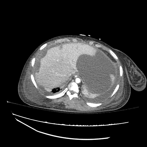
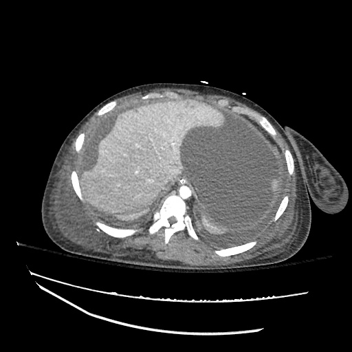
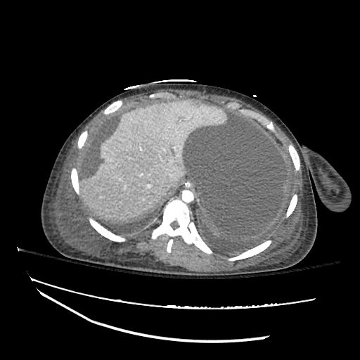
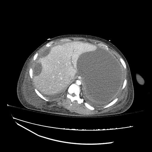
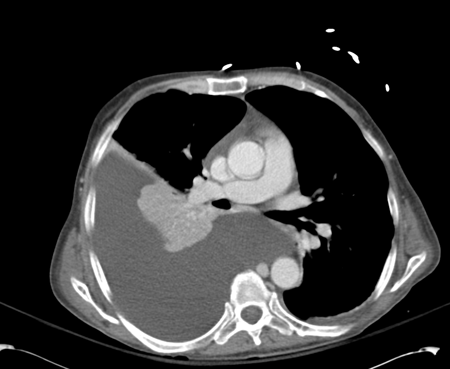
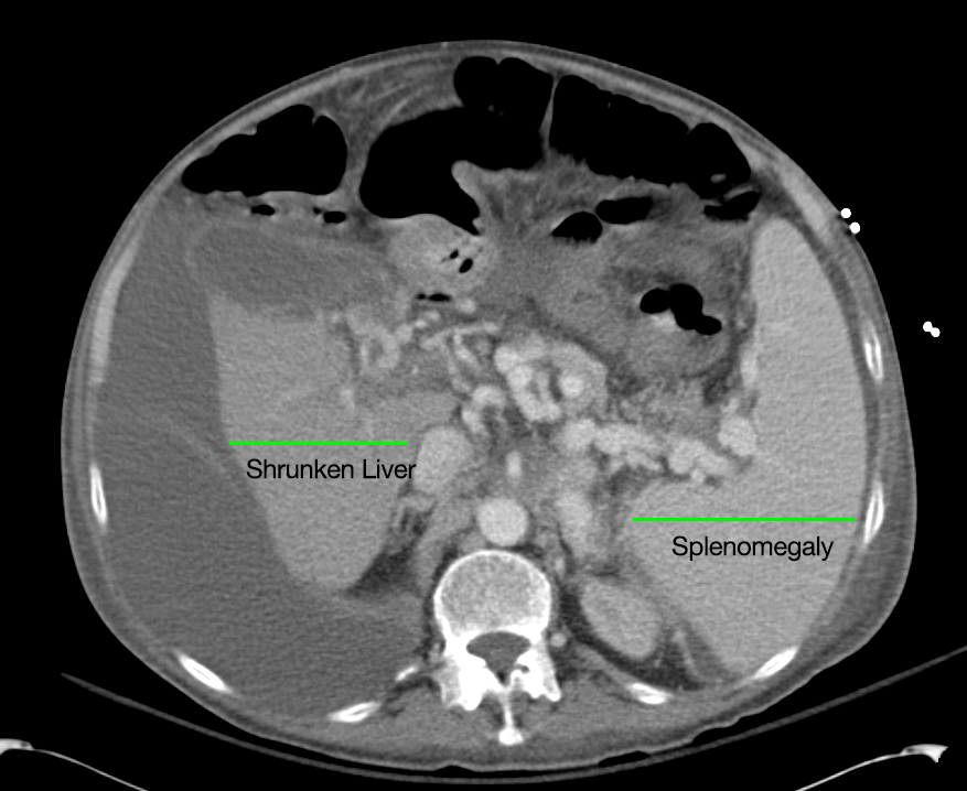
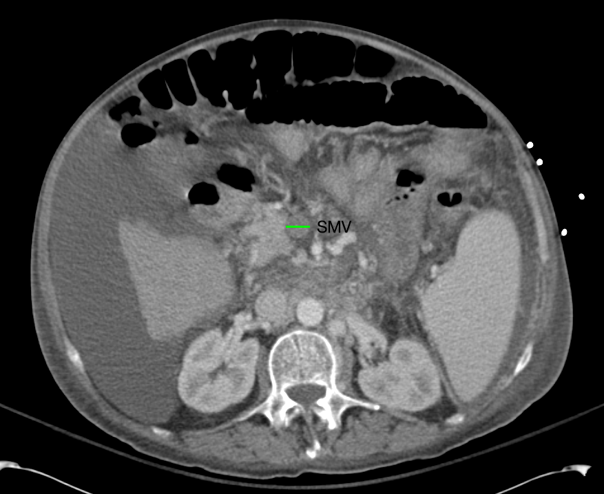
62M with HepC cirrhosis, with e/o decompensation (new-onset ascites) and hyponatremia.

  1. #Hyponatremia: Sodium 114, likely chronic, patient currently asymptomatic without concerning findings on neurological exam. Clinical findings suggestive of hypervolemic hyponatremia 2/2 decompensated cirrhosis resulting in decreased effective arterial blood volume and volume retention. However, the recent initiation of diuretics, mild AKI and early response to isotonic fluids in the ED suggests possible hypovolemic component.
    • 1L fluid restriction
    • q.4.h. sodium check, goal increase of 8mEq per 24h
    • hold diuretics
  2. #Hyperkalemia: Potassium 5.6, asymptomatic, AKI vs. medication-induced (aldactone). Continue monitoring.
  3. #AKI: Elevated creatinine 1.1 from baseline 0.7. Likely pre-renal given recent initiation of diuretics. Consider hepatorenal syndrome given decompensated cirrhosis. Follow-up repeat creatinine after 1L NS bolus in ED.
  4. #Hepatitis C: decompensated with new-onset ascites. No e/o encephalopathy, continue home rifaximin.

Physiology of Hyponatremia: 1,2,3,4

Physiology of Hyponatremia

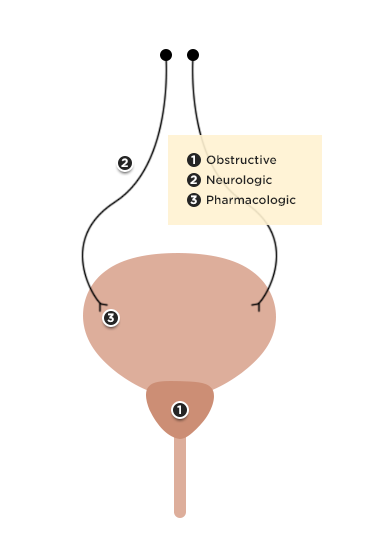
Differential Diagnosis of Hyponatremia: 5

Differential Diagnosis of Hyponatremia

Evaluation of Hyponatremia: 2

  1. Identification of onset (acute vs. chronic)
  2. Presence of symptoms (HA, nausea, confusion, seizures)
  3. Assessment of volume status (edema, JVD, skin turgor, postural BP)
  4. Medical history (cardiac, liver, renal disease), drug history

References:

  1. Freda BJ, Davidson MB, Hall PM. Evaluation of hyponatremia: a little physiology goes a long way. Cleve Clin J Med. 2004;71(8):639–650.
  2. Biswas M, Davies JS. Hyponatraemia in clinical practice. Postgrad Med J. 2007;83(980):373–378. doi:10.1136/pgmj.2006.056515.
  3. Adrogué HJ, Madias NE. Hyponatremia. N. Engl. J. Med. 2000;342(21):1581–1589. doi:10.1056/NEJM200005253422107.
  4. Marx JA, Hockberger RS, Walls RM, Adams JG. Rosen’s emergency medicine: concepts and clinical practice. 2010;1.
  5. Milionis HJ, Liamis GL, Elisaf MS. The hyponatremic patient: a systematic approach to laboratory diagnosis. CMAJ. 2002;166(8):1056–1062.

Thrombocytopenia

Brief HPI:

A middle-aged female with no known medical history is brought to the emergency department with altered mental status. Her family notes worsening confusion over the past 2-3 days associated with vomiting and yellow discoloration of skin and eyes.

Initial vital signs were normal, though with borderline hypotension (99/64mmHg). Examination demonstrated an alert, but lethargic patient with jaundice and scleral icterus, no skin lesions were appreciated. Laboratory studies were obtained:

CBC

  • WBC: 21.3 (N: 83%, Bands: 11%)
  • Hb: 5.5
  • Plt: 6k
  • Marked schistocytes

Coagulation Panel

  • INR: 1.26
  • PTT: Normal
  • Fibrinogen: Normal
  • FDP: Normal
  • D-dimer: >9,000 (normal 250)
  • Haptoglobin: Undetectable
  • LDH: 1493

CMP

  • Creatinine: 1.1
  • AST/ALT: Normal
  • TB: 4.3, DB: 0.8

Imaging:

CT Head: No acute intracranial process.

ttp_ct_01
ttp_ct_02
ttp_ct_03
ttp_ct_04
ttp_ct_05
ttp_ct_06
ttp_ct_07
ttp_ct_08
ttp_ct_09
ttp_ct_10
ttp_ct_11
ttp_ct_12
ttp_ct_13
ttp_ct_14
ttp_ct_15
ttp_ct_16
ttp_ct_17
ttp_ct_18
ttp_ct_19

CT Abdomen/Pelvis with Contrast

Moderate free intra-abdominal fluid, heterogeneous liver with periportal edema, dense right middle lobe consolidation.

ED Course:

The patient developed worsening respiratory failure with hypoxia and tachypnea requiring endotracheal intubation. Thrombotic thrombocytopenic purpura was suspected and while awaiting emergent plasma exchange transfusion, the patient arrested and resuscitation efforts were unsuccessful.

The patient’s ADAMTS13 activity level was <3%. Autopsy demonstrated consolidation of the right middle lobe with possible lymphoproliferative mass, and lung petechial hemorrhages from microvascular thrombi.

Differential Diagnosis of Thrombocytopenia 1-7

Differential Diagnosis of Thrombocytopenia

Algorithm for the Evaluation of Thrombocytopenia 8

Algorithm for the Evaluation of Thrombocytopenia

Definition 9

  • Mild: <150k
  • Moderate: 100-150k
  • Severe: <50k
    • 10-30k: bleeding with minimal trauma
    • <10k: increased risk spontaneous bleeding

History 9,10

  • Prior platelet count
  • Family history bleeding disorders
  • Medications
    • Heparin
    • Quinine, quinidine
    • Rifampin
    • Trimethoprim-sulfamethoxazole
    • Vancomycin
  • Alcohol use
  • Travel-related infections

Physical Examination 9,10

  • Splenomegaly (liver disease)
  • Lymphadenopathy (infection, malignancy)

Workup 10,11

Schistocytes

Red blood cell fragments (schistocytes) 11

  • hCG
  • Repeat CBC
    • Detect spurious measure
    • Neutrophil-predominant leukocytosis: bacterial infection
    • Immature leukocytes (blasts): leukemia, myelodysplasia
  • Peripheral smear
    • Schistocytes: microangiopathic process (DIC, TTP, HUS)
    • Atypical lymphocytes: viral infection
    • Intracellular parasites: malaria
    • Hypersegmented neutrophils: nutritional deficiency
  • Infectious features: HIV, HCV, EBV, H.pylori, blood cultures
  • Autoimmune features: ANA, APL-Ab
  • Suspected occult liver disease: LFT, PT/PTT/INR
  • Suspected thrombotic microangiopathy: PT/PTT/INR, haptoglobin, LDH, fibrinogen, FDP, d-dimer

Specific Conditions 2-6,9,12-20

Disease Cause Presentation Laboratory Findings Treatment
DIC Sepsis
Trauma
Burn
Malignancy
Bleeding
Multi-organ failure
Shock
INR
Fibrinogen
FDP
D-dimer
Directed at underlying cause
Transfusion thresholds for hemorrhage:
FFP for INR >1.5
Platelets if <50k
Cryoprecipitate of fibrinogen <100mg/dL
TTP Insufficient ADAMTS-13 activity Non-specific constitutional symptoms (ex. weakness)
Neuro: headache, AMS, focal neuro deficit
GI: abdominal pain, nausea/vomiting
LDH
Reticulocyte
Unconjugated bilirubin
Haptoglobin
Plasma exchange
HUS Shiga-toxin-producing bacteria, E. coli O157:H7 Bloody diarrhea, anuria, oliguria, and hypertension Aggressive supportive care
HELLP Spectrum of eclampsia Hypertension
Visual symptoms
Headache
RUQ abdominal pain
AST/ALT
Uric acid
Unconjugated bilirubin
LDH
Reticulocyte
Haptoglobin
Delivery, MgSO4
ITP Primary ITP

Secondary ITP
– Drug
– Autoimmune
– Infection
– Malignancy

Usually asymptomatic, may have petechiae or easy bruising Isolated thrombocytopenia Steroids
HIT Exposure to heparin or LMWH Thrombocytopenia or a 50 percent reduction in platelet count between 5-10d exposure
New thrombosis or skin necrosis
4 T’s score
Platelet factor 4 antibodies Withdraw heparin

References

  1. Greinacher A, Selleng S. How I evaluate and treat thrombocytopenia in the intensive care unit patient. Blood. 2016;128(26):3032-3042. doi:10.1182/blood-2016-09-693655.
  2. Joly BS, Coppo P, Veyradier A. Thrombotic thrombocytopenic purpura. Blood. 2017;129(21):2836-2846. doi:10.1182/blood-2016-10-709857.
  3. Leslie SD, Toy PT. Laboratory hemostatic abnormalities in massively transfused patients given red blood cells and crystalloid. Am J Clin Pathol. 1991;96(6):770-773.
  4. Neunert C, Lim W, Crowther M, et al. The American Society of Hematology 2011 evidence-based practice guideline for immune thrombocytopenia. Blood. 2011;117(16):4190-4207. doi:10.1182/blood-2010-08-302984.
  5. Kappler S, Ronan-Bentle S, Graham A. Thrombotic microangiopathies (TTP, HUS, HELLP). Emerg Med Clin North Am. 2014;32(3):649-671. doi:10.1016/j.emc.2014.04.008.
  6. Greinacher A. Heparin-Induced Thrombocytopenia. Solomon CG, ed. N Engl J Med. 2015;373(3):252-261. doi:10.1056/NEJMcp1411910.
  7. Reardon JE Jr., Marques MB. Evaluation of Thrombocytopenia. Lab Med. 2006;37(4):248-250. doi:10.1309/R7P79KERAJHPRHLT.
  8. Stasi R. How to approach thrombocytopenia. Hematology Am Soc Hematol Educ Program. 2012;2012:191-197. doi:10.1182/asheducation-2012.1.191.
  9. Gauer RL, Braun MM. Thrombocytopenia. Am Fam Physician. 2012;85(6):612-622.
  10. Abrams CS. 172 – Thrombocytopenia. Twenty Fifth Edition. Elsevier Inc.; 2016:1159–1167.e2. doi:10.1016/B978-1-4557-5017-7.00172-0.
  11. Wilson CS, Vergara-Lluri ME, Brynes RK. Chapter 11 – Evaluation of Anemia, Leukopenia, and Thrombocytopenia. Second Edition. Elsevier Inc.; 2017:195-234.e195. doi:10.1016/B978-0-323-29613-7.00011-9.
  12. Hui P, Cook DJ, Lim W, Fraser GA, Arnold DM. The frequency and clinical significance of thrombocytopenia complicating critical illness: a systematic review. Chest. 2011;139(2):271-278. doi:10.1378/chest.10-2243.
  13. Jokiranta TS. HUS and atypical HUS. Blood. 2017;129(21):2847-2856. doi:10.1182/blood-2016-11-709865.
  14. Neunert CE. Management of newly diagnosed immune thrombocytopenia: can we change outcomes? Hematology Am Soc Hematol Educ Program. 2017;2017(1):400-405. doi:10.1182/asheducation-2017.1.400.
  15. Lambert MP, Gernsheimer TB. Clinical updates in adult immune thrombocytopenia. Blood. 2017;129(21):2829-2835. doi:10.1182/blood-2017-03-754119.
  16. Arepally GM. Heparin-induced thrombocytopenia. Blood. 2017;129(21):2864-2872. doi:10.1182/blood-2016-11-709873.
  17. Aster RH, Bougie DW. Drug-induced immune thrombocytopenia. N Engl J Med. 2007;357(6):580-587. doi:10.1056/NEJMra066469.
  18. Boral BM, Williams DJ, Boral LI. Disseminated Intravascular Coagulation. Am J Clin Pathol. 2016;146(6):670-680. doi:10.1093/ajcp/aqw195.
  19. Scully M, Hunt BJ, Benjamin S, et al. Guidelines on the diagnosis and management of thrombotic thrombocytopenic purpura and other thrombotic microangiopathies. Br J Haematol. 2012;158(3):323-335. doi:10.1111/j.1365-2141.2012.09167.x.
  20. Levine RL, Hursting MJ, Drexler A, Lewis BE, Francis JL. Heparin-induced thrombocytopenia in the emergency department. Ann Emerg Med. 2004;44(5):511-515. doi:10.1016/j.annemergmed.2004.06.004.

Acute Urinary Retention

Brief H&P:

A 62 year-old male with no significant medical history, presented to the emergency department with several days of vomiting. Examination showed suprapubic fullness with tenderness to palpation and a bedside ultrasound was performed:

RUQ
RUQ

RUQ

Right upper quadrant ultrasound with moderate hydronephrosis.

LUQ
LUQ

LUQ

Left upper quadrant ultrasound with moderate hydronephrosis.

Bladder
Bladder

Bladder

Relatively non-distended bladder.

Bladder Volume
Bladder Volume

Bladder Volume

Post-void bladder volume.

Ultrasound revealed moderate bilateral hydronephrosis with a relatively non-distended bladder. Labs were notable for new renal failure and the patient was admitted for continued evaluation. He was ultimately diagnosed with idiopathic retroperitoneal fibrosis with bilateral distal ureteral obstruction requiring stenting.

Anatomy of Acute Urinary Retention:

Differential Diagnosis of Acute Urinary Retention:1,2,3

Algorithm for the Evaluation of Acute Urinary Retention

Acute Kidney Injury

Hospital Course:

64 year-old female with a history of metastatic colonic adenocarcinoma was initially admitted for PO intolerance secondary to recurrent small bowel obstructions (associated with abdominal tumor burden). On hospital day six, the patient developed tachypnea, hypoxemia, hypotension and was intubated for respiratory distress. In the MICU, the patient was treated for acute hypoxic respiratory failure thought to be caused by aspiration (large volume bilious emesis prior to intubation despite NGT LCWS) vs. accumulating malignant pleural effusions vs. pulmonary embolism. Septic shock of a presumed pulmonary vs. intra-abdominal source was managed with vasopressors and broad-spectrum antimicrobials.

On hospital day fourteen, an elevation in the serum creatinine was noted. Known nephrotoxic agents include iodinated contrast on hospital day five, and vancomycin. The patient’s vasopressor requirement had decreased to norepinephrine 6mcg/kg/min (previously requiring four vasopressors). Over the next six days, the serum creatinine continued to trend upwards associated with a decrease in urine output (0.3-0.5mL/kg/hour). Intravenous crystalloid and colloid administered liberally based on central venous pressure and ultrasound of the inferior vena cava did not impact urine output.

Laboratory Studies

Hospital day 19 18 17 16 15 14 3
Creatinine 1.72 1.59 1.46 1.32 1.24 1.09 0.75
Vancomycin 23.5 28.5 36.3 45.5 47.7 22.1

Urine electrolytes:

  • Una: 10
  • Ucr: 180
  • Uk: 13
  • Ucl: 22
  • Uur: 265
  • FeNa: <1%

UA: 3+LE, 1+ blood, 36WBC, 14RBC, 3+ bacteria, amorphous crystals

Imaging:

IM-0001-0054
IM-0001-0056
IM-0001-0058
IM-0001-0060
IM-0001-0062
IM-0001-0064
IM-0001-0066
IM-0001-0068
IM-0001-0070
IM-0001-0072
IM-0001-0074
IM-0001-0076
IM-0001-0078
IM-0001-0080
IM-0001-0082
IM-0001-0084
IM-0001-0086
IM-0001-0088
IM-0001-0090
IM-0001-0092
IM-0001-0094
IM-0001-0096
IM-0001-0098
IM-0001-0100
IM-0001-0102
IM-0001-0104
IM-0001-0106
IM-0001-0108
IM-0001-0110
IM-0001-0112
IM-0001-0114
IM-0001-0116
IM-0001-0118
IM-0001-0120
IM-0001-0122
IM-0001-0124
IM-0001-0126
IM-0001-0128
IM-0001-0130
IM-0001-0132

CT Abdomen/Pelvis with IV contrast

  • Within the retroperitoneum, the left kidney is small and atrophic and demonstrates limited peripheral enhancement. The left renal artery is also poorly visualized.
  • Severely dilated loops of small bowel, including a segment within the left lower quadrant that may represent a closed loop obstruction.
  • There is a large (16.4 cm in largest diameter) subphrenic fluid collection in the left upper quadrant. A second large (14.2 cm in largest diameter) intraabdominal fluid collection lies inferior and anteriorly.

Assessment:

Oliguric acute renal failure in the setting of convincingly pre-renal urine studies which was not responsive to adequate crystalloid and colloid volume resuscitation. The patient had a normal ejection fraction on a recent echocardiogram, and while the patient was hypoalbuminemic (presumably from poor nutritional status and PO intolerance), urine output was not even transiently responsive to colloid administration. While the patient had recent administration of intravenous contrast, the elevation in serum creatinine occurred more than one week later. Further, the elevated vancomycin trough was likely a consequence rather than the etiology of worsening renal failure. AKI was likely secondary to renal artery compression from mass effect associated with abdominal metastases. There was evidence of a similar process affecting the left kidney, which was severely atrophic. The patient declined further evaluation, which would have included a renal ultrasound.

Definition of Acute Kidney Injury: 1

  • Elevation of serum creatinine > 0.3mg/dL in 48h
  • Elevation of serum creatinine > 1.5x baseline in 7d
  • Oliguria (UOP < 0.5mL/kg/hr) > 6h

Staging of Acute Kidney Injury: 1

Stage Creatinine UOP
1 1.5-1.9x <0.5mL/kg/hr for 6-12h
2 2.0-2.9x <0.5mL/kg/hr for >12h
3 3.0x or RRT <0.3mL/kg/hr for > 24h

Management of Contrast-induced AKI: 2

  • Administer lowest dose
  • Use iso-osmolar, or low-osmolar contrast
  • Volume expansion (NaCl, NaHCO3)
  • PO NAC questionable benefit but likely harmless

Differential Diagnosis of Acute Kidney Injury: 3

Algorithm for the Evaluation of Acute Kidney Injury

NOTE: Algorithm revised in November, 2017. The prior version is no longer supported but remains available here.

Evaluation of AKI: 4

Condition Urinalysis Casts FeNa (%)
Pre-renal Normal Hyaline <1
Intra-renal
ATN Mild proteinuria Pigmented granular >1
AIN Mild proteinuria, Hb, WBC WBC casts, eosinophils >1
GN Moderate/severe proteinuria, Hb RBC casts <1
Post-renal Normal Crystals >1

References:

  1. Kellum, J. A., Lameire, N., KDIGO AKI Guideline Work Group. (2013). Diagnosis, evaluation, and management of acute kidney injury: a KDIGO summary (Part 1). Critical care (London, England), 17(1), 204. doi:10.1186/cc11454
  2. Lameire, N., Kellum, J. A., KDIGO AKI Guideline Work Group. (2013). Contrast-induced acute kidney injury and renal support for acute kidney injury: a KDIGO summary (Part 2). Critical care (London, England), 17(1), 205. doi:10.1186/cc11455
  3. Lameire, N., Van Biesen, W., & Vanholder, R. (2005). Acute renal failure. Lancet, 365(9457), 417–430. doi:10.1016/S0140-6736(05)17831-3
  4. Thadhani, R., Pascual, M., & Bonventre, J. V. (1996). Acute renal failure. New England Journal of Medicine, 334(22), 1448–1460. doi:10.1056/NEJM199605303342207
  5. WikEM: Acute kidney injury

Renal Failure in Cirrhosis

CC:

Consult for acute kidney injury

HPI:

63M with a history of liver cirrhosis of cryptogenic etiology, portal vein thrombosis, and esophageal varices s/p banding (2011) who was admitted to an OSH for altered mental status and hypotension requiring dopamine and was transferred to this facility for a higher level of care.

The nephrology service was consulted for elevated serum creatinine concerning for AKI. The patient has a baseline creatinine of 1.1 (3/2013), 1.9 on transfer and continued worsening to peak of 2.6 today.

PMH:

  • Asthma
  • COPD
  • Cirrhosis (PVT, encephalopathy)
  • Inguinal hernia (recurrent)

PSH:

  • Appendectomy
  • Bilateral inguinal hernia repair

FH:

  • Non-contributory

SHx:

  • Married
  • Denies t/e/d use

Meds:

  • albumin 25g i.v. q.6.h.
  • erythromycin 1,000mg p.o. q.1.h.
  • fluticasone-salmeterol 1 puff b.i.d.
  • lactulose 45g p.o. q.6.h.
  • neomycin 1,000mg p.o. q.1.h.
  • pantoprazole 40mg i.v. daily
  • rifaximin 550mg p.o. b.i.d.
  • sodium benzoate 5g p.o. b.i.d.

Allergies:

  • Sulfa

Physical Exam:

VS: T 37.4 HR 90 RR 15 BP 86/48 O2 97% RA
Gen: Chronically ill-appearing.
HEENT: PERRL, scleral icterus, MMM
CV: RRR
Lungs: CTAB
Abd: +BS, soft, non-tender, non-distended
GU: Large ascites filled scrotum, testicles/inguinal canal not easily palpated
Ext: Warm, well-perfused
Skin: No palmar erythema, no vascular spiders
Neuro: AAOx4, CN II-XII grossly intact

Labs:

  • BMP: 134/4.5/103/20/41/3.0/106 (Ca 9.3, Mg 3.7, PO4 2.4)
  • LFT: AST 89, ALT 33, TB 26.6, CB 16.1, Alb 2.7
  • NH4 167

Imaging:

Pleural Effusion

Pleural Effusion

Large right pleural effusion with underlying compressive atelectasis.

Cirrhosis and Portal Hypertension

Cirrhosis and Portal Hypertension

Shrunken/nodular liver with sequelae of portal hypertension including perisplenic collaterals, and splenomegaly.

SMV Thrombosis

SMV Thrombosis

Near-total thrombosis of the portal vein extending down to superior mesenteric vein.

B/L Inguinal Hernias

B/L Inguinal Hernias

Large volume abdominal ascites with a large amount of fluid extending into the bilateral inguinal canals.

Large Right Inguinal Hernia

Large Right Inguinal Hernia

Large volume abdominal ascites with a large amount of fluid extending into the bilateral inguinal canals.

CT Abdomen/Pelvis (PVT)

CT Abdomen/Pelvis (PVT)

Assessment/Plan:

63M with a history of liver cirrhosis of cryptogenic etiology, recently with hypotension prior to transfer to this facility and increase in creatinine from 1.9-3.0 on current admission (from baseline 1.1).

These findings indicate acute kidney injury, likely hepatorenal syndrome vs. acute tubular necrosis 2/2 prolonged hypotension. Plan to discontinue diuretics and start albumin challenge (1g/kg/day divided q6h x2d). Will also check UA, urine Na/cr/urea/eos, renal US (evaluate obstruction, kidney size). Start midodrine/octreotide for underlying HRS.

  1. Neuro: Intermittent confusion. Lactulose, rifaximin, benzoate.
  2. Resp: 2L NC. ABG 7.36/51/87/27.7/+2. CXR: Large R effusion.
  3. CV: Levo 0.075. Midodrine 15 TID. MAPs 60, HR 80s.
  4. GI: NPO/NGT. TPN.
  5. Renal: See above.
  6. Heme: Coagulopathy, keep INR <2.5
  7. ID: Afebrile. No abx.
  8. Endo: Euglycemic

Renal Failure in Cirrhosis:

Renal failure in cirrhosis is associated with higher mortality both before and after transplant. The main causes of renal failure in cirrhosis are detailed below, with particular attention to an entity unique to cirrhosis: the hepatorenal syndrome.1

Disorder Pathogenesis Diagnosis Management
HRS Dilation of splanchnic arteries initially compensated by increased CO eventually decompensates with activation of mechanisms to preserve ECBV (RAAS, SNS, ADH) leading to fluid retention (ascites, edema) and renal failure due to intrarenal vasoconstriction.Bacterial translocation and the resulting inflammatory response may contribute to splanchnic vasodilation (through production of vasoactive factors like NO).
  • Serum creatinine > 1.5mg/dl-  Not reduced with 1g/kg albumin
  • No confounding factors (2d off diuretics, no nephrotoxic agents, no shock, no e/o intrinsic renal disease)
  • Type 1: doubling creatinine > 2.5mg/dL in <2wk
  • Type 2: stable, slower progression
  • Vasoconstrictor therapy-  Albumin
  • Portasystemic shunting
  • Renal replacement therapy
  • Prevention
    • Norfloxacin
    • Albumin
Intrinsic renal Some causes of liver disease are also associated with intrinsic renal pathology (ex. GN associated with HBV, HCV).
  • Proteinuria, hematuria
  • Renal bx
  • Active urinary sediment
  • Antiviral therapy if appropriate
Pre-renal AKI Hemorrhage (GIB), fluid losses (excess diuresis, diarrhea from lactulose).
  • Suspected from patient history
  • Low FENa, bland urine sediment
  • Hemorrhage: replace volume with fluids, blood products. Control bleeding.
  • Discontinue diuretics, administer fluids if tolerated
ATN Severe ischemic or toxic (NSAID’s, nephrotoxic medications)
  • Renal tubular epithelial cells favor ATN (granular casts common in ATN, HRS)
  • Withdraw therapy
  • Avoid nephrotoxic agents

Pathophysiology of Hepatorenal Syndrome:

Pathophysiology of Hepatorenal Syndrome

Evaluation:

The evaluation of suspected renal failure in patients with cirrhosis involves assessment of renal function for evidence of acute impairment, as well as analaysis of urine for protein or active sediment to suggest intrinsic renal disease (possibly warranting renal ultrasonography or biopsy). Additionally, patients should be evaluated for evidence of bacterial infection including assessment of ascites if present as SBP produces a more severe form of the inflammatory vasodilation mechanism suspected to play a role in HRS.

Treatment:

For renal failure not caused by the hepatorenal syndrome, identification and management of the underlying cause is critical (intrinsic renal disease, hypovolemia/hemorrhage, nephrotoxicity, infection). For suspected HRS, management is dependent on the acuity and setting. In the intensive care unit, vasoconstrictor therapy (norepinephrine, vasopressin) in association with albumin is effective in the treatment of HRS.2,3  In less acute settings, a combination of midodrine, octreotide and albumin improves renal function and is associated with lower short-term mortality.4 Alternatives for patients who do not respond to medical therapy include TIPS, dialysis and transplant.

Summary:

Renal failure in ESLD is due to the causes, complications or management of cirrhosis and has important implications, with HRS in particular offering the worst prognosis.5 Early recognition and management is critical to improving outcomes.

References:

  1. Ginès, P., & Schrier, R. W. (2009). Renal failure in cirrhosis. The New England journal of medicine, 361(13), 1279–1290. doi:10.1056/NEJMra0809139
  2. Singh, V., Ghosh, S., Singh, B., Kumar, P., Sharma, N., Bhalla, A., Sharma, A. K., et al. (2012). Noradrenaline vs. terlipressin in the treatment of hepatorenal syndrome: a randomized study. Journal of hepatology, 56(6), 1293–1298. doi:10.1016/j.jhep.2012.01.012
  3. Kiser, T. H., Fish, D. N., Obritsch, M. D., Jung, R., MacLaren, R., & Parikh, C. R. (2005). Vasopressin, not octreotide, may be beneficial in the treatment of hepatorenal syndrome: a retrospective study. Nephrology, dialysis, transplantation, 20(9), 1813–1820. doi:10.1093/ndt/gfh930
  4. Esrailian, E., Pantangco, E. R., Kyulo, N. L., Hu, K.-Q., & Runyon, B. A. (2007). Octreotide/Midodrine therapy significantly improves renal function and 30-day survival in patients with type 1 hepatorenal syndrome. Digestive diseases and sciences, 52(3), 742–748. doi:10.1007/s10620-006-9312-0
  5. Alessandria, C., Ozdogan, O., Guevara, M., Restuccia, T., Jiménez, W., Arroyo, V., Rodés, J., et al. (2005). MELD score and clinical type predict prognosis in hepatorenal syndrome: relevance to liver transplantation. Hepatology (Baltimore, Md.), 41(6), 1282–1289. doi:10.1002/hep.20687