Emphysematous Urinary Tract Infections

Brief HPI:

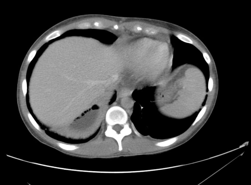
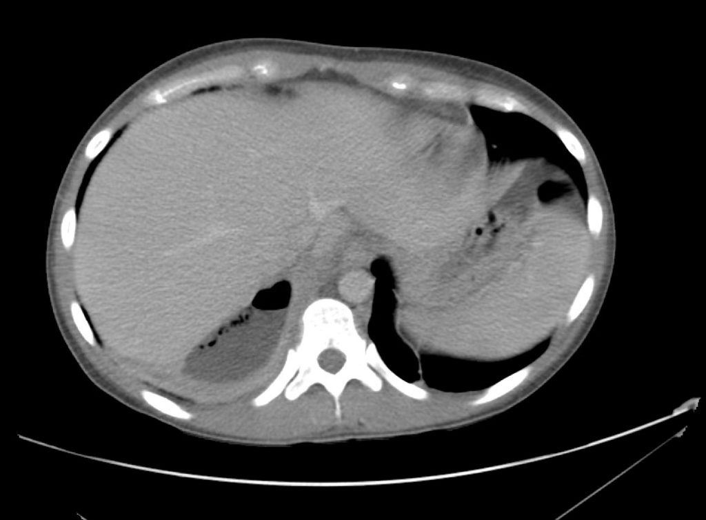
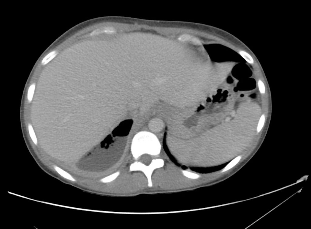
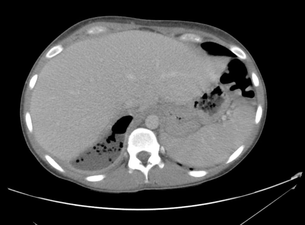
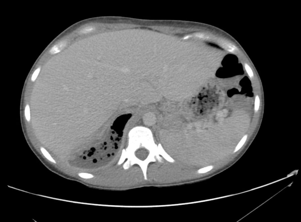
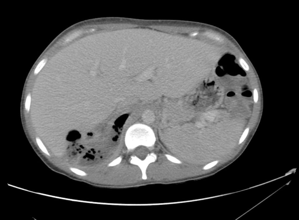
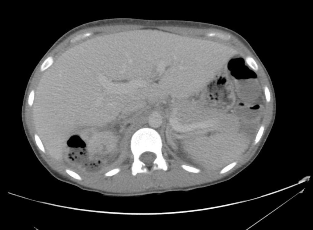
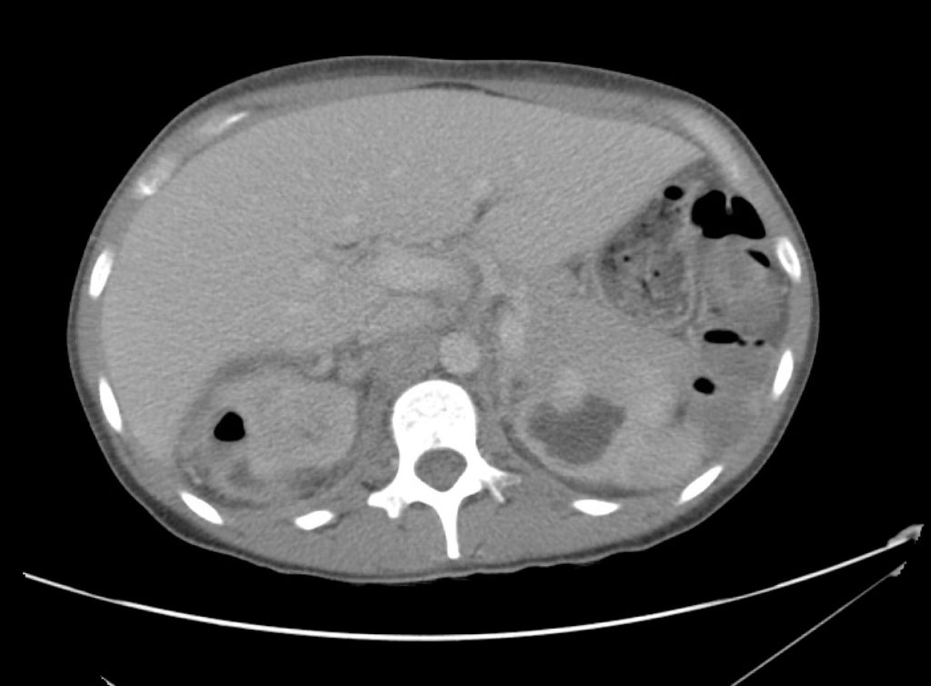
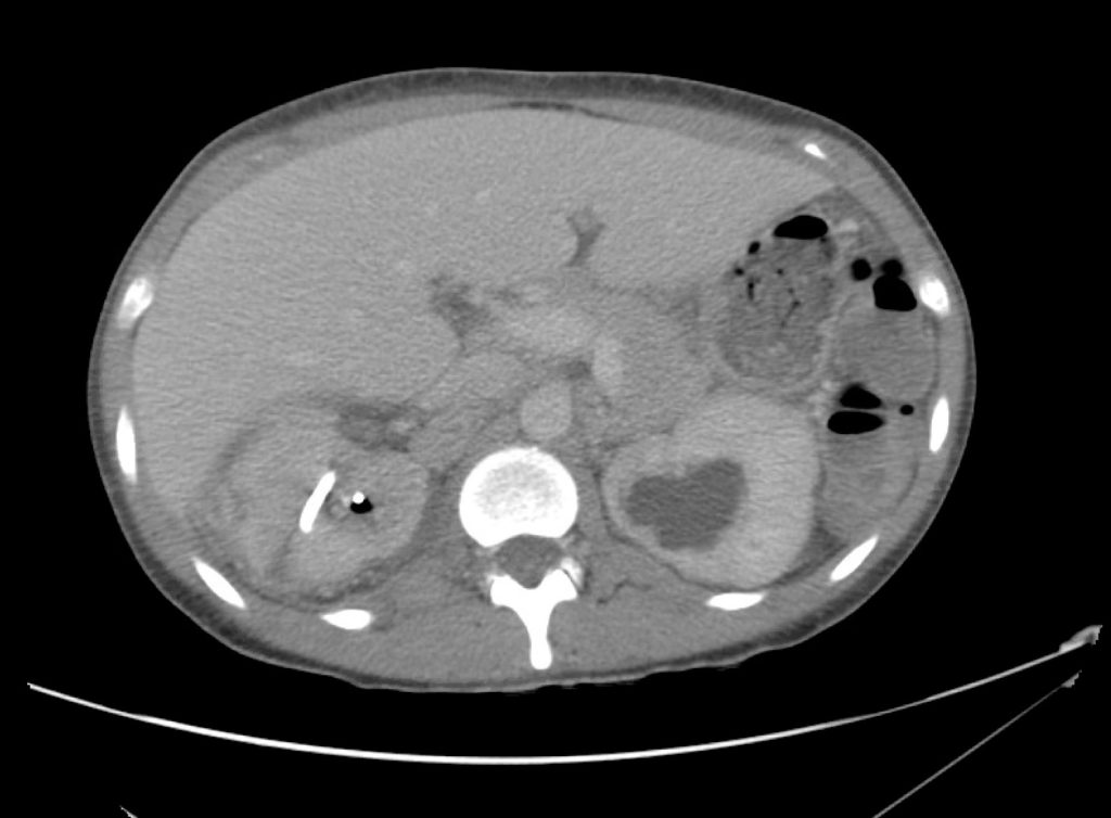
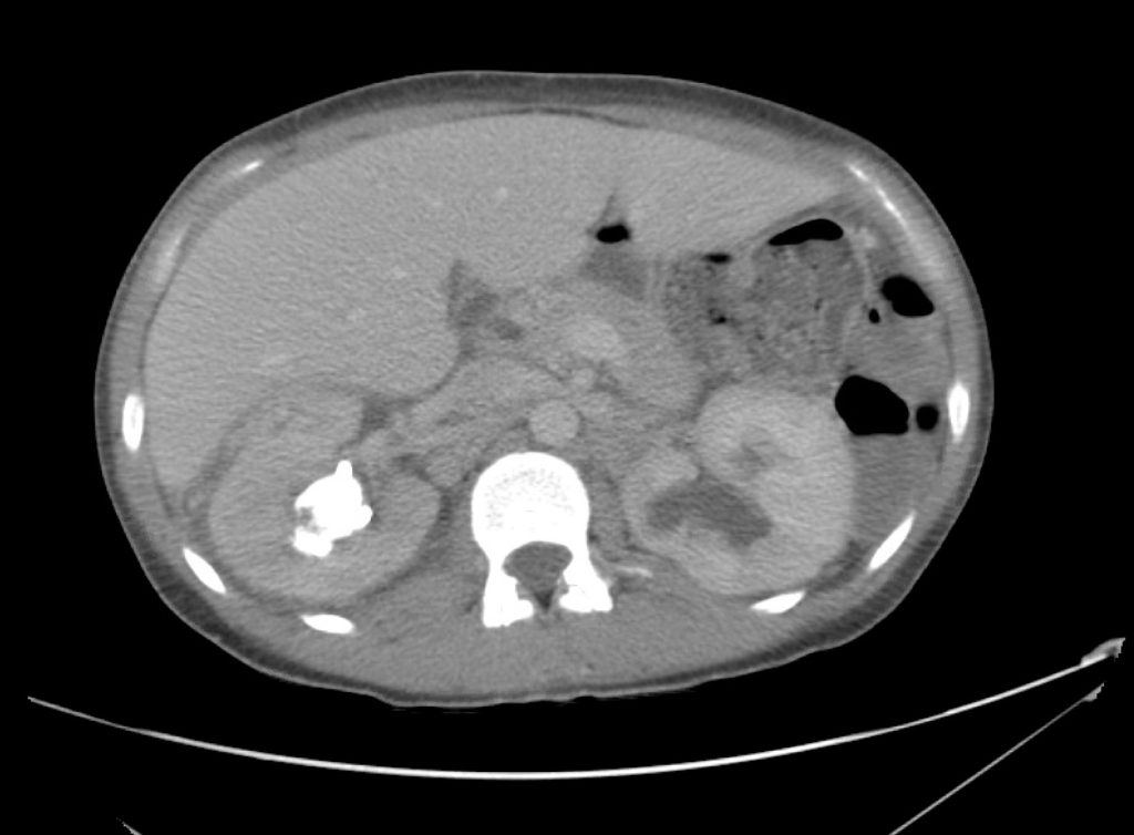
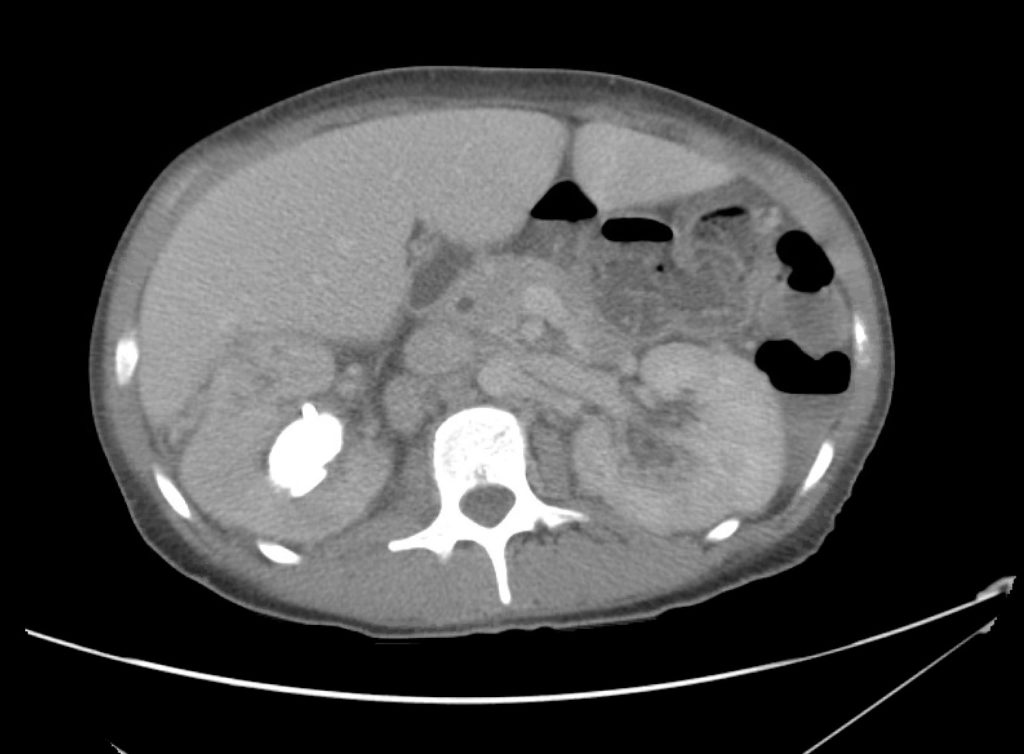
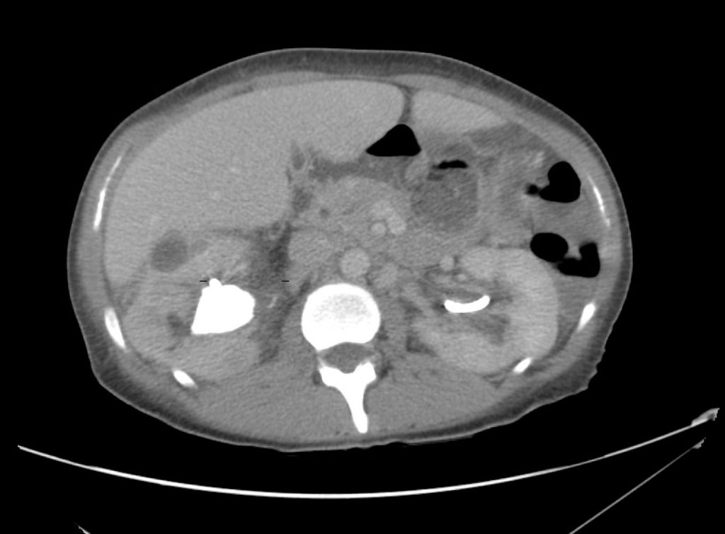
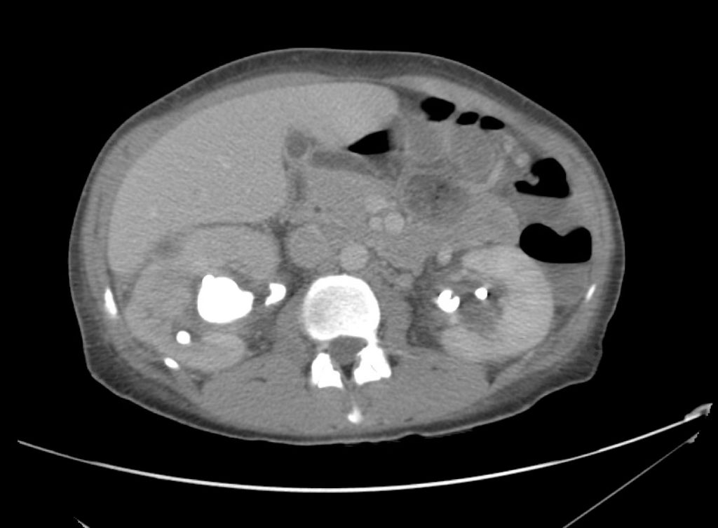
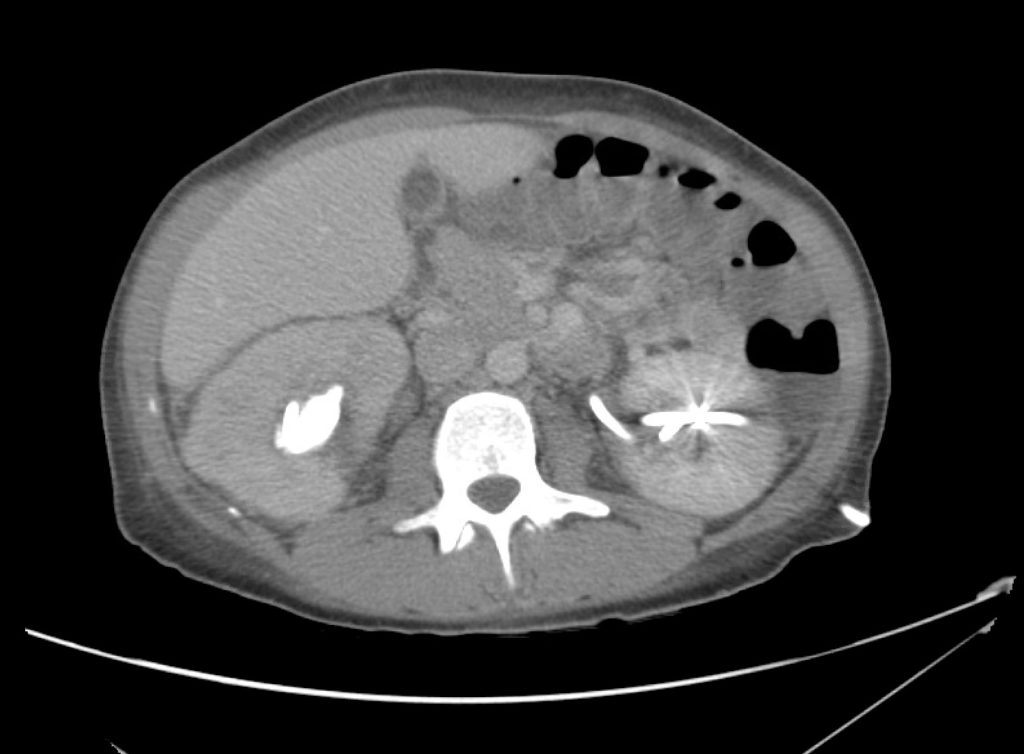
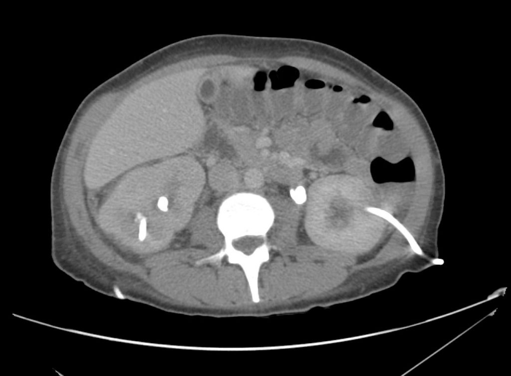
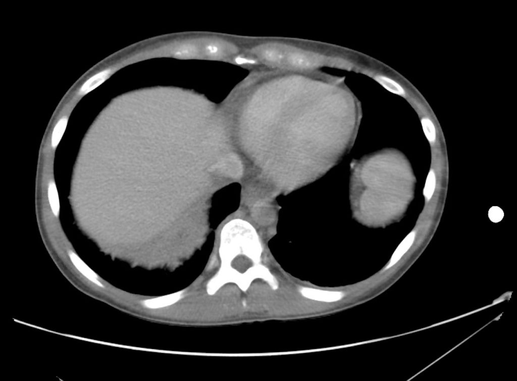
A 45 year-old female with a history of ureterolithiasis s/p bilateral percutaneous nephrostomies, hypertension and diabetes presents to the emergency department with flank pain and dysuria for two days. She noted that output from her right nephrostomy had diminished. On evaluation, her vital signs are notable for fever and tachycardia but are otherwise normal. Examination demonstrates right costovertebral angle tenderness to percussion. Drain sites appeared normal, without overlying erythema. Urinalyses from both nephrostomy collection bags were submitted. Computed tomography of the abdomen and pelvis was obtained to evaluate for nephrostomy malposition.

01_pyelo
03_pyelo
04_pyelo
05_pyelo
06_pyelo
07_pyelo
08_pyelo
09_pyelo
10_pyelo
11_pyelo
12_pyelo
13_pyelo
14_pyelo
15_pyelo
16_pyelo
17_pyelo

CT Abdomen/Pelvis Interpretation

Complex perirenal fluid collection with gas suggestive of emphysematous pyelonephritis with abscess.

Hospital Course

The patient was treated with parenteral antibiotics based on prior culture data and was admitted to the intensive care unit with urology consultation and plan for interventional radiology percutaneous drainage. The patient underwent uncomplicated perinephric drain placement and nephrostomy exchange and was discharged on hospital day five to complete a course of oral antibiotics.

An Algorithm for the Evaluation and Management of Emphysematous Urinary Tract Infections

An Algorithm for the Evaluation and Management of Emphysematous Urinary Tract Infections

References

  1. Evanoff GV, Thompson CS, Foley R, Weinman EJ. Spectrum of gas within the kidney. Emphysematous pyelonephritis and emphysematous pyelitis. Am J Med. 1987;83(1):149-154.
  2. Wan YL, Lee TY, Bullard MJ, Tsai CC. Acute gas-producing bacterial renal infection: correlation between imaging findings and clinical outcome. Radiology. 1996;198(2):433-438. doi:10.1148/radiology.198.2.8596845.
  3. Shokeir AA, El-Azab M, Mohsen T, El-Diasty T. Emphysematous pyelonephritis: a 15-year experience with 20 cases. Urology. 1997;49(3):343-346. doi:10.1016/S0090-4295(96)00501-8.
  4. Chen MT, Huang CN, Chou YH, Huang CH, Chiang CP, Liu GC. Percutaneous drainage in the treatment of emphysematous pyelonephritis: 10-year experience. JURO. 1997;157(5):1569-1573.
  5. Huang JJ, Tseng CC. Emphysematous pyelonephritis: clinicoradiological classification, management, prognosis, and pathogenesis. Arch Intern Med. 2000;160(6):797-805.
  6. Roy C, Pfleger DD, Tuchmann CM, Lang HH, Saussine CC, Jacqmin D. Emphysematous pyelitis: findings in five patients. Radiology. 2001;218(3):647-650. doi:10.1148/radiology.218.3.r01fe14647.
  7. Park BS, Lee S-J, Kim YW, Huh JS, Kim JI, Chang S-G. Outcome of nephrectomy and kidney-preserving procedures for the treatment of emphysematous pyelonephritis. Scand J Urol Nephrol. 2006;40(4):332-338. doi:10.1080/00365590600794902.
  8. Grupper M, Kravtsov A, Potasman I. Emphysematous cystitis: illustrative case report and review of the literature. Medicine (Baltimore). 2007;86(1):47-53. doi:10.1097/MD.0b013e3180307c3a.
  9. Mokabberi R, Ravakhah K. Emphysematous urinary tract infections: diagnosis, treatment and survival (case review series). Am J Med Sci. 2007;333(2):111-116.
  10. Yao J, Gutierrez OM, Reiser J. Emphysematous pyelonephritis. Kidney Int. 2007;71(5):462-465. doi:10.1038/sj.ki.5002001.
  11. Thomas AA, Lane BR, Thomas AZ, Remer EM, Campbell SC, Shoskes DA. Emphysematous cystitis: a review of 135 cases. BJU Int. 2007;100(1):17-20. doi:10.1111/j.1464-410X.2007.06930.x.
  12. Falagas ME, Alexiou VG, Giannopoulou KP, Siempos II. Risk factors for mortality in patients with emphysematous pyelonephritis: a meta-analysis. JURO. 2007;178(3 Pt 1):880–5–quiz1129. doi:10.1016/j.juro.2007.05.017.
  13. Somani BK, Nabi G, Thorpe P, et al. Is percutaneous drainage the new gold standard in the management of emphysematous pyelonephritis? Evidence from a systematic review. J Urol. 2008;179(5):1844-1849. doi:10.1016/j.juro.2008.01.019.
  14. Aswathaman K, Gopalakrishnan G, Gnanaraj L, Chacko NK, Kekre NS, Devasia A. Emphysematous pyelonephritis: outcome of conservative management. Urology. 2008;71(6):1007-1009. doi:10.1016/j.urology.2007.12.095.
  15. Kapoor R, Muruganandham K, Gulia AK, et al. Predictive factors for mortality and need for nephrectomy in patients with emphysematous pyelonephritis. BJU Int. 2010;105(7):986-989. doi:10.1111/j.1464-410X.2009.08930.x.
  16. Ubee SS, McGlynn L, Fordham M. Emphysematous pyelonephritis. BJU Int. 2011;107(9):1474-1478. doi:10.1111/j.1464-410X.2010.09660.x.
  17. Lu Y-C, Chiang B-J, Pong Y-H, et al. Predictors of failure of conservative treatment among patients with emphysematous pyelonephritis. BMC Infect Dis. 2014;14(1):418. doi:10.1186/1471-2334-14-418.

Febrile Seizure

Brief HPI:

An 8-month old female, fully-immunized, otherwise healthy is brought in by paramedics after 1 minute of witnessed generalized tonic-clonic shaking. The patient had otherwise been well, eating and behaving normally earlier that day. On EMS arrival, the patient was post-ictal but grew increasingly responsive en-route and upon presentation to the pediatric emergency department she was crying and appeared normal to her parents. Capillary glucose was 118g/dL. On examination the patient was noted to be febrile with a rectal temperature of 39.4°C. The remainder of the physical examination was normal.

ED Course:

The patient received anti-pyretics and a urinalysis was obtained which was not suggestive of urinary tract infection. During the 3-hour period of observation in the emergency department the patient remained at her normal baseline, had no further seizure activity, and tolerated oral intake with difficulty. The patient was suspected to have a simple febrile seizure and was discharged home.

Algorithm for the Diagnosis of Febrile Seizure

Algorithm for the Evaluation of Febrile Seizure

References

  1. Syndi Seinfeld DO, Pellock JM. Recent Research on Febrile Seizures: A Review. J Neurol Neurophysiol. 2013;4(165). doi:10.4172/2155-9562.1000165.
  2. Whelan H, Harmelink M, Chou E, et al. Complex febrile seizures-A systematic review. Dis Mon. 2017;63(1):5-23. doi:10.1016/j.disamonth.2016.12.001.
  3. Millichap JJ, Gordon Millichap J. Methods of investigation and management of infections causing febrile seizures. Pediatr Neurol. 2008;39(6):381-386. doi:10.1016/j.pediatrneurol.2008.07.017.
  4. Subcommittee on Febrile Seizures, American Academy of Pediatrics. Neurodiagnostic evaluation of the child with a simple febrile seizure. Pediatrics. 2011;127(2):389-394. doi:10.1542/peds.2010-3318.

Hyperthermia

Brief H&P

A young male with unknown medical history is brought in by ambulance with altered mental status. EMS reports that the patient was agitated, requiring restraints for transportation. On arrival, the patient is agitated, uncooperative and unable to provide history. Vital signs are notable for tachycardia, tachypnea and hypertension. Physical examination demonstrates diaphoresis and mydriasis, as well as increased muscle tone – particularly in the lower extremities with ankle clonus. A core temperature is obtained and noted to be elevated at 41.5°C. Point-of-care glucose is normal.

Rapid external cooling measures were instituted and several doses of intravenous benzodiazepines were administered with improvement in agitation. Laboratory studies were notable for a modest leukocytosis (WBC 18.4 without immature forms), serum sodium was 135 without osmolar gap, creatine kinase was slightly elevated without renal dysfunction, and thyroid function tests were normal. Toxicology screen was negative. ECG revealed sinus tachycardia but was otherwise normal and non-contrast computed tomography of the head was normal.

After a brief admission in the intensive care unit, the patient’s mental status improved and he reported MDMA use on the evening of presentation, he also described a history of major depression and was taking paroxetine.

Evaluation of Elevated Temperature

The designation of 38°C as “suspicious” for fever dates to 1868 and the analysis of over one million (axillary) temperature measurements by Carl Wunderlich1. Any cutoff is arbitrary and requires recognition of the clinical context and normal daily variations (with nadir in the morning and peak in evening) 2,3. What is clear is that peripheral thermometry (unless demonstrating fever) is unreliable and a core temperature should be sought4.

Thermoregulation

Temperature homeostasis is a balance between heat production and dissipation maintained by the anterior hypothalamus. Heat production is a byproduct of normal metabolic processes and skeletal muscle activity. Conservation, maintenance or dissipation of heat is aided by cutaneous vasodilation, sweating, or behavioral responses.

Fever is caused by endogenous or exogenous pyrogens which alter the homeostatic set-point, inducing thermogenesis and elevating the body temperature. Precipitants of fever are usually infectious, however non-infectious processes (ex. malignancy, tissue ischemia/infarction, auto-immune disease) resulting in inflammation can provoke a similar response 5-7.

There is no explicit temperature distinction to diagnose hyperthermia, instead the physiologic mechanism is different. In hyperthermia, the body’s homeostatic mechanisms are dysfunctional or overwhelmed due to heat exposure, excess production, ineffective dissipation or hypothalamic malfunction 8.

Algorithm for the Evaluation of Hyperthermia 8-15

Algorithm for the Evaluation of Hyperthermia

Implicated Agents in Drug-Induced Hyperthermic Syndromes 9,10

Serotonin Syndrome

Class Examples
SSRI sertraline, fluoxetine, paroxetine
Other anti-depressants trazodone, venlafaxine, lithium
MAOI phenelzine, isocarboxazid
Anti-epileptic drugs valproate
Analgesics meperidine, fentanyl, tramadol
Anti-emetic ondansetron, metoclopramide
Anti-migraine sumatriptan
Antimicrobial linezolid, ritonavir
Illicit substances MDMA, LSD

Neuroleptic Malignant Syndrome (NMS)

Class Examples
Typical anti-psychotic haloperidol, prochlorperazine
Atypical anti-psychotic risperidone, olanzapine, quetiapine, aripiprazole
Anti-dopaminergic metoclopramide, droperidol

References:

  1. Wunderlich CA. Das Verhalten Der Eigenwärme in Krankheiten. 1870.
  2. Mackowiak PA, Wasserman SS, Levine MM. A critical appraisal of 98.6 degrees F, the upper limit of the normal body temperature, and other legacies of Carl Reinhold August Wunderlich. JAMA. 1992;268(12):1578-1580.
  3. Lee-Chiong TL, Stitt JT. Disorders of temperature regulation. Compr Ther. 1995;21(12):697-704.
  4. Niven DJ, Gaudet JE, Laupland KB, Mrklas KJ, Roberts DJ, Stelfox HT. Accuracy of peripheral thermometers for estimating temperature: a systematic review and meta-analysis. Ann Intern Med. 2015;163(10):768-777. doi:10.7326/M15-1150.
  5. Dinarello CA. Infection, fever, and exogenous and endogenous pyrogens: some concepts have changed. J Endotoxin Res. 2004;10(4):201-222. doi:10.1179/096805104225006129.
  6. Greisman LA, Mackowiak PA. Fever: beneficial and detrimental effects of antipyretics. Curr Opin Infect Dis. 2002;15(3):241-245.
  7. Dinarello CA. Thermoregulation and the pathogenesis of fever. Infect Dis Clin North Am. 1996;10(2):433-449.
  8. Simon HB. Hyperthermia. N Engl J Med. 1993;329(7):483-487. doi:10.1056/NEJM199308123290708.
  9. Boyer EW, Shannon M. The serotonin syndrome. N Engl J Med. 2005;352(11):1112-1120. doi:10.1056/NEJMra041867.
  10. Berman BD. Neuroleptic malignant syndrome: a review for neurohospitalists. Neurohospitalist. 2011;1(1):41-47. doi:10.1177/1941875210386491.
  11. Hayes BD, Martinez JP, Barrueto F. Drug-induced hyperthermic syndromes: part I. Hyperthermia in overdose. Emerg Med Clin North Am. 2013;31(4):1019-1033. doi:10.1016/j.emc.2013.07.004.
  12. Oruch R, Pryme IF, Engelsen BA, Lund A. Neuroleptic malignant syndrome: an easily overlooked neurologic emergency. Neuropsychiatr Dis Treat. 2017;13:161-175. doi:10.2147/NDT.S118438.
  13. Musselman ME, Saely S. Diagnosis and treatment of drug-induced hyperthermia. Am J Health Syst Pharm. 2013;70(1):34-42. doi:10.2146/ajhp110543.
  14. Ahuja N, Cole AJ. Hyperthermia syndromes in psychiatry. Adv psychiatr treat (Print). 2018;15(03):181-191. doi:10.1192/apt.bp.107.005090.
  15. Tomarken JL, Britt BA. Malignant hyperthermia. Ann Emerg Med. 1987;16(11):1253-1265. doi:10.1016/S0196-0644(87)80235-4.

Sinus Tachycardia

Brief History and Physical:

A young female with a history of schizophrenia presents to the emergency department reporting hallucinations. She had been diagnosed with schizophrenia one year previously and was briefly admitted to a psychiatric hospital. She discontinued her anti-psychotic (risperidone) two months ago, and over the past week she reports increasingly prominent auditory and visual hallucinations.

She denies recent illness, vomiting/diarrhea, changes in urinary habits, new medications, alcohol or illicit substance use. She also denies chest pain, palpitations or shortness of breath.

Vital signs are notable for a heart rate of 148bpm and are otherwise normal (including core temperature). Detailed physical examination is normal except for a rapid, regular heart rate. Mental status examination demonstrated normal level of alertness and orientation, linear and cogent responses and occasional response to internal stimuli during which she appeared anxious.

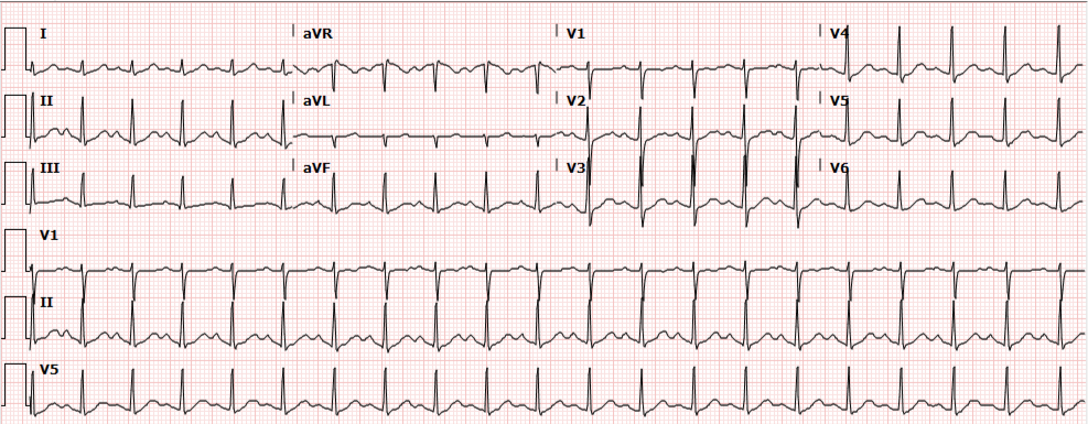
Initial evaluation and management included a 12-lead ECG which showed sinus tachycardia. Multiple boluses of normal saline were initiated while awaiting laboratory workup.

ECG: Sinus Tachycardia

Presentation ECG demonstrates sinus tachycardia.

Update:

Laboratory studies were reviewed and unremarkable. Normal hemoglobin, normal chemistry panel, negative hCG, and negative toxicology screen. The patient remained persistently tachycardic with a heart rate ranging from 140-160bpm (again sinus tachycardia on 12-lead ECG). An atypical antipsychotic and anxiolytic were administered and additional studies were obtained. Serum TSH, troponin and D-dimer were normal and bedside ultrasound did not identify a pericardial effusion. The patient remained asymptomatic, reporting subjective improvement in anxiety and hallucinations. Psychiatry was consulted and the patient was placed in observation for monitoring of sinus tachycardia. Observation course was uneventful as the patient remained asymptomatic. Transthoracic echocardiography was normal. Psychiatry consultation recommended resumption of home anti-psychotic and outpatient follow-up. Tachycardia had improved but not resolved at the time of discharge (heart rate 109bpm) and the patient was instructed to follow-up with her primary care provider.


Algorithm for the Evaluation of Sinus Tachycardia

Algorithm for the Evaluation of Sinus Tachycardia

Any vital sign derangement is concerning and tachycardia may be associated with unanticipated death after discharge home1. The presence of tachycardia suggests one of several categories of hemodynamic, autonomic, or endocrine/metabolic derangement.

Demand for increased cardiac output

A perceived demand for increased cardiac output will prompt chronotropic (and inotropic) amplification before hypotension develops. Causative etiologies include: volume depletion (from hemorrhage, gastrointestinal or renal losses), distributive processes (such as infection), obstruction (pulmonary embolus, or pericardial effusion with impending tamponade), or tissue hypoxia (anemia or lung disease).

Autonomic nervous system

Autonomic nervous system disturbances induced by stimulant, sympathomimetic or anti-cholinergic use, or withdrawal of certain agents such as ethanol or beta-blockers may be at fault.

Endocrine and other causes

Hyperthyroidism and pheochromocytoma should be considered, and as diagnoses of exclusion: anxiety, pain, or inappropriate sinus tachycardia2.

Evaluation:
Core temperature
CBC
Troponin
D-dimer
Bedside cardiac ultrasound
Urine toxicology screen
Ethanol level
TSH/T4

Algorithm for the Evaluation of Narrow-Complex Tachycardia3,4,5,6

Algorithm for the Evaluation of Narrow-Complex Tachycardia

References:

  1. Sklar DP, Crandall CS, Loeliger E, Edmunds K, Paul I, Helitzer DL. Unanticipated Death After Discharge Home From the Emergency Department. Ann Emerg Med. 2007;49(6):735-745. doi:10.1016/j.annemergmed.2006.11.018.
  2. Olshansky B, Sullivan RM. Inappropriate sinus tachycardia. J Am Coll Cardiol. 2013;61(8):793-801. doi:10.1016/j.jacc.2012.07.074.
  3. Yusuf S, Camm AJ. Deciphering the sinus tachycardias. Clin Cardiol. 2005;28(6):267-276.
  4. Katritsis DG, Josephson ME. Differential diagnosis of regular, narrow-QRS tachycardias. Heart Rhythm. 2015;12(7):1667-1676. doi:10.1016/j.hrthm.2015.03.046.
  5. Bibas L, Levi M, Essebag V. Diagnosis and management of supraventricular tachycardias. CMAJ. 2016;188(17-18):E466-E473. doi:10.1503/cmaj.160079.
  6. Link MS. Clinical practice. Evaluation and initial treatment of supraventricular tachycardia. N Engl J Med. 2012;367(15):1438-1448. doi:10.1056/NEJMcp1111259.

Kawasaki Disease

Brief H&P:

An 8-month old male is brought to the emergency department with fever. He has had four days of fever (temperature ranging from 37-40°C), rash on trunk and extremities, white-colored tongue discoloration, and irritability with decreased oral intake. Temperature on presentation was 39.4°C, examination revealed an erythematous maculopapular rash on the extremities and trunk including soles of the feet. Mucous membrane involvement was noted with oropharyngeal erythema and bilateral conjunctival injection. Neck examination demonstrated right-sided cervical adenopathy.

Labs:

  • WBC: 23.4 (N: 59%, B: 21%)
  • ESR: 100mm/hr
  • CRP: 7.59mg/dL
  • Albumin: 3.3g/dL
  • AST/ALT: 78U/L, 65U/L
  • UA: 7WBC, no bacteria

Hospital Course

The patient was admitted with a diagnosis of Kawasaki Disease and was treated with IVIG and high-dose aspirin. The patient demonstrated marked improvement with treatment and had a normal echocardiogram. He was discharged on hospital day three.

Epidemiology1,2

  • Age: 6 months to 5 years
  • Northeast Asian
  • Possible heritable component
  • Seasonal (winter/spring)

Course

  • Acute febrile (T > 39°C refractory to anti-pyretics)
  • Subacute (coronary vasculitis)
  • Convalescent

Diagnosis

  • Fever >5d
  • Criteria (4/5)
    • Conjunctivitis (bilateral, non-exudative)
    • Oropharynx changes (strawberry tongue, erythema, perioral)
    • Cervical lymphadenopathy (unilateral, >1.5cm)
    • Rash
    • Extremity changes (erythema, edema, palm/sole involvement)
  • Incomplete (2-3 criteria)

Labs

  • CBC: Elevated WBC (neutrophil predominant)
  • Urinalysis: Sterile pyuria
  • Acute phase reactants: Elevated ESR (>40-60mm/hr), CRP (>3.0-3.5mg/dL)
  • CMP: Hyponatremia, hypoalbuminemia, hypoproteinemia, elevated transaminases
  • ECG: AV block, ischemia/infarction (aneurysm/thrombosis)
  • Echocardiography: Decreased LVEF, MR, pericardial effusion

Management

  • Hospital admission
  • IVIG (2g/kg)
  • Aspirin (80mg/kg/day)

Algorithm for the Evaluation of Kawasaki and Incomplete Kawasaki Disease3,4

Algorithm for the Evaluation of Kawasaki and Incomplete Kawasaki Disease

References:

  1. Shiari R. Kawasaki Disease; A Review Article. Arch Pediatr Infect Dis. 2014;2(1 SP 154-159).
  2. Yu JJ. Diagnosis of incomplete Kawasaki disease. Korean J Pediatr. 2012;55(3):83-87. doi:10.3345/kjp.2012.55.3.83.
  3. Newburger JW, Takahashi M, Gerber MA, et al. Diagnosis, treatment, and long-term management of Kawasaki disease: a statement for health professionals from the Committee on Rheumatic Fever, Endocarditis, and Kawasaki Disease, Council on Cardiovascular Disease in the Young, American Heart Association. Pediatrics. 2004;114(6):1708-1733. doi:10.1542/peds.2004-2182.
  4. Yellen ES, Gauvreau K, Takahashi M, et al. Performance of 2004 American Heart Association recommendations for treatment of Kawasaki disease. Pediatrics. 2010;125(2):e234-e241. doi:10.1542/peds.2009-0606.

Sore Throat

Evaluation of Sore Throat

Evaluation of Sore Throat

Physical Examination:

Neck
Stiffness, limitation of extension suggestive of retropharyngeal abscess.
Jaw
Trismus associated with peritonsillar cellulitis or abscess.
Oral Cavity
Dry mucous membranes suggest dehydration (from odynophagia) and indicates severity of symptoms.
Tongue elevation, sublingual/submental induration, poor dentition (particularly of mandibular molars) associated with Ludwig Angina.
Unilateral tonsillar enlargement with contralateral uvular deviation suggests peritonisllar abscess. Fluctuance may be palpated.
Tonsilar exudates suggest infectious pharyngitis (non-specific).
Palatal petechiae suggest bacterial pharyngitis.
Ulcerations of the anterior oral cavity are associated with herpes infection, lesions on the soft palate are suggestive of coxsackievirus infection.
Rarely, a grey membrane in the posterior pharynx will suggest diphtheria.
Lymphadenopathy
Tender anterior cervical lymphadenopathy may suggest bacterial pharyngitis.
Posterior cervical lymphadenopathy is associated with infectious mononucleosis.
Large, firm, non-mobile lymph nodes may suggest malignancy.
Eyes
Presence of conjunctivitis (also rhinorrhea, exanthema) associated with viral pharyngitis.
Skin
Ulcers involving the hands, feet, in addition to pharyngeal lesions suggest coxsackievirus infection.
Scarlatiniform rash associated with pharyngitis (particularly in school-age children) suggests streptococcal pharyngitis.
Abdomen
Splenomegaly is associated with infectious mononucleosis.

Centor Criteria (Modified)

  • +1: Fever
  • +1: Tonsillar Exudate
  • +1: Tender anterior cervical lymphadenopathy
  • +1: Absence of cough
  • -1: Age >45yo

Incidence of GABHS by Centor Criteria

  • 0, -1: 1%
  • 1: 10%
  • 2: 17%
  • 3: 35%
  • 4: 51%

References:

  1. Newman, D., & Shreves, A. (2013). Sore Throat. In Rosen’s Emergency Medicine – Concepts and Clinical Practice (8th ed., Vol. 1, pp. 198-202). Elsevier Health Sciences.
  2. King, B. R., & Charles, R. A. (2004). Pharyngitis In The ED Diagnostic Challenges And Management Dilemmas. Emergency medicine practice, 6(5), 1–24.

Fever

Causes of Fever

Causes of Fever

Key Features

  • Morbidity and mortality increase with age and comborbidities
  • Most common sources in elderly: respiratory, genitourinary, skin/soft-tissue
  • Atypical presentations: functional decline, altered mental status

Immediate Evaluation and Management

  • Critical Findings

    • Altered mental status
    • Respiratory distress
    • Hemodynamic instability
  • Critical Interventions

    • Airway management, supplemental O2
    • Cardiac monitoring
    • Fluid resuscitation
    • Empiric antibiotics
    • Cooling measures (T>41.0°C)

Pathophysiology of Fever

Production of endogenous or exogenous pyrogens
Increase temperature set point in hypothalamus
Patient experiences chills when core temperature < set point
Vasoconstriction, shivering causes fever
Patient experiences euthermia, though may feel malaise, fatigue
Resolution
Patient experiences sweats until core temperature returns to normal set point

References

  1. Blum, F., & Biros, M. (2013). Fever in the Adult Patient. In Rosen’s Emergency Medicine – Concepts and Clinical Practice (8th ed., Vol. 1, pp. 119-123). Elsevier Health Sciences.

Fever in Systemic Lupus Erythematosus

HPI:

48F with a history of SLE presenting with two weeks of fevers and joint pain. The patient reports progressive development of these symptoms associated with malaise and muscle aches. She also reports two days of cough productive of yellow sputum, but denies chest pain, shortness of breath or hemoptysis. She states that these symptoms are comparable to prior lupus flares but have persisted longer than usual. She has not travelled or been hospitalized recently and has no sick contacts.

PMH:

  • SLE
  • AVN left hip

PSH:

  • Total hip arthroplasty

FH:

Non-contributory

SHx:

  • Denies tobacco, alcohol or drug use.
  • Works as an accountant.
  • No history of TB exposure.

Meds:

  • Methotrexate 50mg p.o. weekly
  • Plaquenil 400mg p.o. daily
  • MVI

Allergies:

NKDA

Physical Exam:

VS: T 39.4 HR 138 RR 19 BP 97/61 O2 98% 2L NC
Gen: Alert, responsive and in no acute distress.
HEENT: PERRL, dry mucous membranes, no oropharyngeal lesions or erythema, TM intact bilaterally, no cervical lymphadenopathy, neck supple.
CV: Tachycardia, regular rhythm without additional heart sounds.
Lungs: Clear to auscultation bilaterally.
Abd: +BS, soft, non-tender, non-distended, no rebound/guarding.
Ext: No peripheral edema, extremities warm and well-perfused, diffuse tenderness to palpation and resisted range of motion of joints with particular involvement of bilateral shoulders, elbows, knees and ankles. No effusion noted, no erythema or warmth.
Neuro: Alert, oriented to self, location and time. PERRL, EOMI, facial sensation intact, facial muscles symmetric, palate rises symmetrically, tongue protrudes midline without fasciculation, peripheral sensation and motor strength grossly intact with normal gait.

Labs/Studies:

  • 0h:
  • CBC: 6.0/7.7/23.5/259
  • BMP: 138/2.8/116/18/12/0.66/71
  • Ca: 5.0 (corrected for hypoalbuminemia: 6.9)
  • Mg: 1.1
  • Lactate: 0.7
  • ESR: 81 (0-20)
  • CRP: 7.99 (0-0.74)
  • C3: 60 (79-152)
  • C4: 13 (16-38)
  • 12h:
  • BMP: 142/4.6/113/26/15/0.75/128
  • Ca: 7.8

Imaging:

  • CXR: Negative for acute cardiopulmonary process.

Assessment/Plan:

48F with a history of SLE presenting with fever and polyarticular arthralgia.

#SIRS: Fever and tachycardia in the setting of immunosuppression. The differential diagnosis includes pneumonia (bacterial, viral, less-likely fungal), which would be community-acquired. Association with polyarticular arthralgia suggests symptoms may represent lupus flare given no leukocytosis and elevated CRP.

#Hypocalcemia: Asymptomatic, likely due to hypoalbuminemia and hypomagnesemia. Improved after IV fluids and correction of hypokalemia, hypomagnesemia.

Hospital Course:

The patient was admitted and completed a 7-day course of ceftriaxone and azithromycin. Rheumatology was consulted for management of lupus flare, which included resuming home medications and a prednisone taper upon discharge.
The patient was admitted ten days later, presenting with fevers, productive cough and pleuritic chest pain. Found to have a left lower lobe sub-segmental pulmonary embolus and antiphospholipid syndrome. She was also treated empirically for healthcare-associated pneumonia with cefepime and vancomycin given fever and productive cough though there were no imaging findings suggestive of consolidation and sputum cultures were negative.

Differential Diagnosis of Hypocalcemia: 1,2

Differential Diagnosis of Hypocalcemia

Laboratory Evaluation of Hypocalcemia: 1,2

Laboratory Evaluation of Hypocalcemia

Clinical Manifestations of Hypocalcemia: 1

  • Neuromuscular
    • Hyperexcitability
    • Perioral paresthesias
    • Muscle weakness, cramps, fasciulations, tetany
  • CNS
    • Depression
    • Irritability
    • Confusion
    • Seizure
  • Cardiac
    • Decreased contractility/conduction
    • QT prolongation

Management of Symptomatic Hypocalcemia: 1

  • 10mL 10% Calcium Gluconate
  • Dilute in 100mL D5W

Fever in SLE:

It is important to differentiate whether fever in a patient with SLE is due to disease activity (flare) or active infection.

Risk Factors for Infection:3

  • Neutropenia/Lymphopenia
  • Hypocomplementemia
  • Immunosuppressive therapy (especially Azathioprine4)

Laboratory Studies:3

  • CRP: sensitivity 100%, specificity 90% >1.35mg/dL 5
  • PCT: sensitivity 75%, specificity 75% 6

References:

  1. Cooper, M. S., & Gittoes, N. J. L. (2008). Diagnosis and management of hypocalcaemia. BMJ (Clinical research ed.), 336(7656), 1298–1302. doi:10.1136/bmj.39582.589433.BE
  2. Hannan, F. M., & Thakker, R. V. (2013). Investigating hypocalcaemia. BMJ (Clinical research ed.), 346(may09 1), f2213–f2213. doi:10.1136/bmj.f2213
  3. Cuchacovich, R., & Gedalia, A. (2009). Pathophysiology and clinical spectrum of infections in systemic lupus erythematosus. Rheumatic diseases clinics of North America, 35(1), 75–93. doi:10.1016/j.rdc.2009.03.003
  4. Zhou, W. J., & Yang, C.-D. (2009). The causes and clinical significance of fever in systemic lupus erythematosus: a retrospective study of 487 hospitalised patients. Lupus, 18(9), 807–812. doi:10.1177/0961203309103870
  5. Kim, H.-A., Jeon, J.-Y., An, J.-M., Koh, B.-R., & Suh, C.-H. (2012). C-reactive protein is a more sensitive and specific marker for diagnosing bacterial infections in systemic lupus erythematosus compared to S100A8/A9 and procalcitonin. The Journal of rheumatology, 39(4), 728–734. doi:10.3899/jrheum.111044
  6. Scirè, C. A., Cavagna, L., Perotti, C., Bruschi, E., Caporali, R., & Montecucco, C. (2006). Diagnostic value of procalcitonin measurement in febrile patients with systemic autoimmune diseases. Clinical and experimental rheumatology, 24(2), 123–128.

Sore Throat

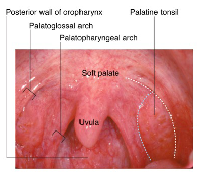
Oropharynx AnatomyID:

17 year-old female presenting to the pediatric ED with sore throat for 2 days.

HPI:

The patient reports steadily worsening sore throat over the past 2 days, associated with a sensation of swelling. The pain is described as sharp, 4/10 in severity, located on the left side of her throat, and worsened with swallowing. She denies inability to swallow or difficulty breathing, she also denies fever, cough, new skin rashes or genital lesions.

She has no PMH/PSH, takes no medications, denies t/e/d use and is not currently sexually active.

PE:

  • VS: 111/65mmHg, 80bpm, 97.8°, 16/min, 100% RA
  • Gen: Well-appearing, NAD
  • HEENT: PERRL, no conjunctival injection, TM clear b/l, minimal pharyngeal erythema on left with 6mm white circular lesion on left tonsil, no tonsillar enlargement, no uvular deviation, no cervical LAD, neck supple no masses, normal neck ROM
  • CV: RRR, no M/R/G, Lungs: CTAB
  • Abdomen: +BS, soft, NT/ND
  • Ext: Warm, well-perfused, normal peripheral pulses

Assessment & Plan:

17yo female with no significant PMH with acute pharyngitis for 2 days. The most likely cause of the patient’s symptoms is viral pharyngitis, potentially herpangina (given the appearance of the tonsillar lesion). A more serious viral/bacterial pharyngitis is less likely given the absence of fever or significant erythema/exudate. There was no uvular deviation to suggest peritonsillar abscess and no evidence of airway obstruction to suggest other acute processes (epiglottitis, retropharyngeal abscess). The plan is to recommend supportive care and ibuprofen for symptomatic relief. The patient will be discharged home in good condition with precautions to return if symptoms worsen or she begins to have difficulty swallowing/breathing.

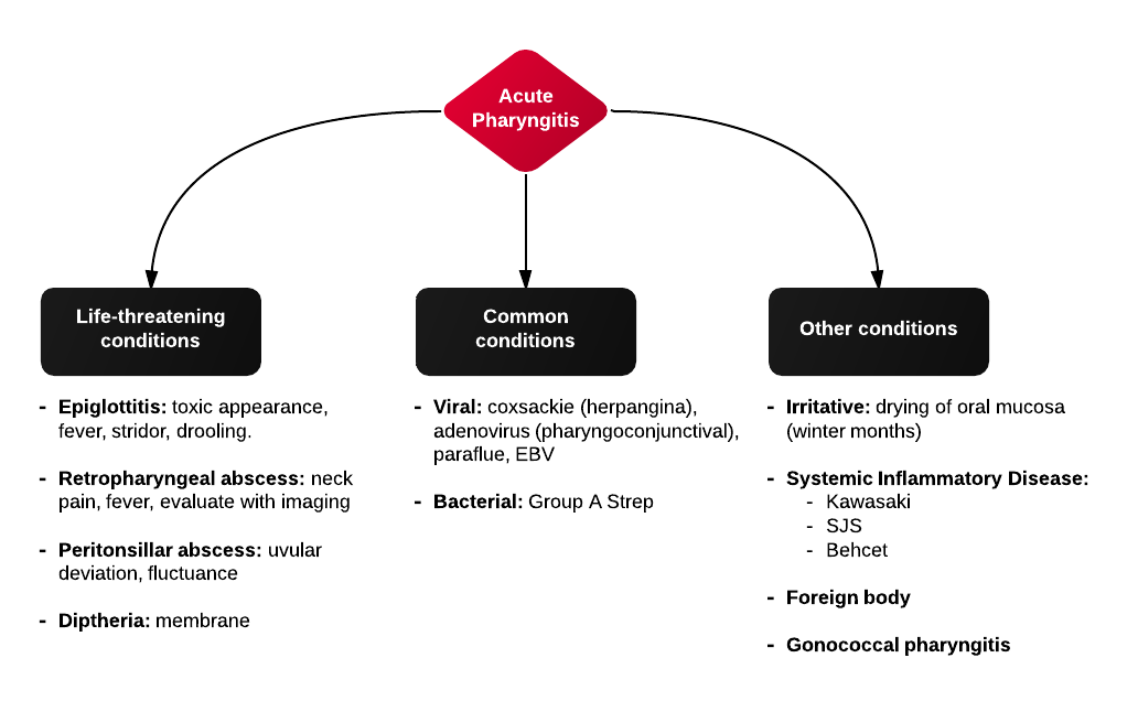
Differential Diagnosis of Acute Pharyngitis:

Acute Pharyngitis


Evaluation (history):

  • Respiratory distress: epiglottitis, retropharyngeal abscess, peritonsillar abscess, EBV (obstruction in or near pharynx)
  • Fatigue: infectious mononucleuosis
  • Abrupt onset: epiglottitis

Evaluation (physical examination):

  • Vesicles anterior: herpetic stomatitis, SJS, Behcet
  • Vesicles posterior: herpangina (± involvement of extremities)
  • Asymmetry: peritonsillar abscess
  • Stridor, drooling, respiratory distress: airway obstruction
  • Generalized inflammation: Kawasaki

Pediatric Fever

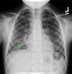
CXR with infiltrates

ID:

5yo girl brought to the pediatric emergency department by her mother due to 3 days of fever.

HPI:

The patient’s fever was first noted 3 days ago, measured at home to 103°F. It is associated with a moist cough, vomiting, and decreased PO intake. Her mother reports that she appears lethargic and has been urinating less frequently. The patient denies headache, changes in vision, burning with urination, or ear pain. No known sick contacts, attends day care.

PMH (Birth History):

No significant medical/surgical history. Ex-term born NSVD with no complications.

PE:

  • VS: 95/65mmHg, 100bpm, 102.6°, 22/min
  • General: Well-appearing, mildly irritated but consolable
  • HEENT: NC/AT, PERRL, oropharynx without erythema, no cervical LAD
  • CV: RRR, no M/G/R
  • Lungs: No evidence of respiratory distress (retractions, flaring), faint crackles over right inferior lung fields
  • Abd: +BS, soft, non-distended, TTP RLQ > LLQ, no rebound/guarding
  • Back: No CVAT

Labs/Imaging:

  • CXR PA/Lateral: RML/RLL infiltrate

Assessment:

5yo with 3 days persistent high fever and cough. These symptoms along with examination findings of crackles warranted further imaging (CXR) which revealed infiltrate in the right inferior lung field. The patient appeared clinically stable and was tolerating PO intake in the ED and was discharged home with azithromycin 5mg/kg/dose (with loading dose), clinic follow-up and strict return precautions.

Evaluation and Management of Pediatric Fever

Algorithm for the Evaluation of Pediatric Fever

A System for Pediatric Fever:

Pathophysiology:

Pathophysiology

Diagnosis:

  • <3mo: 38.0°C, 100.4°F
  • 3-36mo: 39.0°C, 102.2°F
  • Rectal > oral > axillary

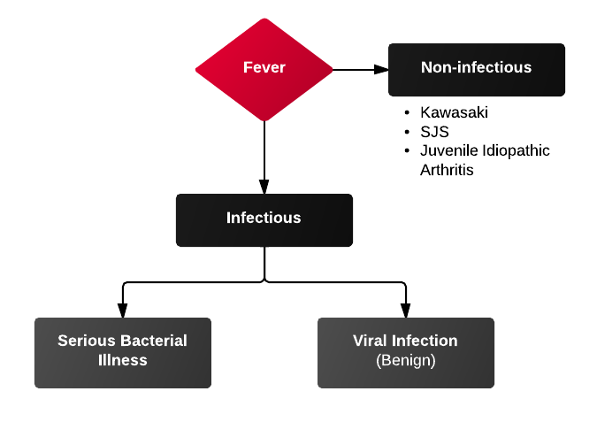
Differential Diagnosis of Pediatric Fever:

Causes Of Fever

Serious Bacterial Illness (SBI):

1) UTI and pyelonephritis

  • Most common cause of SBI
  • Accounts for 3-8% of uncharacterized fevers
  • Female > male, uncircumcised > circumcised
  • Consider BCx, CSF evaluation as 5-10% bacteremic at presentation
  • Urinalysis: LE 75% specificity, Nitrites 97% specificity

2) Pneumonia and sinusitis

  • Sinusitis uncommon <3yo (sinuses unformed)
  • PNA diagnosed with CXR, obtain if findings of respiratory distress (grunting, tachypnea, hypoxemia) or rales on exam

3) Meningitis

  • Diagnose with LP
  • Meningitis suggested if:
    • ANC > 1,000
    • Protein > 80
    • Seizure (particularly complex febrile seizure)

Diagnosis by Age Group:

<3mo

  • Physical exam findings:
    • Tachypnea, hypoxemia → LRT infection
    • Irritability, inconsolability, bulging anterior fontanelle → meningitis
    • Vomiting/diarrhea → non-specific, GE, AOM, UTI, meningitis
  • History
    • Recent immunization: increased risk of SBI (usually UTI) 24-72h after immunization
    • Confirmed bronchiolitis (viral): enterovirus/parainfluenza associated with SBI

3-36mo

  • Physical exam findings:
    • Viral (URTI, GE) → vomiting, diarrhea, rhinorrhea, cough, rash; still playful and responsive
    • UTI → fever, foul-smelling urine, crying when urinating
    • Meningitis → irritability with handling, vomiting, bulging anterior fontanelle, complex febrile seizures

>36mo

  • Physical exam findings: presentation more adult-like
  • Watch for:
    • Group A Streptococcal pharyngitis
    • Infectious mononulceosis
    • Kawasaki: high fever (>5d), strawberry tongue, conjunctivitis, desquamating rash on palms/soles

External Links