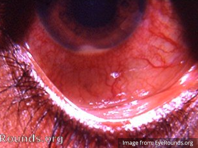
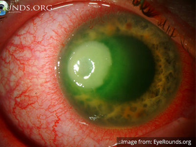
Monocular Painless Vision Loss

Brief H&P:

A 29 year-old female with no known medical history presents with vision changes for two weeks. She describes blurring and darkening of her vision in the left eye without associated pain after which her vision returns to normal. She notes occasional headaches which she describes as her usual migraines.

Physical examination including a detailed neurological examination, visual acuity, and visual fields is normal with the exception of bilateral optic disc edema. A non-contrast head CT is performed and is similarly normal. The patient underwent uncomplicated lumbar puncture with identification of elevated opening pressure with otherwise unremarkable cerebrospinal fluid analyses.

The patient’s evaluation was consistent with idiopathic intracranial hypertension, she was started on acetazolamide 500mg p.o. b.i.d and was discharged with close neurology follow-up.

An Algorithm for the Evaluation of Painless Monocular Vision Loss

An algorithm for the evaluation of painless monocular vision loss

This algorithm was developed by Dr. Lara Samarneh. Lara is an emergency medicine resident at UTHealth Houston. She attended medical school at UTHealth Houston McGovern Medical School and graduated from The University of Texas at Austin with a Bachelor’s degree in Biomedical Engineering. She is passionate about education and is currently serving as Vice Chief of Education.
Special thanks to Dr. Ore-ofe O. Adesina, MD , Associate Professor, Department of Ophthalmology and Visual Science – McGovern Medical School, Medical Director, Cizik Eye Clinic for his expertise and review of this algorithm.

References

  1. Fundoscopy images in the algorithm are sourced from Ophthalmic Atlas Images by EyeRounds.org, The University of Iowa which are licensed under a Creative Commons Attribution-NonCommercial-NoDerivs 3.0 Unported License.
  2. Guluma, K, Lee, J. Ophthalmology. In Rosen’s Emergency Medicine: Concepts and Clinical Practice (10th ed., pp. 750-780). Philadelphia, PA: Elsevier.
  3. Sharma P, Sridhar J, Mehta S. Flashes and floaters. Prim Care. 2015;42(3):425-435.
  4. Bagheri N, Mehta S. Acute vision loss. Prim Care. 2015;42(3):347-361.
  5. Pula JH, Kwan K, Yuen CA, Kattah JC. Update on the evaluation of transient vision loss. Clin Ophthalmol. 2016;10:297-303.
  6. Dehghani A, Giti M, Akhlaghi MR, Karami M, Salehi F. Ultrasonography in distinguishing optic neuritis from nonarteritic anterior ischemic optic neuropathy. Adv Biomed Res. 2012;1:3.
  7. Prasad S, Galetta SL. Approach to the patient with acute monocular visual loss. Neurol Clin Pract. 2012;2(1):14-23.
  8. Abbatemarco JR, Patell R, Buccola J, Willis MA. Acute monocular vision loss: Don’t lose sight of the differential. Rutecki GW, ed. CCJM. 2017;84(10):779-787.
  9. Current management of amaurosis fugax. The amaurosis fugax study group. Stroke. 1990;21(2):201-208.

Red and Painful Eye

Brief HPI

A 60-year-old female with rheumatoid arthritis presents with unilateral eye pain and redness without reported vision changes. Physical examination demonstrates radially-oriented engorged episcleral vessels and normal visual acuity – she was diagnosed with episcleritis and discharged with follow-up.

Algorithm for the Evaluation of the Red and Painful Eye

Algorithm for the Evaluation of the Red and Painful Eye

Overview

The red or painful eye is a common presentation in the emergency department and the rapid identification and management of potentially sight-threatening causes is critical.

The diagnostic approach to the red or painful eye begins with identifying a history of caustic exposure where immediate and copious irrigation (even before detailed examination) may limit further injury. Alkaline agents induce more severe liquefactive necrosis leading to keratoconjunctivitis, while acidic agents are generally less destructive. Management is identical for both: irrigation with lactated Ringer solution through a Morgan lens applied to a topically anesthetized eye for 5-10 minutes repeated until the pH of the eye is neutral1.

Ocular or facial trauma presents a spectrum of differential diagnoses. Suspicion for globe rupture is increased by a suggestive mechanism such as a high-velocity projectile or high-impact blunt facial trauma. Characteristic examination findings include obvious globe deformity, an irregularly-shaped pupil, extrusion of vitreous, markedly decreased visual acuity, or parting of fluorescein (Seidel sign). If globe rupture is suspected, further manipulation is unadvisable and the affected eye should be shielded. Measures should be taken to avoid increases in intra-ocular pressure including elevation of the patient’s head-of-bed, anti-emetics (to prevent intra-ocular hypertension with vomiting), and the avoidance of medications potentially implicated in intra-ocular hypertension (ketamine, succinylcholine). Tetanus and antimicrobial prophylaxis should be provided while awaiting emergent ophthalmology consultation2,3.

Another traumatic diagnosis warranting rapid identification and possible intervention is retrobulbar hematoma – identified by proptosis, eye pain, decreased visual acuity, and elevated intra-ocular pressure. Pressures exceeding 40mmHg warrant lateral cantholysis in conjunction with medical management to prevent optic nerve ischemia and preserve vision4,5.

Diagnostic Approach

The evaluation of the non-traumatic red or painful eye follows a systematic and anatomically-based approach, starting with external components and moving inward:

1. External

The examination begins externally with an assessment of function (visual acuity) and inspection and palpation of the periorbital region. Periorbital edema, erythema, and tenderness to palpation in the setting of systemic illness (fever) is concerning for orbital cellulitis. When associated with elevated intra-ocular pressure or proptosis, a retrobulbar abscess may be present. Both warrant admission and parenteral antibiotics and the latter may require operative management such as aspiration or cantholysis. Less severe features without impact on visual acuity is suggestive of a periorbital cellulitis which may be treated as an outpatient with close follow-up2,6.

2. Lids and Lashes

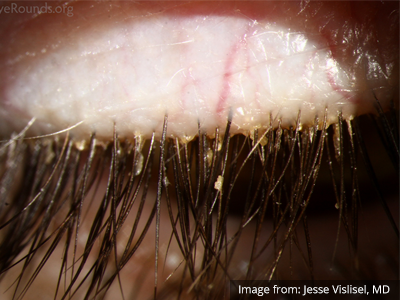
Several non-emergent processes may affect the lids and lashes including blepharitis (inflammation of the eyelid margin), chalazion (inflammation of the Meibomian glands), hordeolum (eyelash follicle abscess), or dacrocystitis (infection of the lacrimal sac)2,7,8.

3. Conjunctiva and Sclera

Again, proceeding from superficial to deeper structures we encounter the epithelial layer (including palpebral and bulbar components) covering the sclera which is subject to allergic or infectious inflammation. Conjunctivitis is characterized by engorgement of superficial conjunctival blood vessels, potentially associated with conjunctival edema (chemosis), or discharge. Most conjunctivitis is self-limited and not sight-threatening, treatment is aimed at symptomatic relief though topical antibiotics have few adverse effects and may be prescribed if the diagnosis of bacterial conjunctivitis is unclear9.

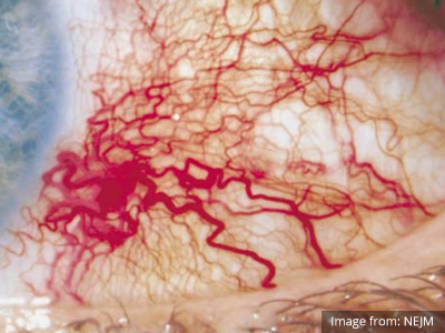
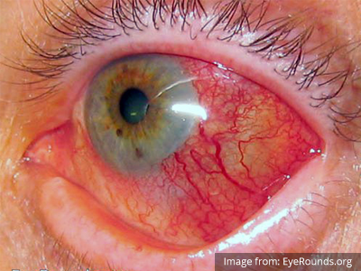
When associated with pain, a deeper inflammatory process is implicated. Scleritis is a frequently immune-mediated inflammatory process (though infection, malignancy and medications have been implicated) associated with pain, photophobia, and examination findings of globe tenderness and engorged scleral blood vessels. Management in the emergency department is trivial (systemic NSAID’s), however ophthalmology consultation should be secured due to the risk of vision-compromising complications, as well as the intimation of an underlying systemic disorder9,10. Episcleritis is similarly immune-mediated, though generally self-limiting. The diagnosis is made by identification of characteristic, radially-oriented engorged episcleral vessels. When the diagnosis of scleritis versus episcleritis or conjunctivitis is in question the application of a topical vasoconstrictor (phenylephrine 2.5%) will blanch vessels in the conjunctival or superficial episcleral plexuses – sparing scleral vessels10,11.

4. Cornea

Keratitis can be caused by infection, ultraviolet light exposure, or contact lens use. Patients may have photophobia and a foreign-body sensation. Gross inspection or slit-lamp examination will show epithelial erosions that stain with fluorescein or the characteristic dendritic pattern accompanying herpes simplex virus infection. Management includes ophthalmology consultation, topical antibiotics if a bacterial process is suggested, and close follow-up7-9,12.

5. Anterior Chamber

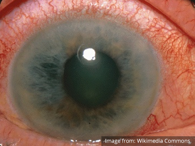
A critical process occurring in the anterior chamber is angle-closure glaucoma. The patient commonly presents with severe pain, circumcorneal injection, and a pupil fixed at mid-dilation. Diagnosis is confirmed by the measurement of elevated intra-ocular pressure (greater than 20mmHg). Reduction of intra-ocular pressure with topical and systemic agents should begin immediately while awaiting emergent ophthalmology consultation13.

The slit-lamp microscope facilitates examination of the anterior chamber. The presence of cells (floating white and red blood cells, or layering hypopyon or hyphema) and flare (protein) suggest inflammation in the anterior segment caused by a systemic inflammatory process, infection, or trauma and warrants close ophthalmologic follow-up2,7,9.

6. Vitreous

An ocular examination mimicking orbital cellulitis with evidence of anterior chamber involvement, particularly in a patient with a history of recent ocular surgery or trauma suggests endophthalmitis. Management requires admission for parenteral antibiotics with ophthalmology consultation2.

Additional Diagnostic Modalities

Advanced imaging may be useful in the diagnosis of traumatic and non-traumatic orbital pathology. Multi-detector computed tomography (MD-CT) is readily available and rapidly performed in the emergency department and can aid in the diagnosis of critical infectious processes, including extension beyond the orbital septum in orbital cellulitis, scleral thickening in endophthalmitis, and characterization of hematoma or abscess in the retrobulbar space. The addition of intravenous contrast media can identify critical vascular processes such as cavernous sinus thrombosis14,15. For traumatic pathology, CT can assist with the evaluation of globe integrity, lens position, vitreous/retinal detachment, and foreign bodies16. Imaging cannot be relied upon exclusively to exclude pathology, and the patient’s presentation and clinician’s examination should determine the need for consultation and evaluation. For globe rupture, for example, in one study of 59 patients with severe ocular trauma and diagnostic uncertainty regarding the presence of globe rupture, CT failed to diagnosed open globe injury in 1/3 of patients (with surgical scleral inspection as a reference standard)17. Another retrospective analysis of 48 eyes sustaining trauma revealed sensitivity ranging from 56-68% for CT identification of open globe injury18.

In addition to potential diagnostic inaccuracy, computed tomography exposes patients to risks including ionizing radiation, and the possibility of contrast-induced nephropathy19. Ultrasound is becoming increasingly accessible and comfortable for the emergency physician, and has the benefit of being relatively non-invasive – including facilitating ocular examination in patients with significant periorbital swelling limiting eye-opening. Ocular ultrasound may aid with the diagnosis of a wide variety of ocular pathology including vitreous hemorrhage, retinal detachment, central retinal arterial/venous occlusions, foreign body identification, lens dislocation and retrobulbar hematoma. In one study of 61 patients presenting with trauma or acute vision changes, ultrasound interpretation agreed with criterion standard (orbital computed tomography or ophthalmology evaluation) for 98% of cases20.

View Ocular Ultrasound Algorithm

References

  1. Messman AM. Ocular Injuries: New Strategies In Emergency Department Management. Emergency Medicine Practice. 2015;17(11):1–21–quiz21–2.
  2. Wright JL, Wightman JM. Red and painful eye. … Concepts and Clinical Practice 8th ed …. 2014.
  3. Romaniuk VM. Ocular trauma and other catastrophes. Emerg Med Clin North Am. 2013;31(2):399-411. doi:10.1016/j.emc.2013.02.003.
  4. Babineau MR, Sanchez LD. Ophthalmologic Procedures in the Emergency Department. Emerg Med Clin North Am. 2008;26(1):17-34. doi:10.1016/j.emc.2007.11.003.
  5. Rowh AD, Ufberg JW, Chan TC, Vilke GM, Harrigan RA. Lateral canthotomy and cantholysis: emergency management of orbital compartment syndrome. J Emerg Med. 2015;48(3):325-330. doi:10.1016/j.jemermed.2014.11.002.
  6. Henderson M, Tierney L, Smetana G. The Patient History: Evidence-Based Approach. McGraw Hill Professional; 2012.
  7. Alteveer JG, Mccans KM, Hemphill RR. The Red Eye, The Swollen Eye, And Acute Vision Loss. … Practice+ Em Practice …. 2002.
  8. Leibowitz HM. The red eye. N Engl J Med. 2000;343(5):345-351. doi:10.1056/NEJM200008033430507.
  9. Mahmood AR, Narang AT. Diagnosis and management of the acute red eye. Emerg Med Clin North Am. 2008. doi:10.1016/j.emc.2007.10.002.
  10. Albini TA, Rao NA, Smith RE. The Diagnosis and Management of Anterior Scleritis. International Ophthalmology Clinics. 2005;45(2):191.
  11. Roscoe M, Landis T. How to diagnose the acute red eye with confidence. JAAPA. 2006;19(3):24–30–quiz45–6.
  12. Deborah S Jacobs MD. Evaluation of the red eye. UpToDate. https://www.uptodate.com/contents/evaluation-of-the-red-eye. Published February 24, 2016. Accessed April 18, 2017.
  13. Prum BE, Herndon LW, Moroi SE, et al. Primary Angle Closure Preferred Practice Pattern(®) Guidelines. Ophthalmology. 2016;123(1):P1-P40. doi:10.1016/j.ophtha.2015.10.049.
  14. LeBedis CA, Sakai O. Nontraumatic orbital conditions: diagnosis with CT and MR imaging in the emergent setting. Radiographics. 2008;28(6):1741-1753. doi:10.1148/rg.286085515.
  15. Platnick J, Crum AV, Soohoo S, Cedeño PA, Johnson MH. The globe: infection, inflammation, and systemic disease. YSULT. 2011;32(1):38-50. doi:10.1053/j.sult.2010.12.003.
  16. Dunkin JM, Crum AV, Swanger RS, Bokhari SAJ. Globe trauma. YSULT. 2011;32(1):51-56. doi:10.1053/j.sult.2010.09.003.
  17. Hoffstetter P, Schreyer AG, Schreyer CI, et al. Multidetector CT (MD-CT) in the diagnosis of uncertain open globe injuries. Rofo. 2010;182(2):151-154. doi:10.1055/s-0028-1109659.
  18. Arey ML, Mootha VV, Whittemore AR, Chason DP, Blomquist PH. Computed tomography in the diagnosis of occult open-globe injuries. Ophthalmology. 2007;114(8):1448-1452. doi:10.1016/j.ophtha.2006.10.051.
  19. Custer PL, Kent TL. Pitfalls of ophthalmic radiographic imaging. Curr Opin Ophthalmol. 2014;25(5):432-435. doi:10.1097/ICU.0000000000000064.
  20. Blaivas M, Theodoro D, Sierzenski PR. A study of bedside ocular ultrasonography in the emergency department. Academic Emergency Medicine. 2002;9(8):791-799.

Diplopia Applied

Brief H&P:

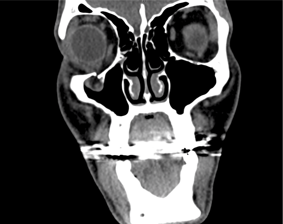
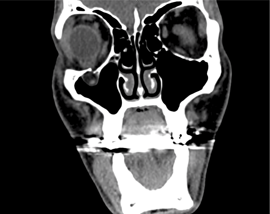
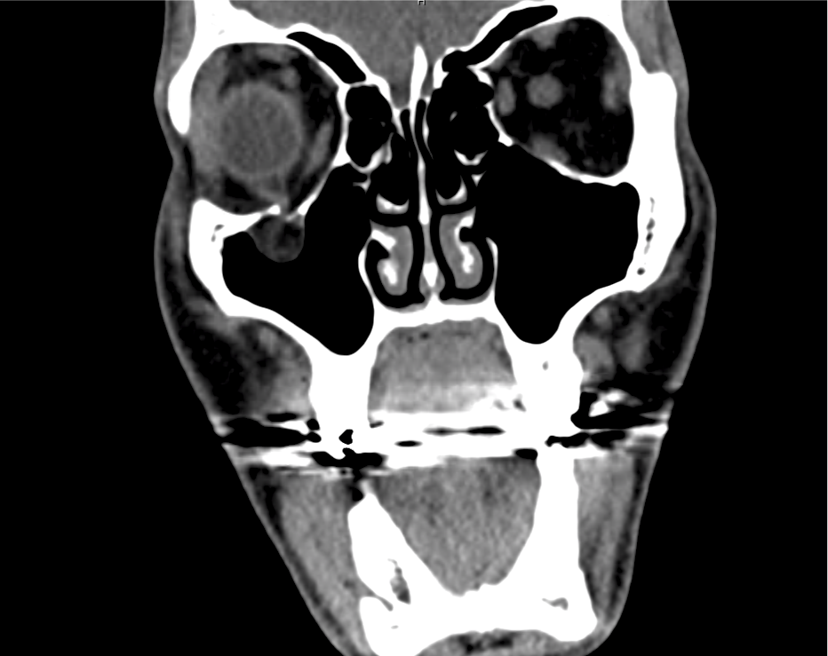
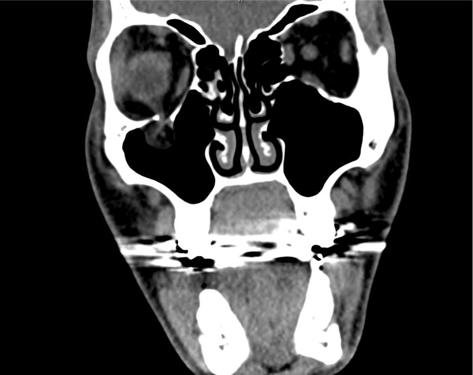
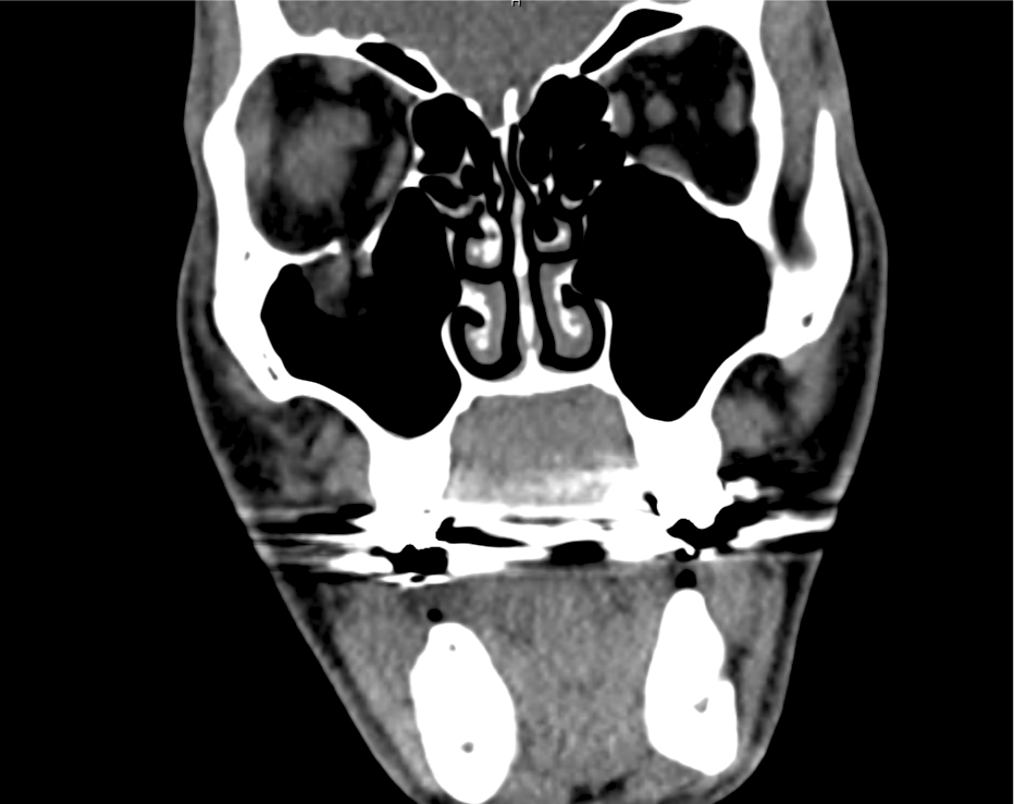
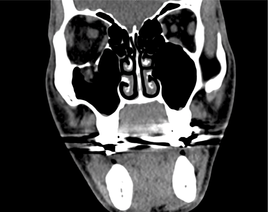
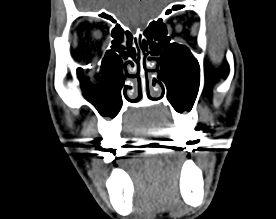
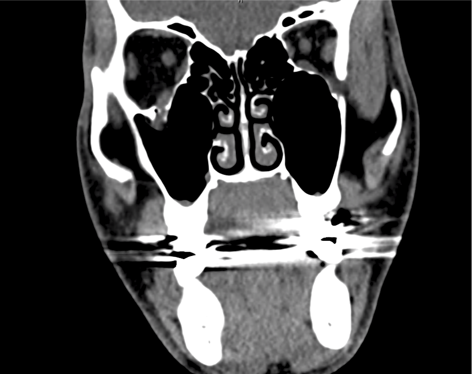
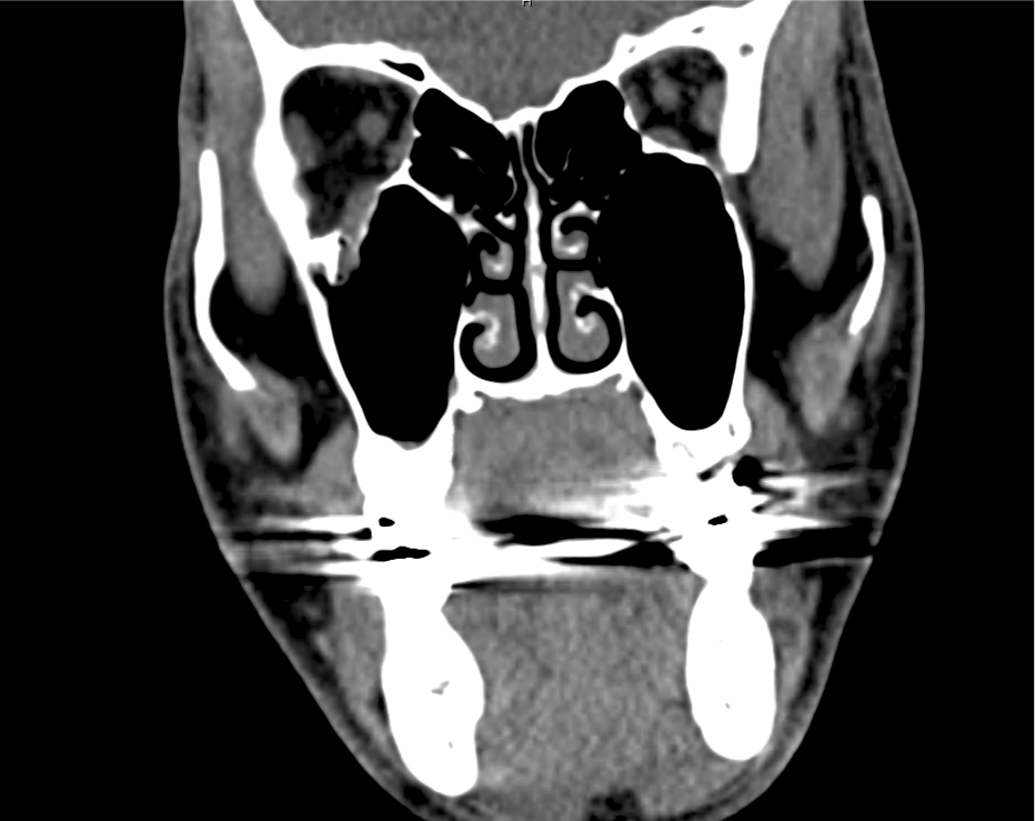
A young male with no past medical history presents to the emergency department after assault. He was punched multiple times in the face and has since noted double vision, worse with upward gaze. Examination revealed right peri-orbital edema with associated limitation to upward gaze.

Imaging:

entrapment_1
entrapment_2
entrapment_3
entrapment_4
entrapment_5
entrapment_6
entrapment_7
entrapment_8
entrapment_9

CT Maxillofacial Non-contrast

Inferior orbital wall fracture with herniation of the inferior rectus muscle.

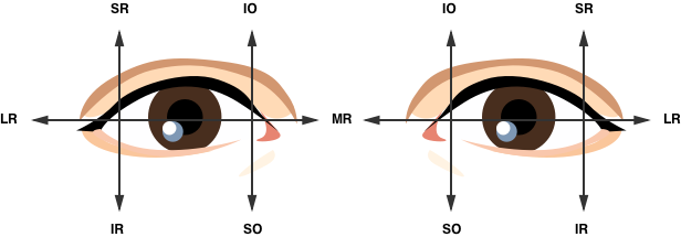
Extraocular Muscle Actions:

Extra-ocular movement actions.

Affected Anatomic Sites in Diplopia:

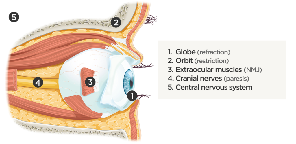
Coordinated eye positioning is affected by voluntary movements (requiring cranial nerve control for conjugate eye movements), vergence (for depth adjustments), as well as reflexive adjustments for head movement (requiring vestibular input). As with any motor activity, neuromuscular control must be normal with unrestricted movement of the globe within the orbit.

Sites causing diplopia

Algorithm for the Evaluation of Diplopia:

Diplopia has been explored previously on ddxof. The earlier algorithm was focused on identifying the paretic nerve. This algorithm uses features of the history and physical examination to identify potential etiologic causes of diplopia.

Algorithm for the Evaluation of Diplopia

References:

  1. Rucker JC, Tomsak RL. Binocular diplopia. A practical approach. Neurologist. 2005;11(2):98-110. doi:10.1097/01.nrl.0000156318.80903.b1.
  2. Friedman DI. Pearls: diplopia. Semin Neurol. 2010;30(1):54-65. doi:10.1055/s-0029-1244995.
  3. Alves M, Miranda A, Narciso MR, Mieiro L, Fonseca T. Diplopia: a diagnostic challenge with common and rare etiologies. Am J Case Rep. 2015;16:220-223. doi:10.12659/AJCR.893134.
  4. Dinkin M. Diagnostic approach to diplopia. Continuum (Minneap Minn). 2014;20(4 Neuro-ophthalmology):942-965. doi:10.1212/01.CON.0000453310.52390.58.
  5. Marx J, Walls R, Hockberger R. Rosen’s Emergency Medicine – Concepts and Clinical Practice. 8 ed. Elsevier Health Sciences; 2013:176-183.
  6. Nazerian P, Vanni S, Tarocchi C, et al. Causes of diplopia in the emergency department: diagnostic accuracy of clinical assessment and of head computed tomography. Eur J Emerg Med. 2014;21(2):118-124. doi:10.1097/MEJ.0b013e3283636120.
  7. Low L, Shah W, MacEwen CJ. Double vision. BMJ. 2015;351:h5385. doi:10.1136/bmj.h5385.
  8. Danchaivijitr C, Kennard C. Diplopia and eye movement disorders. J Neurol Neurosurg Psychiatry. 2004;75 Suppl 4:iv24-iv31. doi:10.1136/jnnp.2004.053413.
  9. Huff JS, Austin EW. Neuro-Ophthalmology in Emergency Medicine. Emerg Med Clin North Am. 2016;34(4):967-986. doi:10.1016/j.emc.2016.06.016.

Diplopia

History and Physical

38F with no medical history, presenting with double vision. The patient reported six weeks of intermittent diplopia for which she had presented to this hospital previously. She was briefly admitted for evaluation of possible cranial nerve IV palsy. Extensive imaging was unremarkable, without mass lesion, infarction, vascular malformation, or meningeal enhancement. She was discharged with outpatient follow-up including ophthalmology clinic and further imaging.

The patient represented due to persistent diplopia that is worse with right gaze. The diplopia is predominantly vertical, alleviated by head tilt. Now associated with three days of right ptosis as well as two weeks of progressive weakness and fatigue – most notable when climbing stairs.

Examination notable for right hypertropia increased on right or downward gaze suggestive of isolated inferior rectus weakness. Pupils were equal and reactive. There was marked fatigable ptosis with 2mm right palpebral fissure compared to 10mm on contralateral side. Symmetrical muscle weakness was noted, 4/5 neck flexion, elbow extension, wrist flexion/extension, shoulder abduction, hip flexion. Gait was wide-based. Application of ice for 5 minutes improved right palpebral fissure opening to >7mm.

Further evaluation included CXR and CT chest with intravenous contrast which did not identify a mediastinal mass. The patient’s respiratory status remained stable throughout hospitalization as assessed by measurements of forced vital capacity. On hospital day one, an edrophonium test was performed which was positive. The patient was started on pyridostigmine, completed a course of IVIG and was discharged with outpatient neurology follow-up.

Evaluation of Diplopia 1

History

  1. Onset/cadence
  2. Direction of gaze with worst diplopia
  3. Orientation (vertical/horizontal)
  4. Associated symptoms (headache, vertigo, dysarthria, eye pain)

Terms Describing Eye Position

Terms describing eye position

Tropias are always present, phorias are identified by cross-cover testing (break fusion)

Algorithm for the Evaluation of Diplopia 2

Algorithm for the Evaluation of Diplopia

Causes of Diplopia 3,4,5,6

Finding EOM Causes Features
Mechanical orbitopathy Variable. Abrupt restriction of movement Orbital cellulitis Pain, erythema
Orbital pseudotumor Autoimmune
Trauma History
Thyroid eye disease Bilateral
Isolated CN III Limited adduction/upgaze/downgaze Microvascular ischemia Pain, risk factors, pupil-sparing
Aneurysm Pupil involvement
Demyelination MRI
Isolated CN IV Limited downgaze (hypertropia) Trauma May be mild
Microvascular ischemia Less common than CN III
ICP Fundoscopy, imaging
Demyelination MRI
Isolated CN VI Limited abduction

(esotropia)

ICP Fundoscopy, imaging
Demyelination MRI
Microvascular ischemia Less common than CN III
INO Limited adduction

(exotropia)

Demyelination MRI
Stroke Dysarthria, ataxia, facial weakness
Multiple CN involvement (III, IV, VI) Variable Cavernous sinus process Retroorbital pain, conjunctival injection or chemosis
Brainstem deficits Variable Brainstem stroke Weakness, dysmetria, tremor
Basilar artery occlusion Vertigo, slurred speech
Wernicke AMS, ataxia, nystagmus
Basilar meningitis Fever, photophobia, meningismus
Miller-Fisher Ataxia, areflexia
Neuromuscular process Variable Myasthenia gravis Fatigability, ice test

References

  1. Alves, M., Miranda, A., Narciso, M. R., Mieiro, L., & Fonseca, T. (2015). Diplopia: a diagnostic challenge with common and rare etiologies. The American journal of case reports, 16, 220–223. doi:10.12659/AJCR.893134
  2. Borooah, S., Wright, M., & Dhillon, B. (2011). Pocket Tutor Ophthalmology. JP Medical Limited. Retrieved from https://books.google.com/books?id=z\_CfWj8-ftoC
  3. Dinkin, M. (2014). Diagnostic approach to diplopia. Continuum (Minneapolis, Minn.), 20(4 Neuro-ophthalmology), 942–965. doi:10.1212/01.CON.0000453310.52390.58
  4. Rucker, J. C., & Tomsak, R. L. (2005). Binocular diplopia. A practical approach. The neurologist, 11(2), 98–110. doi:10.1097/01.nrl.0000156318.80903.b1
  5. Friedman, D. I. (2010). Pearls: diplopia. Seminars in neurology, 30(1), 54–65. doi:10.1055/s-0029-1244995
  6. Guluma, K. (2013). Diplopia. In Rosen’s Emergency Medicine – Concepts and Clinical Practice (8th ed., Vol. 1, pp. 176-183). Elsevier Health Sciences.
  7. WikEM: Diplopia

Conjunctivitis and the Red Eye

Differential diagnosis of Conjunctivitis 1,2,3

Condition Pain Visual Acuity Photophobia Discharge Conjunctiva Lymphadenopathy Laterality Associated Features
Viral conjunctivitis None Unaffected None + watery ++ follicular pattern Pre-auricular Unilateral, spreads bilateral Viral URI
Bacterial conjunctivitis None Unaffected None ++ purulent +++ papillary pattern Occasional Unilateral, spreads bilateral Otitis media
Allergic conjunctivitis None Unaffected None + mucoid + None Bilateral Atopy

Differential Diagnosis of Red Eye 4,5

Condition Comment Hyperemia Pupil Pain Visual Acuity Cornea
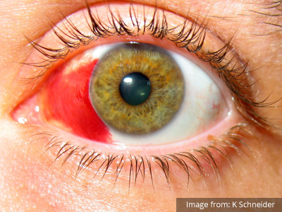
Subconjuntival hemorrhage Subconjunctival Hemorrhage Associated with trauma, coagulopathy, hypertension. Unilateral, sharply circumscribed Unaffected None Unaffected Clear
Blepharitis Blepharitis Acute/chronic inflammation of eyelid. Diffuse Unaffected Foreign body sensation Unaffected Clear
Epislceritis Episcleritis Recurrent, self-limited episodes, possible autoimmune association. Engorged, radially-oriented vessels Unaffected Mild Unaffected Clear
Scleritis Scleritis Vascular or connective tissue disease. Focal or diffuse, pink sclera Unaffected Moderate Reduced Clear
Acute angle-closure glaucoma Acute Angle-Closure Glaucoma Mydriasis leading to decreased outflow of aqueous humor. Circumcorneal injection Semi-dilated Severe Reduced Hazy
Acute anterior uveitis Uveitis Inflammation of iris or ciliary body. Circumcorneal injection Constricted Moderate Reduced Hazy
Keratitis Keratitis Inflammation of corneal epithelium. Caused by infection, contact lenses, UV exposure. Multiple punctate erosions, stain with fluorescein Unaffected Moderate Reduced Hazy

Algorithm for the Evaluation of the Red Eye 6

Algorithm for the Evaluation of the Red Eye

References

  1. Teoh, D. L., & Reynolds, S. (2003). Diagnosis and management of pediatric conjunctivitis. Pediatric emergency care, 19(1), 48–55.
  2. Azari, A. A., & Barney, N. P. (2013). Conjunctivitis. JAMA: the journal of the American Medical Association, 310(16), 1721. doi:10.1001/jama.2013.280318
  3. Cronau, H., Kankanala, R. R., & Mauger, T. (2010). Diagnosis and management of red eye in primary care. American family physician, 81(2), 137–144.
  4. Leibowitz, H. M. (2000). The red eye. New England Journal of Medicine, 343(5), 345–351. doi:10.1056/NEJM200008033430507
  5. Richards, A., & Guzman-Cottrill, J. A. (2010). Conjunctivitis. Pediatrics in review / American Academy of Pediatrics, 31(5), 196–208. doi:10.1542/pir.31-5-196
  6. Borooah, S., Wright, M., & Dhillon, B. (2011). Ophthalmology. JP Medical Limited.