Infographic: CT Chest

Examples

01_dissection-a
03_dissection-a
05_dissection-a
07_dissection-a
09_dissection-a
11_dissection-a
13_dissection-a
15_dissection-a
17_dissection-a
19_dissection-a
21_dissection-a
23_dissection-a
25_dissection-a
27_dissection-a
29_dissection-a
31_dissection-a
33_dissection-a
35_dissection-a
37_dissection-a
39_dissection-a
41_dissection-a
43_dissection-a
45_dissection-a
47_dissection-a
49_dissection-a
51_dissection-a
53_dissection-a
55_dissection-a
57_dissection-a
59_dissection-a
61_dissection-a
63_dissection-a
65_dissection-a
67_dissection-a
69_dissection-a
71_dissection-a
73_dissection-a
75_dissection-a
77_dissection-a
79_dissection-a

This slideshow requires JavaScript.

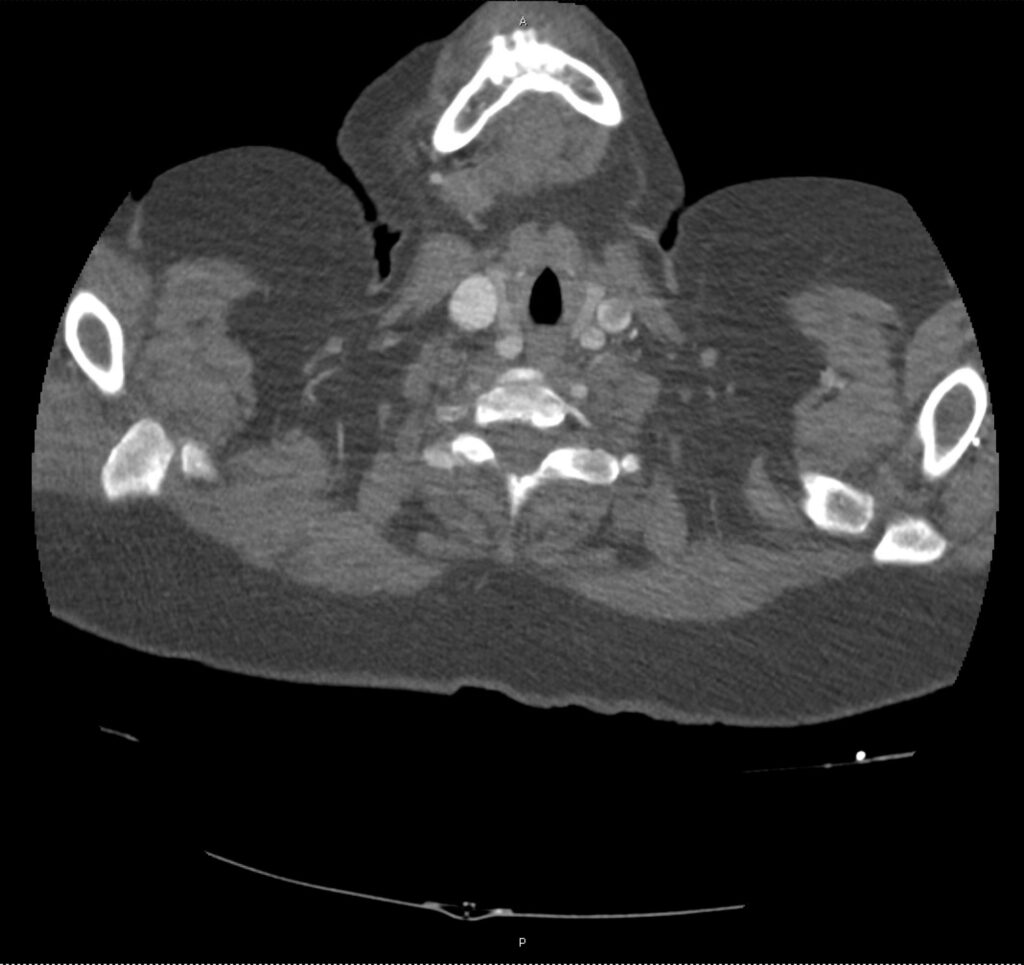
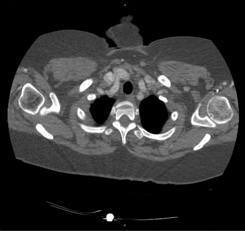
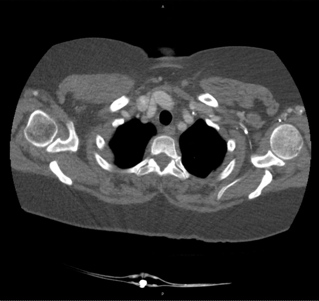
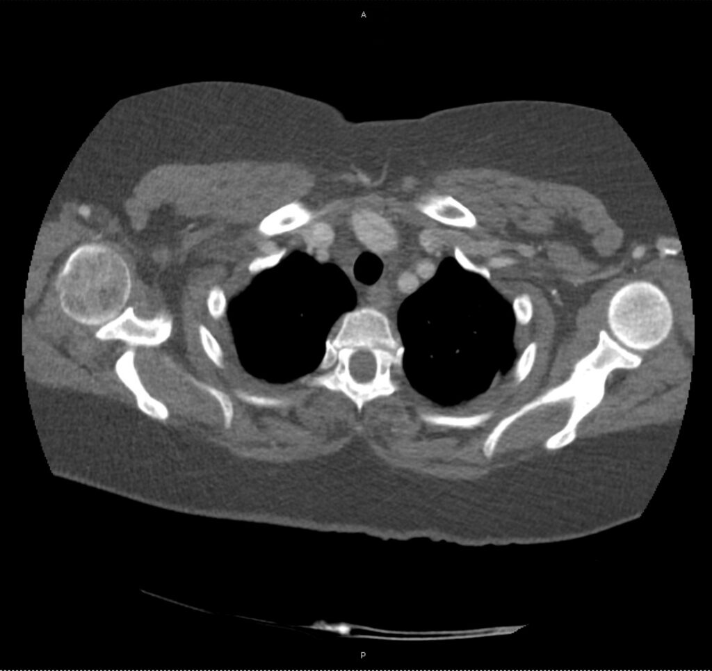
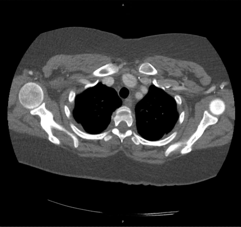
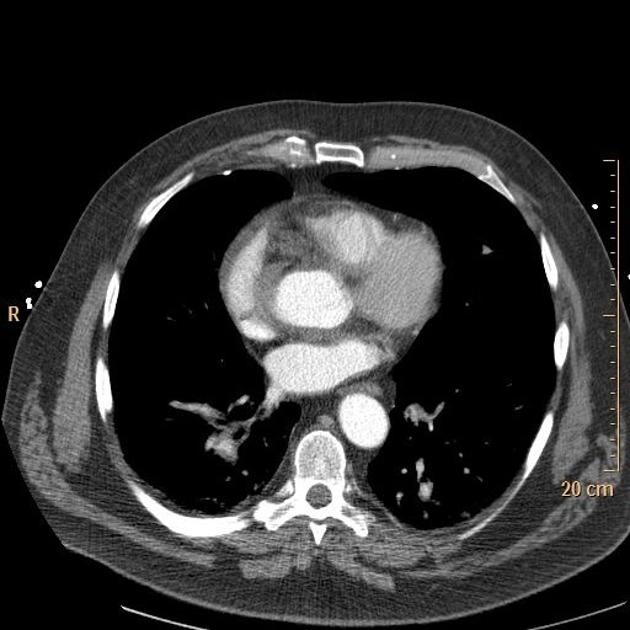
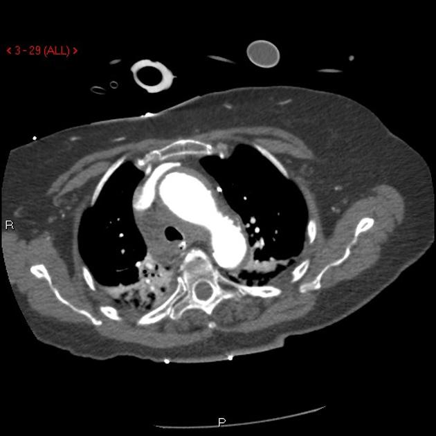
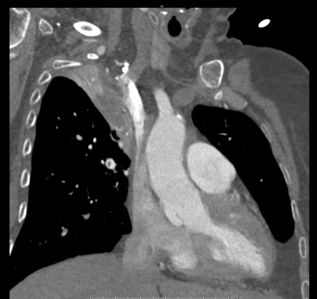
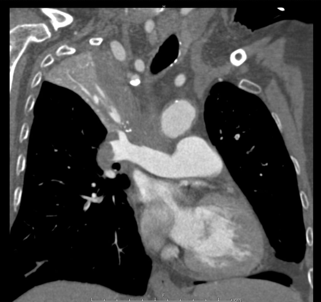
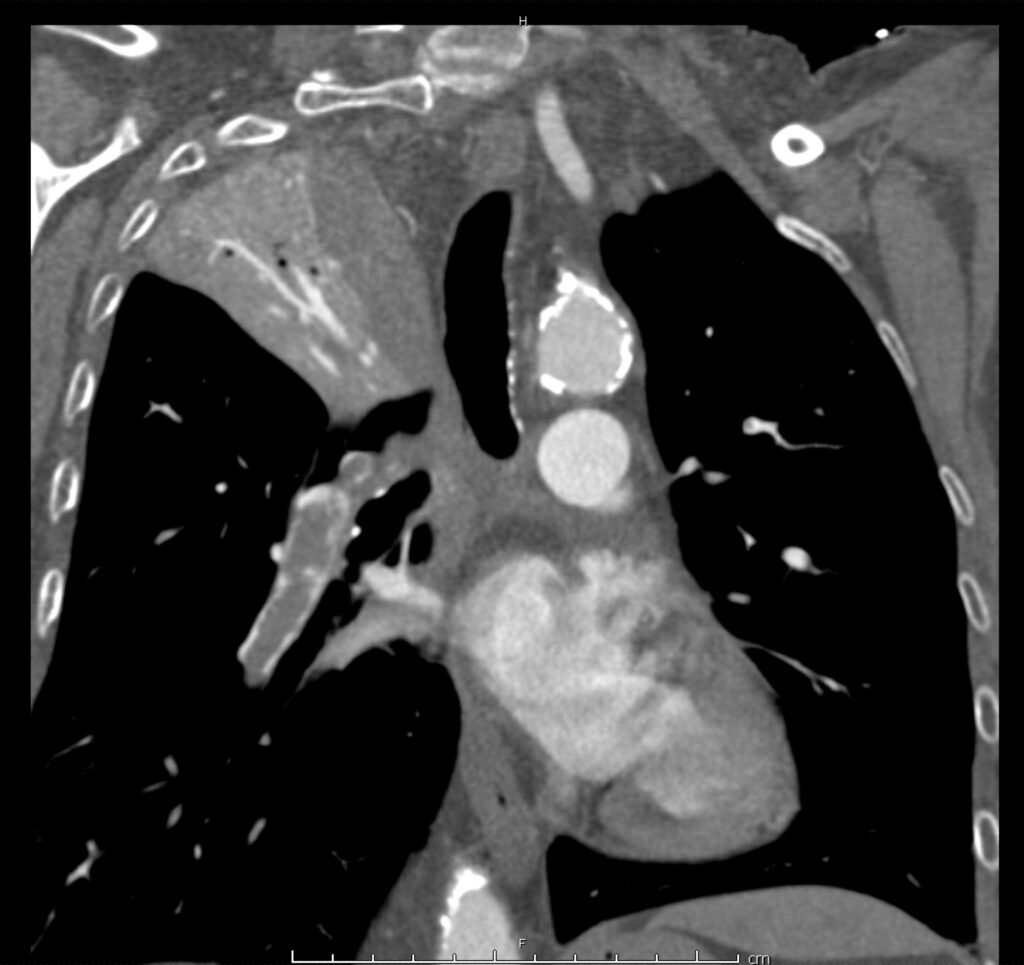
Circulation: Dissection

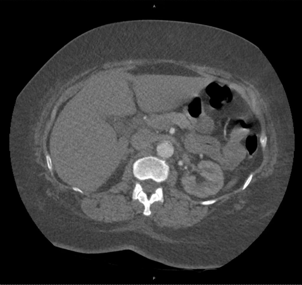
Stanford type A aortic dissection. Flap extends cranially into the common trunk of the innominate/left common carotid arteries and proximal right subclavian artery. Caudally, extends to the abdominal aorta below the level of the superior mesenteric artery.

000-9e30a83281d8cd8b5062cd7be8d3eb_big_gallery
001-172d1633087011c5900b7faa83069f_big_gallery
002-84594cad1b7e3b76fd014e2da99855_big_gallery
003-a80fbdd3d2e19f66370a446f468f8b_big_gallery
004-4aa094723a791bf588e0ed264f772a_big_gallery
005-e4a226a62a909d7c8a047a1b872932_big_gallery
006-7610851bcf1015fed3eb355dd0220e_big_gallery
007-97331e0d369db7c99451d6bc75af21_big_gallery
008-e56c78dad0d766defff840f57c1af6_big_gallery
009-3e42941ea7dce29cec2f9d14cde6d2_big_gallery
010-cee7d6a234bc4e7595279a7a0c35c3_big_gallery
011-9fb3e31fc01b644f00cce6ca6140f5_big_gallery
012-605b792fab054e4f7b0481c1a06688_big_gallery
013-4c11a1e32a569a5942ec14b3225eed_big_gallery
014-c99896363aa54419052802b8208e3e_big_gallery
015-27f195ccc055804f9bf882373988a1_big_gallery
016-af2b52df0aa2f1685ad9ee2ba8e8a4_big_gallery
017-935b535c046a12bab59275019c274d_big_gallery
018-16799bc0f6fad834572ba90aae52d4_big_gallery
019-db2333f97bab39b8bca54366c53a91_big_gallery
020-2bd374348e04799e2ca37a89e64065_big_gallery
021-390d24d581cbf1b544657f4958af87_big_gallery
022-88b6ca275e0757a4096606aa51fa4d_big_gallery
023-4dcda55685c682ad956c7f57f1705c_big_gallery
024-42ab779bbb194554d63fd3892f299c_big_gallery
025-ede3ecd889fbfcda094f809d993ce6_big_gallery
026-0f011a15b3f598caa3df0fb8efb4ea_big_gallery
027-7b67ed104cca44641750d9678f32b5_big_gallery
028-00b5878757c2fee0ae0a0e17a7df2e_big_gallery
029-c08985fc8ea295ab1940932e6a0d0c_big_gallery
030-a6acc1260e0007ce3d6f2e5a9dc068_big_gallery
031-fe6405537d165f3c79c4f9e761adb6_big_gallery
032-7d6ec7fd098546e70179a974e50e7f_big_gallery
033-9ee5dbb0d2d5b31624c611a02eb97e_big_gallery

This slideshow requires JavaScript.

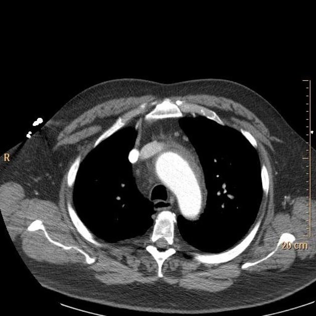
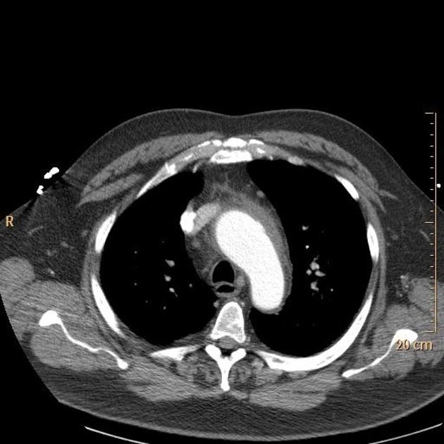
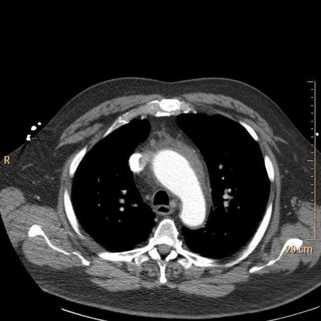
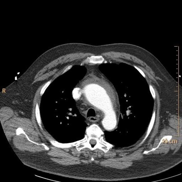
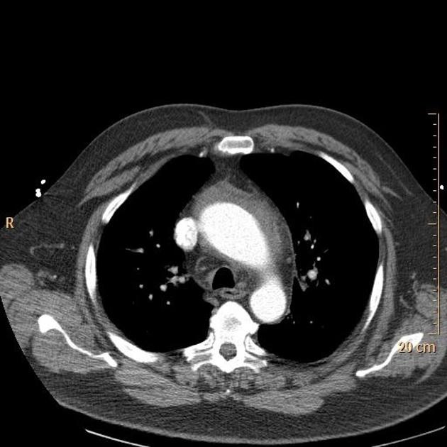
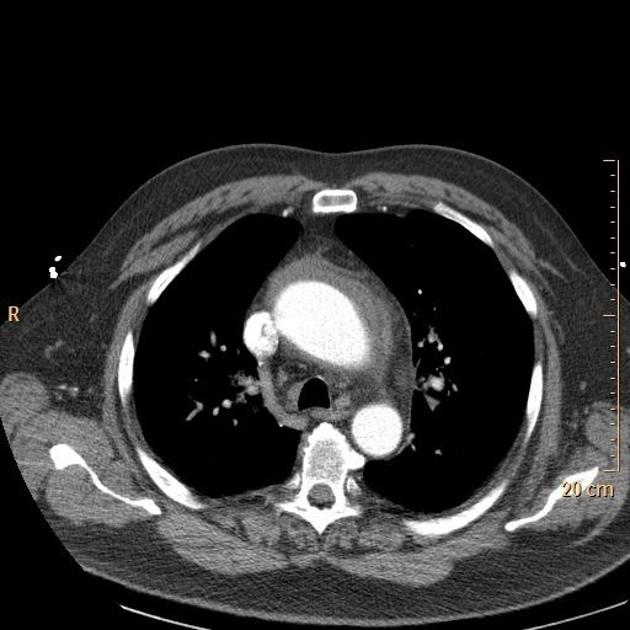
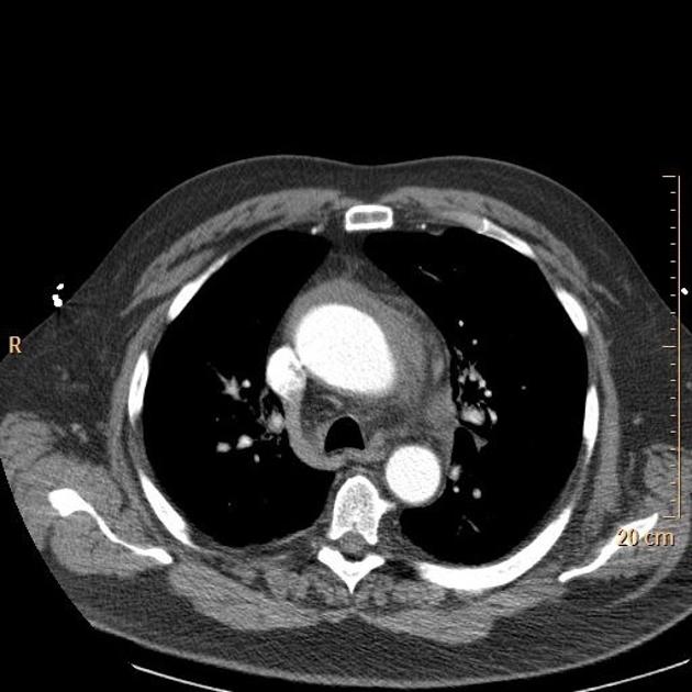
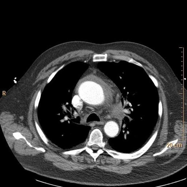
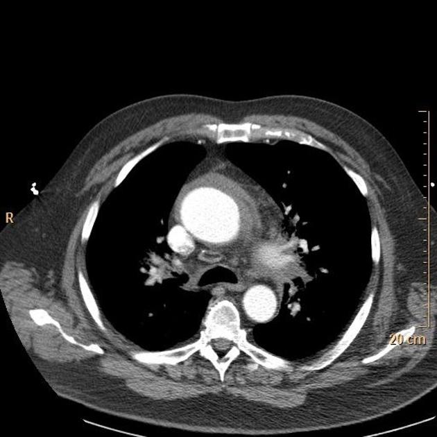
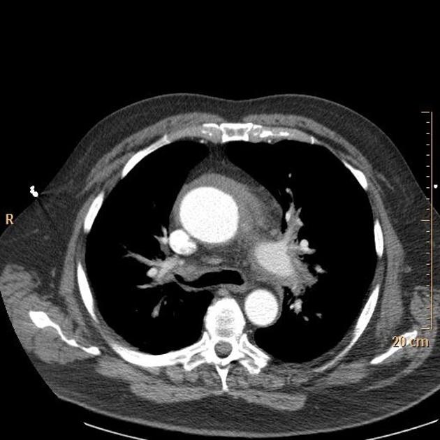
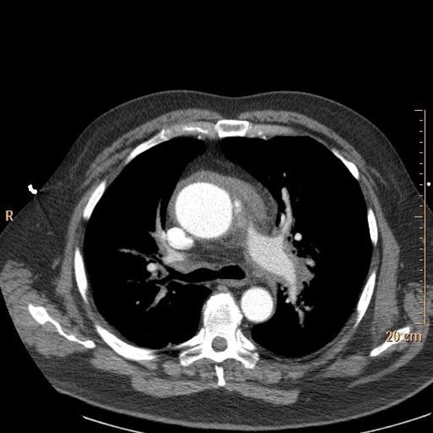
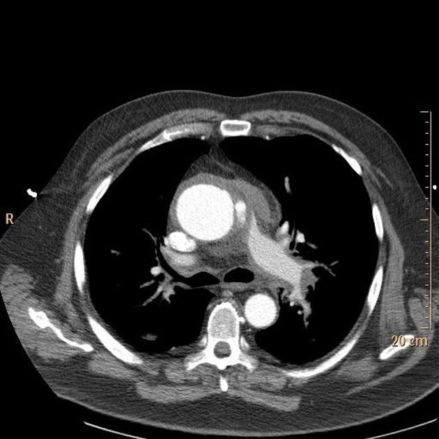
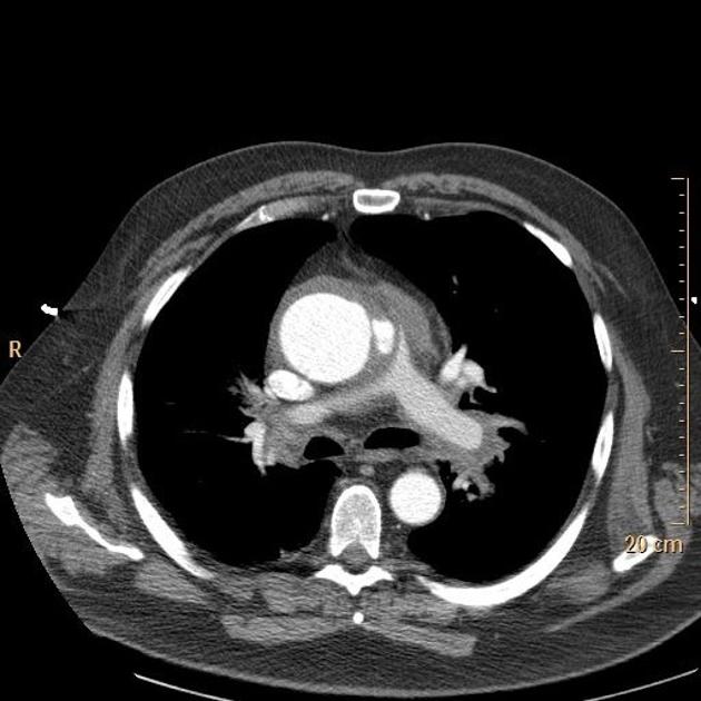
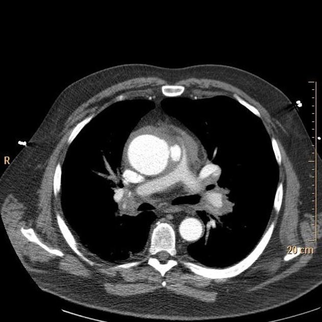
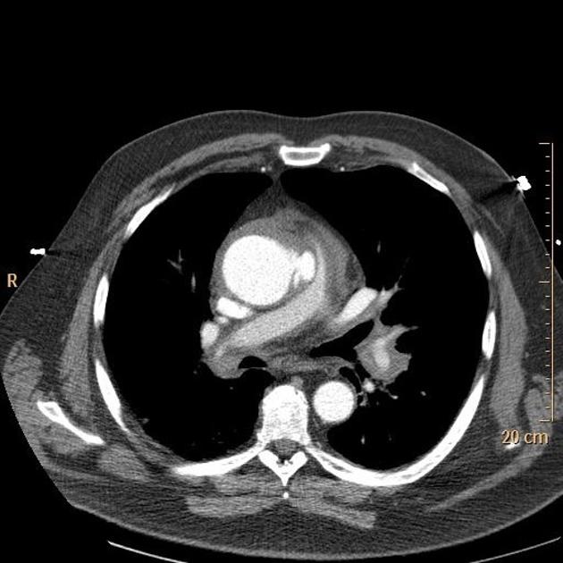
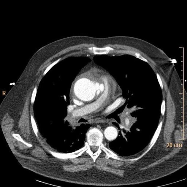
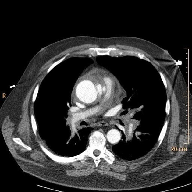
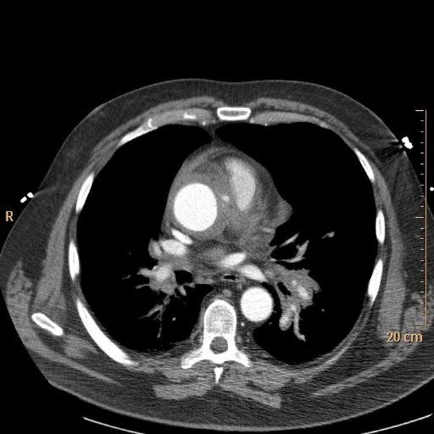
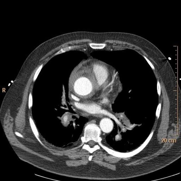
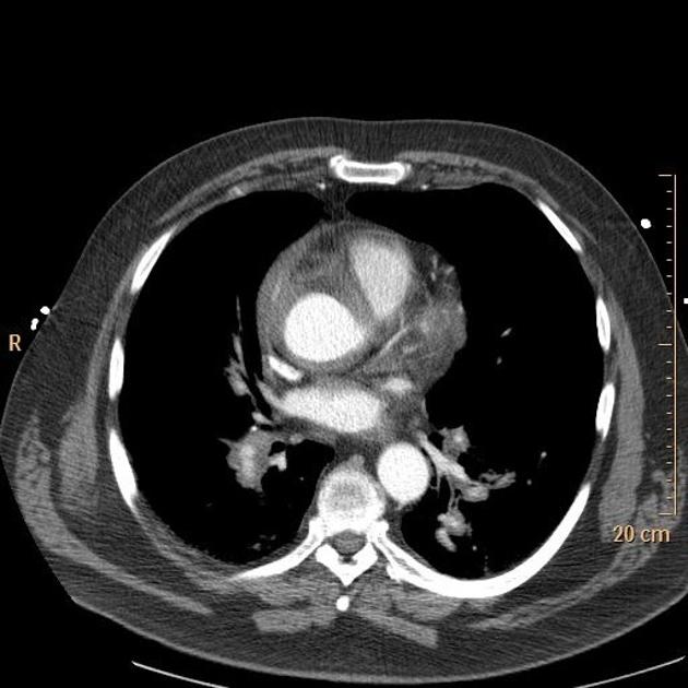
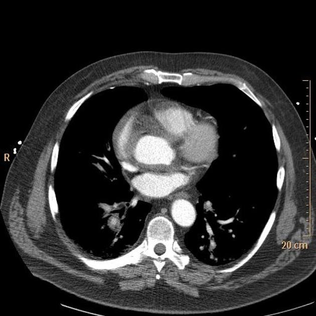
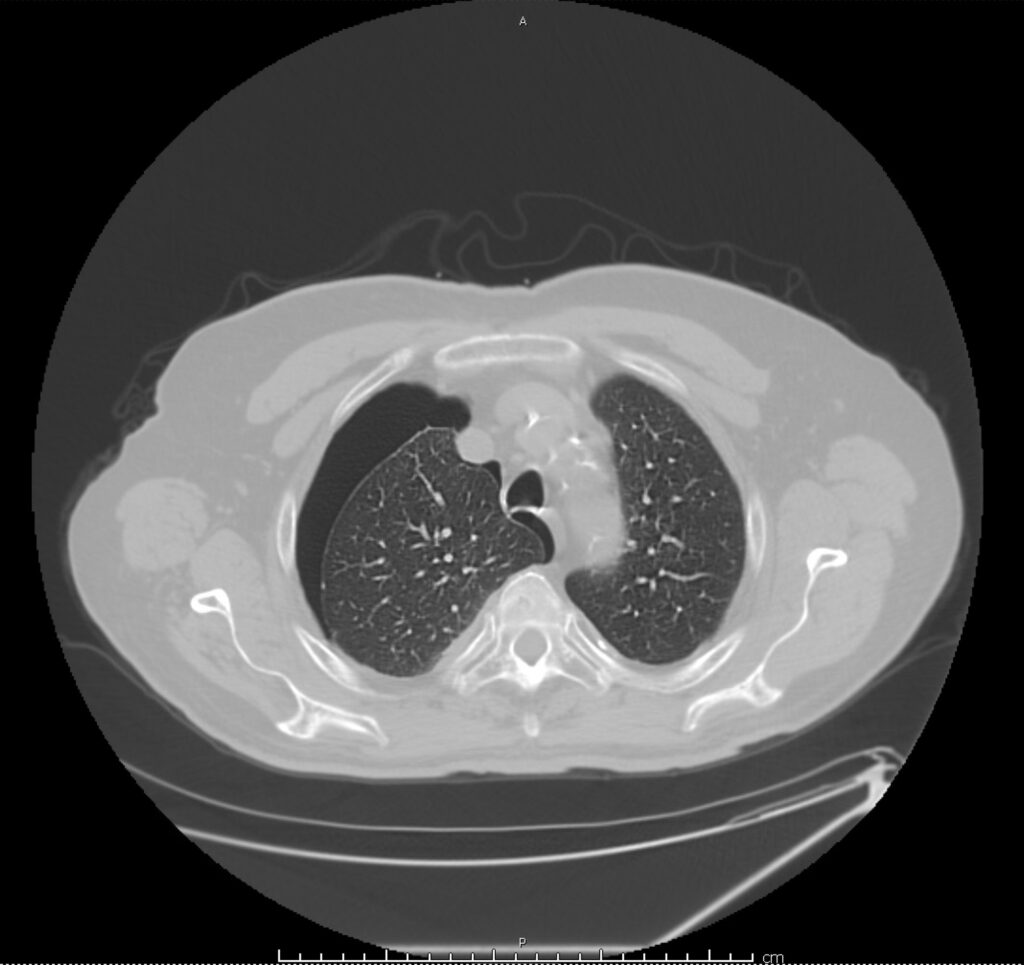
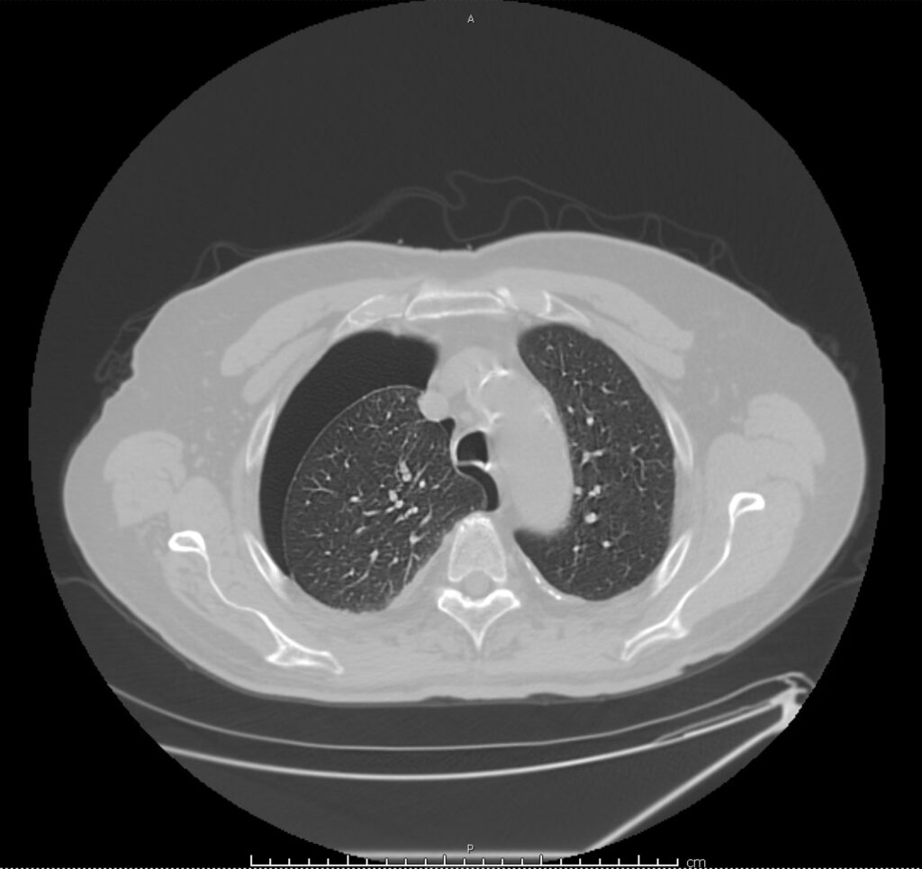
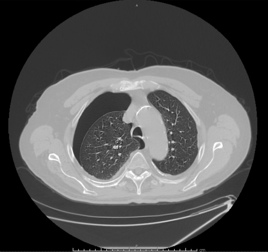
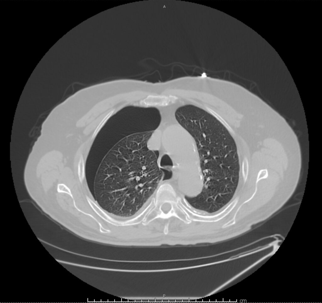
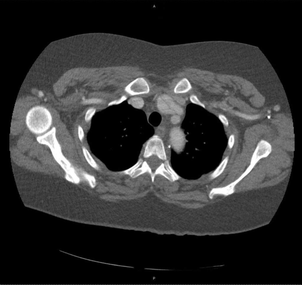
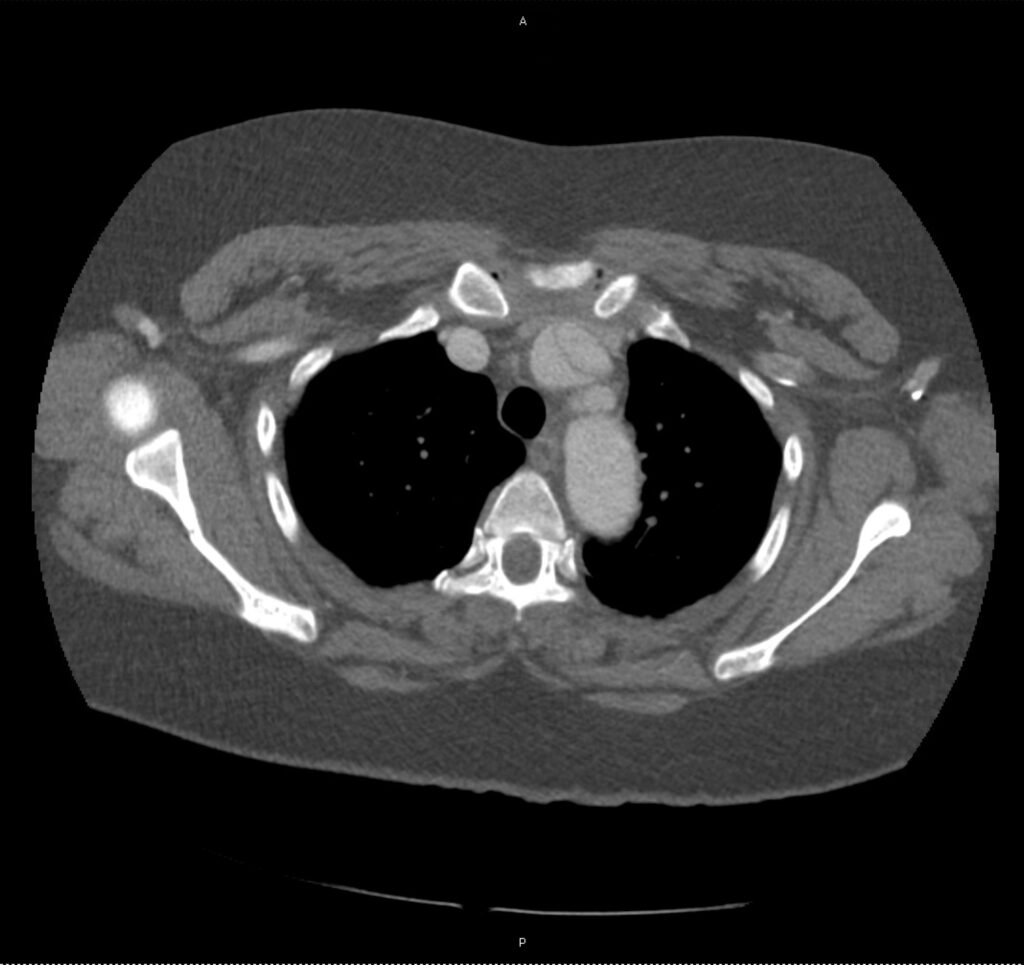
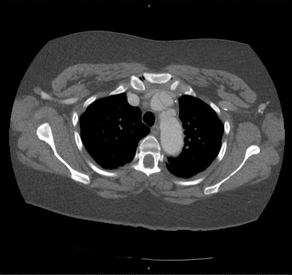
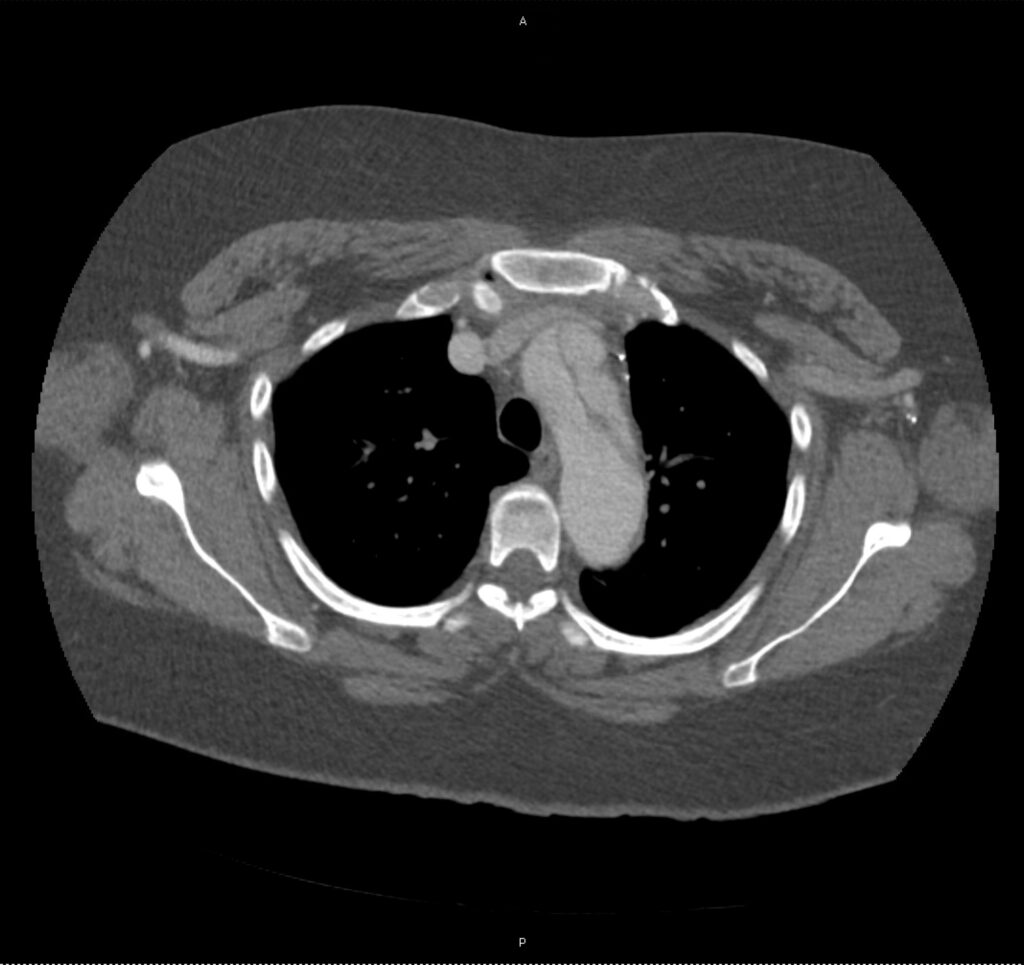
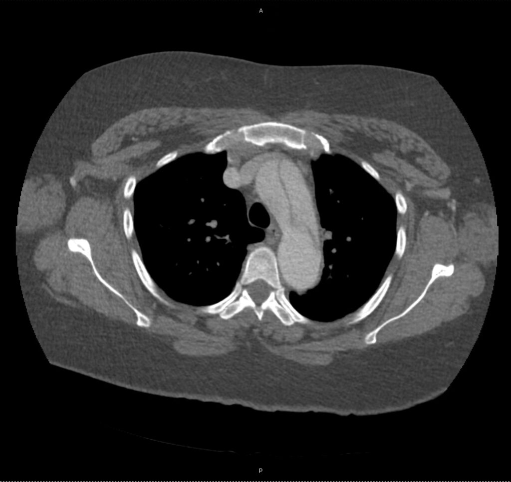
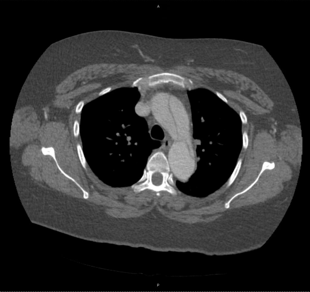
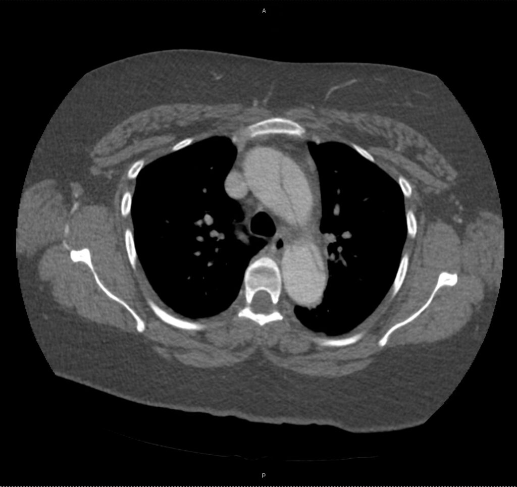
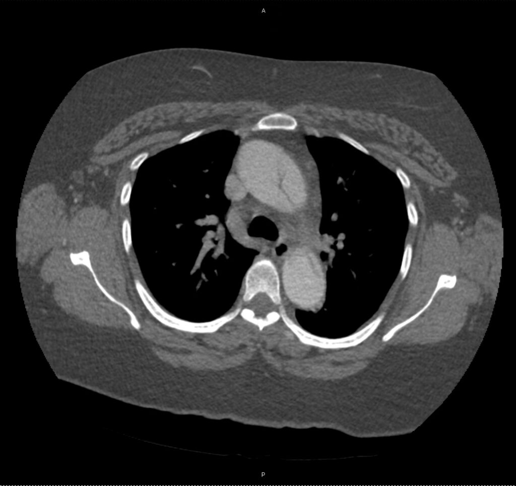
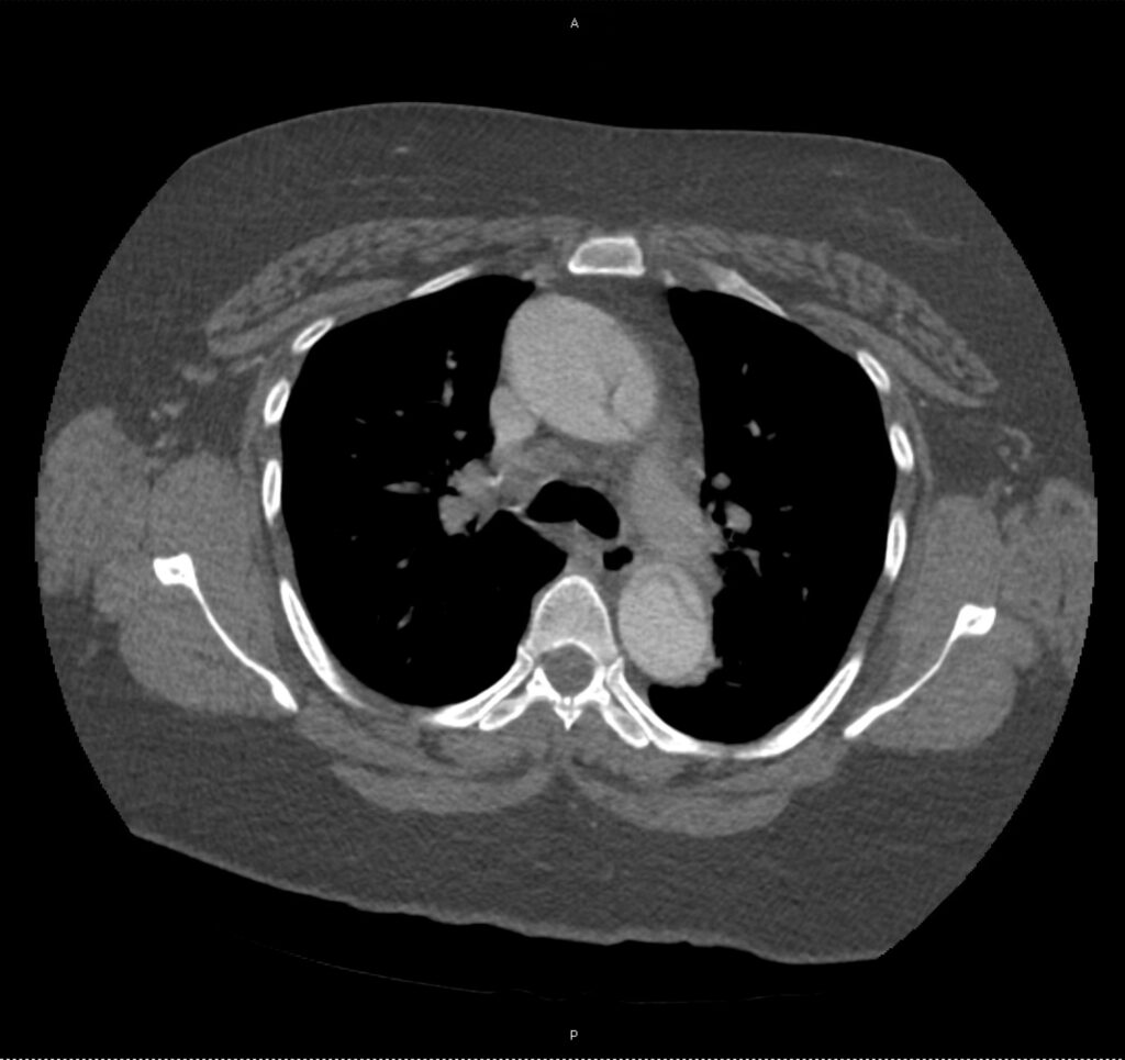
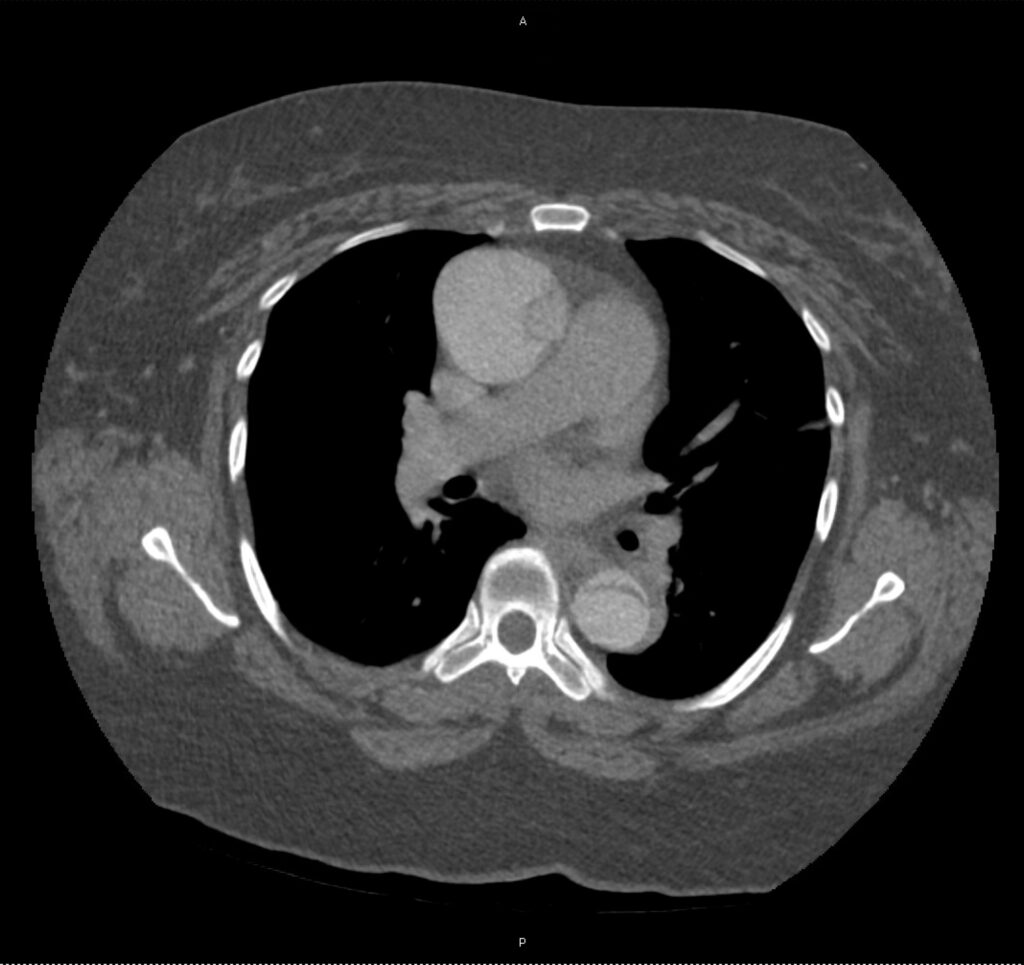
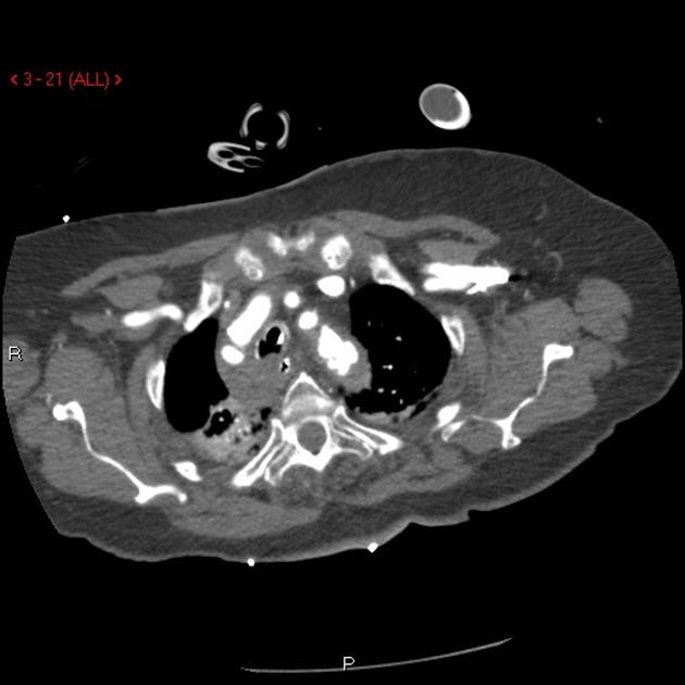
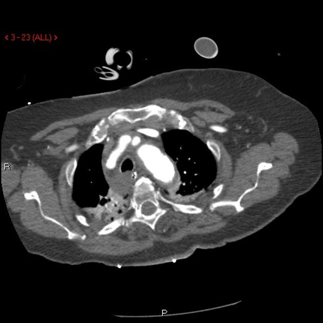
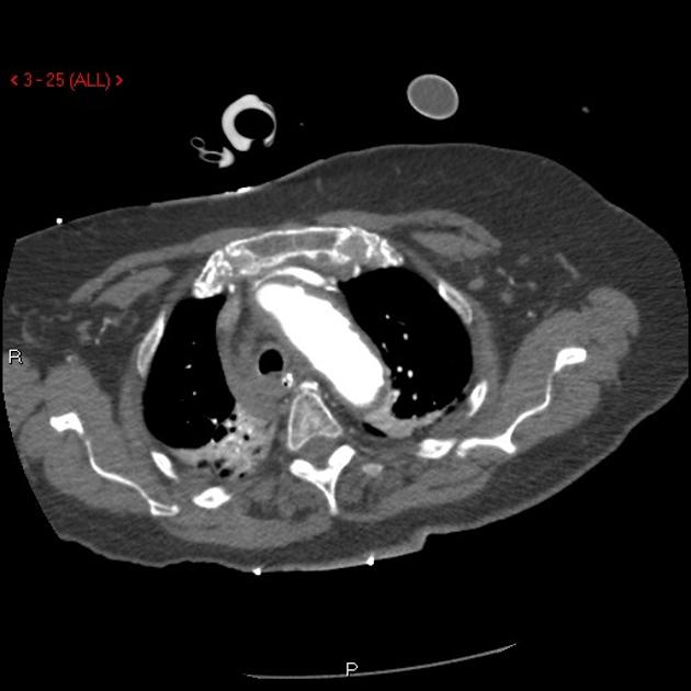
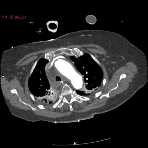
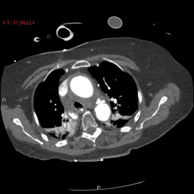
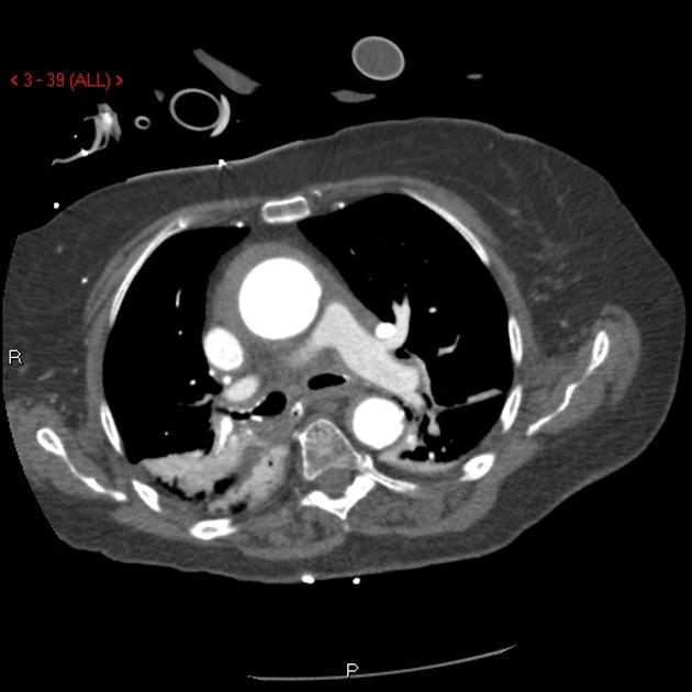
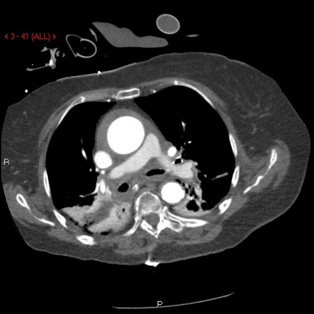
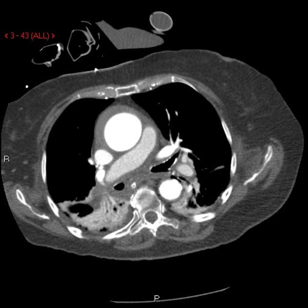
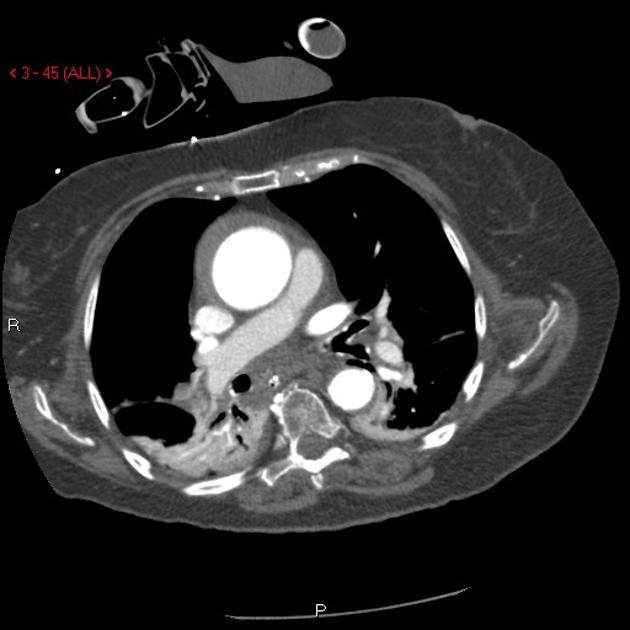
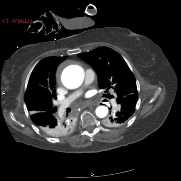
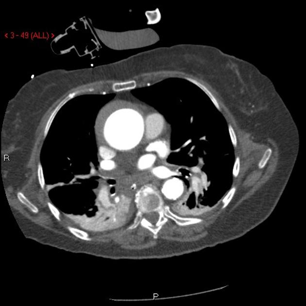
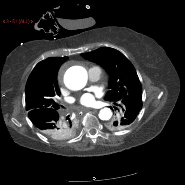
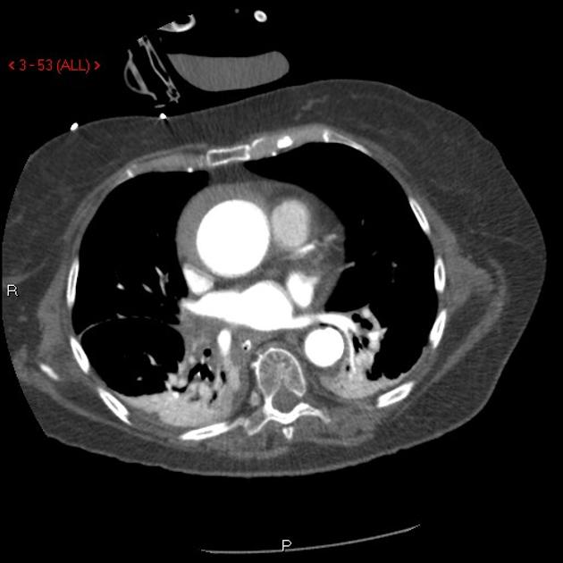
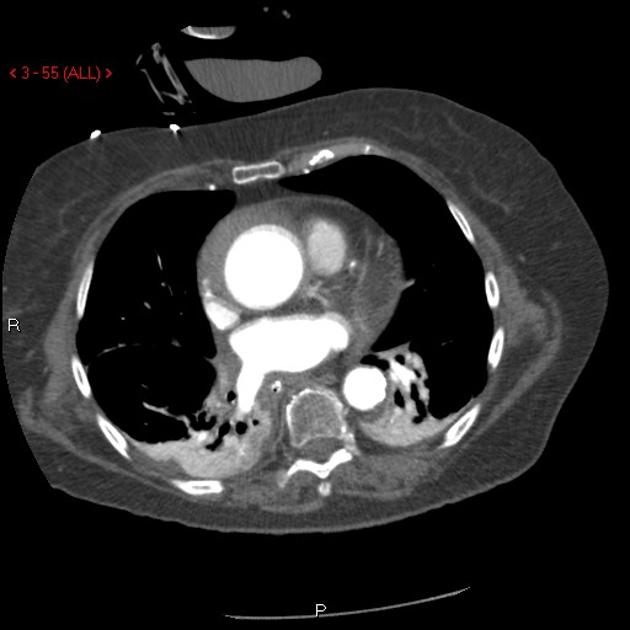
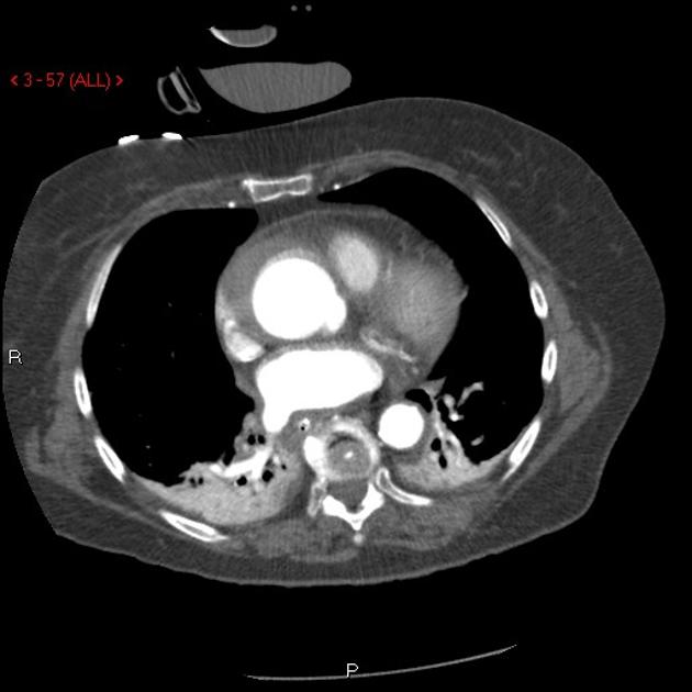
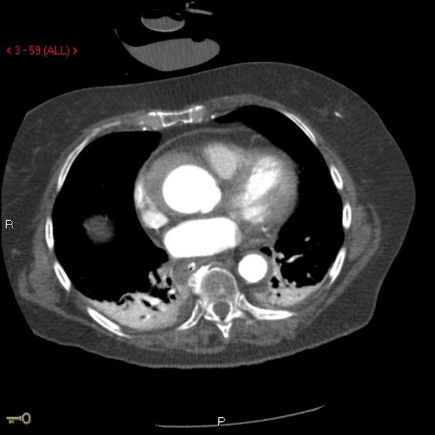
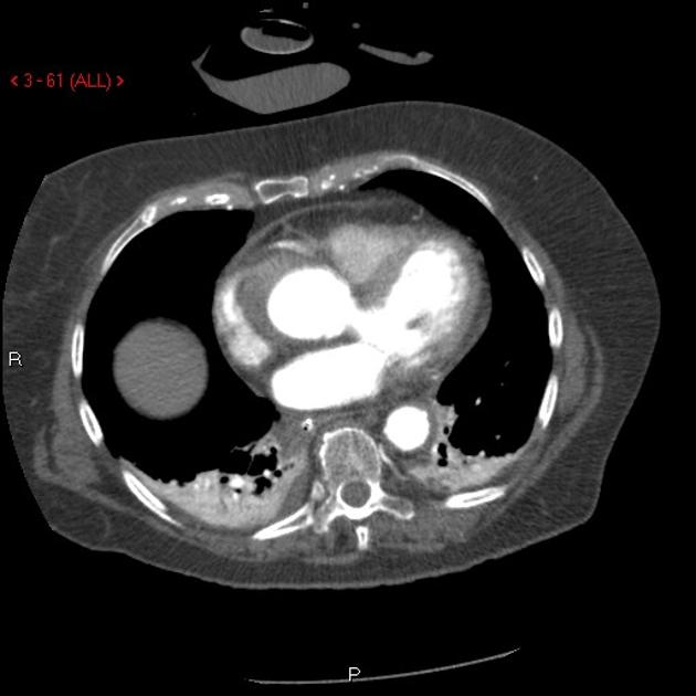
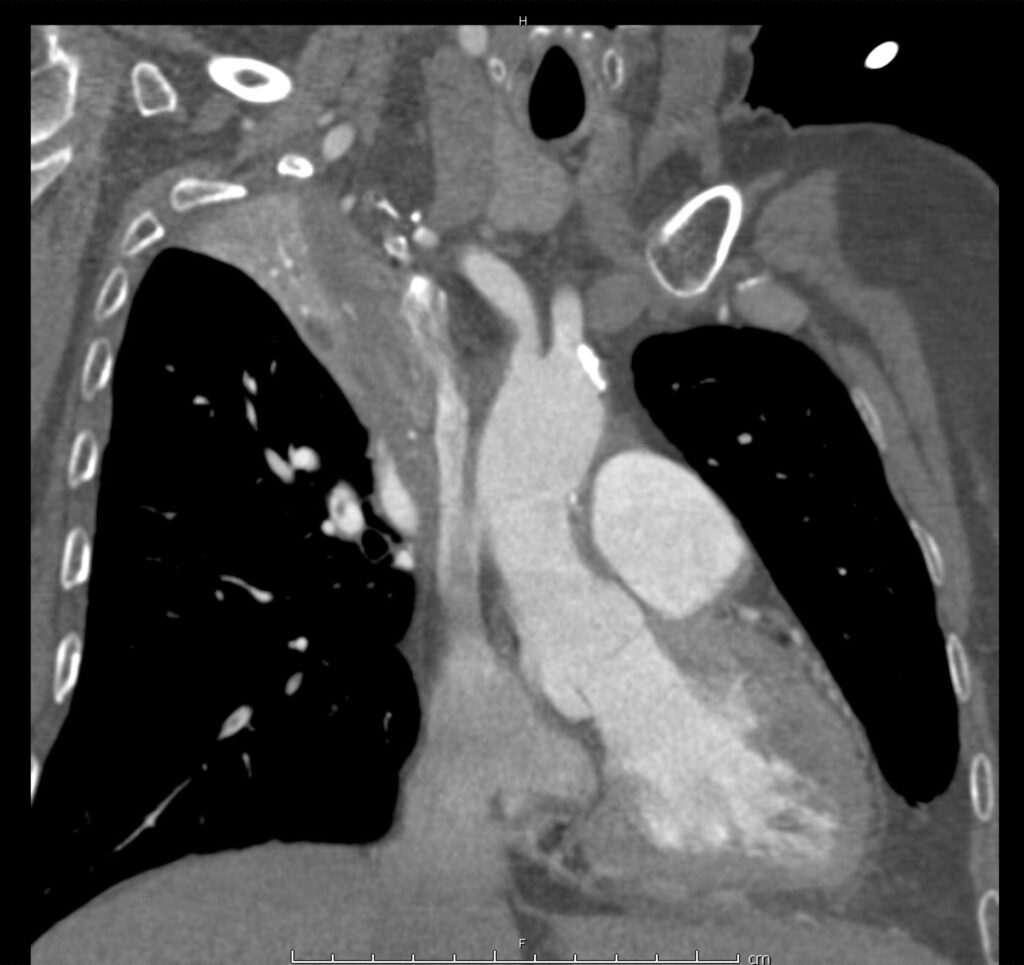
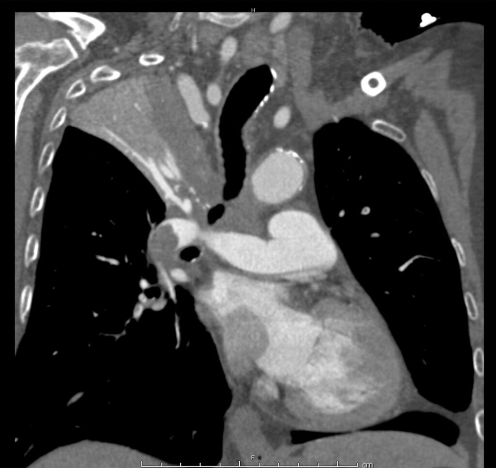
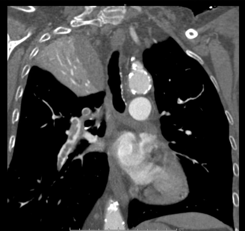
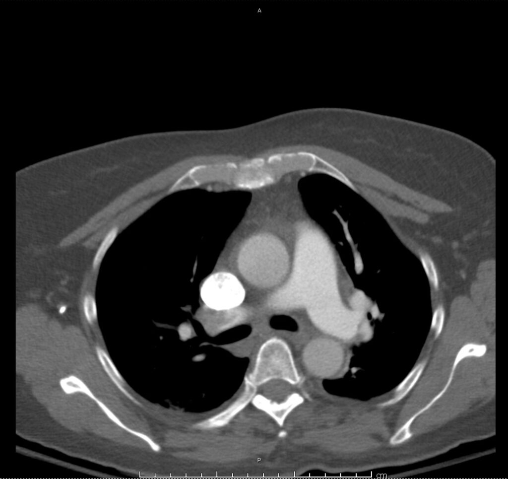
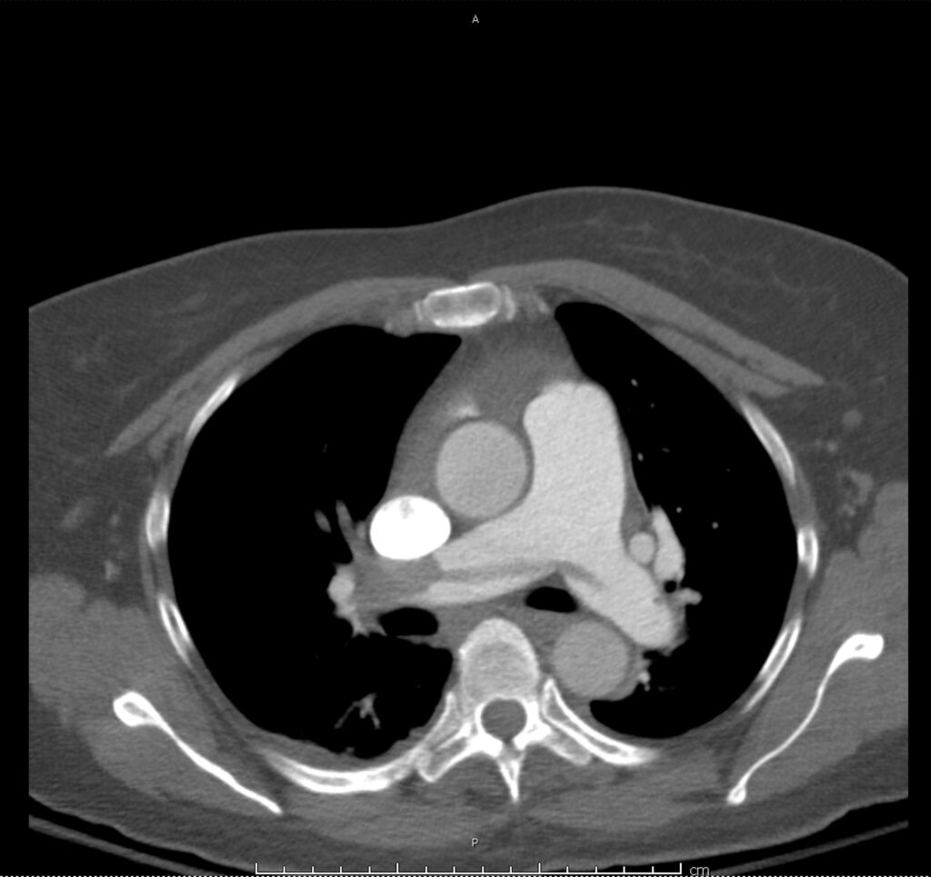
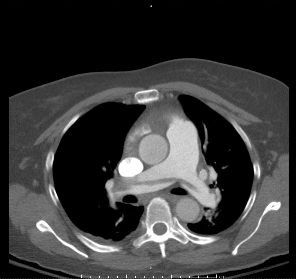
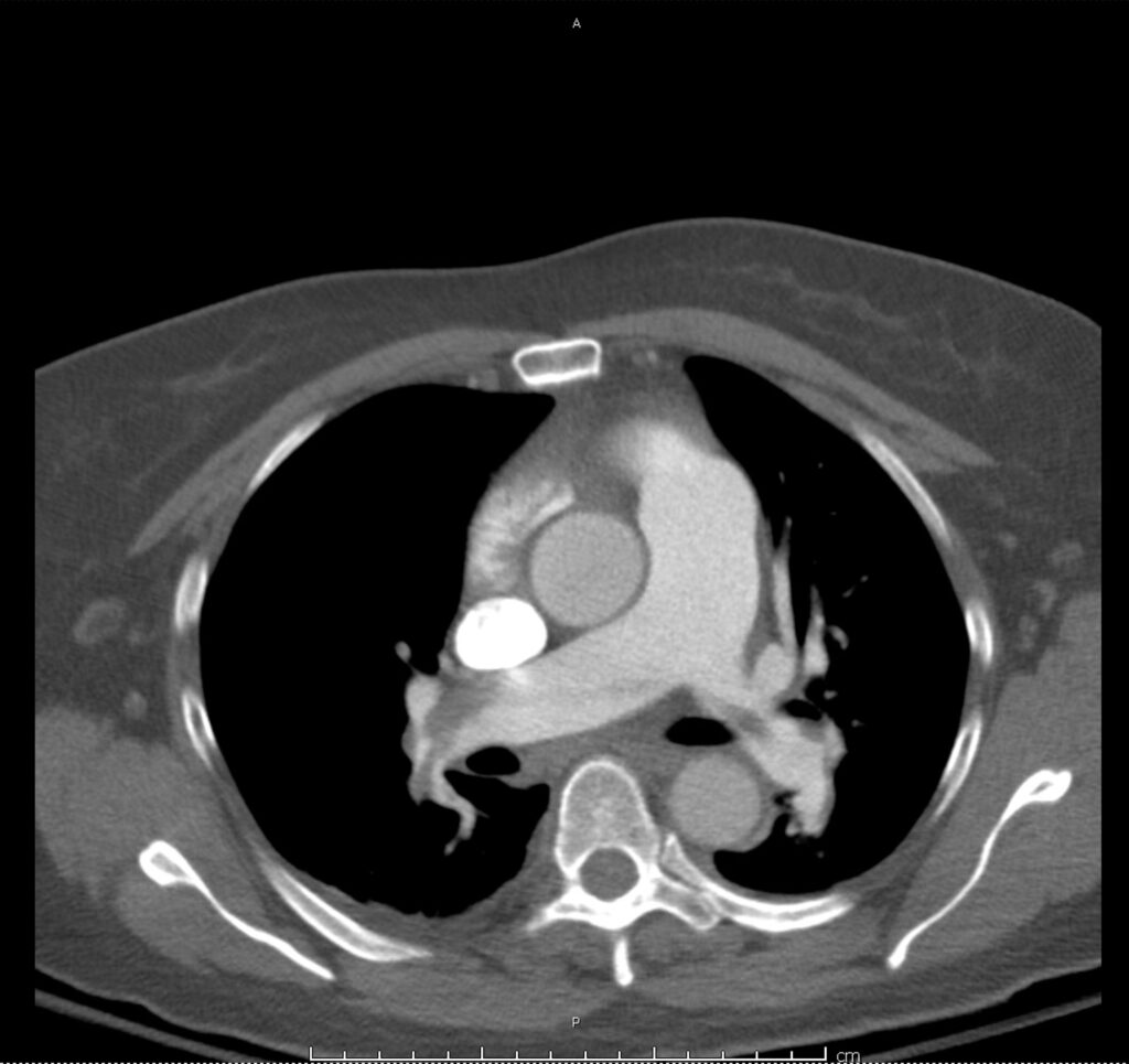
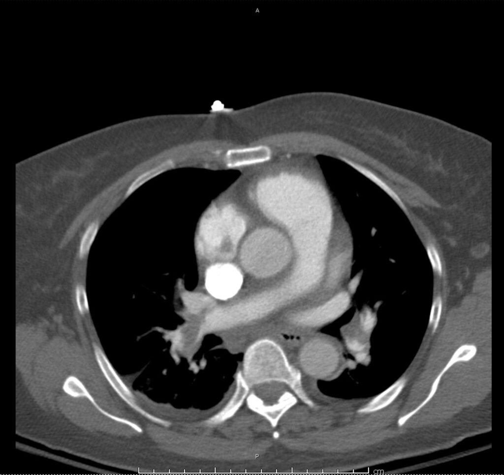
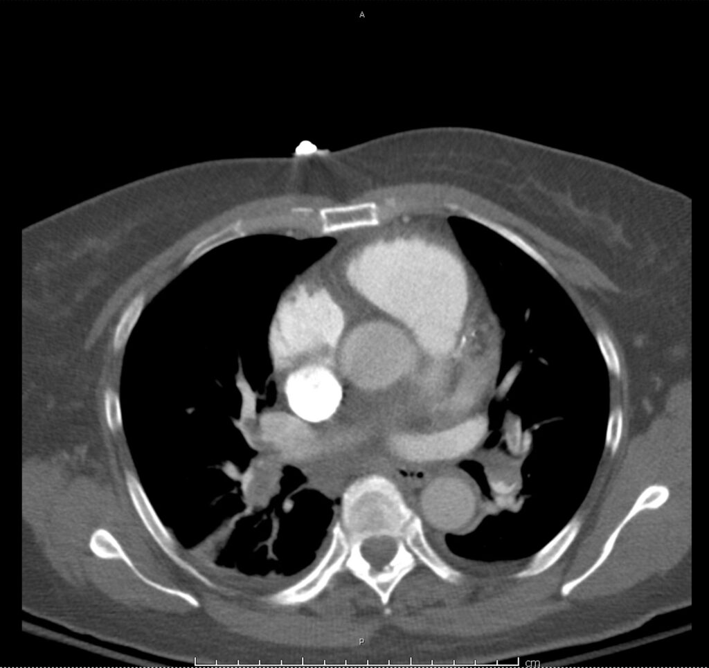
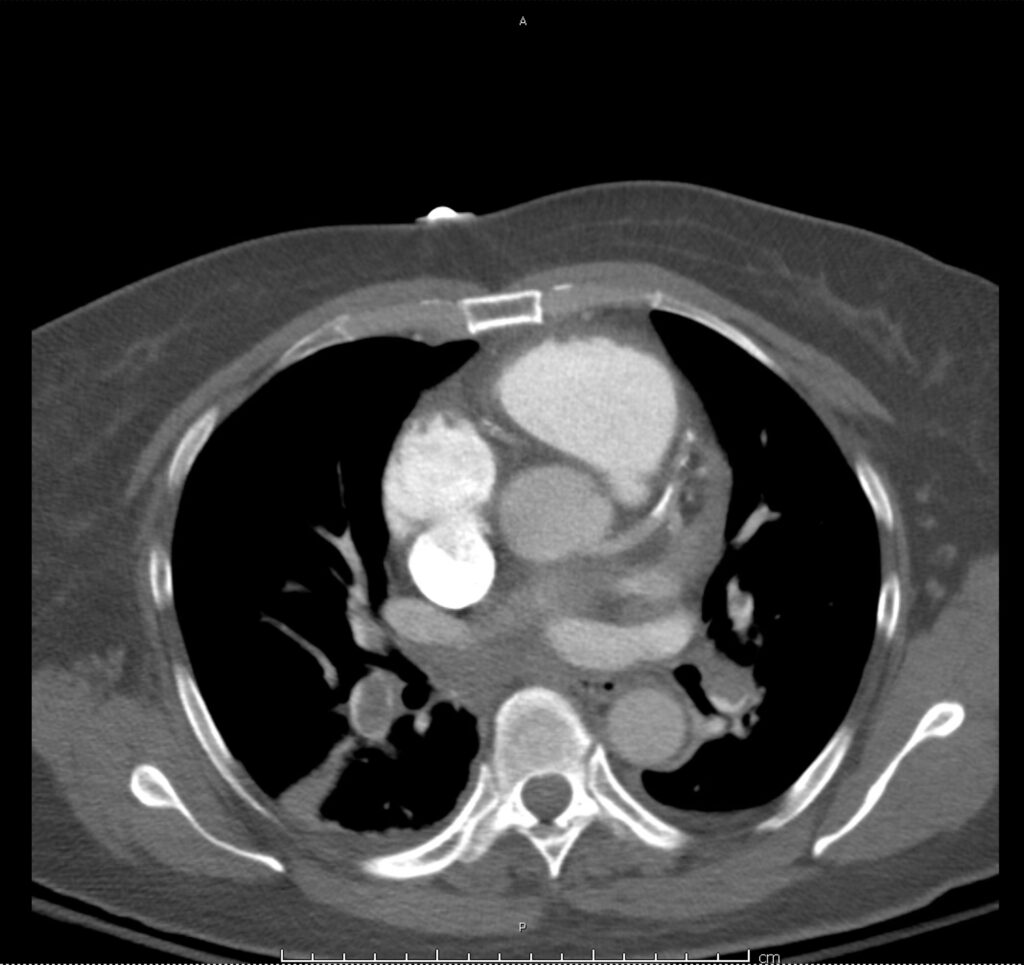
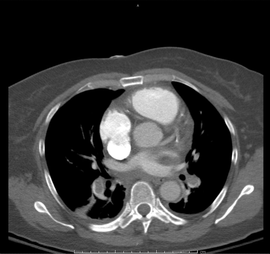
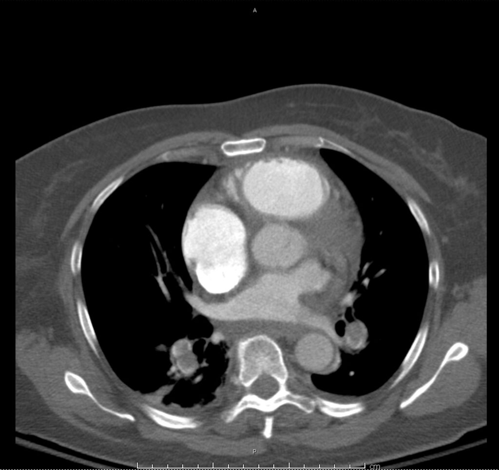
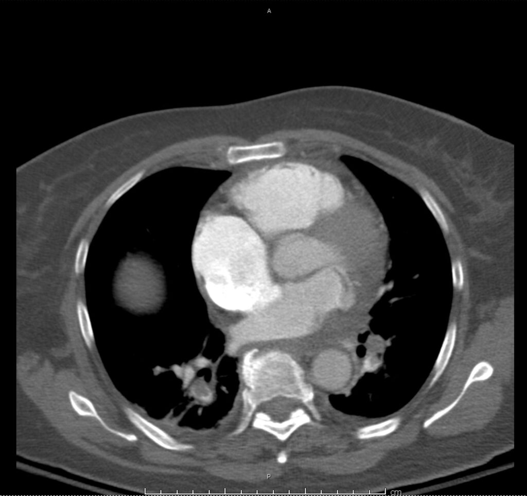
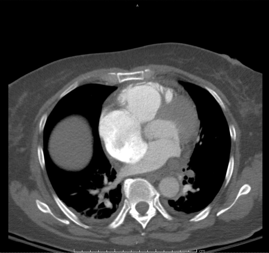
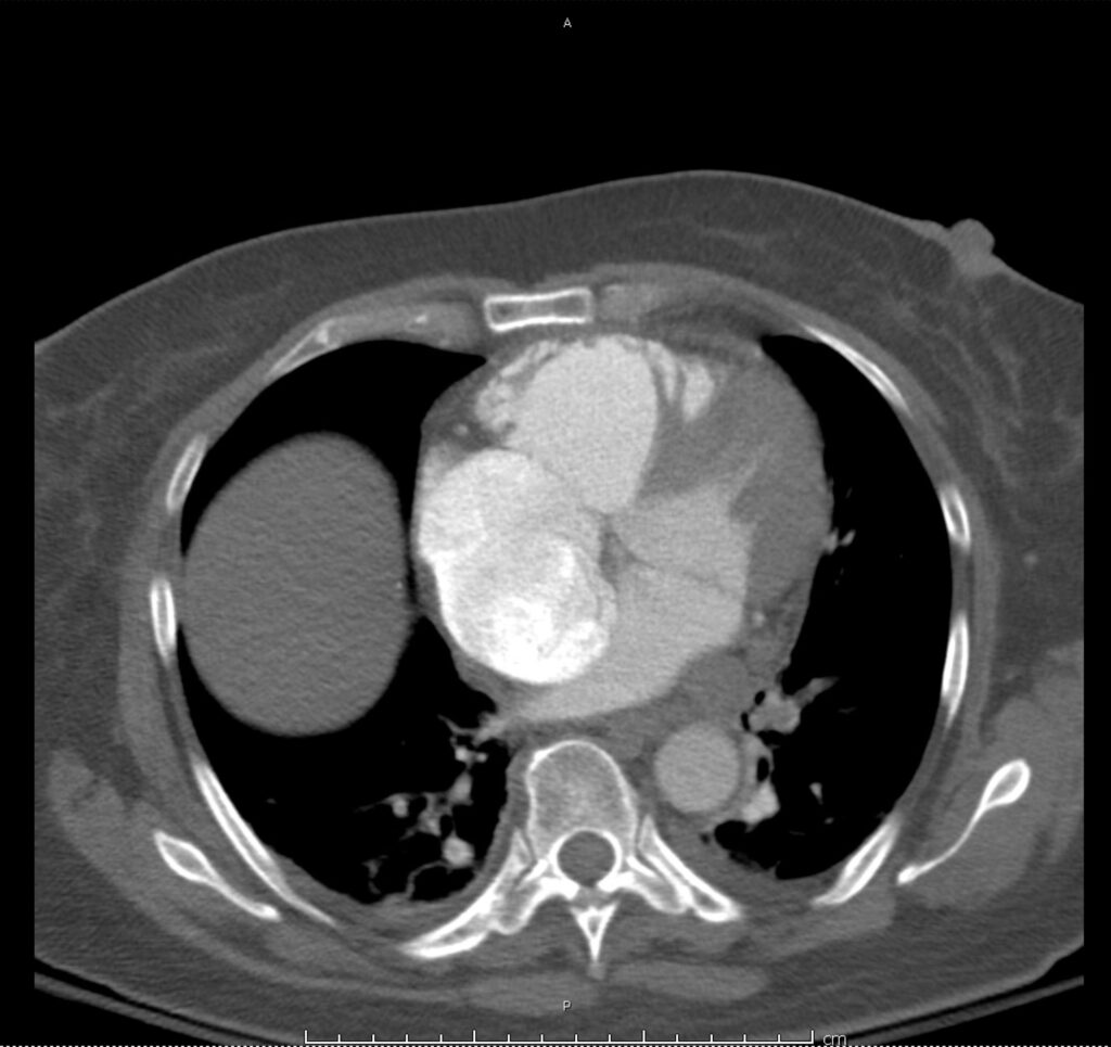
Circulation: Penetrating Atherosclerotic Ulcer

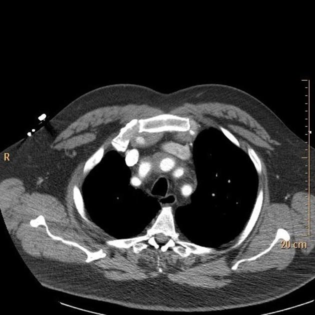
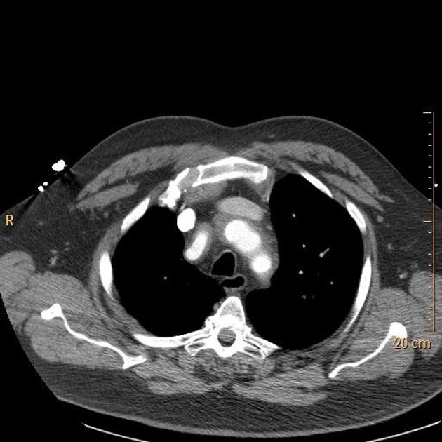
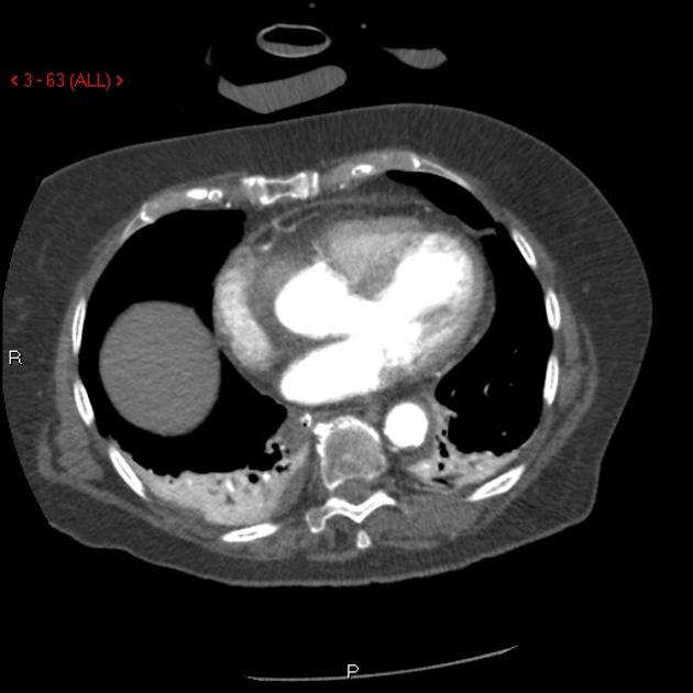
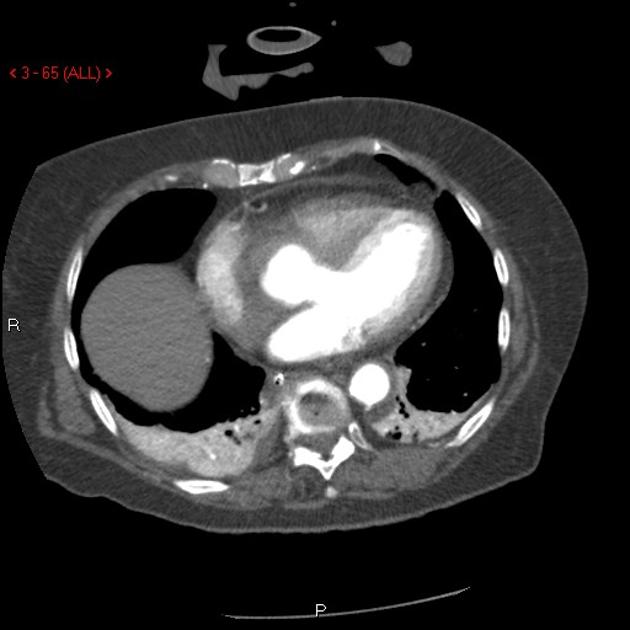
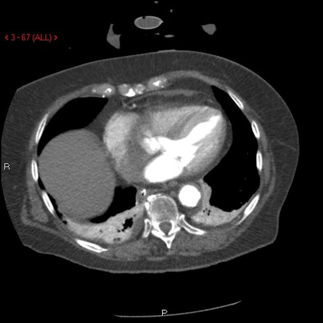
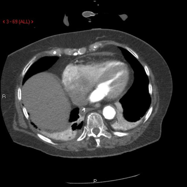
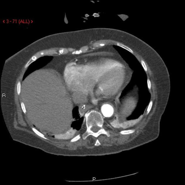
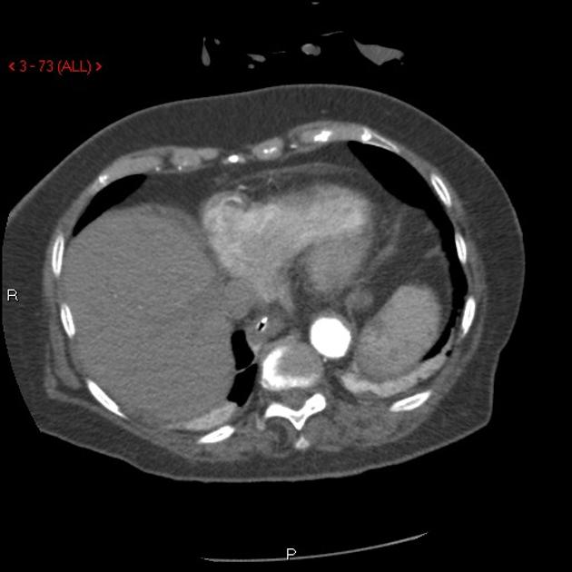
Penetrating atherosclerotic ulcer of the ascending aorta with mediastinal hematoma and compression of the pulmonary artery.
Case courtesy of Dr Jens Christian Fischer, Radiopaedia.org. From the case rID: 12810

000-867587463d372d24b2b85250801c08_big_gallery
001-86341bc69038d7dba6c8b214dbd7c6_big_gallery
002-3ecd031c687b50c7f638b89a07a6b2_big_gallery
003-fc7ae33a714c317405cbdcad8ba928_big_gallery
004-df10ff6032d8778870f4d1bc324239_big_gallery
005-d25bd376a013076542b5e13a3632b7_big_gallery
006-bf4c7f76c923fe9a347ce79834bb8d_big_gallery
007-5644688715e4a142836c83974912b7_big_gallery
008-a4d612629b3734055830a7fba49ce3_big_gallery
009-270b688772d018c5df629238469775_big_gallery
010-cb4fcb36bec3df294eae04e2757bdb_big_gallery
011-e336de8e0329a3a505c03c54af1873_big_gallery
012-209baf0df18e74ac5b8b1d03ae97f5_big_gallery
013-b5de9e8d295cdd23824b6fc6bc2f20_big_gallery
014-59749360eb2b2fe9345dcc4c9ebdb3_big_gallery
015-82a894b7759091f1976ee585abc77f_big_gallery
016-b007313a5e7cf19fee5d6bdcd16c00_big_gallery
017-ba78a9eca498b2d3f269b2b91d0184_big_gallery
018-5017f99f8870a75b17818af58ae3b8_big_gallery
019-c483f0793ac01098b7f623a00ee1cd_big_gallery
020-276239983571b1bda2d7e23c72f317_big_gallery
021-80a2615431ba2c78788120c881e681_big_gallery
022-d042500418422c7c11b19023e1c88e_big_gallery
023-0c098e5f4411c67b2b7695f4d08b7e_big_gallery
024-fe3cbc3a671ec8533d578dc5884430_big_gallery
025-d5b58c845a93a9ecb1297f3da236c5_big_gallery
026-1c9ef17a044b21ffac2708e2dc5941_big_gallery

This slideshow requires JavaScript.

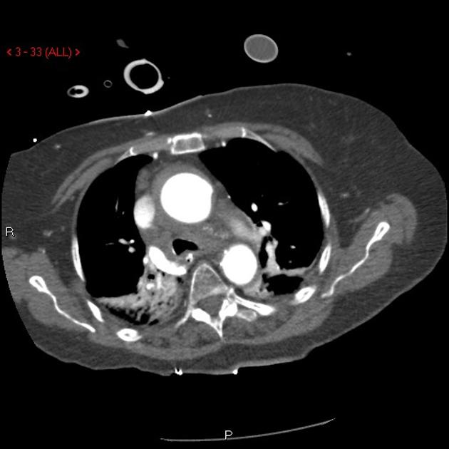
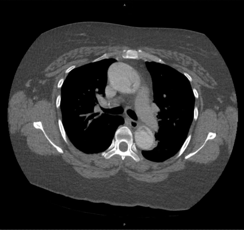
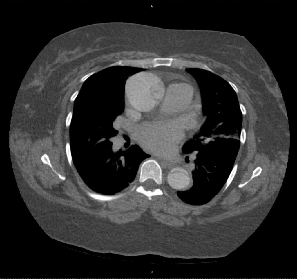
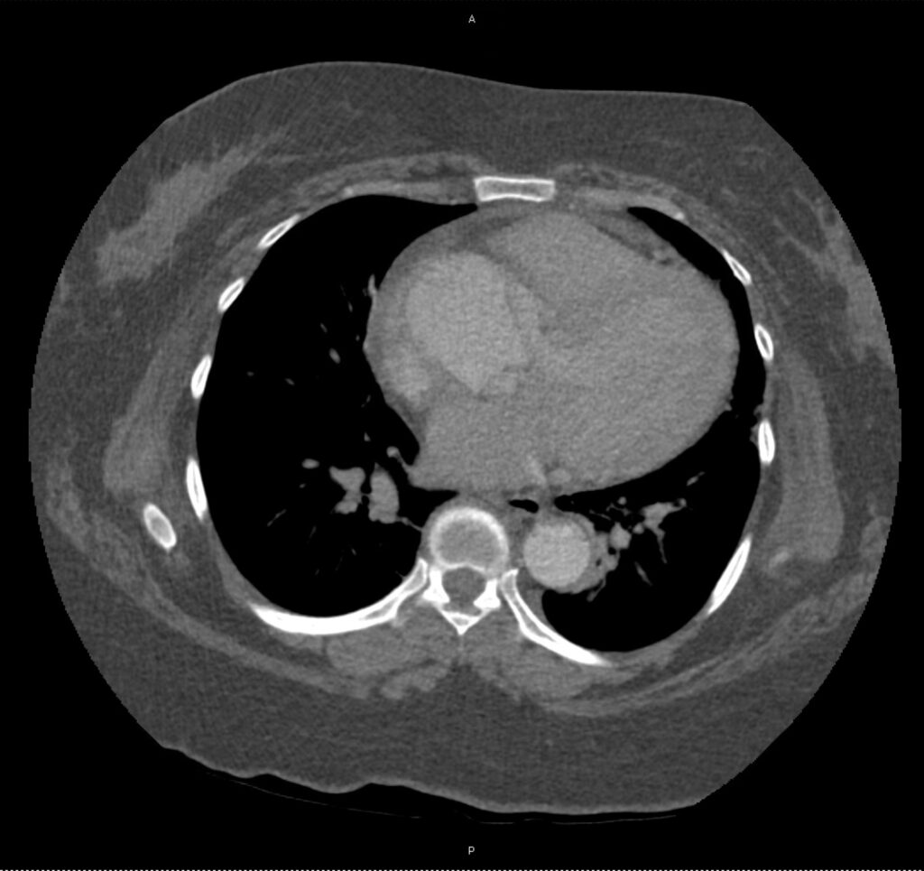
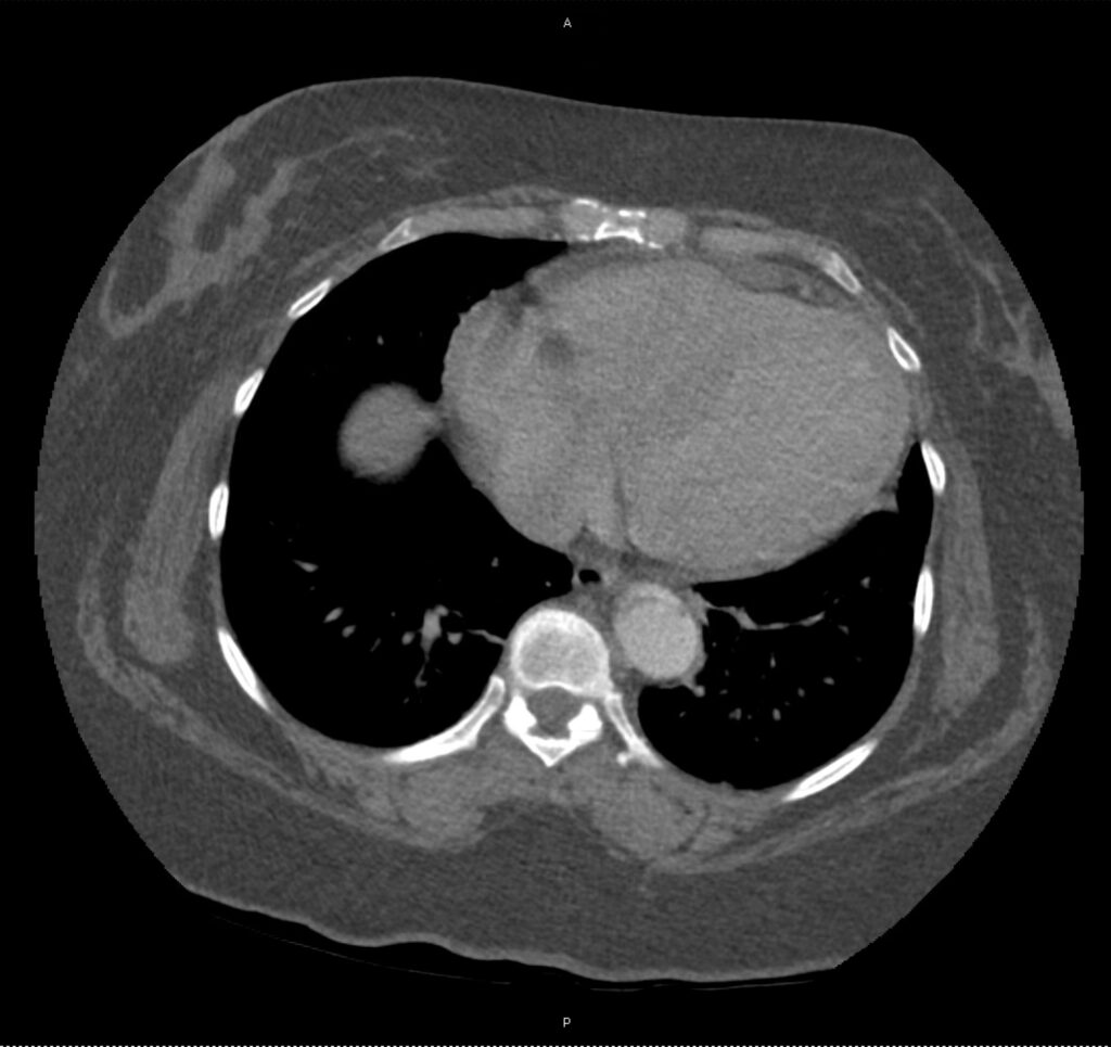
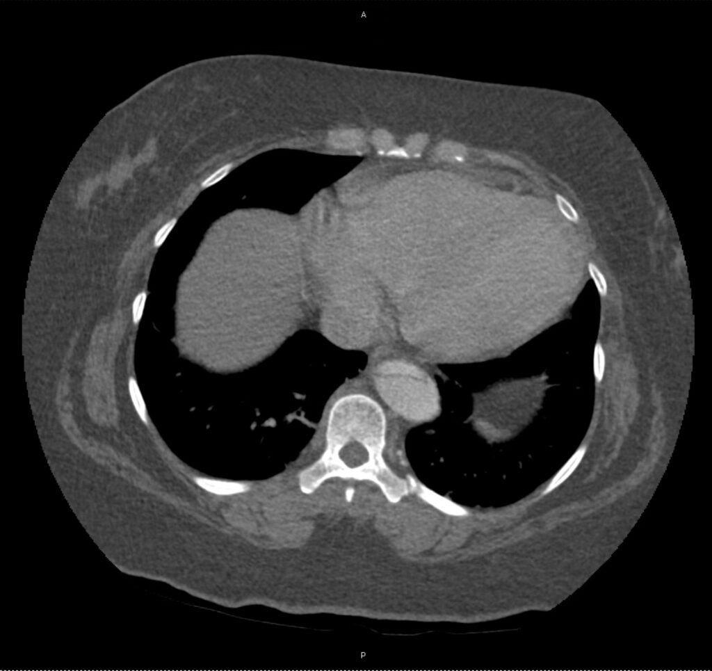
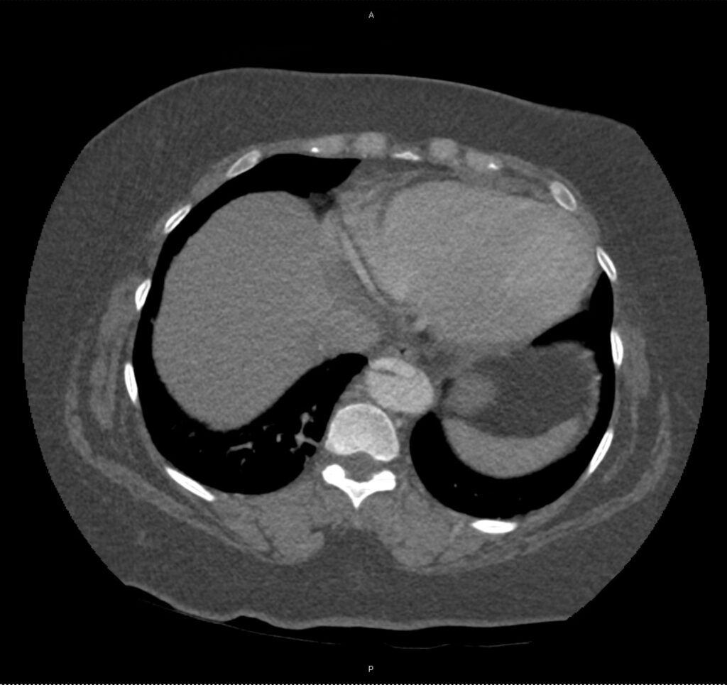
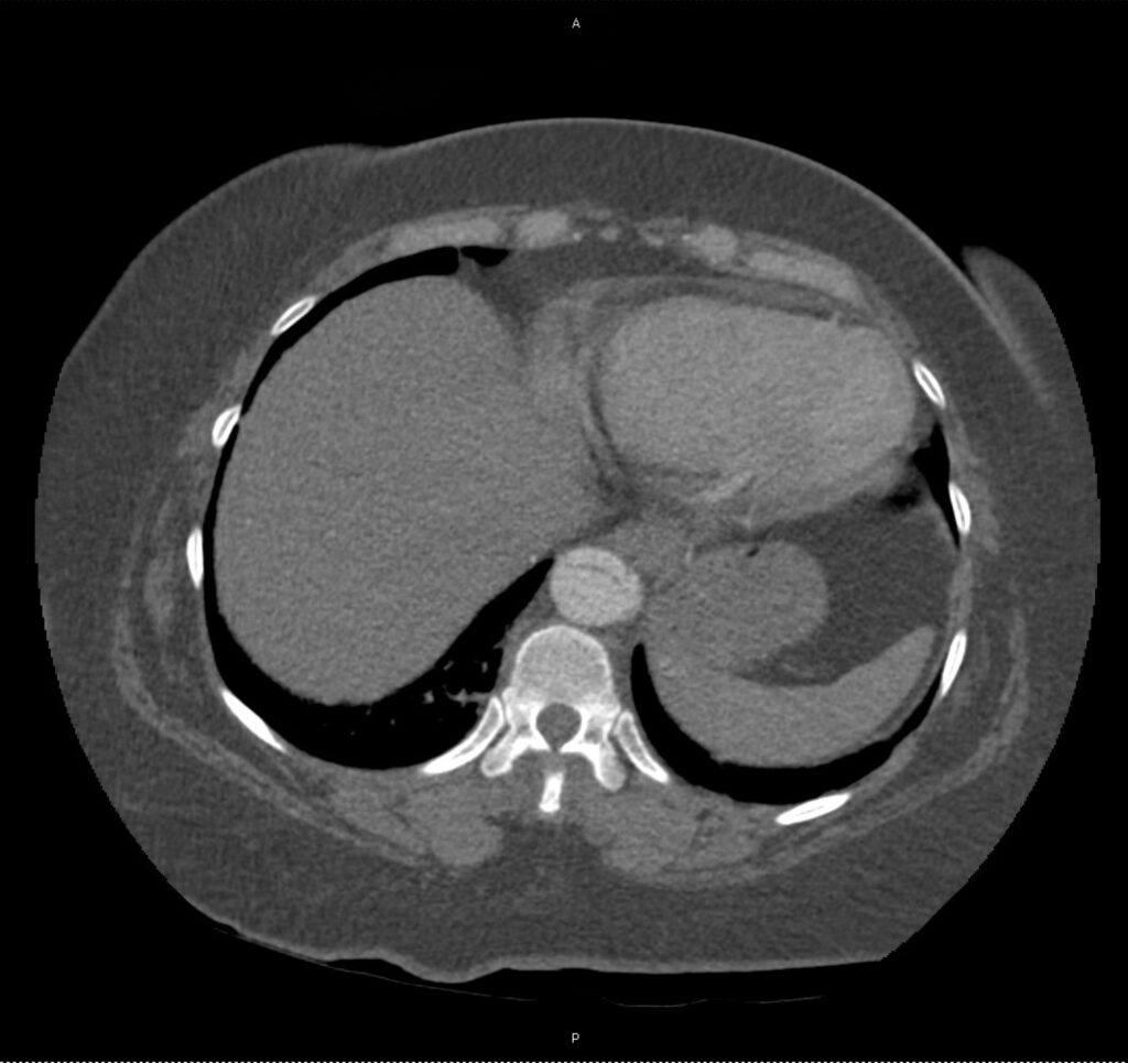
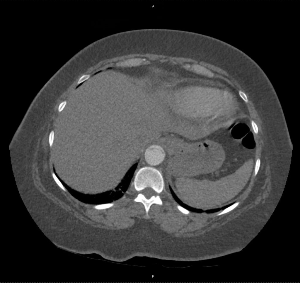
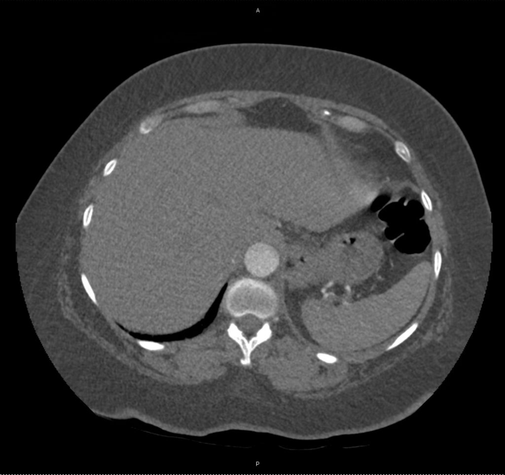
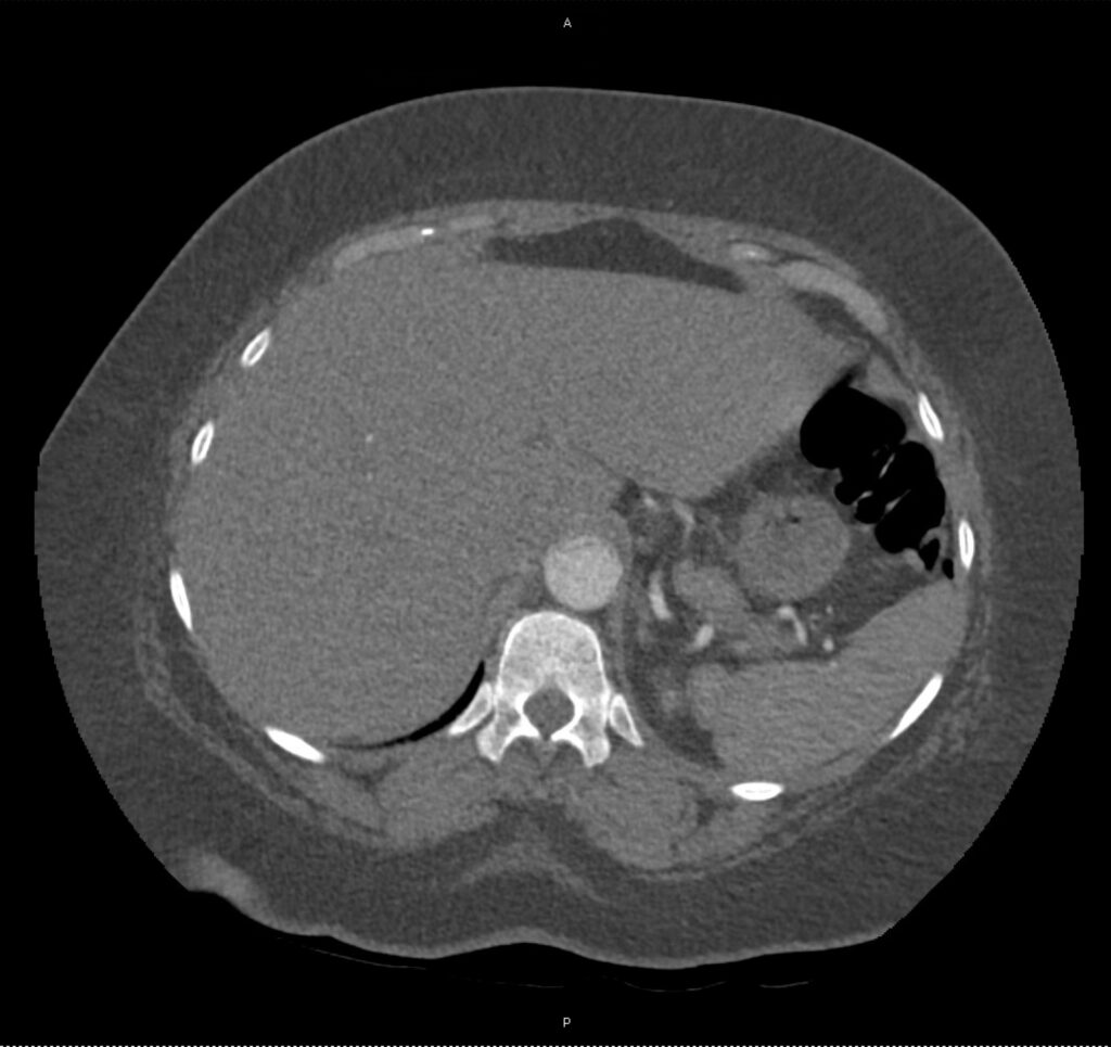
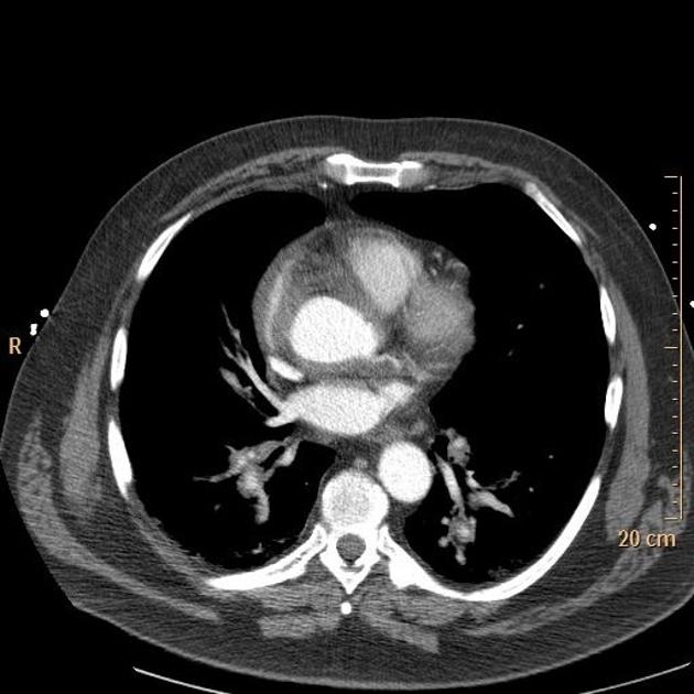
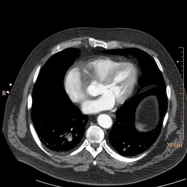
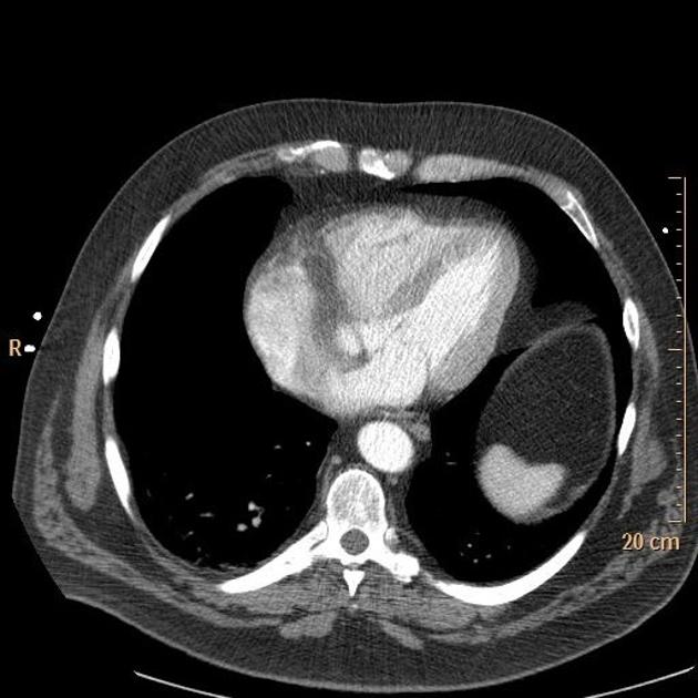
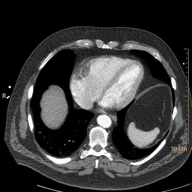
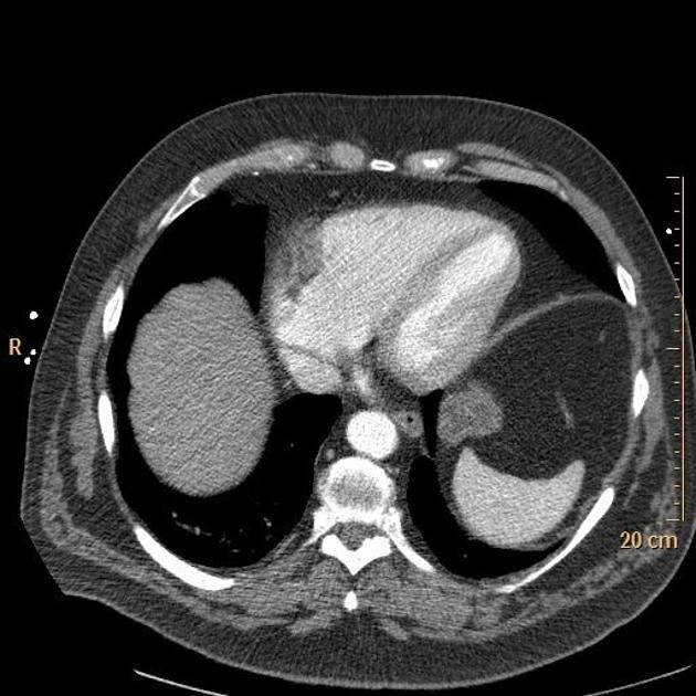
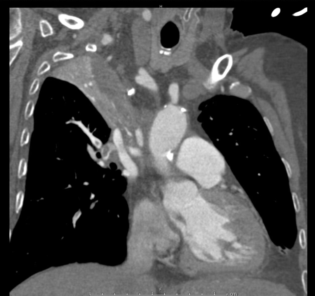
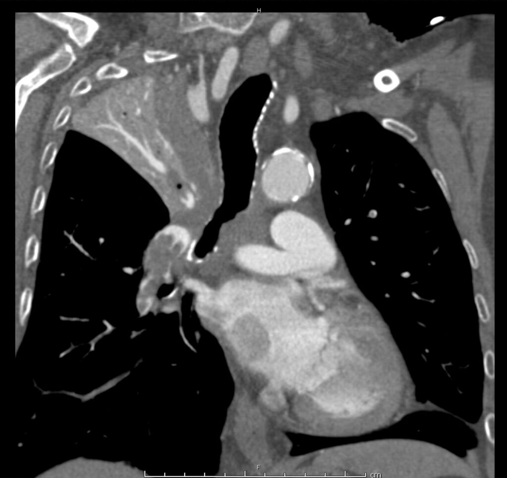
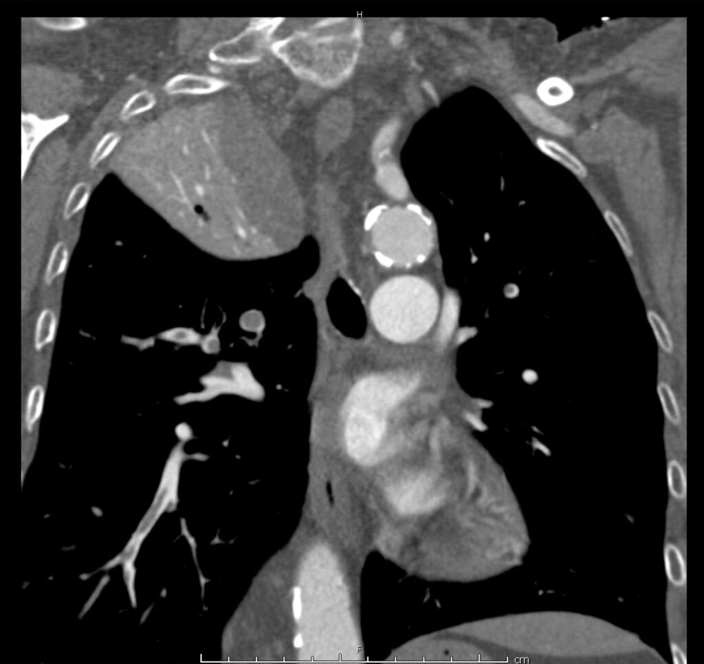
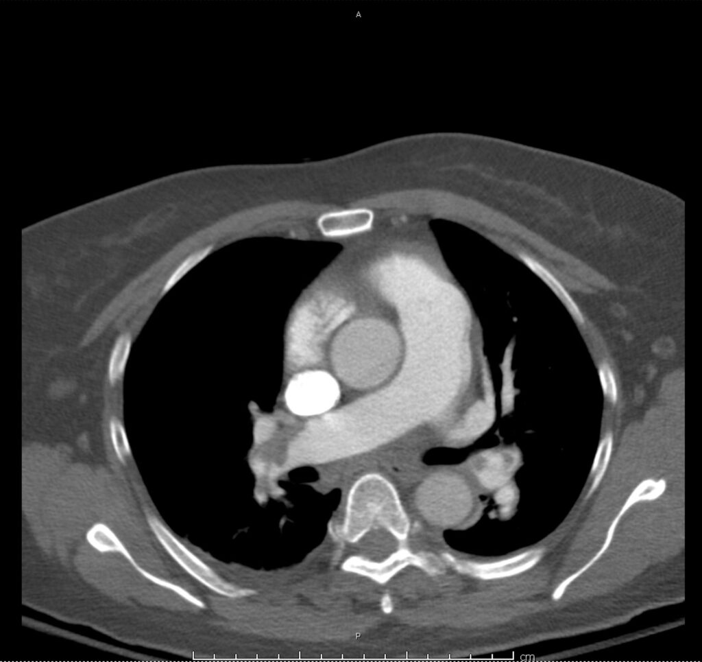
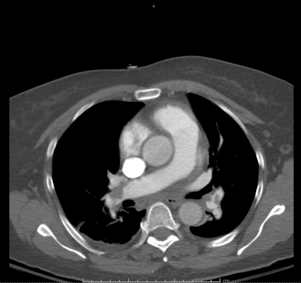
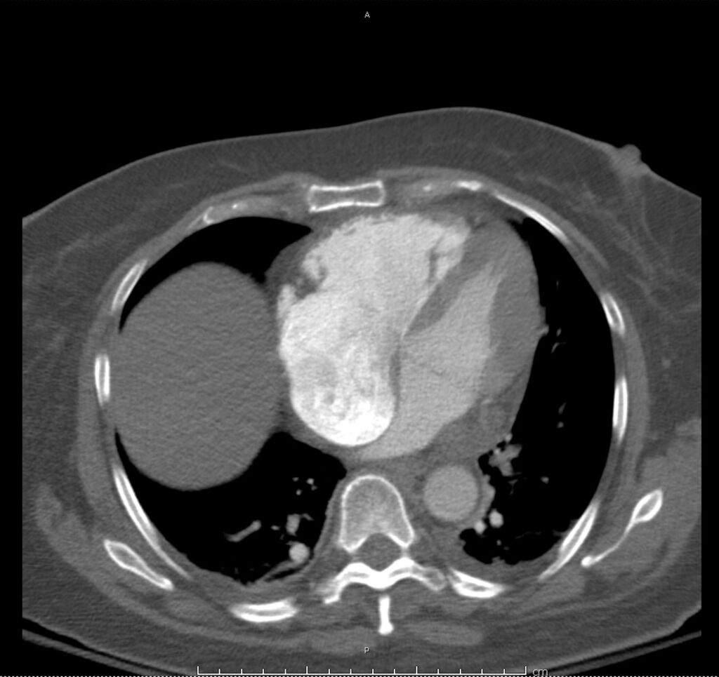
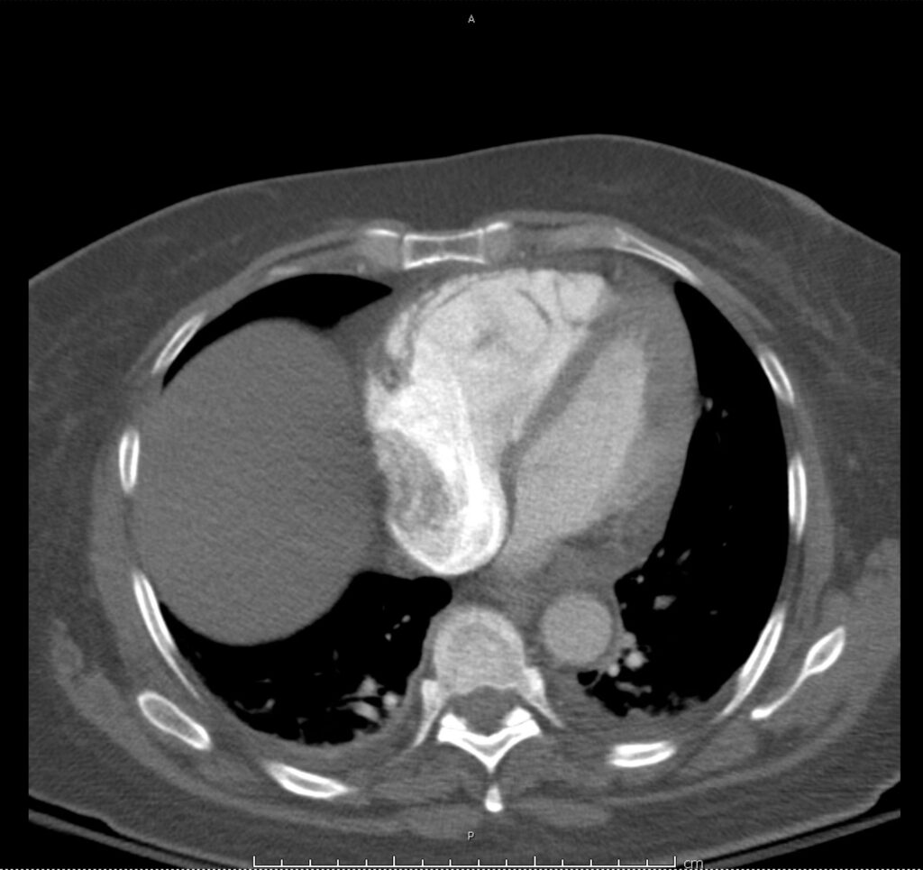
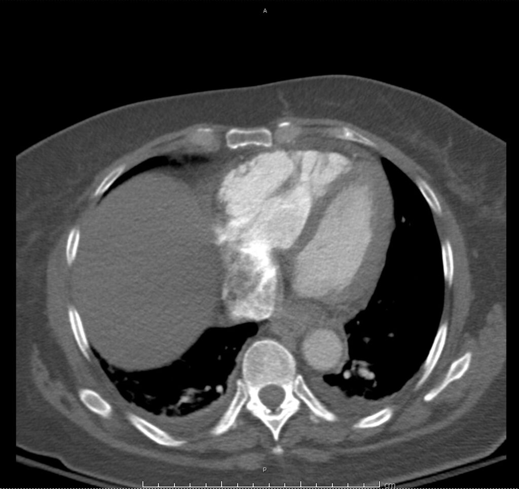
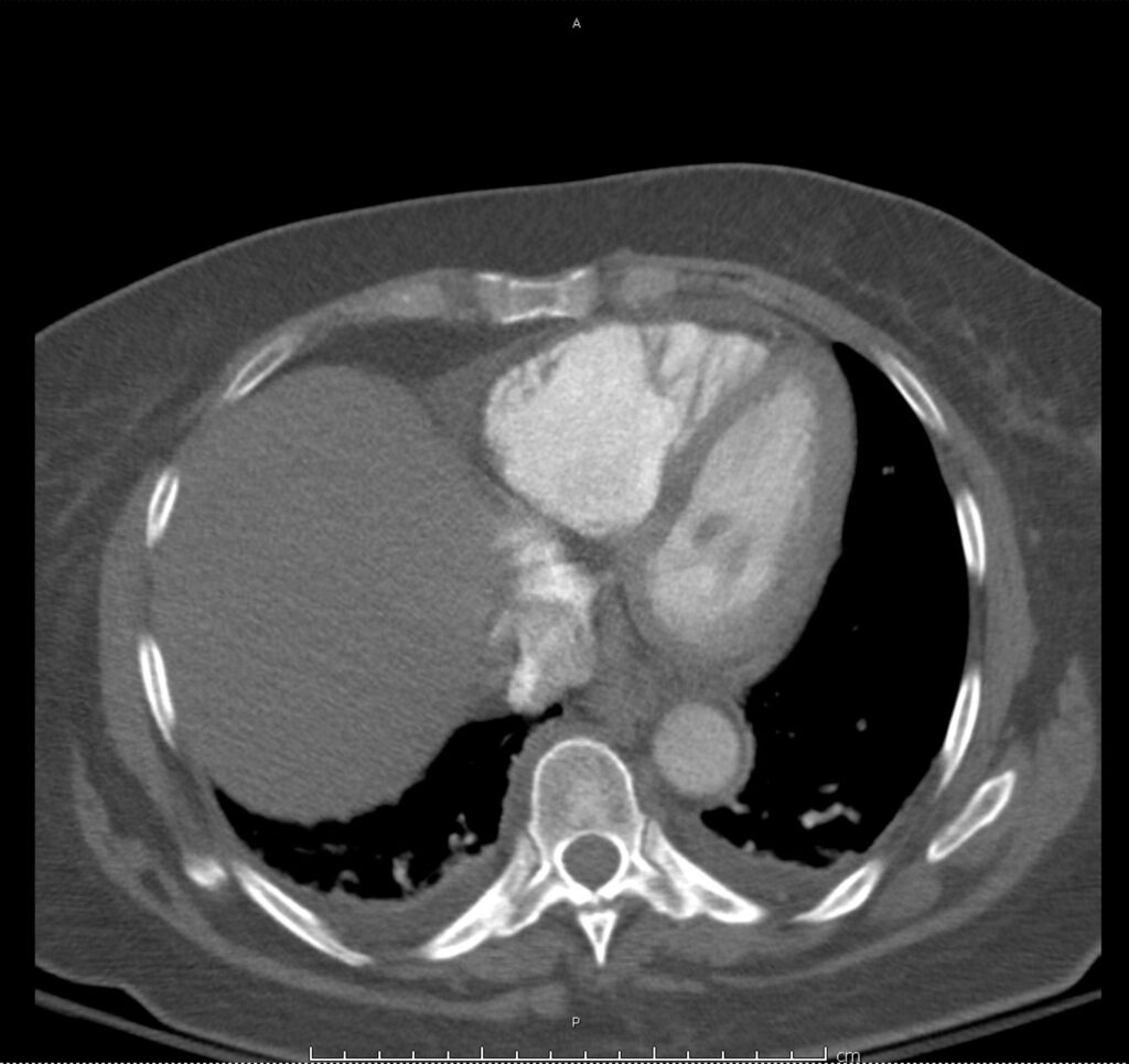
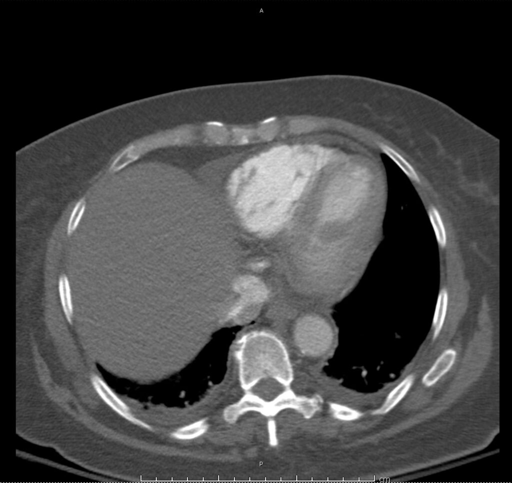
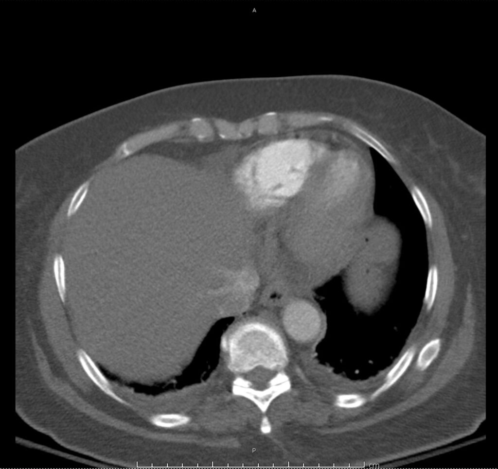
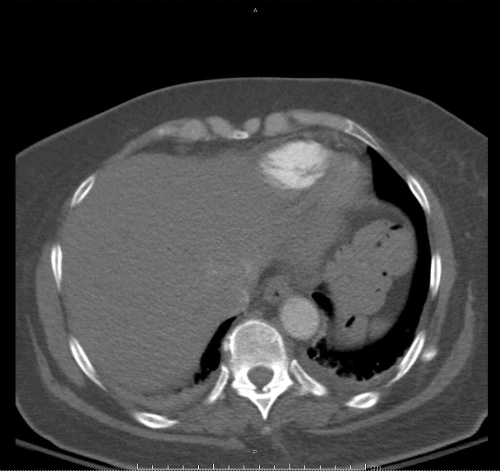
Circulation: Intramural Hematoma

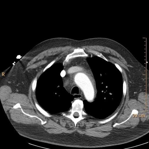
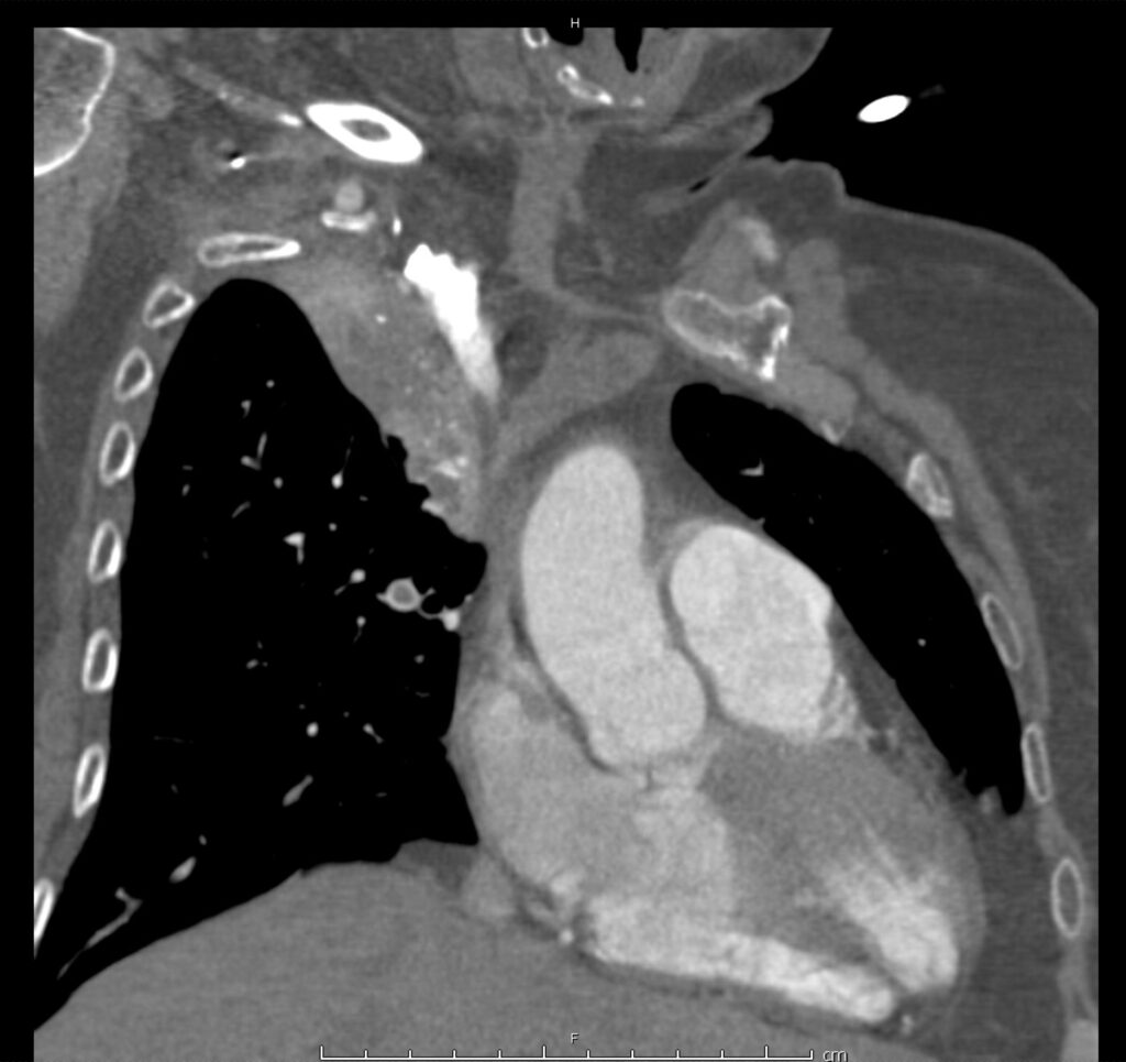
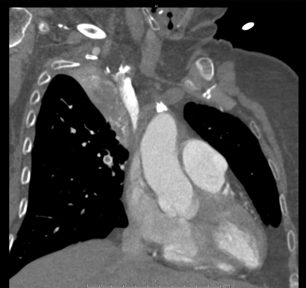
Intramural hematoma surrounding aortic root and ascending aorta with small hemopericardium.
Case courtesy of Dr David Preston, Radiopaedia.org. From the case rID: 27746

01_pe
02_pe
03_pe
04_pe
05_pe
06_pe
07_pe
08_pe
09_pe
10_pe
11_pe
12_pe
13_pe
14_pe
15_pe

This slideshow requires JavaScript.

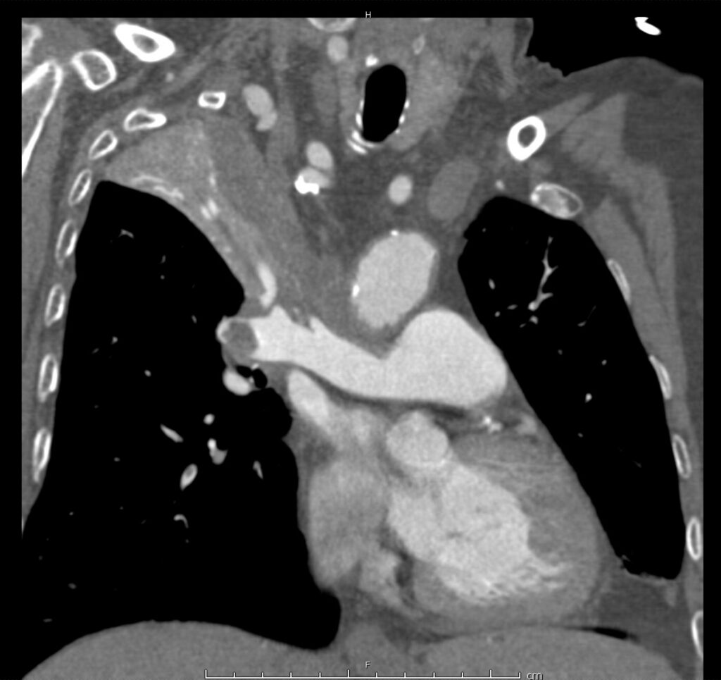
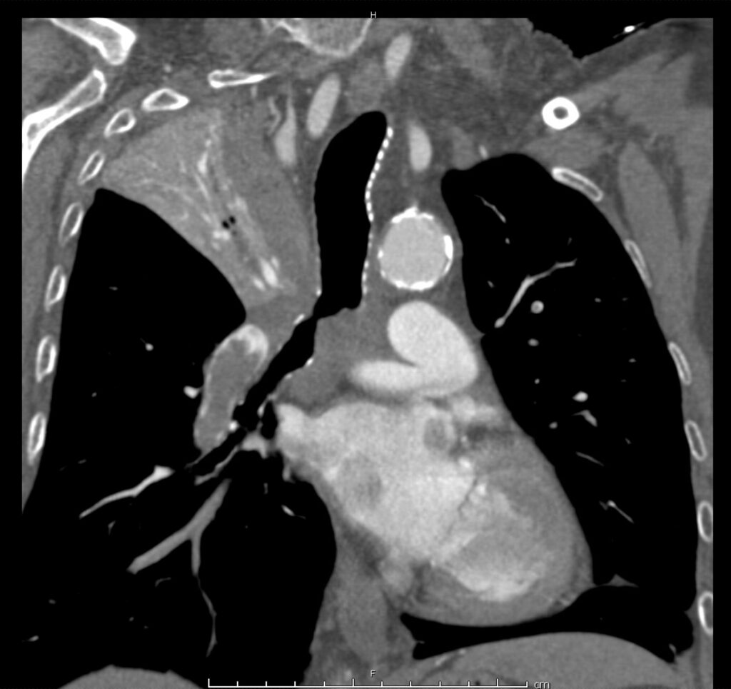
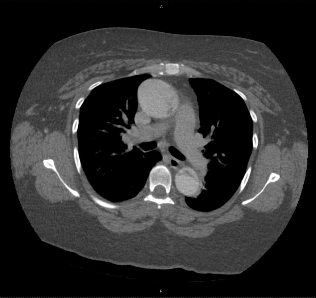
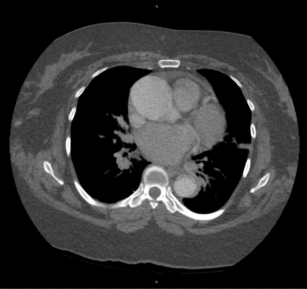
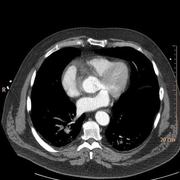
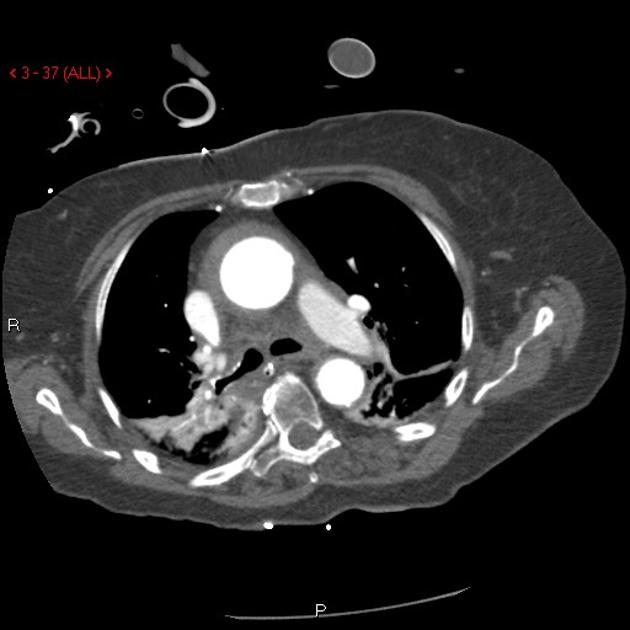
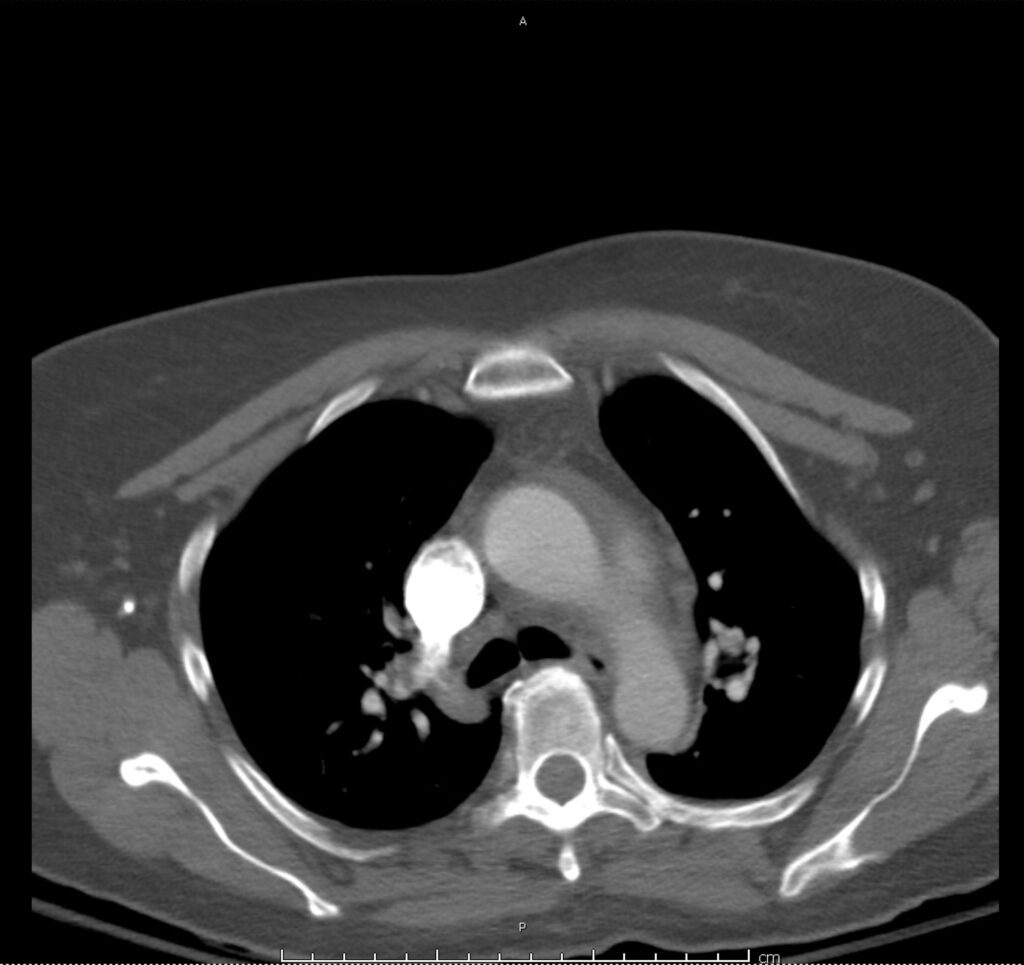
Circulation: Filling Defects

Extensive bilateral pulmonary emboli involving nearly all branches distal to left/right main pulmonary arteries.

01_pe-saddle
02_pe-saddle
03_pe-saddle
04_pe-saddle
05_pe-saddle
06_pe-saddle
07_pe-saddle
08_pe-saddle
09_pe-saddle
10_pe-saddle
11_pe-saddle
12_pe-saddle
13_pe-saddle
14_pe-saddle
15_pe-saddle
16_pe-saddle
17_pe-saddle
18_pe-saddle
19_pe-saddle
20_pe-saddle
21_pe-saddle
22_pe-saddle
23_pe-saddle

This slideshow requires JavaScript.

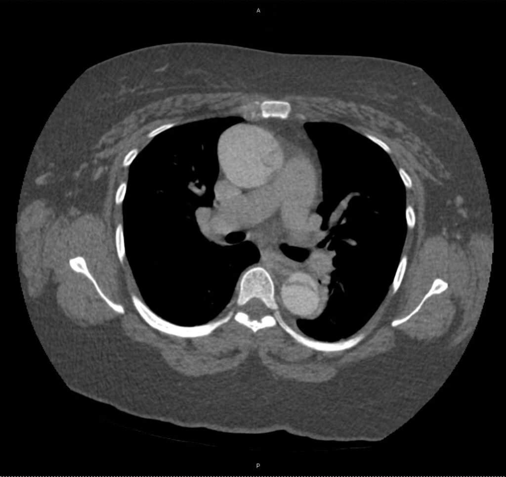
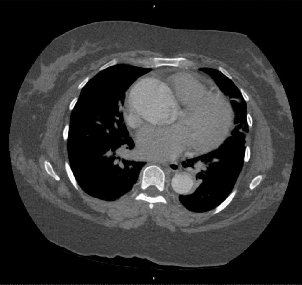
Circulation: Complications

Saddle pulmonary embolism with pulmonary artery dilation and flattening of the interventricular septum suggestive of right ventricular strain.

01_ptx
02_ptx
03_ptx
04_ptx
05_ptx
06_ptx
07_ptx
08_ptx
09_ptx
10_ptx
11_ptx
12_ptx
13_ptx
14_ptx

Breathing: Pleura

Pneumothorax