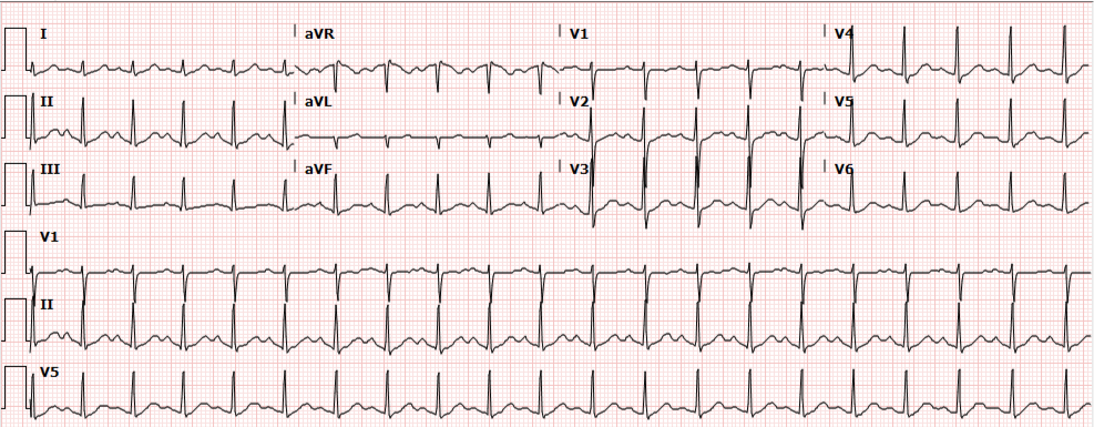
Sinus Tachycardia

Brief History and Physical:

A young female with a history of schizophrenia presents to the emergency department reporting hallucinations. She had been diagnosed with schizophrenia one year previously and was briefly admitted to a psychiatric hospital. She discontinued her anti-psychotic (risperidone) two months ago, and over the past week she reports increasingly prominent auditory and visual hallucinations.

She denies recent illness, vomiting/diarrhea, changes in urinary habits, new medications, alcohol or illicit substance use. She also denies chest pain, palpitations or shortness of breath.

Vital signs are notable for a heart rate of 148bpm and are otherwise normal (including core temperature). Detailed physical examination is normal except for a rapid, regular heart rate. Mental status examination demonstrated normal level of alertness and orientation, linear and cogent responses and occasional response to internal stimuli during which she appeared anxious.

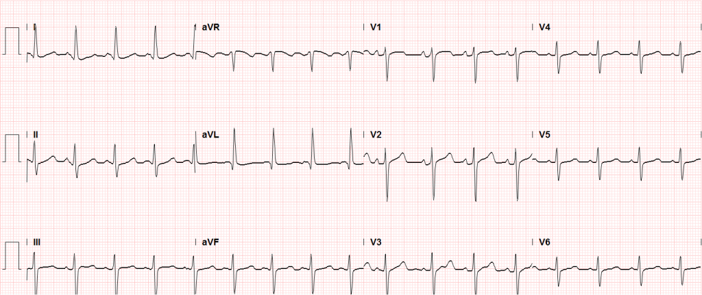
Initial evaluation and management included a 12-lead ECG which showed sinus tachycardia. Multiple boluses of normal saline were initiated while awaiting laboratory workup.

ECG: Sinus Tachycardia

Presentation ECG demonstrates sinus tachycardia.

Update:

Laboratory studies were reviewed and unremarkable. Normal hemoglobin, normal chemistry panel, negative hCG, and negative toxicology screen. The patient remained persistently tachycardic with a heart rate ranging from 140-160bpm (again sinus tachycardia on 12-lead ECG). An atypical antipsychotic and anxiolytic were administered and additional studies were obtained. Serum TSH, troponin and D-dimer were normal and bedside ultrasound did not identify a pericardial effusion. The patient remained asymptomatic, reporting subjective improvement in anxiety and hallucinations. Psychiatry was consulted and the patient was placed in observation for monitoring of sinus tachycardia. Observation course was uneventful as the patient remained asymptomatic. Transthoracic echocardiography was normal. Psychiatry consultation recommended resumption of home anti-psychotic and outpatient follow-up. Tachycardia had improved but not resolved at the time of discharge (heart rate 109bpm) and the patient was instructed to follow-up with her primary care provider.


Algorithm for the Evaluation of Sinus Tachycardia

Algorithm for the Evaluation of Sinus Tachycardia

Any vital sign derangement is concerning and tachycardia may be associated with unanticipated death after discharge home1. The presence of tachycardia suggests one of several categories of hemodynamic, autonomic, or endocrine/metabolic derangement.

Demand for increased cardiac output

A perceived demand for increased cardiac output will prompt chronotropic (and inotropic) amplification before hypotension develops. Causative etiologies include: volume depletion (from hemorrhage, gastrointestinal or renal losses), distributive processes (such as infection), obstruction (pulmonary embolus, or pericardial effusion with impending tamponade), or tissue hypoxia (anemia or lung disease).

Autonomic nervous system

Autonomic nervous system disturbances induced by stimulant, sympathomimetic or anti-cholinergic use, or withdrawal of certain agents such as ethanol or beta-blockers may be at fault.

Endocrine and other causes

Hyperthyroidism and pheochromocytoma should be considered, and as diagnoses of exclusion: anxiety, pain, or inappropriate sinus tachycardia2.

Evaluation:
Core temperature
CBC
Troponin
D-dimer
Bedside cardiac ultrasound
Urine toxicology screen
Ethanol level
TSH/T4

Algorithm for the Evaluation of Narrow-Complex Tachycardia3,4,5,6

Algorithm for the Evaluation of Narrow-Complex Tachycardia

References:

  1. Sklar DP, Crandall CS, Loeliger E, Edmunds K, Paul I, Helitzer DL. Unanticipated Death After Discharge Home From the Emergency Department. Ann Emerg Med. 2007;49(6):735-745. doi:10.1016/j.annemergmed.2006.11.018.
  2. Olshansky B, Sullivan RM. Inappropriate sinus tachycardia. J Am Coll Cardiol. 2013;61(8):793-801. doi:10.1016/j.jacc.2012.07.074.
  3. Yusuf S, Camm AJ. Deciphering the sinus tachycardias. Clin Cardiol. 2005;28(6):267-276.
  4. Katritsis DG, Josephson ME. Differential diagnosis of regular, narrow-QRS tachycardias. Heart Rhythm. 2015;12(7):1667-1676. doi:10.1016/j.hrthm.2015.03.046.
  5. Bibas L, Levi M, Essebag V. Diagnosis and management of supraventricular tachycardias. CMAJ. 2016;188(17-18):E466-E473. doi:10.1503/cmaj.160079.
  6. Link MS. Clinical practice. Evaluation and initial treatment of supraventricular tachycardia. N Engl J Med. 2012;367(15):1438-1448. doi:10.1056/NEJMcp1111259.

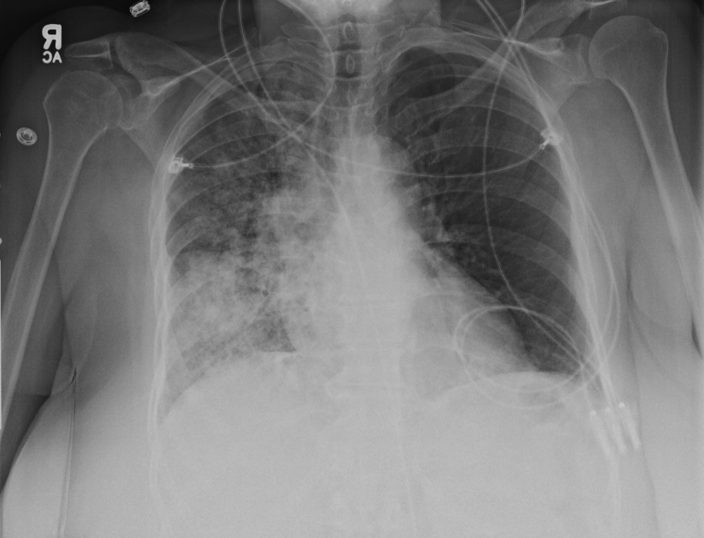
Anemia

HPI

70M with a history of dementia presenting with 3 days of fatigue. The patient was unable to provide detailed history, however family members reported worsening fatigue with the patient requiring assistance with ambulation for several days. The patient was referred from an outside clinic after point-of-care hemoglobin of 6.7. No reported history of anemia, and no history suggestive of obvious external bleeding.

Vital signs stable, tachycardia and tachypnea noted with minimal exertion but saturating well on ambient air and in no acute distress. Examination notable for conjunctival pallor without scleral icterus, systolic flow murmur, brown stool guaiac negative.

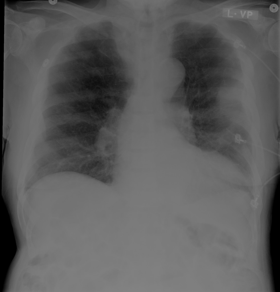
CBC with hemoglobin of 7.5 , MCV 80.3 , RDW 22.4 , no leukocytosis and normal platelets. Also noted was an alkaline phosphatase of 828 , normal total and direct bilirubin, and undetectable serum troponin. Chest x-ray showed a possible pleural-based mass.

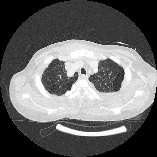
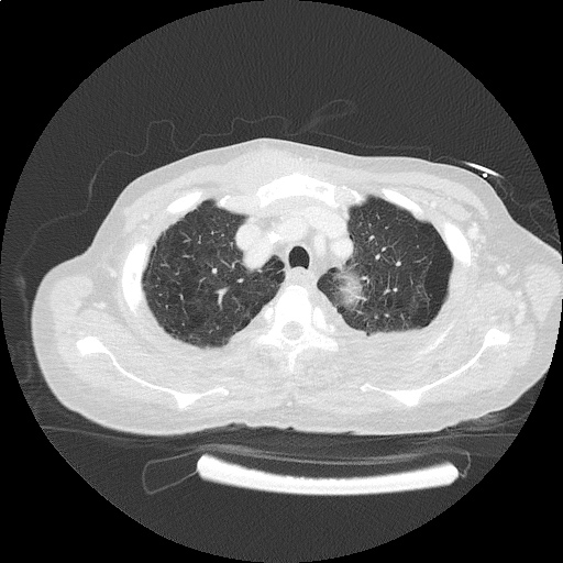
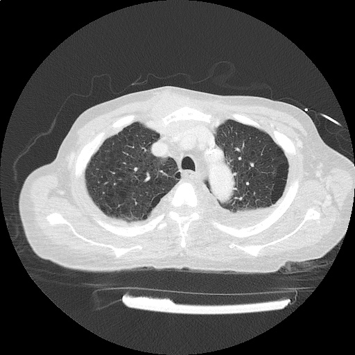
The patient was transfused two units of PRBC’s and admitted for further evaluation. CT chest/abdomen/pelvis revealed sternal and rib-based pleural soft-tissue mass, prostate mass, pelvic and retroperitoneal lymphadenopathy as well as extensive bony metastatic disease consistent with primary prostate cancer with diffused metastasis. Serum PSA was 2,087 . Iron studies suggested anemia of chronic disease. Reticulocytes were not obtained but may have suggested inadequate production index given extensive bony metastases and possible associated myelosuppression. The patient was symptomatically improved after transfusion and discharged with outpatient follow-up for discussions regarding possible biopsy and treatment.

Images

Chest x-ray with pleural-based mass

Areas of pleural thickening. Possible pleural based mass in left mid lung.

ct_chest_anemia_01
ct_chest_anemia_02
ct_chest_anemia_03
ct_chest_anemia_04
ct_chest_anemia_05
ct_chest_anemia_06
ct_chest_anemia_07
ct_chest_anemia_08
ct_chest_anemia_09
ct_chest_anemia_10
ct_chest_anemia_11
ct_chest_anemia_12
ct_chest_anemia_13

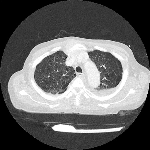
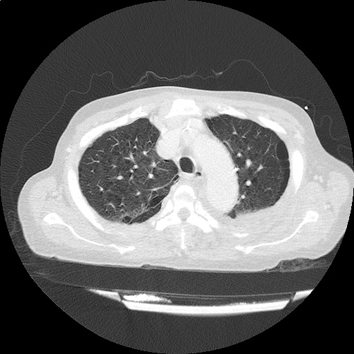
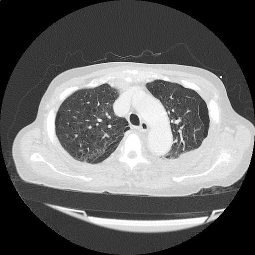
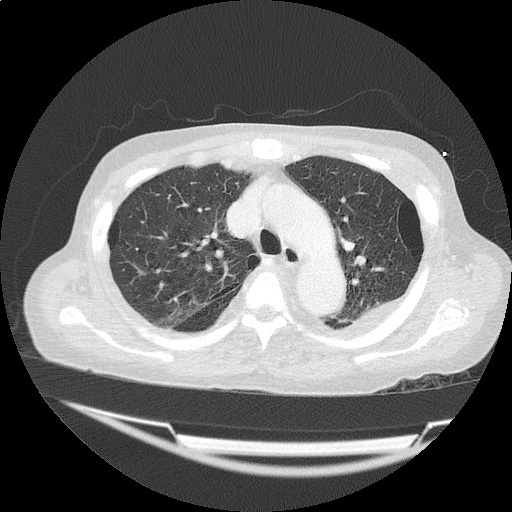
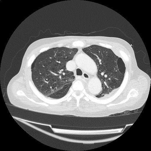
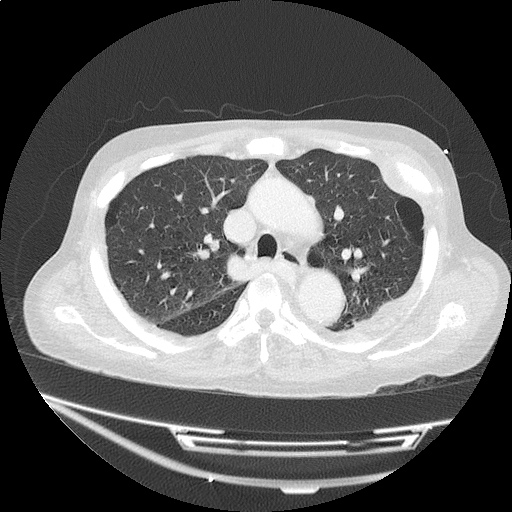
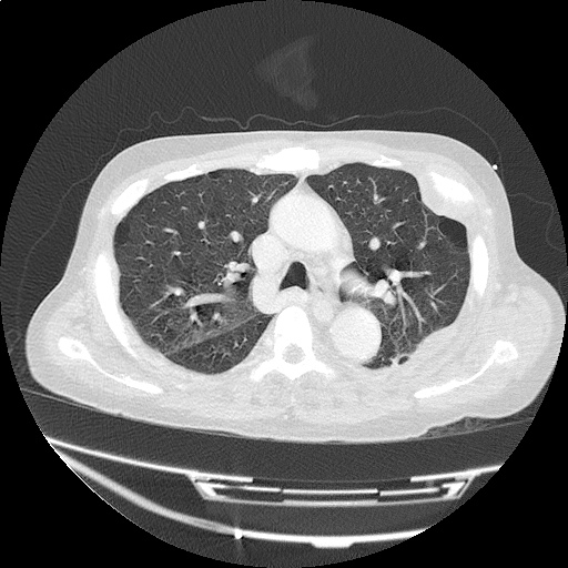
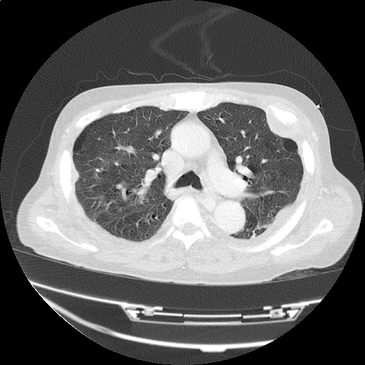
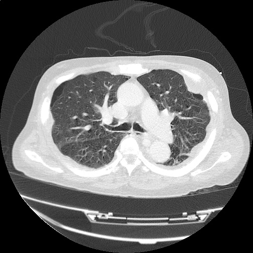
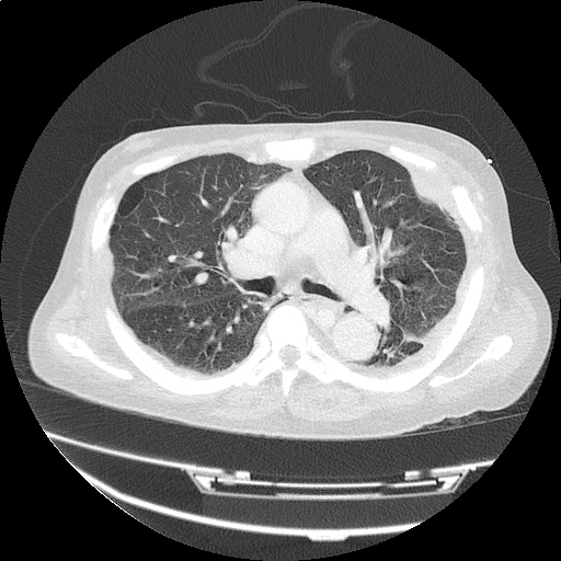
CT Chest: Lung Window

  • Rib-based pleural soft tissue masses.
  • Large 5.6 x 4.4cm anterior sternal soft-tissue mass.
ct_bone_anemia_01
ct_bone_anemia_02
ct_bone_anemia_03
ct_bone_anemia_04
ct_bone_anemia_05
ct_bone_anemia_06
ct_bone_anemia_07
ct_bone_anemia_08

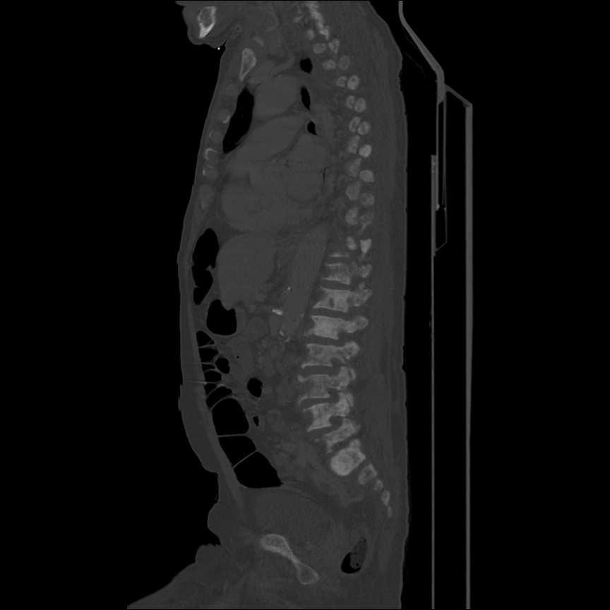
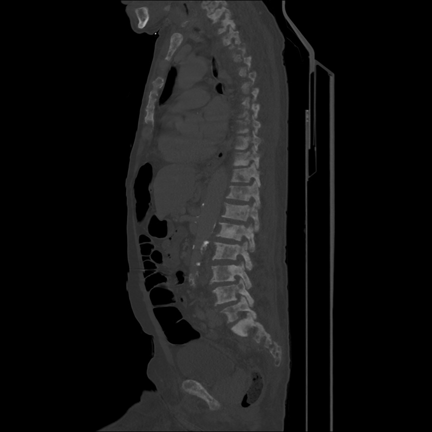
CT Body: Bone Window

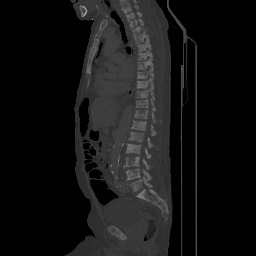
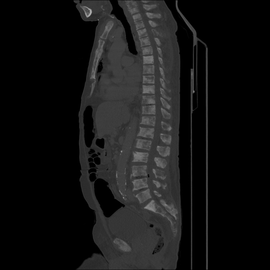
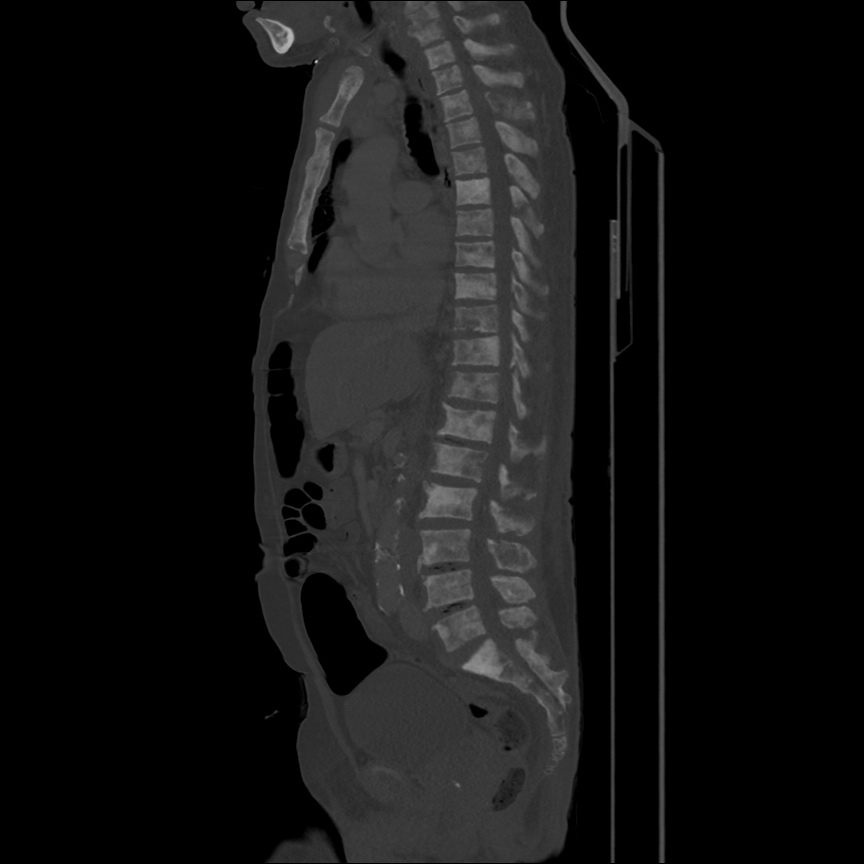
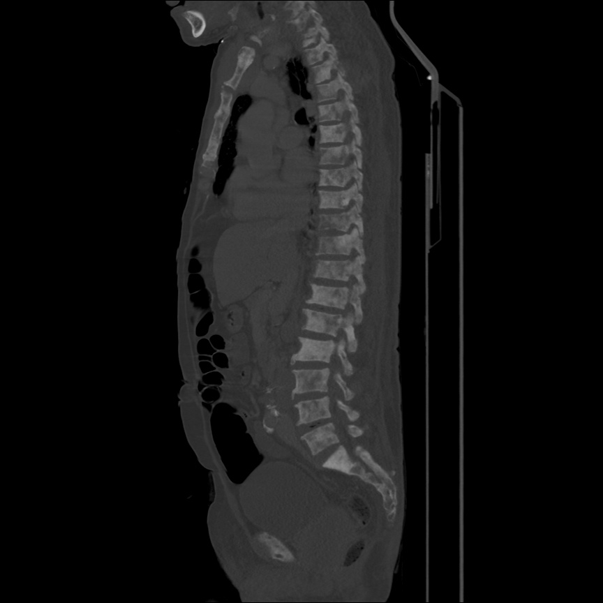
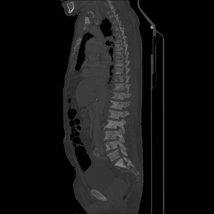
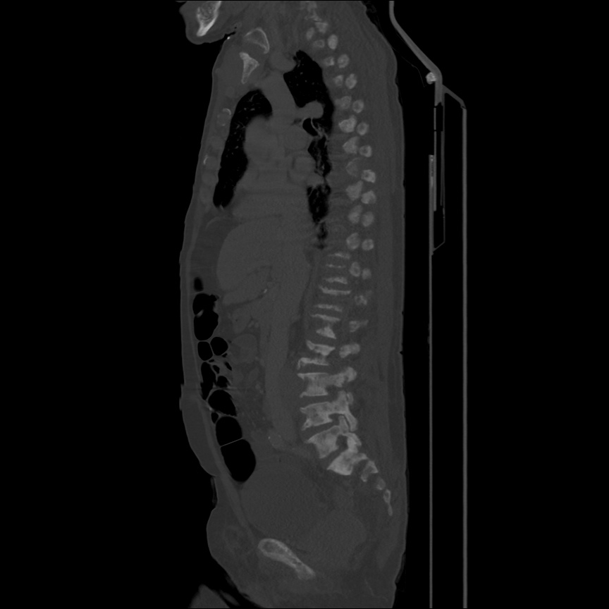
  • Extensive bony metastatic disease.
  • Prostate mass, large pelvic and retroperitoneal lymphadenopathy.
  • Consistent with primary prostate cancer with diffuse metastasis.

Algorithm for the Evaluation of Anemia 1,2

Algorithm for the Evaluation of Anemia

References:

  1. Zaiden R, Rana F. Evaluation of Anemia. BMJ Best Practice. Oct 2014. http://us.bestpractice.bmj.com/best-practice/monograph/93/overview.html. Last accessed 15 May 2015.
  2. Janz, T. G., Johnson, R. L., & Rubenstein, S. D. (2013). Anemia in the emergency department: evaluation and treatment. Emergency medicine practice, 15(11), 1–15– quiz 15–6.
  3. WiKEM: Anemia

Lactic Acidosis

HPI:

59F with a reported history of congestive heart failure, presenting with intermittent chest discomfort for three days.

She characterized this discomfort as “heartburn”, describing a mid-epigastric burning sensation radiating up her neck, not associated with exertion, lasting 1-2 hours and resolving with antacids. The patient has poor exercise tolerance at baseline and for the past several years has been able to ambulate only short distances around her home, and states that these symptoms have been worsening in the past week. She denies chest pain on exertion, orthopnea or paroxysmal nocturnal dyspnea. She states that she was diagnosed with congestive heart failure five years ago, but was never prescribed medications.

On further questioning, the patient reports several weeks of mouth and lip pain which has limited oral intake, though no dysphagia to solids or liquids. She otherwise denies fevers/chills, abdominal pain, nausea/vomiting, cough, changes in urinary or bowel habits.

In the emergency department, the patient was noted to have an elevated serum troponin, though ECG showed no changes of acute ischemia/infarction.

PMH:

  • Congestive heart failure

PSH:

  • None

FH:

  • Mother with diabetes
  • Father with MI at age 65

SHx:

  • 4-5 drinks of alcohol/day
  • No tobacco or drug use

Meds:

  • None

Allergies:

NKDA

Physical Exam:

VS: T 37.4 HR 106 RR 18 BP 145/82 O2 100% RA
Gen: Morbidly obese female, lying in bed, in no acute respiratory distress, speaking in complete sentences.
HEENT: Dry, cracked lips, slightly erythematous, otherwise moist mucous membranes, poor dentition. Mild scleral icterus. No cervical lymphadenopathy.
CV: Rapid rate, regular rhythm, normal S1/S2, II/VI systolic ejection murmur at LUSB, no radiation appreciated. No jugular venous distension.
Lungs: Clear to auscultation in posterior lung fields bilaterally, no crackles appreciated.
Chest: Well-circumscribed erythematous patch in folds beneath left breast, no underlying fluctuance, no significant tenderness to palpation. On contralateral breast, some hyperpigmentation but no erythema.
Abdomen: Obese, non-tender, non-distended. Patch of erythema below pannus, mildly tender to palpation.
Ext: Bilateral lower extremities with marked edema and overlying scaly plaques, some slightly ulcerated weeping serous fluid. Peripheral pulses are difficult to palpate, capillary refill difficult to assess.

Labs/Studies:

  • CBC: 11.1/11.1/34.5/212 (MCV 114.2)
  • BMP: 140/4.5/97/20/10/1.14/64
  • Anion Gap: 23
  • LFT: AST: 73, ALT: 26, AP: 300, TB: 4.6, DB: 2.1, Alb: 3.0, INR 1.3
  • BNP: 158
  • Troponin: 1.284
Sinus tachycardia, LVH, secondary repolarization abnormalities

Sinus tachycardia, LVH, secondary repolarization abnormalities

Imaging:

CT Pulmonary Angiography:
No evidence of central pulmonary embolism, thoracic aortic dissection, or thoracic aortic aneurysm. Evaluation of the peripheral vessels is limited due to motion artifact. No focal consolidation or pneumothorax.

CT Abdomen/Pelvis non-contrast:
No evidence of intra-abdominal abscess or definite source of infection. Marked hepatic steatosis.

CT Lower Extremity non-contrast:
Diffuse circumferential subcutaneous edema involving both lower extremities from the level of the mid thighs distally through the feet. There are bilateral subcutaneous calcifications which are likely venous calcifications in the setting of chronic venous stasis disease. There is some overlying skin thickening.

TTE:
There is moderate concentric left ventricular hypertrophy with hyperdynamic LV wall motion. The Ejection Fraction estimate is >70%. Grade I/IV (mild) LV diastolic dysfunction. No hemodynamically significant valve abnormalities.

US Abdomen:
Hepatomegaly, echogenic liver suggesting fatty infiltration. Moderately blunted hepatic vein waveforms suggesting decreased hepatic parenchymal compliance.

Assessment/Plan:

The patient was admitted to the cardiology service for management of NSTEMI and evaluation of undiagnosed CHF. She was started on a heparin continuous infusion. In addition, a CT pulmonary angiogram was obtained to evaluate for pulmonary embolism as an explanation of her progressive dyspnea on exertion. No PE, consolidation or effusion was identified.

Despite the patient’s reported history of congestive heart failure, there was no evidence that her symptoms were a result of an acute exacerbation with only a mildly elevated BNP but no jugular venous distension or evidence of pulmonary edema. The patient’s significant lower extremity edema was more suggestive of chronic venous stasis.

One notable laboratory abnormality that was explored was her elevated anion gap metabolic acidosis. Studies submitted included serum lactate, salicylates, osmolarity, CK, and urinalysis for ketonuria. This evaluation was notable for an elevated serum lactate of 13.2mmol/L and an arterial blood gas that showed adequate respiratory compensation (and no A-a gradient). Given the patient’s modest leukocytosis (with neutrophil predominance), and tachycardia, the concern for sepsis was increased though the source remained unclear. Prominent possibilities included a skin and soft-tissue infection vs. less likely intra-abdominal source though the patient’s physical examination was not suggestive of a process that would produce such a substantial lactic acidosis. Blood cultures were drawn and the patient was started on empiric antibiotics for the suspected sources. In addition, the patient was cautiously volume resuscitated given her reported history of CHF while pending a transthoracic echocardiogram to evaluate cardiac function. Additional imaging including CT abdomen/pelvis and lower extremities was obtained (though without contrast due to the patient’s recent exposure), and no obvious source was identified.

Over the next two days, the patient’s serum lactate downtrended to normal range, as did the serum troponin. A transthoracic echocardiogram showed an LVEF >70% with mild concentric hypertrophy and diastolic dysfunction. Blood and urine cultures were without growth.

Additional issues managed during the hospitalization included elevated serum transaminases (AST > ALT), conjugated hyperbilirubinemia and evidence of decreased hepatic synthetic function with hypoalbuminemia and elevated INR. Given the patient’s history of EtOH use, as well as other corroborating findings including macrocytic anemia, hypomagnesemia, folate and B12 deficiency, this was attributed to alcoholic hepatitis (discriminant function <32). Infectious hepatitis serologies were negative. The patient was started on nutritional supplements. Finally, the patient persistently complained of lip and oral mucosal pain. Examination was without discrete lesions but some mucosal redness was identified. Despite poor dentition, there was no evidence of abscess and HSV/HIV testing was negative. This was thought to be stomatitis caused by her identified nutritional deficiencies.

Differential Diagnosis of Elevated Serum Lactate 1,2

Differential Diagnosis of Elevated Serum Lactate

Algorithm for Evaluation of Acidemia 3,4

Algorithm for Evaluation of Acidemia

Algorithm for Evaluation of Alkalemia 3,4

Algorithm for Evaluation of Alkalemia

References:

  1. Fall, P. J., & Szerlip, H. M. (2005). Lactic acidosis: from sour milk to septic shock. Journal of intensive care medicine, 20(5), 255–271. doi:10.1177/0885066605278644
  2. Luft, F. C. (2001). Lactic acidosis update for critical care clinicians. Journal of the American Society of Nephrology : JASN, 12 Suppl 17, S15–9.
  3. Ingelfinger, J. R., Berend, K., de Vries, A. P. J., & Gans, R. O. B. (2014). Physiological Approach to Assessment of Acid–Base Disturbances. The New England journal of medicine, 371(15), 1434–1445. doi:10.1056/NEJMra1003327
  4. Ingelfinger, J. R., & Seifter, J. L. (2014). Integration of Acid–Base and Electrolyte Disorders. The New England journal of medicine, 371(19), 1821–1831. doi:10.1056/NEJMra1215672

Hemoptysis

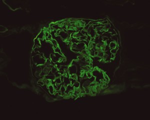
Source: Mulpuru, S., Touchie, C., Karpinski, J., & Humphrey-Murto, S. (2010). Coexistent Wegener“s granulomatosis and Goodpasture”s disease. The Journal of rheumatology, 37(8), 1786–1787. doi:10.3899/jrheum.091404

Linear IgG deposits consistent with anti-GBM disease.

CC:

“bad cough”

HPI:

61yo African American female w/hx of HTN presenting with 1mo of persistent cough productive of green-yellow sputum, noticed streaks of blood for the past 5 days. She came to the ED today because she has been feeling increasingly fatigued. She reports subjective fevers at the onset of symptoms which has resolved. She denies shortness of breath, chest pain, chills, night sweats. She sought medical care for this problem 2wk ago and was treated with amoxicillin and a cough suppressant. She recalls a coworker was ill one month ago. She is US-born, had a negative PPD in the past and has no known exposures to tuberculosis.

Of note, the patient reports her urine had a foamy appearance and has been darker in color beginning 3 weeks ago, but this had resolved. She denied dysuria, or frank hematuria.

PMH:

  • HTN
  • Asthma – last required medications >30yrs ago

 PSH:

  • None

FH:

  • Non-contributory

 SHx:

  • No t/e/d
  • Works as librarian

Meds:

  • benazepril
  • amlodipine
  • amoxicillin
  • promethazine

Allergies:

  • NKDA

Physical Exam:

VS: T 99.4 HR 97 BP 132/60 RR 20 O2 92%
Gen: Well-appearing, pleasant, speaking in complete sentences
HEENT: PERRL, MMM no lesions, no cervical lymphadenopathy
CV: RRR, normal S1/S2, no murmur appreciated
Lungs: Crackles in posterior: right middle/inferior and left inferior fields, no wheezing, no dullness to percussion
Abd: +BS, soft, non-tender, no CVAT
Ext: Warm, well-perfused, 2+ peripheral pulses, 1+ pitting edema to knee
Skin: No lesions on exposed skin
Neuro: AAO

Labs:

  • CBC: 12.3/6.7/19.8/52.3 (S: 94, B: 1, L: 4, M: 1, MCV: 92.3); baseline Hb/Hct (1/11/2012) 13.4
  • BMP: 136/3.6/101/25/46/3.43/126; baseline creatinine (1/11/2012) 1.18
  • UA: brown, trace LE, – nitrites, 2+ protein, 81 RBC

Imaging:

CXR PA

  • Right mid-lung zone consolidation is present, suggests pneumonia if acute.
  • Mild asymmetric right parenchymal increased density is seen diffusely as well.

Assessment/Plan:

65AAF w/hx HTN presents with persistent productive cough, recently with hemoptysis.

# Cough: Symptoms and physical findings of abnormal breath sounds (crackles, though no strict consolidation) concerning for community-acquired pneumonia. Addition of hemoptysis raises concern for TB, particularly when taking into consideration the duration of cough and presence of constitutional symptoms. CBC shows leukocytosis with left shift, CXR with right mid/lower lob infiltrates consistent with pneumonia. Recommend admission and isolation to rule out TB, start empiric therapy for community acquired pneumonia with ceftriaxone, azithromycin. Obtain induced sputum samples for culture, AFB smear and culture.

# Abnormal urine: Patient describes changes in urine suggestive of proteinuria and hematuria. Acuity of onset and apparent spontaneous resolution suggests a chronic kidney injury 2/2 hypertension is unlikely. Absence of dysuria, or tenderness (suprapubic, costovertebral) suggests complicated UTI unlikely. Urinalysis notable for 2+ protein and significant RBC’s, possible nephritic syndrome. In the setting of hemoptysis, this raises concern for anti-GBM disease vs. vasculitis.

# Anemia: Normocytic anemia. No evidence of acute, life-threatening hemorrhage as patient is currently hemodynamically stable. Possible sites of blood loss include alveoli, glomeruli. Given that patient sought care today for worsening fatigue, will monitor hemoglobin closely and consider transfusion. Obtain iron studies.

# HTN: BP stable, hold home medications.

Interval History:

The patient was admitted for management of community-acquired pneumonia and isolation to rule out TB. Empiric therapy with CTX + azithromycin was continued. On HOD1, the patient was transfused two units of PRBC’s. On HOD2, the patient underwent CT chest/abdomen/pelvis due to worsening respiratory status despite antimicrobial therapy. On HOD3, the patient went into atrial fibrillation with RVR which was converted to sinus rhythm with metoprolol 5mg IV x3. On HOD5, nephrology consult recommended starting steroid therapy, plasmapheresis and obtaining a renal biopsy, however the biopsy was delayed due to worsening respiratory status.

Interval Labs:

  • Iron studies: Fe 8, TIBC 203, Ferritin 468, haptoglobin 333, retics 2.7
  • Inflammatory markers: ESR 120, CRP 34
  • Micro: BCx NGTD, RCx moderate Candida, sputum AFB smear negative x3
  • LFT: AST 34, ALT 29, ALP 52, protein 6.3, albumin 2.4, T.bil 0.8, D.bil 0.2
  • Quant-gold: negative
  • Anti-GBM 1.2 (nl <1.0)
  • p-ANCA: positive 1:640, [ELISA pending]
  • ANA: positive 1:320, speckled
  • HIV: negative

Interval Imaging:

CT Chest

  • Diffuse right lung, tree and bud opacities, ground-glass opacities and areas of confluence with scattered air bronchograms. Less severe similar pattern in the left lung as well particular at the base.
  • Right paratracheal, subcarinal and perihilar LAD.
  • Findings concerning for primary TB in the right clinical setting. DDx nonspecific bacterial PNA and fungal PNA.

CT Abdomen/Pelvis

  • Mild nonspecific R > L perinephric stranding.

Interval Assessment/Plan:

# Acute respiratory failure: Unlikely simple CA-PNA given worsening status while on appropriate antibiotic therapy. Active tuberculosis possible given history of chronic productive cough with hemoptysis, constitutional symptoms and imaging findings. IGRA’s of limited utility in diagnosis of active disease, further, while three negative sputum AFB smears decreases the likelihood of TB, additional testing with NAAT and culture is required. Another possibility is a vasculitic process given concomitant hematuria and acute renal failure, with respiratory symptoms now 2/2 alveolar hemorrhage. This was evaluated with ANCA assay which was positive for p-ANCA with high titer. This is often suggestive of primary vasculitis (in this case likely microscopic polyangiitis vs. Churg-Strauss), however ELISA for target antigen is of particular importance as p-ANCA with specificity for antigens other than MPO can be associated with another condition on the differential: Goodpasture’s syndrome. This patient was found to have elevated anti-GBM antibodies which are highly suggestive of Goodpasture’s syndrome, and can be associated with ANCA-positivity (often suggesting a poorer prognosis with decreased likelihood of recovery of renal function).1

# Acute kidney injury: The patient had significant elevation of serum creatinine compared to last-recorded baseline. She also described darkening and foamy appearance of urine 3 weeks prior to admission, suggestive of proteinuria/hematuria of relatively acute onset. This was supported by urinalysis findings of protein and RBC’s (with casts). Given presence of anti-GBM antibodies, high specificity of such findings, and correlation with glomerulonephritis with evidence of pulmonary alveolar hemorrhage, this appears to be the most likely cause at this time. Definitive diagnosis with renal biopsy to be obtained following stabilization of respiratory status. Patient will be started on plasmapheresis and immunosuppressive therapy (corticosteroids, cyclophosphamide).

# Normocytic Anemia: Likely combination of acute blood loss (2/2 hematuria, pulmonary alveolar hemorrhage) and chronic disease. Normocytic anemia with some reticulocytosis suggestive of acute blood loss, however iron studies with low Fe, TIBC and elevated ferritin suggest chronic disease as an associated factor.

Differential Diagnosis of Hemoptysis: 2, 3

A System for Hemoptysis

A System for the Diagnosis of Tuberculosis: 4, 5

A System for the Diagnosis of Tuberculosis


A System for Vasculitides: 8, 9

A System for Vasculitidies


Vasculitis Mimics: 9

Vasculitis Mimics


Interpretation of antineutrophil cytoplasmic autoantibodies (ANCA): 10

Pattern Target Associated vasculitis Other diseases
C-ANCA PR3
  • Granulomatosis with polangiitis (Wegener’s)
  • Eosinophilic granulomatosis with polyangiitis (Churg-Strauss)
  • Microscopic polyangiitis
  • Pauci-immune glomerulonephritis
C-ANCA (atypical) BPIMPO
  • IBD
  • Cystic fibrosis


P-ANCA MPO
  • Microscopic polyangiitis
  • Eosinophilic granulomatosis with polyangiitis (Churg-Strauss)
  • Pauci-immune glomerulonephritis
Non-MPO
  • Autoimmune hepatitis
  • IBD, PSC
  • SLE, RA
  • Drugs
  • Infection (HIV, fungal)

Differential Diagnosis of Anemias: 11

A System for Anemias

References:

  1. Levy, J. B., Hammad, T., Coulthart, A., Dougan, T., & Pusey, C. D. (2004). Clinical features and outcome of patients with both ANCA and anti-GBM antibodies. Kidney international, 66(4), 1535–1540. doi:10.1111/j.1523-1755.2004.00917.x
  2. Bidwell, J. L., & Pachner, R. W. (2005). Hemoptysis: diagnosis and management. American family physician, 72(7), 1253–1260.
  3. Hirshberg, B., Biran, I., Glazer, M., & Kramer, M. R. (1997). Hemoptysis: etiology, evaluation, and outcome in a tertiary referral hospital. Chest, 112(2), 440–444. doi:10.1378/chest.112.2.440
  4. Campbell, I. A., & Bah-Sow, O. (2006). Pulmonary tuberculosis: diagnosis and treatment. BMJ (Clinical research ed.), 332(7551), 1194–1197. doi:10.1136/bmj.332.7551.1194
  5. Zumla, A., Raviglione, M., Hafner, R., & Reyn, von, C. F. (2013). Tuberculosis. The New England journal of medicine, 368(8), 745–755. doi:10.1056/NEJMra1200894
  6. Diagnostic Standards and Classification of Tuberculosis in Adults and Children. American journal of respiratory and critical care medicine. doi:10.1164/ajrccm.161.4.16141
  7. Laraque, F., Griggs, A., Slopen, M., & Munsiff, S. S. (2009). Performance of nucleic acid amplification tests for diagnosis of tuberculosis in a large urban setting. Clinical infectious diseases : an official publication of the Infectious Diseases Society of America, 49(1), 46–54. doi:10.1086/599037
  8. Gross, W. L., Trabandt, A., & Reinhold-Keller, E. (2000). Diagnosis and evaluation of vasculitis. Rheumatology (Oxford, England), 39(3), 245–252.
  9. Suresh, E. (2006). Diagnostic approach to patients with suspected vasculitis. Postgraduate medical journal, 82(970), 483–488. doi:10.1136/pgmj.2005.042648
  10. Rus, V., & Handwerger, B. S. (2000). Clinical value of antineutrophil cytoplasmic antibodies. Current rheumatology reports, 2(5), 383–389.
  11. Goljan, E. (2011). Pathology. Philadelphia, PA: Mosby/Elsevier.