Dysphagia

Brief H&P

A 47-year-old male with no known medical history presents with dysphagia. He reports 3 weeks of symptoms, describing difficulty predominantly with swallowing solid foods which is aided by the concomitant ingestion of liquids. He points to his throat as the area of discomfort, but has not noted any choking or coughing after attempts at swallowing. He occasionally suffers from “heartburn”, describing a burning sensation in his chest provoked by certain foods and was previously prescribed omeprazole which he has not taken for several years. He denies any prior surgeries, tobacco or alcohol use, relevant family history or similar symptoms in the past.

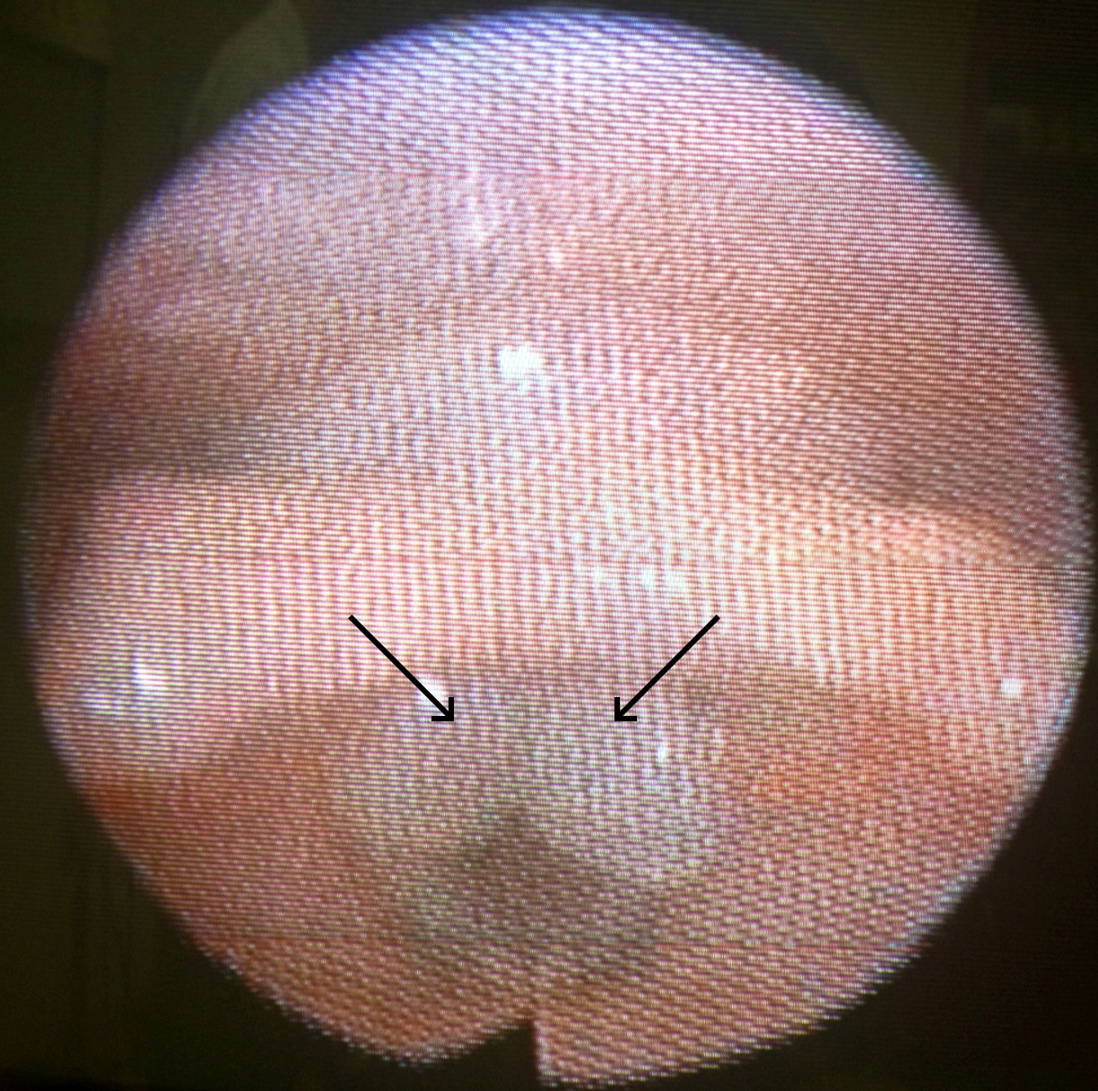
Physical examination was unrevealing, demonstrating a normal neurological examination, normal phonation, normal oropharynx and no appreciable neck masses. The patient was observed to comfortably swallow water.

He was discharged with gastroenterology follow-up and ultimately underwent esophagogastroduodenoscopy which demonstrated narrowing of the distal esophagus suggestive of a peptic stricture. Dilation was deferred in favor of resumption of proton pump inhibitor therapy.


Types of Dysphagia1,2

Oropharyngeal3
Characterized by difficulty initiating swallowing and accompanied by choking/coughing, nasopharyngeal regurgitation or aspiration.
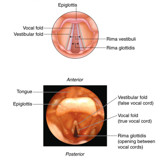
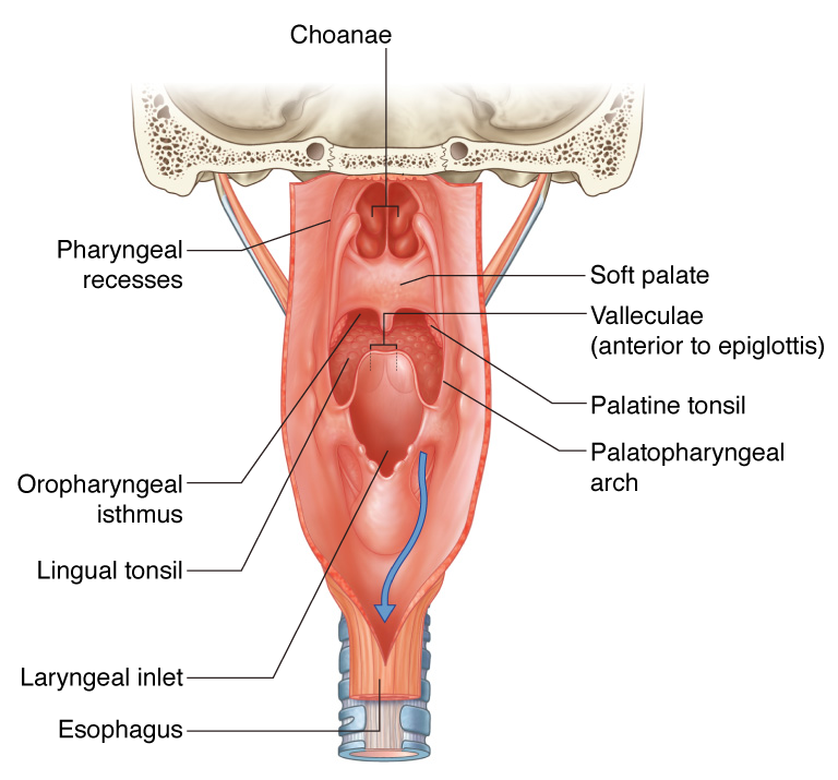
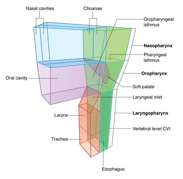
Involved anatomy: Tongue, muscles of mastication, soft palate (elevation to close nasopharynx), suprahyoid muscles (elevate larynx), epiglottis (occlude airway), cricopharyngeus muscle (release upper esophageal sphincter). Neurological control predominantly coordinated by cranial nerves (V, VII, IX, X, XII)
Esophageal4
Delayed after initiating swallowing and characterized by a sensation of food bolus arresting in transit.
Involved anatomy: Skeletal and smooth muscle along the esophagus and lower esophageal sphincter. Neurological control predominantly coordinated by medulla

Important Historical Features5,6

  • Difficulty with liquids suggests motility problem
  • Difficulty with solids only or solids progressing to liquids suggests mechanical obstruction
  • Identify a history of head and neck surgery or radiation therapy
  • Identify a personal or family history of connective tissue disorder (scleroderma, RA, SLE) which may be associated with esophageal dysmotility
  • Review home medications (NSAID, bisphosphonate, potassium chloride, ferrous sulfate)
  • Immunocompromised patients are at risk for infectious esophagitis (Candida, CMV, HSV) which are generally associated with odynophagia
  • A history of heartburn may be associated with reflux-mediated complications such as erosive esophagitis, peptic stricture, and adenocarcinoma of the esophagus
  • Young patients are more likely to be affected by eosinophilic esophagitis
  • Patient localization of site of obstruction is generally accurate, patients are more accurate at localizing proximal than distal obstructions7

Algorithm for the Evaluation of Dysphagia8

Algorithm for the Evaluation of Dysphagia

Management9-11

Patients who are safely tolerating oral intake can be referred for outpatient gastroenterology evaluation. Admission should be considered for patients at high-risk for aspiration.

References

  1. Spieker MR. Evaluating dysphagia. Am Fam Physician. 2000;61(12):3639-3648.
  2. Abdel Jalil AA, Katzka DA, Castell DO. Approach to the patient with dysphagia. Am J Med. 2015;128(10):1138.e17-.e23. doi:10.1016/j.amjmed.2015.04.026.
  3. Shaker R. Oropharyngeal Dysphagia. Gastroenterol Hepatol (N Y). 2006;2(9):633-634.
  4. Galmiche JP, Clouse RE, Bálint A, et al. Functional esophageal disorders. Gastroenterology. 2006;130(5):1459-1465. doi:10.1053/j.gastro.2005.08.060.
  5. McCullough GH, Martino R. Clinical Evaluation of Patients with Dysphagia: Importance of History Taking and Physical Exam. In: Manual of Diagnostic and Therapeutic Techniques for Disorders of Deglutition. New York, NY: Springer New York; 2012:11-30. doi:10.1007/978-1-4614-3779-6_2.
  6. Cook IJ. Diagnostic evaluation of dysphagia. Nat Clin Pract Gastroenterol Hepatol. 2008;5(7):393-403. doi:10.1038/ncpgasthep1153.
  7. Wilcox CM, Alexander LN, Clark WS. Localization of an obstructing esophageal lesion. Is the patient accurate? Dig Dis Sci. 1995;40(10):2192-2196.
  8. Trate DM, Parkman HP, Fisher RS. Dysphagia. Evaluation, diagnosis, and treatment. Prim Care. 1996;23(3):417-432.
  9. American Gastroenterological Association medical position statement on management of oropharyngeal dysphagia. Gastroenterology. 1999;116(2):452-454. doi:10.1016/S0016-5085(99)70143-5.
  10. Spechler SJ. American Gastroenterological Association medical position statement on treatment of patients with dysphagia caused by benign disorders of the distal esophagus. Gastroenterology. 1999;117(1):229-232. doi:10.1016/S0016-5085(99)70572-X.
  11. Varadarajulu S, Eloubeidi MA, Patel RS, et al. The yield and the predictors of esophageal pathology when upper endoscopy is used for the initial evaluation of dysphagia. Gastrointest Endosc. 2005;61(7):804-808.


Dysphonia (Hoarseness)

Case 1

HPI:

36 year-old female with no significant medical history who presents after referral for voice hoarseness. According to the patient, she underwent a C-section 3 months ago (at an outside hospital) complicated by bleeding requiring a second operation (L salpingoophorectomy); however, neither procedure required emergent intubation. She reports that she had some vomiting associated with anesthesia which ultimately required intubation and admission to the MICU for 5-6d. She was discharged 10 days after the initial operation, and both she and her baby were in good health. Two weeks after discharge, she began experiencing throat irritation and 1 month after discharge she noticed voice hoarseness which has been persistent. Today, she denies difficulty swallowing or breathing, F/C, N/V, abdominal pain.

PMH:

None

PSH:

Cesarean x2, L salpingoophorectomy

FH:

Non-contributory

SHx:

Lives at home taking care of 3 children, denies t/e/d

Meds:

None

Allergies:

NKDA

Physical Exam:

Gen: WA, NAD
Head: NC/AT
OC: MMM, no lesions, no pharyngeal erythema/exudates, hoarse voice
Ears: EAC clear, TMI b/l
Flex: Posterior commissure edema, cobblestoning, b/l TVC with shiny white masses
Flexible nasolaryngoscopy image showing trauma granulomata.

Flexible nasolaryngoscopy image showing trauma granulomata.

Assessment/Plan:

36F, no significant PMH, recent Cesarean and L salpingoophorectomy c/b likely aspiration requiring intubation and mechanical ventilation for several days with onset of progressive voice hoarseness 1mo later. History and flexible nasolaryngoscopy consistent with trauma granulomata, as well as laryngopharyngeal reflux.

  • Discussed treatment options with patient, recommend voice rest x2mo and continued monitoring
  • Laryngopharyngeal reflux, given dietary/lifestyle education, start omeprazole 20mg p.o. b.i.d.
  • RTC 1mo

Case 2

HPI:

51 year-old female with a history of Hepatitis C, COPD and an 80 pack-year smoking history presents with concern about progressive voice hoarseness x2mo. She reports quitting smoking two months ago and is not sure if the hoarseness preceded or followed quitting. She has occasional throat discomfort which is mild. She otherwise denies difficulty or pain with swallowing, worsening shortness of breath, unintentional weight loss.

She also reports a new mass on her neck which she first noticed yesterday. Denies associated pain, or surrounding skin changes.

PMH:

  • Hepatitis C
  • COPD

PSH:

  • Hysterectomy

FH:

Non-contributory

SHx:

80 pack-year smoking history, no current EtOH, drug use (previously used heroin and opiates)

Meds:

  • Methadone
  • Elavil
  • Multiple unknown inhaled medications

Allergies:

  • Naproxen (swelling)

Physical Exam:

Gen: WA, NAD
Head: NC/AT
Eyes: PERRL, EOMI
Ears: b/l EAC erythema, TMI, no lesions/exudates
OC: MMM, no lesions
Neck: Supple, no thyroid enlargement, no cervical lymphadenopathy, 5x6cm soft, round, mobile, non-tender mass on left lateral neck w/o overlying skin changes
Flex: Diffuse laryngeal damage, thickened posterior commissure, right TVC with area of leukoplakia, left TVC appears irregular

Assessment/Plan:

51F hx HepC, COPD, 80py smoking, presenting with voice hoarseness x2mo. History concerning for malignancy, exam today shows significant laryngeal damage and vocal cord irregularities warranting further evaluation. Possible component of fungal infection 2/2 inhaled steroid use for COPD, plan to reduce potentially aggravating factors (treat fungal infection, voice rest) and repeat evaluation. Neck mass possibly lipoma however will evaluate further given concern for malignancy.

  • Start fluconazole 100mg two tables p.o. on day1, 100mg p.o. daily x7d
  • Start nystatin 100,000 units/mL 10mL gargle and swallow t.i.d. x2wks
  • Advised voice rest
  • RTC in 3wks
  • CT neck/soft tissue w/wo IV contrast to evaluate neck mass

Anatomy of the Pharynx/Larynx:

Vocal cord anatomy
Pharyngeal wall anatomy
Structure of the pharynx

Physiology of Voice Production: 1

Voice is produced through the passive vibration of vocal folds in an air stream and requires:

  1. Adequate Air Stream
  2. Smooth vocal fold edges
  3. Vocal folds with normal vibratory properties
  4. Appropriate vocal fold positioning

Differential diagnosis of dysphonia (hoarseness): 1,2,3

 Differential Diagnosis of Dysphonia (hoarseness)

Characteristics of Hoarse Voice: 5

Characteristic Likely cause
Breathy Vocal cord paralysis
Hoarse Vocal cord lesion, LPR
Low-pitched Reinke’s edema, vocal abuse, LPR

References:

  1. Mau, T. (2010). Diagnostic Evaluation and Management of Hoarseness. Medical Clinics of North America, 94(5), 945–960. doi:10.1016/j.mcna.2010.05.010
  2. Feierabend, R. H., & Shahram, M. N. (2009). Hoarseness in adults. American family physician, 80(4), 363–370.
  3. Schwartz, S. R., Cohen, S. M., Dailey, S. H., Rosenfeld, R. M., Deutsch, E. S., Gillespie, M. B., Granieri, E., et al. (2009, September). Clinical practice guideline: hoarseness (dysphonia). Otolaryngology. doi:10.1016/j.otohns.2009.06.744
  4. Bruch, J.W., Kamani D.V. Diaphragmatic pacing. In: UpToDate, Basow, DS (Ed), UpToDate, Waltham, MA, 2013.
  5. Rosen, C. A., Anderson, D., & Murry, T. (1998). Evaluating hoarseness: keeping your patient’s voice healthy. American family physician, 57(11), 2775–2782.