Weakness

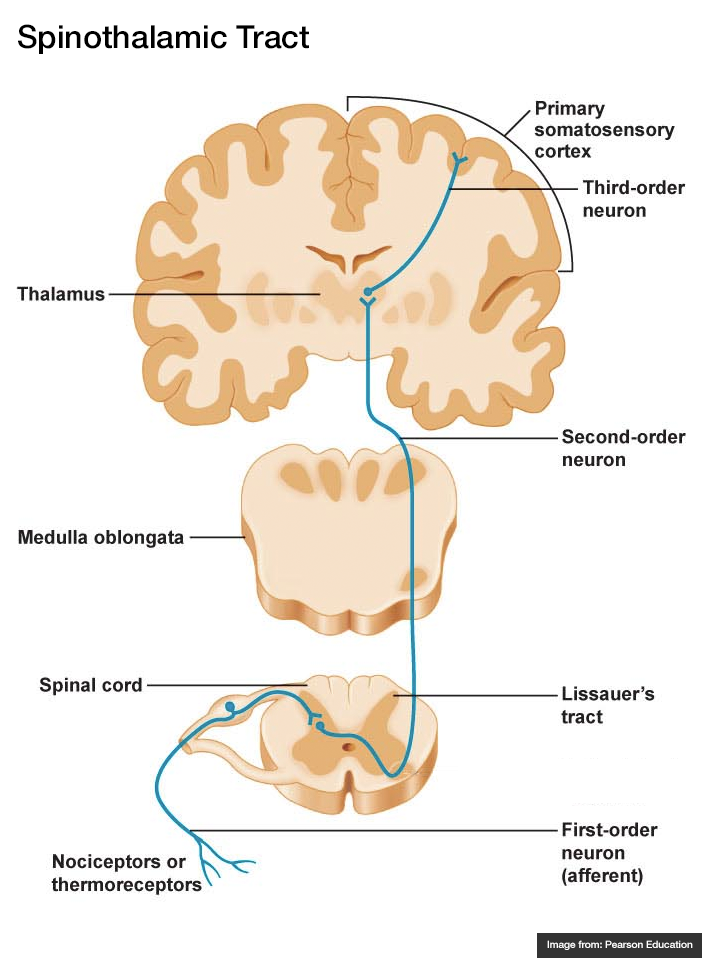
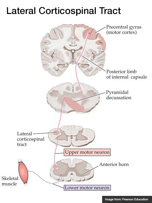
A systematic approach to motor weakness progresses along an anatomic tract from the cerebral cortex to individual sarcomeres. Impulses are generated in the primary motor cortex mapped to the homunculus, then aggregate as they descend through the internal capsule. Fibers decussate in the medulla and descend in the contralateral lateral corticospinal tract. These upper motor neurons (UMN) synapse with the lower motor neuron (LMN) in the anterior horn of the spinal cord. The lower motor neuron is bundled with neighboring fibers into a peripheral nerve and activates the target muscle fibers at the neuromuscular junction.

Algorithm for the Evaluation of Weakness

Algorithm for the Evaluation of Weakness

Upper Versus Lower Motor Neuron Findings

Finding Upper Lower
Reflexes
Atrophy
Weakness
Fasciculation
Tone
Summary
Recalling these findings can be simplified by understanding the underlying process. Denervation near the target muscle fibers (lower motor neuron disease) results in dampening of the efferent limb of spinal reflexes, resulting in hyporeflexia. The absence of nourishing stimulation leads to muscle atrophy and disorganized interpretation of proximal activity produces fasciculation.

Comparison Between Myopathy, Neuropathy and Neuromuscular Junction Processes

Finding Myopathy Neuropathy Neuromuscular Junction
Example Polymyositis Guillain-Barre Syndrome Myasthenia Gravis
Distribution Proximal > Distal Distal > Proximal Diffuse, Bulbar
Reflexes Normal
Sensory
Fatigue
CK Normal Normal

Motor Strength Grading

Grade Description
5 Normal
4 Reduced, moves against resistance
3 Moves against gravity
2 Moves only with elimination of gravity
1 Fasciculation only
0 None

Reflex Grading

Grade Description
4 Increased amplitude, spread to adjacent, clonus
3 Increased
2 Normal
1 Decreased
0 None

References

  1. Ganti L, Rastogi V. Acute Generalized Weakness. Emerg Med Clin North Am. 2016;34(4):795-809. doi:10.1016/j.emc.2016.06.006.
  2. Asimos AW. Weakness: A Systematic Approach To Acute, Non-traumatic, Neurologic And Neuromuscular Causes. Emergency Medicine Practice. 2002;4(12):1-28.
  3. Morchi R. Weakness. In: Rosen’s Emergency Medicine. Elsevier Inc.; 2014:2521.

Weakness

View Algorithm
There is a ddxof algorithm for the evaluation of weakness. View it here.

Motor Neuron Signs

Upper Motor Neuron:
Spasticity
Hyperreflexia
Pronator drift
Babinski
Lower Motor Neuron:
Flaccidity
Hyporeflexia
Fasciculation
Atrophy

Causes of Weakness

Lesion Critical Emergent
Non-neurological Shock (VS, clinical assessment)
Hypoglycemia (POC glucose)
Electrolyte derangement (BMP)
Anemia (POC Hb, CBC)
MI (ECG, troponin)
CNS depression (Utox, EtOH)
 
Cortex Stroke Tumor
Abscess
Demyelination
Brainstem Stroke Demyelination
Spinal Cord Ischemia
Compression (disk, abscess, hematoma)
Demyelination (transverse myelitis)
Peripheral Acute demyelination (GBS) Compressive plexopathy
Muscle Rhabdomyolysis Inflammatory myositis

Weakness Syndromes

Unilateral weakness, ipsilateral face
Lesion: Contralateral cortex, internal capsule
Causes: Stroke (sudden onset), demyelination/mass (gradual onset)
Symptoms: Neglect, visual field cut, aphasia
Findings: UMN signs
Key features: Association with headache suggests hemorrhage or mass
Unilateral weakness, contralateral face
Lesion: Brainstem
Causes: Vertebrobasilar insufficiency, demyelination
Symptoms: Dysphagia, dysarthria, diplopia, vertigo, nausea/vomiting
Findings: CN involvement, cerebellar abnormalities
Unilateral weakness, no facial involvement
Lesion: Contralateral medial cerebral cortex, discrete internal capsule
Causes: Stroke
Rare Cause: Brown-Sequard if contralateral hemibody pain and temperature sensory disturbance
Unilateral weakness single limb (monoparesis/plegia)
Lesion: Spinal cord, peripheral nerve, NMJ
UMN signs: Brown-Sequard if contralateral pain and temperature sensory disturbance
LMN signs: Radiculopathy if associated sensory disturbance
Normal reflexes, normal sensation: Consider NMJ disorder
Bilateral weakness of lower extremities (paraparesis/plegia)
Lesion: Spinal cord, peripheral nerve
UMN signs: Anterior cord syndrome (compression, ischemia, demyelination) if contralateral pain and temperature sensory disturbance
Cauda equina: Loss of perianal sensation, loss of rectal tone, or urinary retention
GBS: If no signs of cauda equina and sensory disturbances paralleling ascending weakness (with hyporeflexia)
Bilateral weakness of upper extremities
Lesion: Central cord syndrome
Causes: Syringomyelia, hyperextension injury
Findings: Pain and temperature sensory disturbances in upper extremities (intact proprioception)
Bilateral weakness of all four extremities (quadriparesis/plegia)
Lesion: Cervical spinal cord
Findings: UMN signs below level of injury, strength/sensory testing identifies level
Bilateral weakness, proximal groups
Lesion: Muscle
Causes: Rhabdomyolysis, polymyositis, dermatomyositis, myopathies
Findings: Muscle tenderness to palpation, no UMN signs, no sensory disturbances
Facial weakness, upper and lower face
Lesion: CNVII
Causes: Bell’s palsy, mastoiditis, parotitis
Other CN involvement suggests brainstem lesion, multiple cranial neuropathies, or NMJ

Review of Spinal Cord Anatomy

  • Dorsal Column – Medial Lemniscus (fine touch, proprioception)
    1. Afferent sensory fibers with cell body in DRG
    2. Ascend in ipsilateral posterior column
    3. Synapse in medulla, decussate, ascend in contralateral medial lemniscus
    4. Synapse in thalamus (VPL)
    5. Synapse in sensory strip of post-central gyrus
  • Spinothalamic Tract (pain, temperature)

    1. Afferent sensory fibers with cell body in DRG
    2. Ascends 1-2 levels
    3. Synapse in ipsilateral spinal cord, decussate, ascend in contralateral lateral spinothalamic tract
    4. Synapse in thalamus (VPL)
    5. Synapse in sensory strip of post-central gyrus
  • Lateral Corticospinal Tract (motor)

    1. Efferent cell body in motor strip of pre-central gyrus
    2. Descends through internal capsule
    3. Decussates in pyramid of medulla, descends in contralateral lateral corticospinal tract
    4. Synapse in anterior horn, lower motor neuron to muscle fiber
Spinal Cord Syndromes
Spinothalamic Tract
Dorsal Column / Medial Lemniscus
Lateral Corticospinal Tract

References

  1. Morchi, R. (2013). Weakness. In Rosen’s Emergency Medicine – Concepts and Clinical Practice (8th ed., Vol. 1, pp. 124-128). Elsevier Health Sciences.