Pleural Effusion

HPI:

64F with a history of CAD, MI, CHF, CLL and rheumatoid arthritis who presented to the emergency department after transfer from a rehabilitation facility for respiratory distress. The patient reported several days of progressive shortness of breath with dyspnea on exertion. She also noted some associated orthopnea and lower extremity edema. The patient was recently hospitalized for similar symptoms and was diagnosed with CHF at the time. At the rehab facility, the patient became hypoxemic and hypertensive, reporting shortness of breath and chest pain prior to presentation.

Hospital Course:

The patient was initially managed with BiPAP and nitroglycerin continuous infusion, but was then stable on supplemental O2 via nasal cannula and was transitioned to long-acting nitrates and anti-hypertensives. The patient’s hypoxemic respiratory failure was initially attributed to acute exacerbation of left-ventricular heart failure, and the patient was managed with spot diuresis. However, there was no symptomatic improvement and the patient became hypernatremic so diuresis was held as alternative diagnoses were explored.

A transthoracic echocardiogram showed preserved LVEF (50-55%), but some diastolic dysfunction and elevated PAP/RAP. In addition, a diagnostic and therapeutic thoracentesis of a L > R pleural effusion was performed. Pleural fluid studies were suggestive of a transudative process, though with some abnormal characteristics (including lymphocyte predominance, as well as presence of signet cells).

Rheumatology and pulmonology services were consulted for input and recommendations for further evaluation were appreciated. Per rheumatology, the patient’s diagnosis of rheumatoid arthritis may not be consistent with her presentation or prior serologic studies. Her pleural fluid analysis was also not consistent with rheumatoid disease. According to pulmonary consult, the patient’s hypoxemia remains most consistent with left ventricular dysfunction though primary pulmonary processes cannot be excluded (and would warrant further evaluation with imaging and pulmonary function testing).

PMH:

  • CHF
  • CAD
  • CVA
  • Myocardial Infarction
  • HTN
  • Hypothyroidism
  • CLL
  • Anemia

PSH:

  • TAH-BSO

FH:

  • No family history of autoimmune disease.
  • Mother: DM

SHx:

  • Denies tobacco/EtOH/drug use
  • Lives at home, at SNF since discharge

Meds:

  • Furosemide 20mg p.o. daily
  • Gabapentin 300mg p.o. t.i.d.
  • Hydralazine 50mg p.o. t.i.d.
  • Hydrochlorothiazide 25mg p.o. b.i.d.
  • Hydroxychloroquine 200mg p.o. daily
  • Levothyroxine 25mcg p.o. daily
  • Minoxidil 2.5mg p.o. b.i.d.
  • Pantoprazole 40mg p.o. daily
  • Prednisone 15mg p.o. daily

Allergies:

  • Shellfish
  • Physical Exam:

    VS: T 37.2 HR 84 RR 15 BP 147/75 O2 97% 4LNC
    Gen: Elderly female, alert and oriented to self and place, responding appropriately to questions.
    HEENT: Mucous membranes moist, sclera anicteric, no cervical lymphadenopathy.
    CV: Regular rate and rhythm, normal S1/S2, no additional heart sounds. III/VI mid-systolic murmur heard best at LLSB with diastolic component, no radiation appreciated. Non-displaced PMI. JVP measured to 14cm.
    Lungs: Decreased breath sounds in left lung field to inferior 2/3 with crackles above, on right crackles to inferior 1/2 of lung fields posteriorly. Dullness to percussion of inferior left lung field posteriorly.
    Abdomen: Soft, non-tender, non-distended, no hepatosplenomegaly, no appreciable fluid wave.
    Ext: Bilateral lower extremities with 2+ pitting edema to knees, some hyperpigmentation to right lower extremity.

    Labs/Studies:

    • CBC: 11.2/10.2/32.7/179
    • BMP: 141/3.9/103/30/15/0.85/107
    • INR: 0.9
    • BNP: 1857
    • UA: WBC 4, RBC 29 , +Bacteria, UCr 90, UPr 14
    • Rheum: CCP <16, ANCA neg, RF 936
    • Pleural Fluid: LDH 98 (serum 237), Protein 2.8 (serum 6.0), Glucose 107
    • Cytology: Reactive mesothelial cells, histiocytes, lymphocytes, signet cells

    Imaging

    CXR
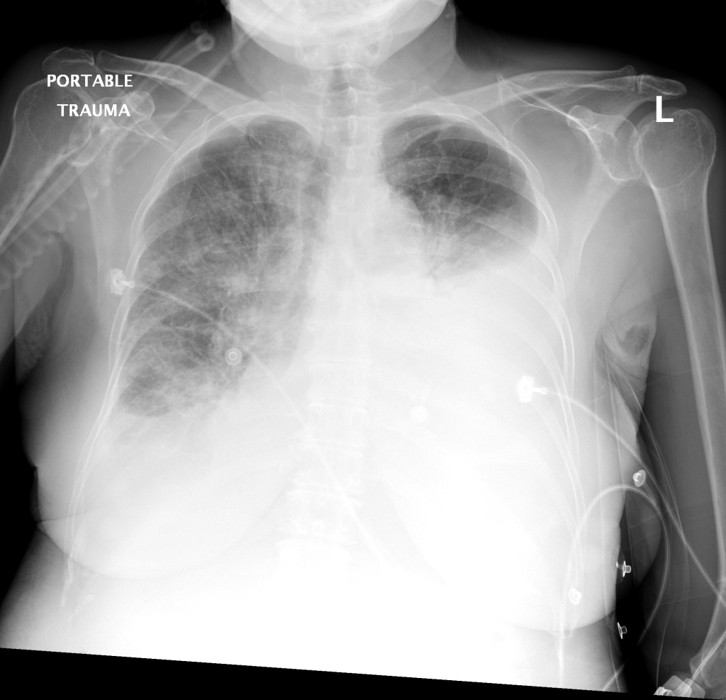
    CXR: Pleural Effusion

    There is a large left pleural effusion obscuring the lower half of the left hemi thorax. The cardiac silhouette is also obscured. There is pulmonary venous vascular congestion. There is also a right pleural effusion with fluid tracking into the minor fissure. Pulmonary interstitial edema is also noted.

    CT Chest (High-Resolution):

    • Bilateral, left greater than right, pleural effusions with adjacent atelectasis and collapse versus consolidation of the left lower lobe.
    • Prominent main pulmonary artery measuring 3.3 cm in diameter, which can be seen with pulmonary arterial hypertension.

    TTE:

    • LVEF is 50-55%.
    • Impaired left ventricular relaxation, which is associated with grade I/IV or mild diastolic dysfunction.
    • Moderate aortic stenosis with mild regurgitation (AVA 1.4 cm3, mean gradient 14mmHg, peak velocity 2.4 m/s).
    • Severe pulmonary hypertension (est PASP 52-62mmHg).
    • The inferior vena cava appeared dilated and decreased <50% with respiration (RAP 10-20 mmHg).
    • Minimal pericardial effusion without echocardiographic evidence of tamponade.

    Assessment/Plan:

    64F with history of CAD (prior MI), CHF, hypertension, CLL, hypothyroidism presented from a SNF with progressive shortness of breath, orthopnea and LE swelling, found to have bilateral (L>R) pleural effusion now s/p thoracentesis with transudative fluid.

    #Acute hypoxic respiratory failure: Large pleural effusions, s/p thoracentesis with pleural fluid suggestive of transudative process. Most likely secondary to left ventricular diastolic dysfunction. Improved after thoracentesis and diuresis. High-resolution CT chest performed without evidence of autoimmune-related pulmonary fibrosis or ILD (though persistent pleural effusions, pulmonary vascular congestion).

    #Pleural fluid signet cells: Identified on cytology, potentially related to history of untreated CLL or alternative primary malignancy.

    #Left ventricular diastolic dysfunction, decompensated: Associated with pleural effusions and hypoxemic respiratory failure. Management with diuresis.

    #Pulmonary Hypertension: Severe, noted on transthoracic echocardiography, may be secondary to hypoxemia associated with pleural effusions, consider repeat imaging once euvolemic or right-heart catheterization.

    #Microscopic Hematuria: No evidence of infection, no symptoms suggestive of nephrolithiasis. No casts identified or significant proteinuria. Plan for renal ultrasound.

    #Rheumatoid Arthritis: History of rheumatoid arthritis, on prednisone and hydroxychloroquine. Imaging without evidence of inflammatory arthropathy, RF elevated but CCP negative. Per rheumatology, the patient’s symptoms are not consistent with RA, continuing home medications while evaluation is ongoing. Pleural effusions unlikely associated with RA as transudative, and without monocyte predominance or low glucose.

    Case 2: Malignant Pleural Effusion

    malig_01
    malig_02
    malig_03
    malig_04
    malig_05
    malig_06
    malig_07
    malig_08
    malig_09
    malig_10
    malig_11
    malig_12
    malig_13
    malig_14
    Within the lungs there are ground-glass opacities bilaterally, and a left pleural effusion with adjacent consolidation vs compressive atelectasis.
    • Protein: 2.6 (serum: 4.9)
    • LDH: 1275 (serum: 219)
    • Cytology: Malignant cells

    Case 3: Traumatic Thoracentesis

    trauma_01
    trauma_02
    trauma_03
    trauma_04
    trauma_05
    trauma_06
    trauma_07
    trauma_08
    trauma_09
    trauma_10
    trauma_11
    trauma_12
    trauma_13
    trauma_14
    trauma_15
    trauma_16
    trauma_17
    trauma_18
    trauma_19
    Moderate right pleural effusion, some fluid in non-dependent portions suggestive of loculation. Diffuse nodules and opacification in right lung with compressive atelectasis. Left pleural effusion with high density material at the posterior costophrenic angle. Left chest tube.
    • Protein: 2.7 (serum 6.4)
    • LDH: 344 (serum 236)
    • Cell count: 100,000 RBC

    Case 4: Pneumonia

    pna_01
    pna_02
    pna_03
    pna_04
    pna_05
    pna_06
    pna_07
    pna_08
    pna_09
    pna_10
    pna_11
    pna_12
    pna_13
    pna_14
    pna_15
    Loculated right pleural effusion with foci of atelectasis and consolidative changes concerning for pneumonia. Minimal left-sided pleural effusion with consolidative changes. Enlarged mediastinal lymph nodes, possibly reactive.
    • Protein: 4.3 (serum 6.7)
    • LDH: 377 (serum 108)
    • pH: 7.46
    • Glucose: 153
    • Neutrophils: 84%

    Etiology of Pleural Effusions: 1

    Etiology Frequency (%)
    CHF 35
    Pneumonia 22
    Malignancy 15
    Pulmonary Embolism 11

    Clinical Features in the Diagnosis of Pleural Effusions and Identifying Etiology: 1,2

    Pleural effusions can be easily identified on chest radiography, physical examination findings include dullness to percussion, decreased tactile fremitus and decreased (or absent) breath sounds.

    • Hemoptysis: Malignancy, PE, TB
    • Weight Loss: Malignancy, TB
    • Ascites: Cirrhosis, ovarian cancer
    • Unilateral Leg Swelling: PE
    • Bilateral Leg Swelling: CHF, cirrhosis, nephrotic syndrome
    • Jugular Venous Distension: CHF

    Differential Diagnosis of Pleural Effusions: 1,2,3,4

    Differential Diagnosis of Pleural Effusions

    References:

    1. Light, R. W. (2002). Clinical practice. Pleural effusion. The New England journal of medicine, 346(25), 1971–1977. doi:10.1056/NEJMcp010731
    2. McGrath, E. E., & Anderson, P. B. (2011). Diagnosis of pleural effusion: a systematic approach. American journal of critical care : an official publication, American Association of Critical-Care Nurses, 20(2), 119–27– quiz 128. doi:10.4037/ajcc2011685
    3. Thomsen, T. W., DeLaPena, J., & Setnik, G. S. (2006). Videos in clinical medicine. Thoracentesis. The New England journal of medicine (Vol. 355, p. e16). doi:10.1056/NEJMvcm053812
    4. Wilcox, M. E., Chong, C. A. K. Y., Stanbrook, M. B., Tricco, A. C., Wong, C., & Straus, S. E. (2014). Does this patient have an exudative pleural effusion? The Rational Clinical Examination systematic review. JAMA : the journal of the American Medical Association, 311(23), 2422–2431. doi:10.1001/jama.2014.5552
    5. WikEM: Pleural effusion

    Lower Extremity Edema

    HPI:

    51 year-old male with a history of HTN, DM and chronic alcohol abuse presenting with lower extremity swelling. He notes one month of progressive, bilateral lower extremity swelling, in the past two weeks associated with increasing pain and redness and is now no longer able to ambulate due to pain. He denies fevers/chills, chest pain or shortness of breath. He also denies orthopnea and paroxysmal nocturnal dyspnea. He states that he has not had these symptoms prior to one month ago. On review of systems he denies nausea/vomiting, abdominal pain, and changes in bowel or urinary habits. He has a history of GI bleeding (unknown treatment) but denies hematemesis, hematochezia or melena. He has previously experienced alcohol withdrawal, which manifested as tremors, but no hallucinations or seizures.

    PMH:

    • HTN
    • DM
    • Chronic EtOH abuse

    PSH:

    None

    FH:

    Unknown

    SHx:

    • Drinks 1-2 pints of alcohol daily, last drink this morning.
    • Denies current tobacco or drug abuse, no prior IVDA.

    Meds:

    None

    Allergies:

    NKDA

    Physical Exam:

    VS: T 37.6 HR 86 RR 16 BP 128/84 O2 99% RA
    Gen: Adult, non-obese male, lying in bed. Tremors noted in upper extremities.
    HEENT: PERRL, EOMI, no scleral icterus. Mucous membranes moist.
    CV: RRR, normal S1/S2, no additional heart sounds, JVP 3cm above sternal angle at 30°.
    Lungs: CTAB, no crackles.
    Abd: Soft, non-distended, with normoactive bowel sounds. Liver edge palpated 1cm below costal margin at mid-clavicular line, non-tender. No rebound/guarding.
    Ext: Warm, well-perfused with 2+ distal pulses (PT, DP). 3+ pitting edema symmetric in bilateral lower extremities to knee. Erythema and warmth bilaterally extending from ankles to mid-shin. Mild tenderness to palpation. No pain with passive dorsiflexion. 3x3cm shallow ulceration below medial malleolus on right lower extremity without underlying fluctuance or expression of purulent material. No venous varicosities noted. Decreased sensation to light touch below knee bilaterally.
    Rectal: Normal rectal tone, brown stool, guaiac negative.
    Neuro: Alert and oriented, CN II-XII intact, gait intact, normal FTN/RAM.

    Labs/Studies:

    • CBC: 7.4/13.1/39/180
    • Creatinine: 0.84
    • Albumin: 4.3
    • BNP: 28

    Imaging:

    Venous Lower Extremity Ultrasound

    1. No DVT.
    2. Pulsatile flow in bilateral EIV (external iliac veins) suggestive of elevated right heart pressure.

    Assessment/Plan:

    51M with HTN, DM, EtOH abuse presenting with lower extremity edema. Chronic bilateral lower extremity edema likely secondary to chronic venous insufficiency perhaps related to OSA given ultrasound findings of pulsatile flow in EIV’s. Doubt systemic cause: no evidence of heart failure on exam and normal BNP, no stigmata of cirrhosis and normal albumin, normal creatinine. Also, no evidence of DVT on ultrasound though bilateral DVT unlikely. Bilateral cellulitis also unlikely as the patient is afebrile without leukocytosis, however the patient was started on antibiotics including ceftriaxone and TMP/SMX given erythema, warmth and tenderness to palpation. The patient received benzodiazepines which eased withdrawal symptoms and he was admitted for continued treatment.

    Mechanisms of Lower Extremity Edema: 1

    Mechanisms of Lower Extremity Edema

    Differential Diagnosis of Lower Extremity Edema: 1,2

    Differential Diagnosis of Lower Extremity Edema

    Evaluation:

    History 1,2

    • Duration: acute (<72h) vs. chronic
    • Pain: DVT, CRPS, less severe in venous insufficiency
    • Systemic Disease
      • Cardiac: orthopnea, PND
      • Renal: proteinuria
      • Hepatic: jaundice, ascites
    • Malignancy: lymphedema
    • Improvement with elevation/recumbency: venous insufficiency
    • OSA: snoring, daytime somnolence
    • Medications: B-blocker, CCB, hormones, NSAID’s

    Physical Exam 1,2

    • Distribution: unilateral, bilateral, generalized
    • Quality: pitting, non-pitting
    • TTP: DVT, cellulitis
    • Varicose veins: venous insufficiency
    • Kaposi-Stemmer: inability to pinch dorsum of foot at base of 2nd toe (lymphedema)
    • Systemic Disease
      • Cardiac: JVD, crackles
      • Hepatic: ascites, scleral icterus, spider angiomas
    • Brawny, medial maleolar involvement: venous insufficiency

    Key Features Distinguishing Cellulitis: 3

    • Typically unilateral and acute
    • Often with systemic symptoms (fever, leukocytosis)
    • Risk Factors: immunosuppression, previous episodes, DM, PVD

    References:

    1. Trayes, K. P., Studdiford, J. S., Pickle, S., & Tully, A. S. (2013). Edema: diagnosis and management. American family physician, 88(2), 102–110.
    2. Ely, J. W., Osheroff, J. A., Chambliss, M. L., & Ebell, M. H. (2006). Approach to leg edema of unclear etiology. Journal of the American Board of Family Medicine : JABFM, 19(2), 148–160.
    3. Keller, E. C., Tomecki, K. J., & Alraies, M. C. (2012). Distinguishing cellulitis from its mimics. Cleveland Clinic journal of medicine, 79(8), 547–552. doi:10.3949/ccjm.79a.11121
    4. WikEM: Pedal edema

    Quick Case: Pleuritic Chest Pain

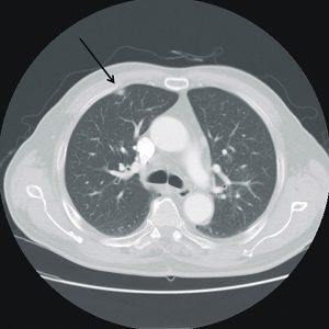
    Image from: Maeng, C. H., Chin, S. O., Yang, B. H., Kim, S.-Y., Youn, H.-J., Cho, K. S., Baek, S. K., et al. (2007). A case of organizing pneumonia associated with rituximab. Cancer research and treatment : official journal of Korean Cancer Association, 39(2), 88–91. doi:10.4143/crt.2007.39.2.88

    30yo male presenting with forearm cellulitis, also complaining of right-sided sharp chest pain worse with deep inspiration and some movements of the ipsilateral shoulder. Found to have multiple pulmonary nodules (suggestive of metastasis) with the largest being a subpleural nodule in the superior/anterior portion of the RUL (roughly the location of the patient’s pain).


    Differential Diagnosis of Pleuritic Chest Pain

    Causes of Pleuritic CP

    Location of Referred Pain

    Referred Pain


    Syncope

    ID:

    A 50 year-old male with a reported two-year history of infrequent spells, presenting with two spells in the past two days.

    HPI:

    The patient’s spells began two years ago, he recounts that he was watching television when he lost consciousness and a friend noted he started shaking; he does not recall the event, and awoke in the hospital. The next spell occurred one year later, though the patient is unable to recall much about this episode. The patient remained spell-free until yesterday when he was on a bus, lost consciousness and awoke in a hospital. He notes that he had bit his tongue and lost control of his bladder. He was discharged hours later with a prescription for an AED which he was unable to fill. This morning, the patient had another spell while in the bathroom. His roommate heard him fall, found him on the ground, and noted that his mouth was moving but did not see any other movements.

    The patient’s episodes are all associated with loss of consciousness and are followed by 5-10 minutes of disorientation after which he recovers fully. The episodes are sometimes preceded by a feeling of “euphoria”, though this feeling sometimes occurs without subsequent LOC.

    The patient denies any associated palpitations, dizziness/LH, chest pain or muscle pain.  He has not had any recent fevers/chills, dysuria, cough, headache, changes in vision, numbness/tingling, weakness, difficulty speaking or swallowing or weight loss. He also denies any history of head trauma.

    Physical Examination:

    • VS: Stable and WNL
    • General: Well-appearing, pleasant, and in NAD.
    • HEENT: NC/AT. MMM. Small lesion on tongue.
    • Lungs: CTAB.
    • CV: RRR with occasional ectopic beats, no M/R/G.
    • Abdomen: S/NT/ND. Bowel sounds present.
    • Neurological exam: AAOx4, CN II-XII intact, motor/sensation/reflexes/coordination/gait WNL

    Imaging/Studies:

    • EKG: Occasional PAC/PVC
    • CT Brain: Unremarkable except for mild age-related cerebral atrophy

    Assessment & Plan:

    50 year-old male with a history of HTN and a reported two-year history of infrequent spells presenting with two spells in the past two days. The description of the patient’s episodes could be consistent with seizures. Aspects supporting this notion include loss of consciousness and period of confusion following each episode. One of the recent episodes was also associated with tongue-biting and loss of bladder control. Additionally, some episodes are associated with a sensation of euphoria rising from the abdomen to the head which could be indicative of an aura. Characteristics that suggest other causes include the absence of noted convulsions and non-stereotyped nature of each episode which could be due to the patient’s poor recollection of these events and absence of reliable witnesses. In the case of true seizures, the possible etiologies in this patient include a mass, metabolic abnormalities, substance use, or concomitant infection exacerbating an existing propensity for seizure activity. Other, non-seizure causes warranting evaluation include cardiogenic syncope particularly given the evidence of ectopic beats on examination and electrocardiogram.

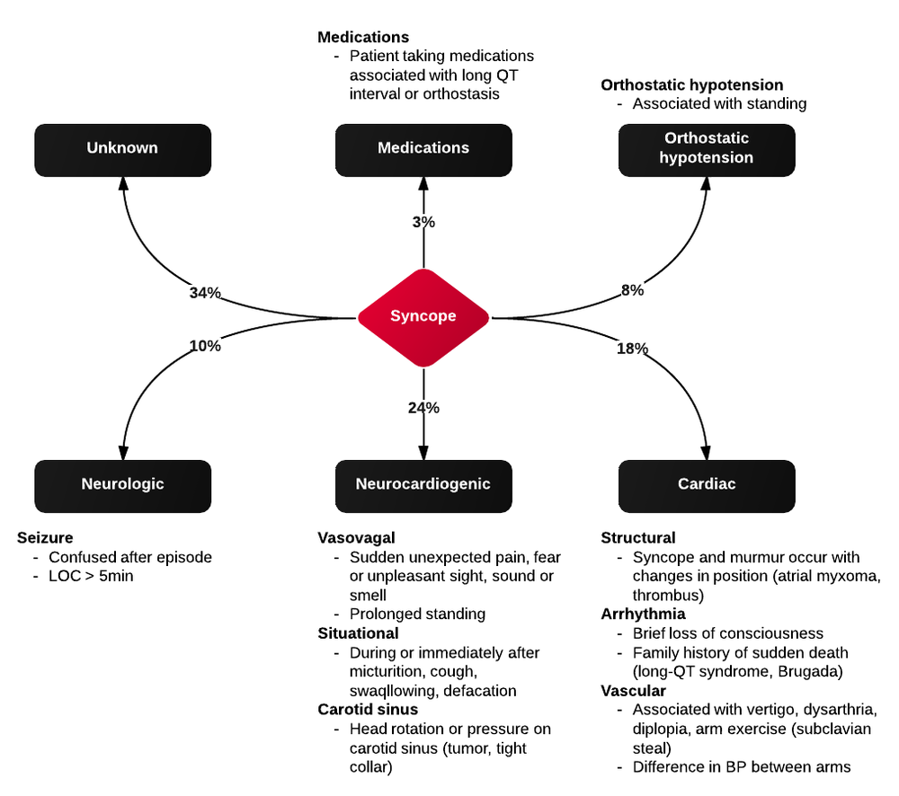
    Differential Diagnosis of Syncope

    First, is it syncope? History is very important for distinguishing syncope from other causes (seizure, dizziness, vertigo, presyncope). Ask about precipitating events, prodromal symptoms, post-ictal confusion. Common causes of syncope and their associated symptoms are detailed in the figure below.

    References:

    1. Kapoor, W. N. (2000). Syncope. The New England journal of medicine, 343(25), 1856–1862. doi:10.1056/NEJM200012213432507