Brief H&P:
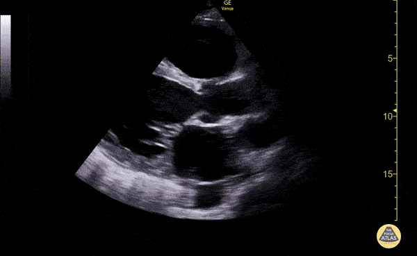
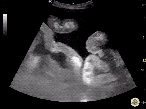
Depressed ejection fraction, image from The POCUS Atlas
An 44 year-old male with no reported medical history (though limited access to medical care) presents with lower extremity swelling. He states that the symptoms have been gradually worsening over the past 3 months. He notes occasional fatigue while at work but denies chest pain, shortness of breath, leg pain or changes in urination.
A point-of-care ultrasound is performed showing decreased left ventricular ejection fraction. The patient was admitted for further evaluation and management of new-onset congestive heart failure.
Algorithm for the Evaluation of Lower Extremity Edema with Ultrasound

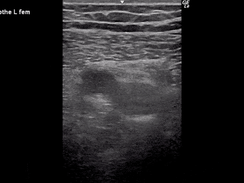
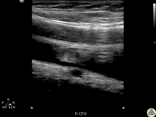
Gallery
The POCUS Atlas
The ultrasound images and videos used in this post come from The POCUS Atlas, a collaborative collection focusing on rare, exotic and perfectly captured ultrasound images.
The ultrasound images and videos used in this post come from The POCUS Atlas, a collaborative collection focusing on rare, exotic and perfectly captured ultrasound images.
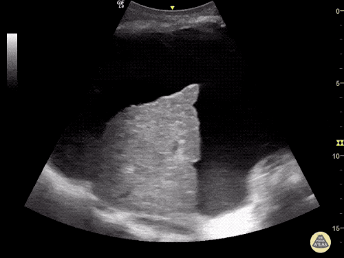
Nodular liver contour, ascites
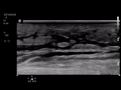
Ascites
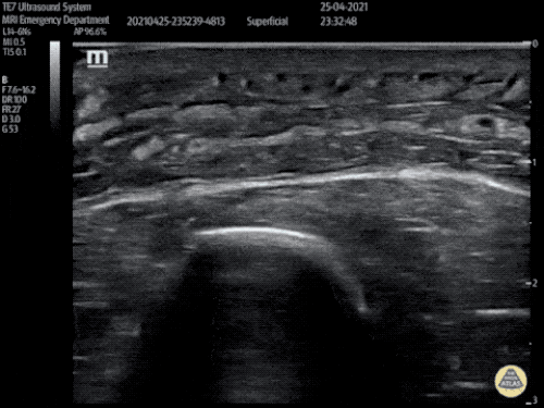
Cobblestoning
Cobblestoning
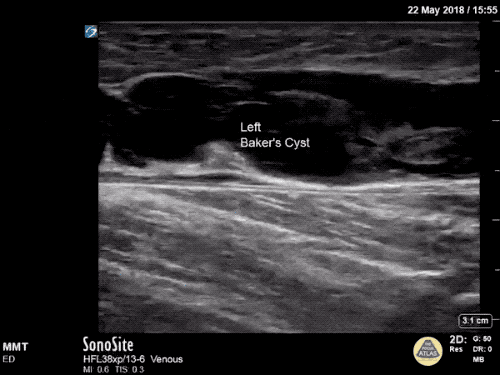
Longitudinal view of a ruptured Baker cyst
References
- Trayes KP, Studdiford JS, Pickle S, Tully AS. Edema: diagnosis and management. Am Fam Physician. 2013;88(2):102-110.
- Goyal A, Cusick AS, Bhutta BS. Peripheral Edema. [Updated 2022 Nov 19]. In: StatPearls [Internet]. Treasure Island (FL): StatPearls Publishing; 2022 Jan-. Available from: https://www.ncbi.nlm.nih.gov/books/NBK554452/
- Smith, C. Clinical manifestations and evaluation of edema in adults. Post TW, ed. UpToDate. Waltham, MA: UpToDate Inc. http://www.uptodate.com. Accessed 2/11/2023.
This algorithm was developed by Dr. Huakang Huang. Huakang is an emergency medicine resident at UTHealth Houston.