Brief H&P
A young patient with no past medical history is brought in by ambulance after a high-speed motor vehicle accident. Trauma survey demonstrates absent motor/sensation in bilateral lower extremities with sensory level at T3-T4. Computed tomography of the cervical spine was obtained and is shown below.
Imaging
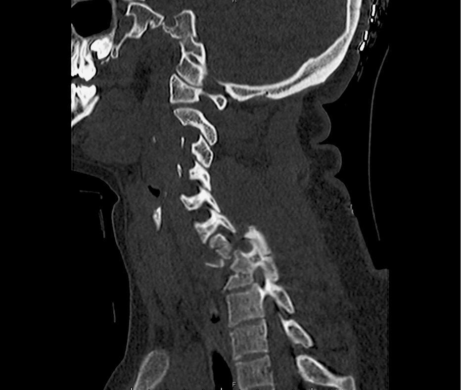
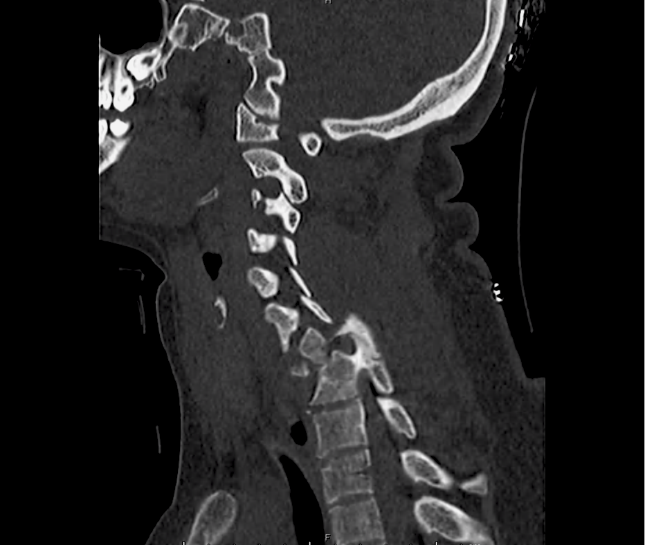
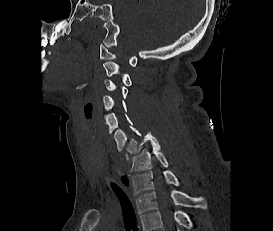
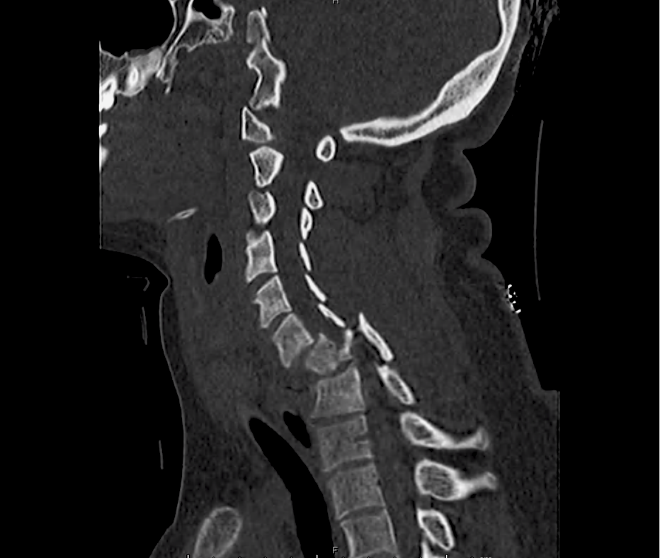
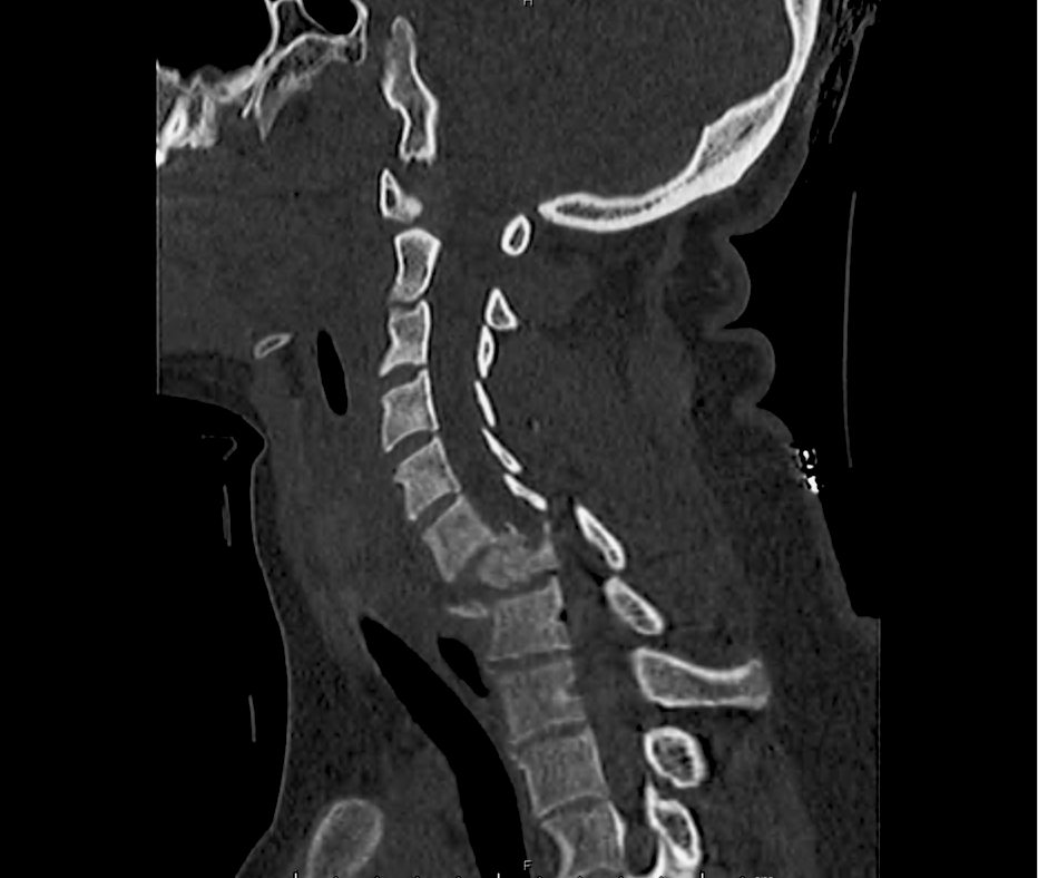
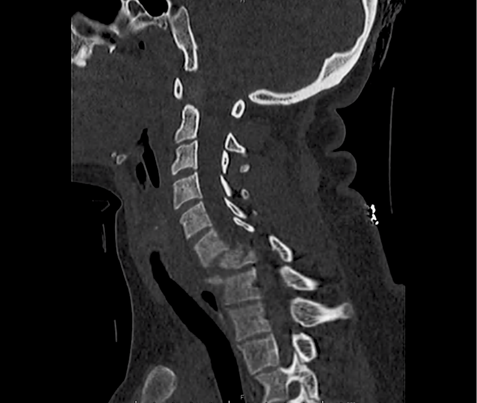
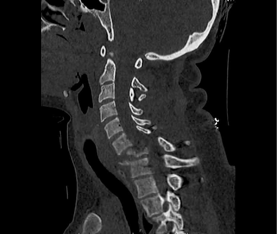
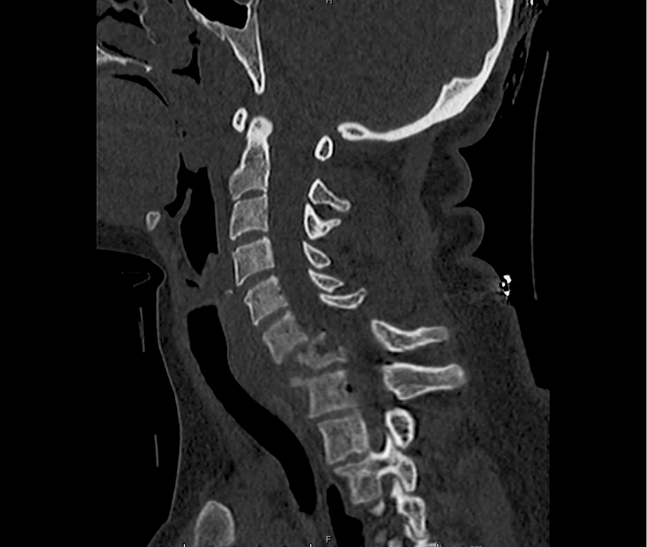
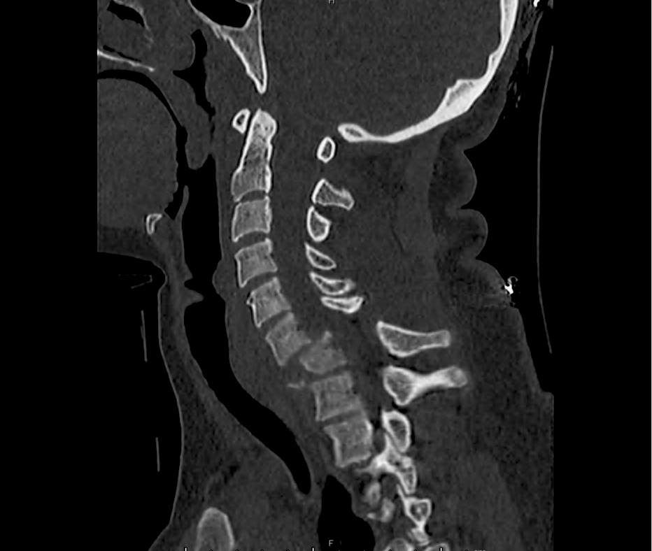
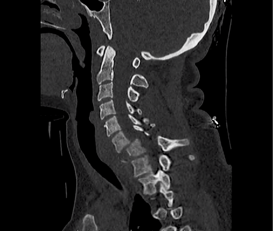
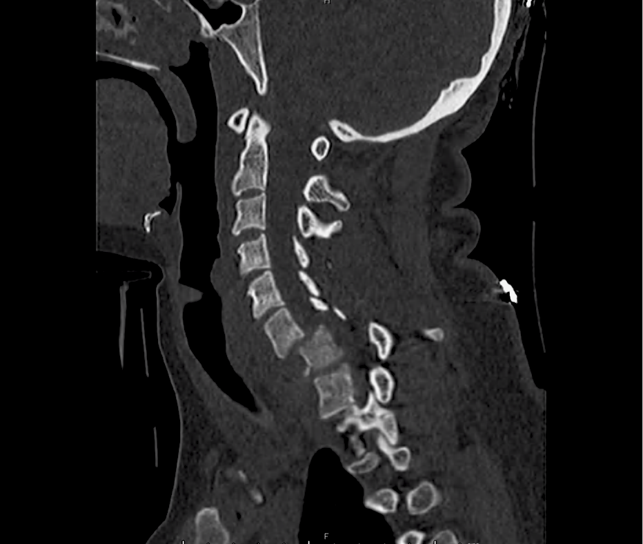
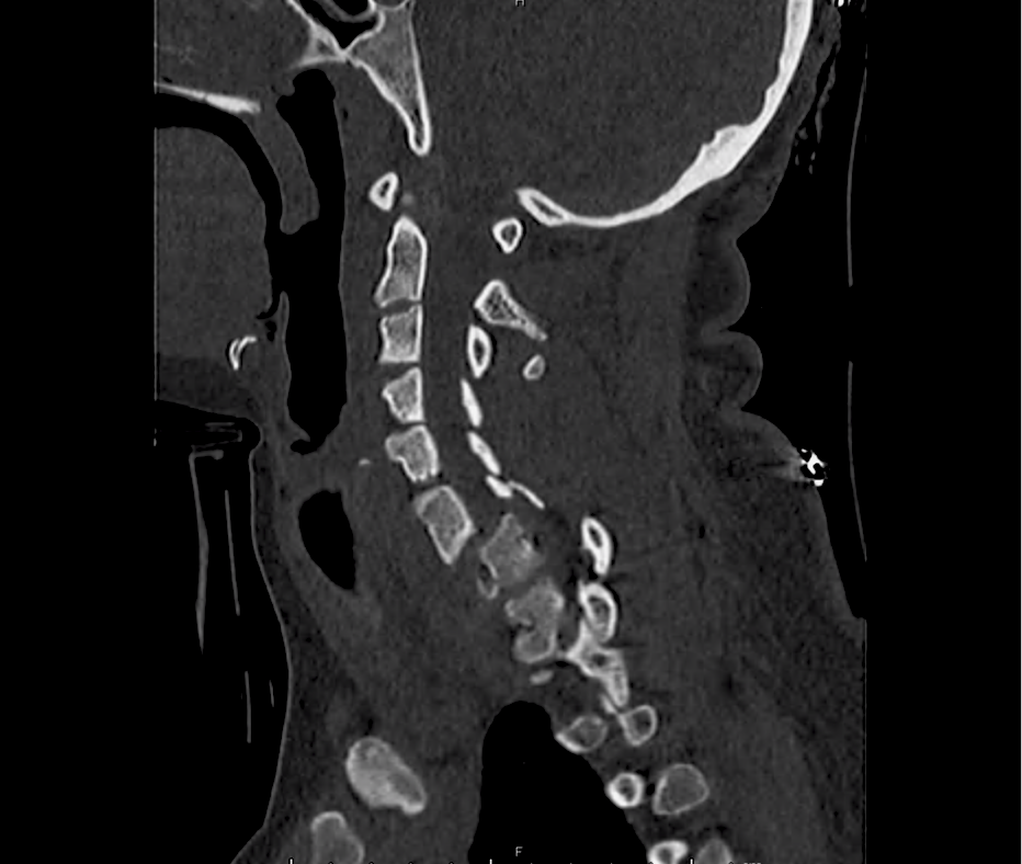
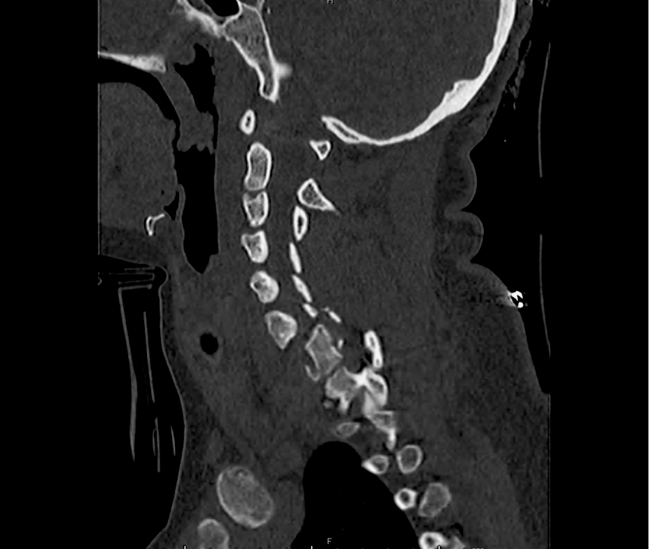
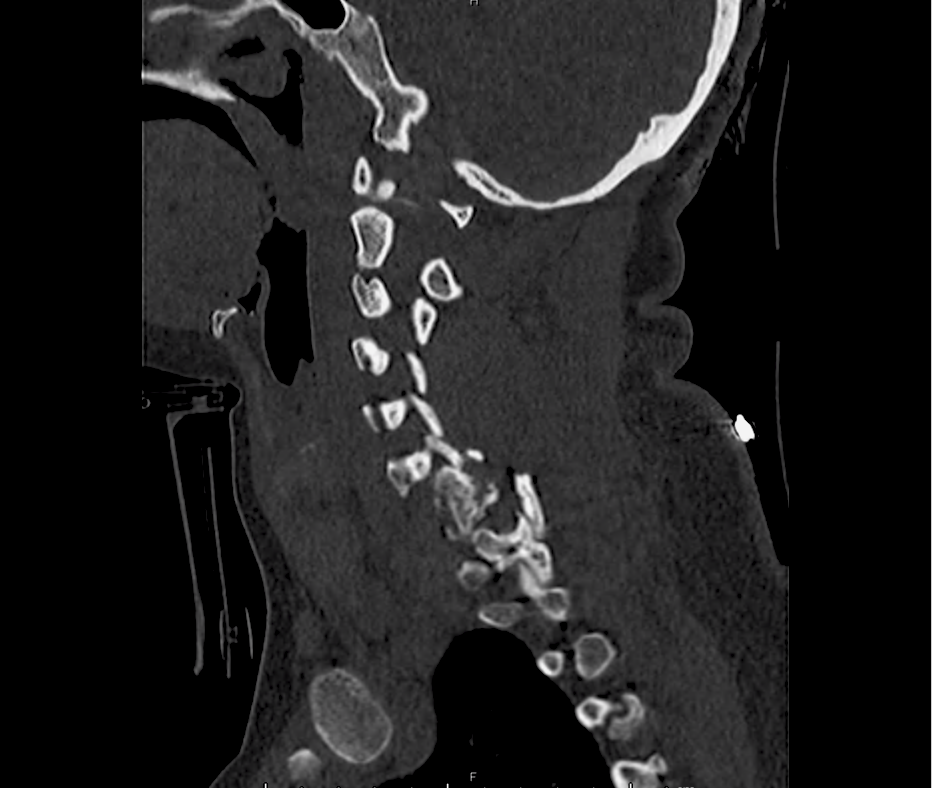
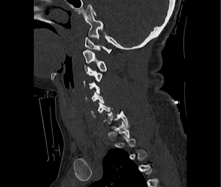
CT C-Spine
Fracture-dislocation at C6-C7 and C7-T1 with comminuted burst fracture to C7 and locked facet joint with resultant anterior migration of C6 over C7, unstable cervical spine fracture.
Anatomy
Flexion
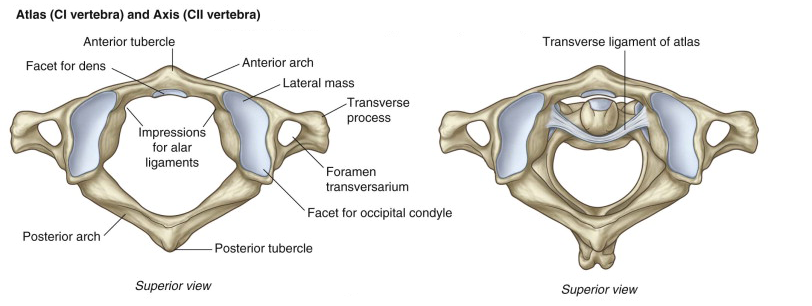
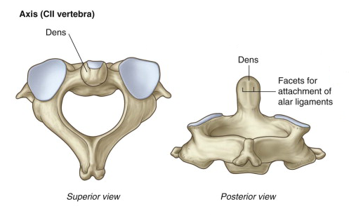
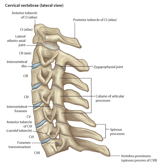
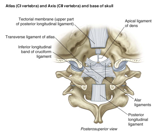
C1/C2
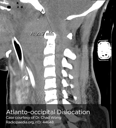
- Atlanto-occipital dislocation , atlantoaxial dislocations , potentially associated with odontoid fracture.
- Imaging: Basion-dens interval (BDI) >10mm,
- Stability: Unstable.
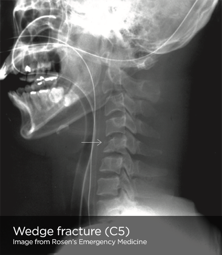
Wedge fracture
- Stretch on strong nuchal ligament transmits force to vertebral body.
- Stability: Generally stable unless >50% compression or multiple contiguous.
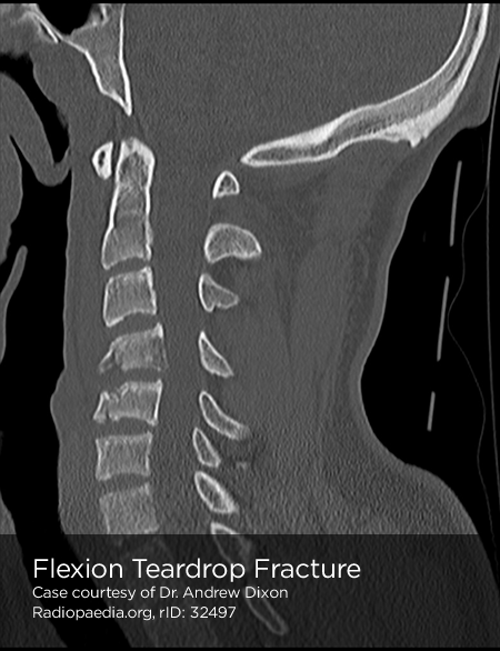
Flexion-teardrop fracture
- Severe flexion force, avulsion of fragment of anterior/inferior portion of vertebral body.
- Stability: Unstable, involves anterior/posterior ligamentous disruptions.
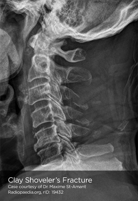
Clay shoveler’s fracture
- Oblique fracture of spinous process of lower cervical spine.
- Stability: Stable
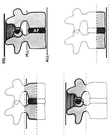
Subluxation
- Pure ligamentous injury without associated fracture.
- Imaging: Widening of interspinous and intervertebral spaces on lateral.
- Stability: Potentially unstable.
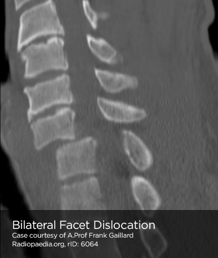
Bilateral facet dislocation
- Anterior displacement of spine above level of injury caused by dislocation of upper inferior facet from lower superior facet.
- Imaging: Anterior displacement greater than ½ AP diameter of vertebral body.
- Stability: Unstable
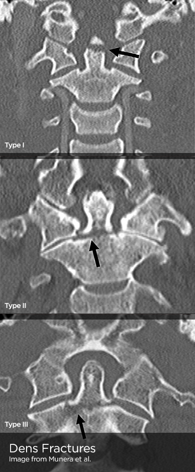
Odontoid process fracture
- Head trauma with shear force directed at odontoid.
- Sub-classification: Type I (above transverse ligament), type II (odontoid base), type III (extension to body of C2)
- Stability: Types II, III unstable.
Flexion/Rotation
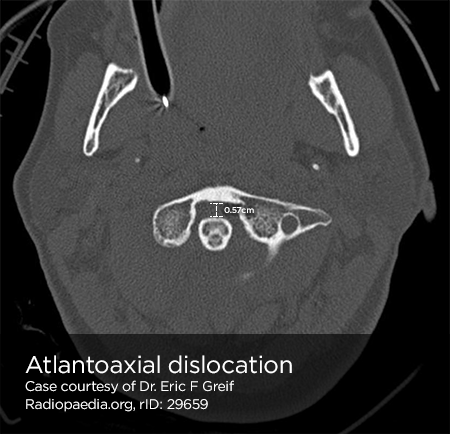
Rotary atlantoaxial dislocation
- Imaging: Open-mouth odontoid, asymmetric lateral masses of C1.
- Stability: Unstable
Unilateral facet dislocation
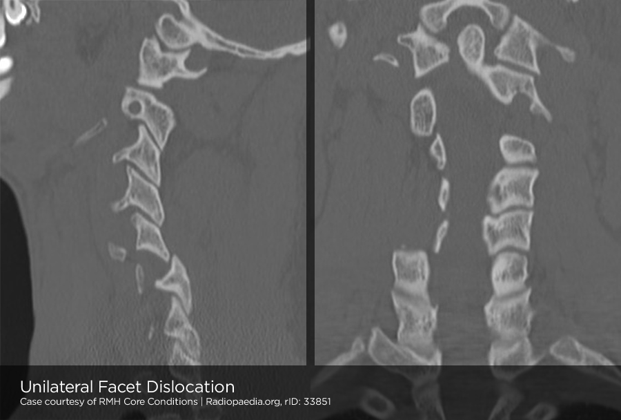
- Flexion and rotation centered around single facet results in contralateral facet dislocation.
- Imaging: AP radiograph shows spinous processes above dislocation displaced from midline, lateral radiograph shows anterior displacement of lower vertebra (less than ½ AP diameter of vertebral body).
Extension
Posterior neural arch fracture (C1)
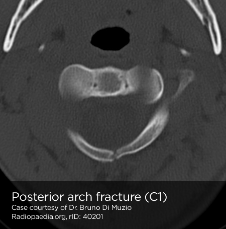
- Forced extension causes compressive force on posterior elements of C1 between occiput and C2.
- Stability: Unstable
Hangman’s fracture (spondylolysis C2)
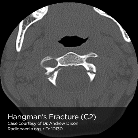
- Abrupt deceleration causes fracture of bilateral pedicles of C2, potentially with associated subluxation. Rarely associated with SCI due to large diameter of neural canal at C2.
- Imaging: May be associated with retropharyngeal space edema.
- Stability: Unstable
Extension-teardrop fracture
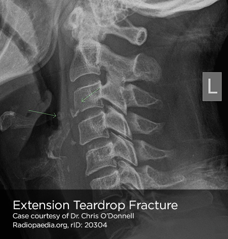
- Abrupt extension (ex. diving) results in stretch along anterior longitudinal ligament with avulsion of anterior/inferior fragment of vertebral body (usually C5-C7).
- Imaging: May be radiographically similar to flexion-teardrop fracture.
- Complications: Central cord syndrome
- Stability: Unstable in extension
Vertical compression
Burst fracture
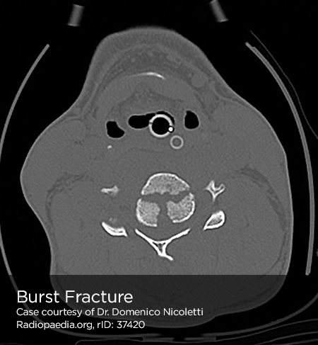
- Force applied from above or below causes transmission of force to intervertebral disc and vertebral body.
- Imaging: Comminuted vertebral body, >40% compression of anterior vertebral body.
- Complications: Fracture fragments may impinge on spinal cord.
- Stability: Stable
Jefferson fracture (C1)
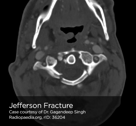
- Vertical force transmitted from occipital condyles to superior articular facets of atlas, resulting in fractures of anterior and posterior arches.
- Imaging: Widening of predental space. Open-mouth odontoid view may reveal bilateral offset distance of >7mm between lateral masses of C1/C2.
- Stability: Unstable
Cervical Spine Imaging Decision Rule (Canadian)

References:
- MD RK, MD BED, CAQ-SM KHM, MD WF. Emergency Department Evaluation and Treatment of Cervical Spine Injuries. Emergency Medicine Clinics of NA. 2015;33(2):241-282. doi:10.1016/j.emc.2014.12.002.
- Denis F. Spinal instability as defined by the three-column spine concept in acute spinal trauma. Clin Orthop Relat Res. 1984;(189):65-76.
- Munera F, Rivas LA, Nunez DB, Quencer RM. Imaging evaluation of adult spinal injuries: emphasis on multidetector CT in cervical spine trauma. Radiology. 2012;263(3):645-660. doi:10.1148/radiol.12110526.