HPI:
42M with 1.5 weeks of fevers. Initially presented to ER 1wk ago and treated for possible otitis media, however follow-up ENT appointment showed no evidence of OM on exam. Fevers persisted and he developed headaches and went to urgent care where a CT head and LP were negative. A mild elevation of serum transaminases was noted and the following CT abdomen/pelvis was obtained. He denied GI symptoms.
Imaging:

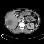
Hepatic Abscess - Axial
- 7.4 cm cystic lesion in the inferior right lobe of the liver most consistent in appearance with hepatic abscess.
- Multiple calcified
gallstones with a 10 mm gallstone in the neck of the gallbladder or possibly in the cystic duct.
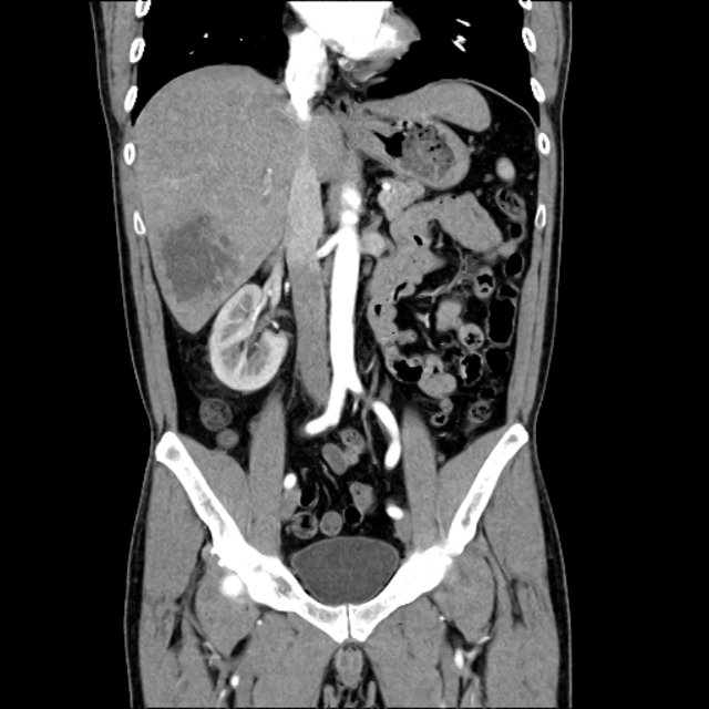
Hepatic Abscess - Coronal
- 7.4 cm cystic lesion in the inferior right lobe of the liver most consistent in appearance with hepatic abscess.
- Multiple calcified
gallstones with a 10 mm gallstone in the neck of the gallbladder or possibly in the cystic duct.
Assessment & Plan:
# Liver abscess: likely pyogenic s/p CT-guided drainage with 60cc purulent fluid removed. Gram stain showed GNR and WBC’s, culture grew Klebsiella pneumonia. Treated with ceftriaxone 2g IV q24h, metronidazole 500mg PO TID.
Differential Diagnosis of Hepatic Abscess:1

References:
- Krige J, Beckingham IJ. Liver abscesses and hydatid disease. BMJ. 2001;322(7285):537–540.