Fever

Causes of Fever

Causes of Fever

Key Features

  • Morbidity and mortality increase with age and comborbidities
  • Most common sources in elderly: respiratory, genitourinary, skin/soft-tissue
  • Atypical presentations: functional decline, altered mental status

Immediate Evaluation and Management

  • Critical Findings

    • Altered mental status
    • Respiratory distress
    • Hemodynamic instability
  • Critical Interventions

    • Airway management, supplemental O2
    • Cardiac monitoring
    • Fluid resuscitation
    • Empiric antibiotics
    • Cooling measures (T>41.0°C)

Pathophysiology of Fever

Production of endogenous or exogenous pyrogens
Increase temperature set point in hypothalamus
Patient experiences chills when core temperature < set point
Vasoconstriction, shivering causes fever
Patient experiences euthermia, though may feel malaise, fatigue
Resolution
Patient experiences sweats until core temperature returns to normal set point

References

  1. Blum, F., & Biros, M. (2013). Fever in the Adult Patient. In Rosen’s Emergency Medicine – Concepts and Clinical Practice (8th ed., Vol. 1, pp. 119-123). Elsevier Health Sciences.

Weakness

View Algorithm
There is a ddxof algorithm for the evaluation of weakness. View it here.

Motor Neuron Signs

Upper Motor Neuron:
Spasticity
Hyperreflexia
Pronator drift
Babinski
Lower Motor Neuron:
Flaccidity
Hyporeflexia
Fasciculation
Atrophy

Causes of Weakness

Lesion Critical Emergent
Non-neurological Shock (VS, clinical assessment)
Hypoglycemia (POC glucose)
Electrolyte derangement (BMP)
Anemia (POC Hb, CBC)
MI (ECG, troponin)
CNS depression (Utox, EtOH)
 
Cortex Stroke Tumor
Abscess
Demyelination
Brainstem Stroke Demyelination
Spinal Cord Ischemia
Compression (disk, abscess, hematoma)
Demyelination (transverse myelitis)
Peripheral Acute demyelination (GBS) Compressive plexopathy
Muscle Rhabdomyolysis Inflammatory myositis

Weakness Syndromes

Unilateral weakness, ipsilateral face
Lesion: Contralateral cortex, internal capsule
Causes: Stroke (sudden onset), demyelination/mass (gradual onset)
Symptoms: Neglect, visual field cut, aphasia
Findings: UMN signs
Key features: Association with headache suggests hemorrhage or mass
Unilateral weakness, contralateral face
Lesion: Brainstem
Causes: Vertebrobasilar insufficiency, demyelination
Symptoms: Dysphagia, dysarthria, diplopia, vertigo, nausea/vomiting
Findings: CN involvement, cerebellar abnormalities
Unilateral weakness, no facial involvement
Lesion: Contralateral medial cerebral cortex, discrete internal capsule
Causes: Stroke
Rare Cause: Brown-Sequard if contralateral hemibody pain and temperature sensory disturbance
Unilateral weakness single limb (monoparesis/plegia)
Lesion: Spinal cord, peripheral nerve, NMJ
UMN signs: Brown-Sequard if contralateral pain and temperature sensory disturbance
LMN signs: Radiculopathy if associated sensory disturbance
Normal reflexes, normal sensation: Consider NMJ disorder
Bilateral weakness of lower extremities (paraparesis/plegia)
Lesion: Spinal cord, peripheral nerve
UMN signs: Anterior cord syndrome (compression, ischemia, demyelination) if contralateral pain and temperature sensory disturbance
Cauda equina: Loss of perianal sensation, loss of rectal tone, or urinary retention
GBS: If no signs of cauda equina and sensory disturbances paralleling ascending weakness (with hyporeflexia)
Bilateral weakness of upper extremities
Lesion: Central cord syndrome
Causes: Syringomyelia, hyperextension injury
Findings: Pain and temperature sensory disturbances in upper extremities (intact proprioception)
Bilateral weakness of all four extremities (quadriparesis/plegia)
Lesion: Cervical spinal cord
Findings: UMN signs below level of injury, strength/sensory testing identifies level
Bilateral weakness, proximal groups
Lesion: Muscle
Causes: Rhabdomyolysis, polymyositis, dermatomyositis, myopathies
Findings: Muscle tenderness to palpation, no UMN signs, no sensory disturbances
Facial weakness, upper and lower face
Lesion: CNVII
Causes: Bell’s palsy, mastoiditis, parotitis
Other CN involvement suggests brainstem lesion, multiple cranial neuropathies, or NMJ

Review of Spinal Cord Anatomy

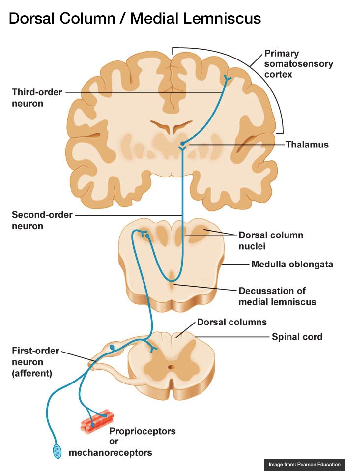
  • Dorsal Column – Medial Lemniscus (fine touch, proprioception)
    1. Afferent sensory fibers with cell body in DRG
    2. Ascend in ipsilateral posterior column
    3. Synapse in medulla, decussate, ascend in contralateral medial lemniscus
    4. Synapse in thalamus (VPL)
    5. Synapse in sensory strip of post-central gyrus
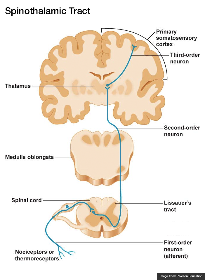
  • Spinothalamic Tract (pain, temperature)

    1. Afferent sensory fibers with cell body in DRG
    2. Ascends 1-2 levels
    3. Synapse in ipsilateral spinal cord, decussate, ascend in contralateral lateral spinothalamic tract
    4. Synapse in thalamus (VPL)
    5. Synapse in sensory strip of post-central gyrus
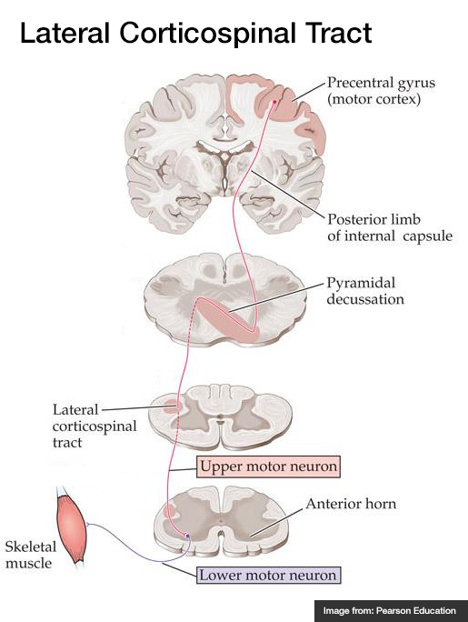
  • Lateral Corticospinal Tract (motor)

    1. Efferent cell body in motor strip of pre-central gyrus
    2. Descends through internal capsule
    3. Decussates in pyramid of medulla, descends in contralateral lateral corticospinal tract
    4. Synapse in anterior horn, lower motor neuron to muscle fiber
Spinal Cord Syndromes
Spinothalamic Tract
Dorsal Column / Medial Lemniscus
Lateral Corticospinal Tract

References

  1. Morchi, R. (2013). Weakness. In Rosen’s Emergency Medicine – Concepts and Clinical Practice (8th ed., Vol. 1, pp. 124-128). Elsevier Health Sciences.

Syncope

Causes of Syncope

Causes of Syncope

History

  • Rate of onset
  • Position at onset
  • Duration, rate of recovery
  • Preceding features

    • Obstruction: associated with exertion
    • Neurocardiogenic: associated with emotion, micturition, bowel movement, emesis, neck movement
  • Following features

    • Seizure: Postictal confusion
    • Hypotension: Initial VS
    • Associated trauma

Physical Examination

  • VS: rhythm, BP, temperature
  • HEENT: mucous membranes (laceration, dry), trauma, papilledema
  • CV: murmur (AS), rub (pericarditis), bruit (cerebrovascular disease), JVD (obstruction)
  • Lungs: crackles (CHF)
  • Abdomen: pulsatile mass (AAA)
  • Extremities: pulse discrepancy (dissection)
  • Neuro: focal findings (stroke, mass, seizure)

Evaluation

  • ECG: arrhythmia (PR, QT, Brugada, unanticipated hypertrophy, RV strain, pericarditis)
  • Orthostatic VS
  • CBC: anemia
  • BMP: electrolyte abnormalities (hyponatremia, hyper/hypokalemia)
  • Glucose: hypoglycemia
  • Troponin: ischemia
  • B-hCG: ectopic
  • Utox: drugs
  • CXR: dissection
  • CT head: focal neurological findings
  • CT PA: concern for PE
  • US abdomen: AAA

San Francisco Syncope Rules (CHESS)

  • CHF
  • Hematocrit <30%
  • ECG abnormality
  • SBP <90mmHg
  • SOB

References

  1. De Lorenzo, R. (2013). Syncope. In Rosen’s Emergency Medicine – Concepts and Clinical Practice (8th ed., Vol. 1, pp. 135-141). Elsevier Health Sciences.

Altered Mental Status

Components of Consciousness

Components of Consciousness

Causes of Altered Mental Status

Causes of Altered Mental Status

History

Rate of onset
Abrupt: CNS
Gradual: Systemic

Physical Examination

  • Vital Signs

    • Blood Pressure: low (shock), high (SAH, stroke, ICP)
    • Heart Rate: low (medication overdose, conduction block), high (hypovolemia, infection, anemia, thyrotoxicosis, drug/toxin)
    • Temperature: low/high (infection, drug/toxin, environmental)
    • Respiratory Rate: low/high (CNS, drug/toxin, metabolic derangement)
  • Eyes

    • Unilateral dilation: CNS/structural cause
    • Papilledema: ICP
    • EOM: cranial nerve dysfunction
    • Oculocephalic: brainstem function
  • Head: trauma
  • Mucous membranes: hydration, laceration
  • Neck: meningeal irritation
  • Pulmonary: respiratory effort
  • CV: murmur, arrhythmia, CO
  • Abdomen: pulsatile mass, sequelae of liver failure
  • Skin: rash, needle tracks

Labs

  • Glucose
  • ECG: arrhythmia, ischemia, electrolyte abnormalities
  • BMP: electrolytes, renal failure, anion gap
  • ABG: hypoxemia, hypercarbia
  • Urinalysis: infection, SG
  • Utox
  • CBC: leukocytosis, leukopenia, severe anemia, thrombocytopenia
  • Ammonia: hepatic encephalopathy
  • TFT: thyrotoxicosis, myxedema coma
  • CSF: meningitis, encephalitis

Imaging

  • CT head: Non-contrast sufficient to identify ICH. Use contrast if mass/infection suspected
  • CTA head/neck: If aneurysm, AVM, venous sinus thrombosis or vertebrobasilar insufficiency suspected
  • CXR: PNA

References

  1. Bassin, B., & Cooke, J. (2013). Depressed Consciousness and Coma. In Rosen’s Emergency Medicine – Concepts and Clinical Practice (8th ed., Vol. 1, pp. 142-150). Elsevier Health Sciences.

Seizure

Definition

Seizure
Pathologic neuronal activation leading to abnormal function
Epilepsy
Recurrent unprovoked seizures

Classification

  • Cause
    • Primary: Unprovoked
    • Secondary: Provoked, caused by trauma, illness, intoxication, metabolic disturbances, etc.
  • Effect on mentation
    • Generalized: involvement of both hemispheres with associated loss of consciousness (tonic-clonic, absence, atonic, myoclonic)
    • Focal: Involving single hemisphere with preserved level of consciousness
  • Status epilepticus
    • Witnessed convulsions lasting >5min
    • Recurrent seizure without recovery from postictal period

Causes of Seizures

Causes of Seizures

Management of Seizures

Management of Seizures

Medications for Treatment of Seizures

Medication Dose (adult) Dose (peds) Comment
1st Line
Lorazepam 4mg IV <13kg: 0.1mg/kg (max 2mg)
13-39kg: 2mg

>39kg: 4mg
Repeat in 10min
Midazolam 10mg IM 0.2mg/kg IM (max 5mg) Repeat in 10min
Midazolam 10mg buccal 0.5mg/kg buccal (max 5mg) Repeat in 10min
2nd Line
Fosphenytoin 20mg PE/kg IV    
Phenytoin 20mg/kg IV   May cause hypotension
3rd Line
Midazolam 0.05-2mg/kg/hr    
Propofol 1-2mg/kg bolus then 20-200mcg/kg/min    
Pentobarbital 5-15mg/kg bolus then 0.5-5mg/kg/hr    
Special Conditions
Glucose 50mL D50/W   Hypoglycemia
MgSO4 6g IV over 15min   Eclampsia (20wks gestation to 6wks post-partum)
Pyridoxine 0.5g/min until seizures stop, max 5g   INH ingestion
3% saline 100-200mL over 1-2h   Confirmed hyponatremia

History

Points suggestive of seizure over alternative process
Abrupt onset
Duration < 120s
LOC
Purposeless activity: automatisms, tonic-clonic
Provocation: fever in children, substance withdrawal
Postictal state
Retrograde amnesia
Incontinence, oral trauma (buccal maceration, tongue laceration)
Rapidly resolving lactic acidosis
Important historical points for patients with seizure history
Recent illness
Medications (adherence, changes, interactions)
Substance use
Ictogenic factors
Recent/remote head trauma
Developmental abnormalities
Substance use
Sleep deprivation
Pregnancy

Key Physical Examination Findings

  • Vital sign abnormalities persisting beyond immediate postictal state (may suggest drug/toxin exposure, CNS lesion)
  • Nuchal rigidity
  • Signs of IVDA
  • Sequela

    • Head trauma
    • Tongue laceration
    • Shoulder dislocation (posterior)
  • Neurological exam

    • Stroke
    • Elevated ICP
    • Failure to note improvement in postictal confusion (encephalopathy, subclinical seizures)

Labs

  • Glucose
  • BMP (Na, Ca, Mg)
  • AED levels
  • CBC (leukocytosis and bandemia common post-seizure)
  • CSF
  • B-hCG
  • LFT (hepatic dysfunction, alcoholic hepatitis)
  • Lactate (rapidly resolves on repeat)

Indications for Imaging

  • New seizures
  • History of trauma
  • History of malignancy
  • Immunocompromised
  • Headache
  • Anti-coagulation
  • Focal neurological exam
  • Persistent AMS

References

  1. McMullan, J., Davitt, A., & Pollack, C. (2013). Seizures. In Rosen’s Emergency Medicine – Concepts and Clinical Practice (8th ed., Vol. 1, pp. 156-161). Elsevier Health Sciences
  2. WikEM: Seizure

Headache

Brief HPI:

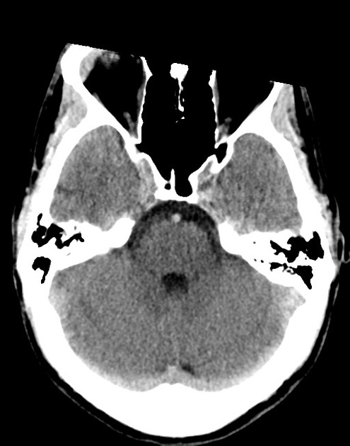
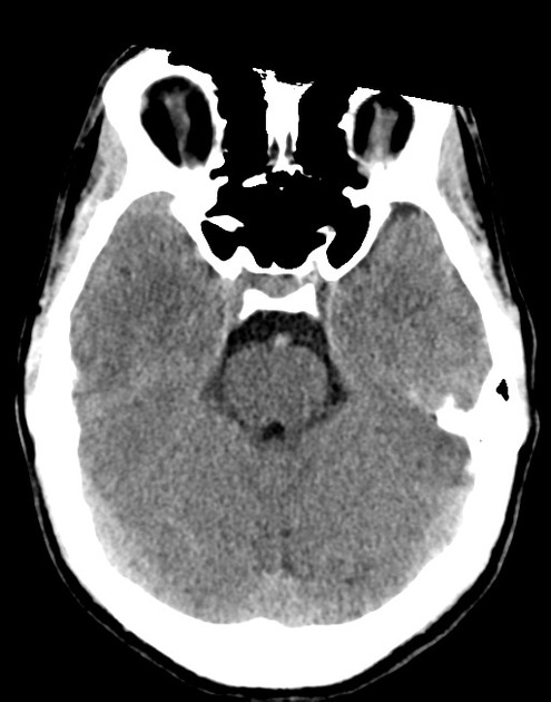
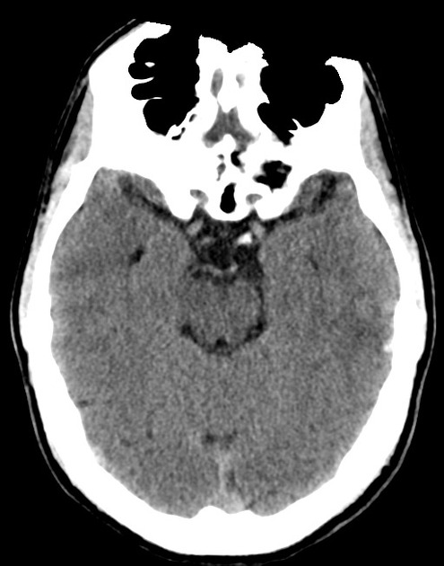
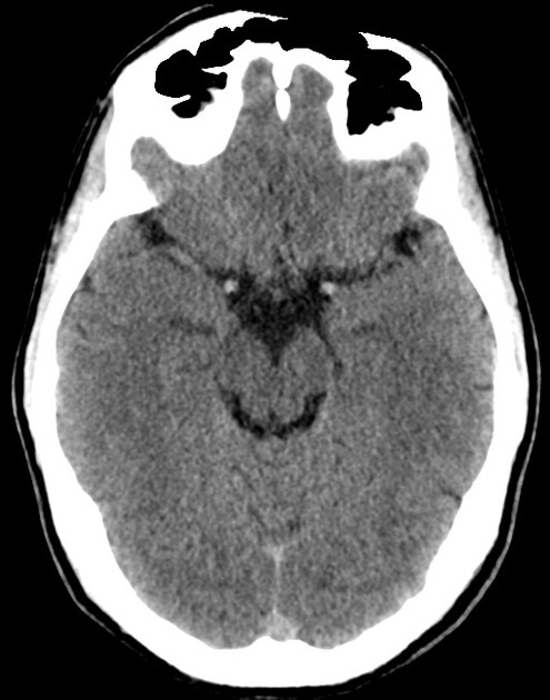
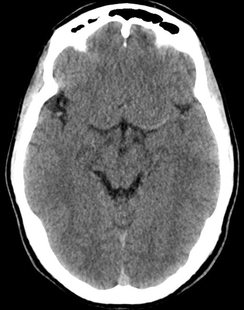
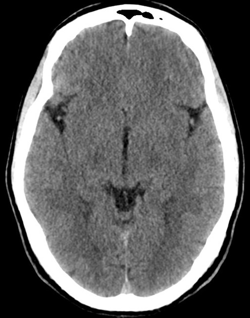
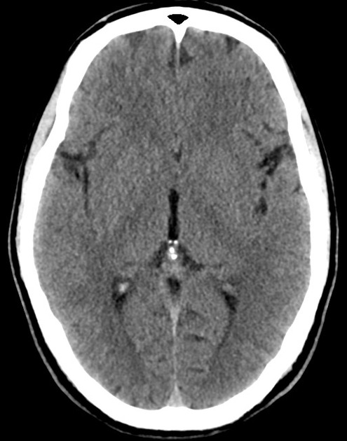
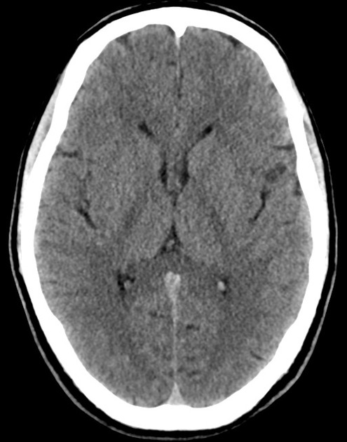
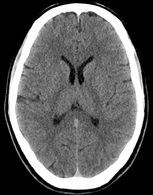
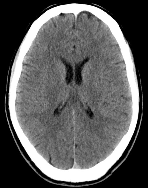
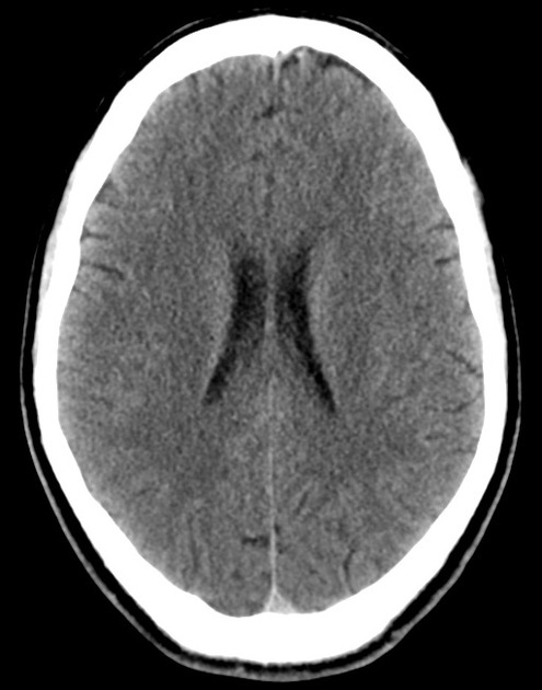
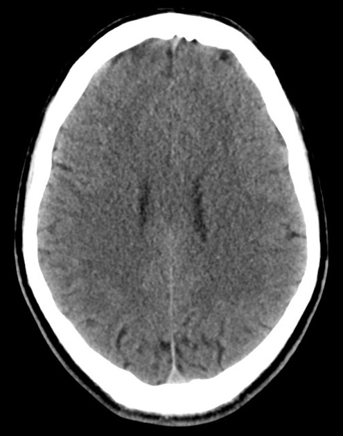
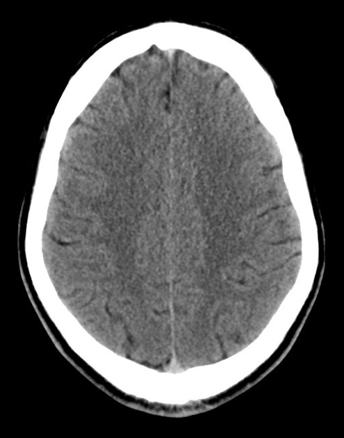
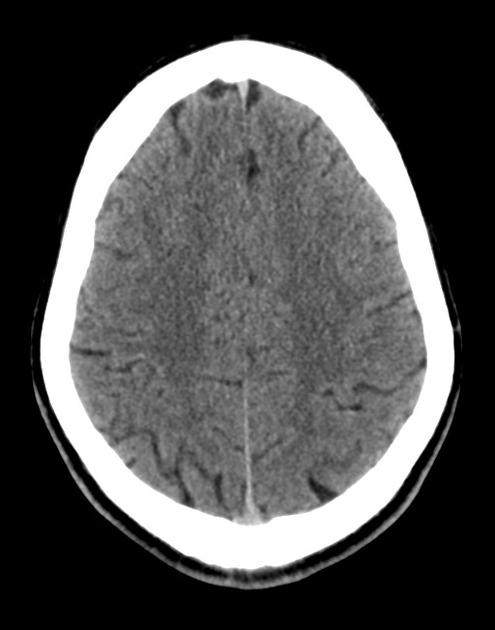
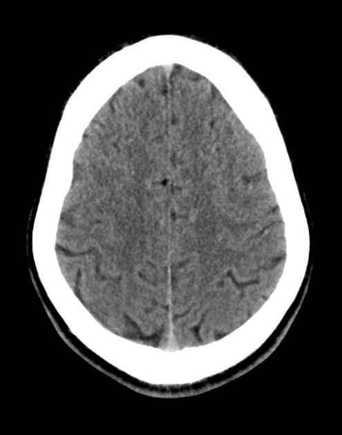
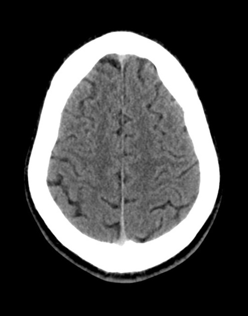
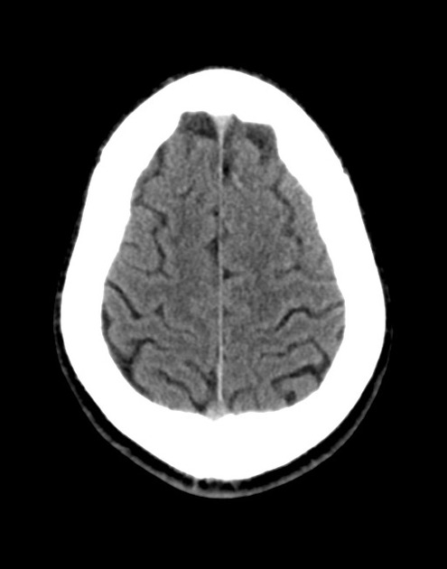
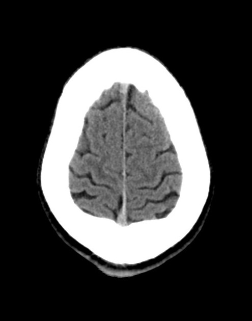
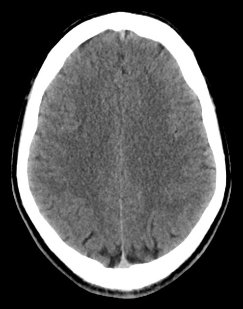
A 48 year-old male with hypertension and hyperlipidemia presents with headache. Notes onset of symptoms 8 hours prior to presentation, reaching maximal severity within seconds. Headache improved with over-the-counter analgesics. On examination, there are no neurological deficits, neck is supple. A CT head non-contrast is obtained:

01_normal
02_normal
03_normal
04_normal
05_normal
06_normal
07_normal
08_normal
09_normal
10_normal
11_normal
12_normal
13_normal
14_normal
15_normal
16_normal
18_normal
19_normal
20_normal

CT Head:

No acute intracranial process. Case courtesy of Assoc Prof Craig Hacking, Radiopaedia.org, rID: 37118

ED Course:

A lumbar puncture is performed, CSF sampling reveals xanthochromia – neurosurgery is consulted and the patient is admitted for angiography and possible intervention.


An Algorithm for the Evaluation of Headache

An Algorithm for the Evaluation of Headache

High-Risk Historical Features

  • Sudden onset (seconds/minutes), patient recalls activity at onset
  • Worst in life or change in character from established headache
  • Fever, neck pain/stiffness
  • Altered mental status
  • Malignancy
  • Coagulopathy: iatrogenic, hepatopathy, dialysis
  • Immunocompromised
  • Rare: CO exposure, jaw claudication, PCKD

Location of Pain
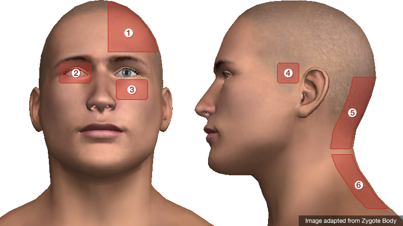
Headache Location

  1. Unilateral: migraine
  2. Periorbital: glaucoma, CVT, optic neuritis, cluster
  3. Facial/maxillary: trigeminal neuralgia, sinusitis
  4. Temporal: GCA
  5. Occipital: cerebellar stroke
  6. Nuchal: meningitis

Characteristics of Primary Headaches

Type Location Duration Quality Associated symptoms Comment
Migraine Unilateral Hours to days Throbbing Photophobia, phonophobia Atypical migraines with neurological findings (basilar, ophthalmoplegic, ophthalmic, hemiplegic)
Tension Bilateral Minutes to days Constricting None
Cluster Unilateral, periorbital Minutes to hours Throbbing Conjunctival injection, lacrimation, rhinorrhea, miosis, eyelid edema Males 90%, triggered by EtOH.

Physical Examination Findings

Vital Signs
Fever: present in 95% of patients with meningitis
Head
Trauma: signs of basilar skull fracture
Temporal artery tenderness/induration: GCA
Pericranial muscle tenderness: tension headache
Trigger point, Tinnel sign: occipital neuralgia
Eyes
Pupillary defects: aneurysm with CN III compression
Papilledema, absence of spontaneous venous pulsations: elevated intracranial pressure
EOM abnormalities: ICH, mass lesion, neuropathy (DM, Lyme)
Horner syndrome (ptosis, miosis, anhidrosis): carotid dissection
Visual field defect: stroke, atypical migraine
Conjunctival injection: glaucoma (fixed, mid-size pupil, elevated intraocular pressure), cluster headache
Mouth
Thrush: immunocompromise
Sinuses
Tenderness to palpation, abnormal transillumination: sinusitis
Neck
Resistance to supine neck flexion: meningitis
Kernig: supine position, hip flexed, knee flexed, resistance to knee extension
Brudzinski: supine position, neck flexion results in knee flexion
Jolt accentuation: patient rotates head side-to-side, 2-3 times/sec exacerbates headache

References:

  1. Russi, C. (2013). Headache. In Rosen’s Emergency Medicine – Concepts and Clinical Practice (8th ed., Vol. 1, pp. 170-175). Elsevier Health Sciences.
  2. Godwin SA, Villa J. “Acute headache in the ED: Evidence-Based Evaluation and Treatment Options.” Emerg Med Pract 2001; 3(6): 1-32.
  3. Edlow, J. A., Panagos, P. D., Godwin, S. A., Thomas, T. L., & Decker, W. W. (2008). Clinical Policy: Critical Issues in the Evaluation and Management of Adult Patients Presenting to the Emergency Department With Acute Headache. Annals of emergency medicine, 52(4), 407–436. doi:10.1016/j.annemergmed.2008.07.001
  4. WikEM: Headache
Dizziness and Vertigo

Types of Dizziness

Types of Dizziness

Distinguishing Central vs. Peripheral Vertigo

Characteristic Peripheral Central
Onset Sudden Gradual
Intensity Severe Mild
Duration Minutes Weeks
Timing Intermittent Continuous
Nystagmus Horizontal Vertical, bidirectional
Exacerbation with head movement +
Auditory symptoms +
Neurological findings +

Causes of Vertigo

Causes of Vertigo

Characteristics of common causes of vertigo

Cause Mechanism Onset Symptoms Findings
Peripheral
BPPV Otolith Brief, positional episodes Nausea, vomiting, absent auditory symptoms. Dix-Hallpike positive
Vestibular neuronitis Viral, post-viral inflammation of vestibular portion of CNVIII Acute and severe, subsiding over days. Nausea, vomiting, absent auditory symptoms. Head thrust abnormal
Meniere Endolymphatic hydrops Recurrent, lasting hours Tinnitus, hearing loss. SNHL
Central
Vertebrobasilar insufficiency Atherosclerosis (vascular risk factors) Acute onset, recurrent episodes if TIA Headache, gait impairment, diplopia, absent auditory symptoms. Neurologic deficits
Cerebellar stroke Atherosclerosis (vascular risk factors) Acute and severe Headache, dysphagia, gait impairment Dysmetria, dysdiadochokinesia, ataxia, CN palsy
Brainstem stroke Atherosclerosis (vascular risk factors), dissection Acute and severe Dysphagia, dysphonia, gait impairment, sensory disturbances Loss of pain/temperature on ipsilateral face, contralateral body, palatal/pharyngeal paralysis
MS Demyelination Subacute onset History of other, variable symptoms INO

History

  • Onset, duration, timing, severity, exacerbating factors
  • Vascular risk factors: age, male, HTN, CAD, DM, atrial fibrillation
  • Vestibulotoxic medications: aminoglycosides, AED

Key Physical Examination Findings

  • VS: Presence of hypotension suggests presyncope
  • Head: Examine for evidence of trauma
  • Neck: Auscultate for carotid bruit
  • Ear: Effusion or perforation suggests peripheral process (possible perilymphatic fistula)
  • Eye: Examine for pupillary defects (CNIII), papilledema, extraoccular muscles
  • Neuro: Cerebellar testing

Positional Testing

Dix-Hallpike
Turn head 45°
Upright sitting → supine (head overhanging bed)
Positive: nystagmus + symptoms on one side
Roll
Supine
Turn head 90°
Positive: nystagmus + symptoms on both sides, more severe on affected

HINTS1

Normal head impulse, direction-changing nystagmus, or skew deviation suggests stroke.

Head impulse
Focus on examiner’s nose
Rapidly turn head 10° in horizontal plan
Presence of corrective saccade suggests defect of peripheral vestibular nerve
Nystagmus
Peripheral: Horizontal, unidirectional. Increases on gaze in direction of fast phase (decreases or resolves opposite)
Central: Direction changing
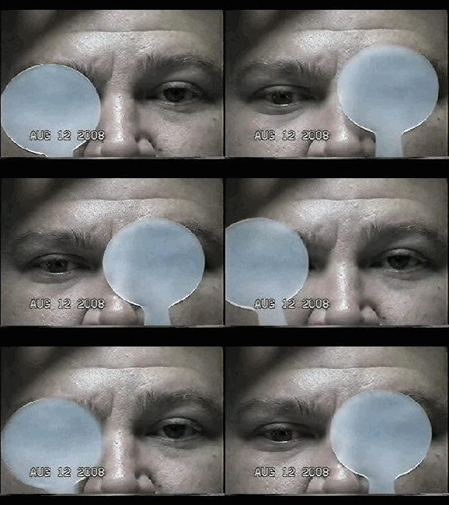
Skew deviation
Cross cover
Presence of vertical disconjugate gaze suggests brainstem dysfunction

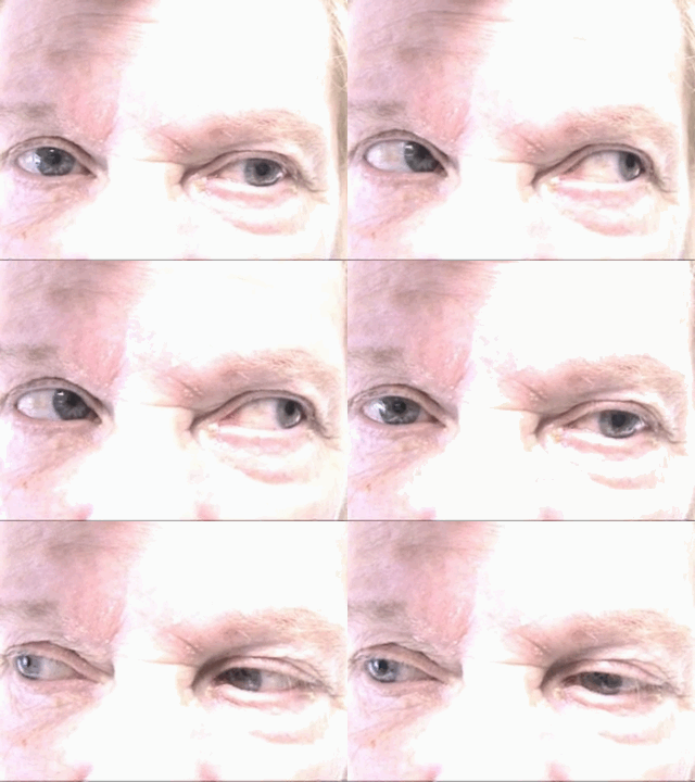
HINTS Gallery

Positive Head Impulse Test
Central Changing Nystagmus
Skew Deviation

Labs

  • Glucose
  • CBC/Chemistry
  • ECG

Imaging

  • Warranted if findings concerning for central process
  • MRI preferred

Management

Specific etiologies
Vestibular neuronitis: steroids
Meniere: dietary changes
BPPV: canalith repositioning
Symptomatic relief
Promethazine (Phenergan) 12.5-25mg PO
Ondansetron (Zofran) 4mg IV
Lorazepam (Ativan) 1-2mg PO/IV
Meclizine (Antivert) 25mg PO q6-8h PRN

References

  1. Kattah, J. C., Talkad, A. V., Wang, D. Z., Hsieh, Y.-H., & Newman-Toker, D. E. (2009). HINTS to diagnose stroke in the acute vestibular syndrome: three-step bedside oculomotor examination more sensitive than early MRI diffusion-weighted imaging. Stroke; a journal of cerebral circulation, 40(11), 3504–3510. doi:10.1161/STROKEAHA.109.551234
  2. Chang, A., & Olshaker, J. (2013). Dizziness and Vertigo. In Rosen’s Emergency Medicine – Concepts and Clinical Practice (8th ed., Vol. 1, pp. 162-169). Elsevier Health Sciences.

Sore Throat

Evaluation of Sore Throat

Evaluation of Sore Throat

Physical Examination:

Neck
Stiffness, limitation of extension suggestive of retropharyngeal abscess.
Jaw
Trismus associated with peritonsillar cellulitis or abscess.
Oral Cavity
Dry mucous membranes suggest dehydration (from odynophagia) and indicates severity of symptoms.
Tongue elevation, sublingual/submental induration, poor dentition (particularly of mandibular molars) associated with Ludwig Angina.
Unilateral tonsillar enlargement with contralateral uvular deviation suggests peritonisllar abscess. Fluctuance may be palpated.
Tonsilar exudates suggest infectious pharyngitis (non-specific).
Palatal petechiae suggest bacterial pharyngitis.
Ulcerations of the anterior oral cavity are associated with herpes infection, lesions on the soft palate are suggestive of coxsackievirus infection.
Rarely, a grey membrane in the posterior pharynx will suggest diphtheria.
Lymphadenopathy
Tender anterior cervical lymphadenopathy may suggest bacterial pharyngitis.
Posterior cervical lymphadenopathy is associated with infectious mononucleosis.
Large, firm, non-mobile lymph nodes may suggest malignancy.
Eyes
Presence of conjunctivitis (also rhinorrhea, exanthema) associated with viral pharyngitis.
Skin
Ulcers involving the hands, feet, in addition to pharyngeal lesions suggest coxsackievirus infection.
Scarlatiniform rash associated with pharyngitis (particularly in school-age children) suggests streptococcal pharyngitis.
Abdomen
Splenomegaly is associated with infectious mononucleosis.

Centor Criteria (Modified)

  • +1: Fever
  • +1: Tonsillar Exudate
  • +1: Tender anterior cervical lymphadenopathy
  • +1: Absence of cough
  • -1: Age >45yo

Incidence of GABHS by Centor Criteria

  • 0, -1: 1%
  • 1: 10%
  • 2: 17%
  • 3: 35%
  • 4: 51%

References:

  1. Newman, D., & Shreves, A. (2013). Sore Throat. In Rosen’s Emergency Medicine – Concepts and Clinical Practice (8th ed., Vol. 1, pp. 198-202). Elsevier Health Sciences.
  2. King, B. R., & Charles, R. A. (2004). Pharyngitis In The ED Diagnostic Challenges And Management Dilemmas. Emergency medicine practice, 6(5), 1–24.

Dyspnea

Causes of Dyspnea

Causes of Dyspnea

Findings in Selected Causes of Dyspnea

Condition History Symptoms Findings Evaluation
Anaphylaxis Exposure to allergen Abrupt onset, facial swelling Stridor, wheezing, hives  
PE Immobilization, malignancy, prior DVT/PE, surgery, OCP Abrupt onset, pleuritic chest pain Tachycardia, hypoxia ECG (RV strain)
CT PA, D-dimer
LE US (DVT)
Pneumonia Exposure, tobacco use Fever, productive cough Focal rales CXR
CBC
Blood/respiratory cultures
Pneumothorax Trauma, thin male Abrupt onset, chest pain Decreased BS, subQ emphysema, JVD and tracheal deviation if tension CXR
US
Fluid overload Dietary indiscretion, medication non-adherence Orthopnea, PND JVD, S3/S4, peripheral edema CXR
US
ECG
BNP
COPD/Asthma Tobacco use, personal/family history Progressive Retractions, accessory muscle use, wheezing CXR
US (distinguish from fluid overload)
Malignancy Tobacco use, weight loss Hemoptysis   CXR
CT Chest

References

  1. Braithwaite, S., & Perina, D. (2013). Dyspnea. In Rosen’s Emergency Medicine – Concepts and Clinical Practice (8th ed., Vol. 1, pp. 206-213). Elsevier Health Sciences.

Chest Pain

An Algorithm for the Evaluation of Chest Pain

Algorithm for the Evaluation of Chest Pain

NOTE: Algorithm revised in November, 2017. The prior version is no longer supported but remains available here.

Guided Lecture

EM Ed
Watch “Chest Pain: It’s Giving Me Angina” from EM Ed. In this lecture Dr. Celedon reviews the critical differential diagnosis for chest pain and how to safely and effectively work up patient’s with this challenging chief complaint.

References

  1. Brown, J. (2013). Chest Pain. In Rosen’s Emergency Medicine – Concepts and Clinical Practice (8th ed., Vol. 1, pp. 214-222). Elsevier Health Sciences.

Abdominal Pain

Pathophysiology of Abdominal Pain

  1. Visceral: distension of hollow organs or capsular stretch of solid organs.
  2. Somatic: parietal peritoneal irritation
  3. Referred

    • Extra-abdominopelvic

      • Epigastric: inferior MI
      • Pelvic: hip
      • Abdominal: lower lobe pneumonia/infarction
    • Abdominopelvic

      • Shoulder: diaphragmatic irritation (ex. perforated duodenal ulcer, splenic pathology)
      • Mid-back: aortopathy, pancreatitis
      • Flank: renal pathology
      • Low back: uterus, rectum

Concerning Historical Features

  • Elderly: increased probability for severe disease with poor clinical diagnostic accuracy
  • Immunocompromised: HIV/AIDS, uncontrolled diabetes, chronic liver disease, chemotherapy, other immunosuppression
  • Pain preceding nausea/vomiting: increased likelihood of surgical process
  • Abrupt onset, duration <48h, constant timing
  • Prior abdominal surgical history: consider bowel obstruction
  • No prior episodes of similar pain
  • Recent antibiotic or steroid use: may mask signs of infection
  • Cardiac risk factors (HTN, vascular disease, atrial fibrillation: increased risk for mesenteric ischemia or aortic aneurysm
  • Heavy NSAID use or anticoagulation: increase concern for gastrointestinal bleeding

Imaging

  • Plain film reserved for those who would otherwise not undergo CT. XR abdomen for bowel obstruction or radiopaque foreign body.
  • CT abdomen/pelvis with IV contrast, particularly if elderly or immunocompromised.
  • Ultrasound preferred for hepatobiliary pathology
  • Bedside ultrasound for identification of IUP, free intraperitoneal fluid, cholecystitis, CBD dilation, ascites, hydronephrosis, aortopathy, volume status.

Causes of Abdominal Pain

Causes of Abdominal Pain

References

  1. Budhram, G., & Bengiamin, R. (2013). Abdominal Pain. In Rosen’s Emergency Medicine – Concepts and Clinical Practice (8th ed., Vol. 1, pp. 223-231). Elsevier Health Sciences.

Nausea and Vomiting

Pathophysiology of Nausea and Vomiting

Pathophysiology of Nausea and Vomiting

Complications of Nausea and Vomiting

  • Hypovolemia: activates RAAS
  • Metabolic alkalosis: loss of hydrogen ions in vomitus
  • Hypokalemia: RAAS promotes sodium retention and potassium excretion
  • Esophageal injury: Mallory-Weiss tear, Boerhaave syndrome
  • Aspiration

Key Historical Findings

Duration of vomiting
Acute: Episodic and occurring for <1 week. Suggestive of obstructive, toxic/metabolic, infectious, ischemic or neurologic process.
Chronic: Episodic and occurring for >1 month. Suggestive of partial obstruction, motility disorder or neurologic process.
Onset
Acute onset: suggests pancreatitis, gastroenteritis, or cholecystitis.
Timing
Post prandial: delayed >1 hour suggests gastric outlet obstruction or gastroparesis.
Contents
Bile: presence of bile suggests patent connection between duodenum and stomach (no GOO)
Undigested food: suggests upper GI tract process (achalasia, esophageal stricture, Zenker)
Feculent: suggests distal bowel obstruction
Associated symptoms
Headache: suggests elevated ICP

Causes of Nausea and Vomiting

Causes of Nausea and Vomiting

References

  1. Zun, L. (2013). Nausea and Vomiting. In Rosen’s Emergency Medicine – Concepts and Clinical Practice (8th ed., Vol. 1, pp. 238-247). Elsevier Health Sciences.

Gastrointestinal Bleeding

Evaluation and Management of Gastrointestinal Bleeding

Evaluation and Management of Gastrointestinal Bleeding

Key Historical Features

Quantity
Patient’s estimate
Symptoms suggestive of anemia/volume depletion: (pre)syncope, dyspnea
Appearance/Location
Distinguish upper from lower GI bleding
PMH
Prior episodes and source
History of aortic aneurysm graft
Comorbidities: presence of CAD, CHF, liver disease or diabetes increases mortality
Medications/substance use
Gastrotoxic, anti-coagulants, anti-platelet agents
Alcohol abuse

Key Physical Findings

Vital signs
Tachycardia or hypotension
Eyes
Conjuntival pallor suggests anemia
Scleral icterus suggests liver disease
Abdomen
Hyperactive bowel sounds may be present in UGIB (blood is cathartic)
Epigastric tenderness to palpation suggests PUD
Diffuse tenderness suggests bowel ischemia, obstruction/ileus, or perforation
Rectal (digital, anoscopy)
May reveal fissures, hemorrhoids or polyps

Labs/Diagnostic Tests

  • CBC: consider transfusion for Hb <8-10g/dL particularly in elderly or those with CAD
  • BMP: BUN:creatinine > 36 in the absence of renal failure suggests UGIB
  • PT/PTT/INR: coagulopathy
  • Lactate: elevated in bowel ischemia or systemic hypoperfusion
  • T&S or T&C
  • ECG: screen for myocardial ischemia

Blatchford Scoring System

Item Value Points
BUN 18-22 2
22-28 3
28-70 4
>70 6
Hb (male) 12-13 1
10-12 3
<10 6
Hb (female) 10-12 1
<10 6
SBP 100-109 1
90-99 2
<90 3
Other HR > 100 1
Melena 1
Syncope 2
Liver disease 2
Heart failure 2

References:

  1. Goralnick, E., & Meguerdichian, D. (2013). Gastrointestinal Bleeding. In Rosen's Emergency Medicine – Concepts and Clinical Practice (8th ed., Vol. 1, pp. 248-253). Elsevier Health Sciences.

Acute Pelvic Pain

Evaluation of Acute Pelvic Pain

Acute Pelvic Pain

Key Historical Findings

Location
Lateralized: suggests process related to tube or ovary, consider unilateral urinary tract process. On right, add appendicitis to differential; on left, add diverticulitis (particularly if age >40.
Central: suggests process involving uterus, bladder or bilateral adnexa
Diffuse: suggests PID
Radiation
Radiation to rectum suggests pooling of fluid or blood in cul-de-sac
Onset
Abrupt: suggests acute intrapelvic hemorrhage (from ruptured ectopic or ovarian cyst), ovarian torsion, urolithiasis
Gradual: inflammatory process such as PID
Chronic/recurrent: suggests endometriosis, recurrent ovarian cyst, ovarian mass
Associated Symptoms
Fevers/chills: suggests infectious process
Nausea/vomiting: suggests process involving gastrointestinal tract, though may accompany pregnancy or severe pain associated with ovarian torsion, urolithiasis.
Dysuria: suggests process involving urinary tract, though may be associated with local vulvar/vaginal process
Urinary urgency: more specific for bladder or urethral irritation
Obstetric History
History of recurrent spontaneous abortions or prior ectopic pregnancy increases likelihood of recurrence.
Ongoing fertility treatments increase likelihood for ectopic/heterotopic (occurs in 1:100 with assisted reproduction compared to 1:8000 in general population)
Vaginal Bleeding
In non-pregnant: suggests PID, DUB, cervical or uterine cancer
In early pregnancy: may be associated with ectopic pregnancy, non-viable IUP, or subchorionic hemorrhage
In late pregnancy: may be associated with placental pathology (previa, abruption)

Key Physical Findings

  • Pelvic examination: assists with localization of lateralized process. Should be preceded by ultrasound if >20 weeks.
  • Abnormal vaginal discharge: suggests vaginitis, cervicitis, PID, or retained foreign body.
  • Cervical motion tenderness: suggests reproductive tract inflammation or irritation of adjacent structures (appendicitis, cystitis)
  • Unilateral adnexal mass/tenderness: associated with ovarian cyst/mass, TOA, ectopic, or ovarian torsion.

References:

  1. Hart, D., & Lipsky, A. (2013). Acute Pelvic Pain in Women. In Rosen's Emergency Medicine – Concepts and Clinical Practice (8th ed., Vol. 1, pp. 266-272). Elsevier Health Sciences.
  2. WikEM: Pelvic pain

Back Pain

Causes of Back Pain

Causes of Back Pain

Key Historical Findings

Onset
Acute onset with associated activity suggests mechanical process
Acute onset without trigger, particularly if severe pain may suggest vascular process
Progressive onset without trigger suggests non-mechanical process (i.e. malignancy)
Aggravating/Alleviating Factors
Worsening with cough/valsalva suggests herniated disk
Relief with flexion associated with spinal stenosis
Location/Radiation
Radicular pain typically extends below knee, associated with nerve root involvement
Radiation to/from chest or abdomen suggests visceral source
Flank location suggests retroperitoneal source
History/Associated Symptoms
Fever
Medications (particularly anti-coagulants)
Hematuria
Malignancy
IVDA
Vascular disease

Key Physical Findings

  • Abnormal vital signs

    • Fever: abscess, osteomyelitis, discitis
    • Hypertension: dissection
    • Shock: AAA
  • Localize point of greatest tenderness
  • Examine abdomen for pulsatile mass
  • Perform thorough neurological examination including rectal tone and perianal sensation
  • Positive straight leg raise associated with sciatic nerve root irritation and is sensitive (but not specific) for disk disease.

References

  1. Mahoney, B. (2013). Back Pain. In Rosen’s Emergency Medicine – Concepts and Clinical Practice (8th ed., Vol. 1, pp. 278-284). Elsevier Health Sciences.
  2. WikEM: Lower back pain