Sinus Tachycardia

Brief History and Physical:

A young female with a history of schizophrenia presents to the emergency department reporting hallucinations. She had been diagnosed with schizophrenia one year previously and was briefly admitted to a psychiatric hospital. She discontinued her anti-psychotic (risperidone) two months ago, and over the past week she reports increasingly prominent auditory and visual hallucinations.

She denies recent illness, vomiting/diarrhea, changes in urinary habits, new medications, alcohol or illicit substance use. She also denies chest pain, palpitations or shortness of breath.

Vital signs are notable for a heart rate of 148bpm and are otherwise normal (including core temperature). Detailed physical examination is normal except for a rapid, regular heart rate. Mental status examination demonstrated normal level of alertness and orientation, linear and cogent responses and occasional response to internal stimuli during which she appeared anxious.

Initial evaluation and management included a 12-lead ECG which showed sinus tachycardia. Multiple boluses of normal saline were initiated while awaiting laboratory workup.

ECG: Sinus Tachycardia

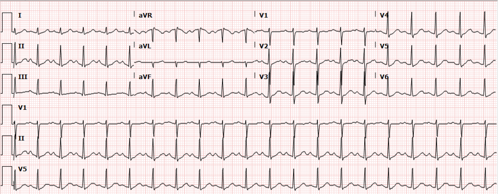
Presentation ECG demonstrates sinus tachycardia.

Update:

Laboratory studies were reviewed and unremarkable. Normal hemoglobin, normal chemistry panel, negative hCG, and negative toxicology screen. The patient remained persistently tachycardic with a heart rate ranging from 140-160bpm (again sinus tachycardia on 12-lead ECG). An atypical antipsychotic and anxiolytic were administered and additional studies were obtained. Serum TSH, troponin and D-dimer were normal and bedside ultrasound did not identify a pericardial effusion. The patient remained asymptomatic, reporting subjective improvement in anxiety and hallucinations. Psychiatry was consulted and the patient was placed in observation for monitoring of sinus tachycardia. Observation course was uneventful as the patient remained asymptomatic. Transthoracic echocardiography was normal. Psychiatry consultation recommended resumption of home anti-psychotic and outpatient follow-up. Tachycardia had improved but not resolved at the time of discharge (heart rate 109bpm) and the patient was instructed to follow-up with her primary care provider.


Algorithm for the Evaluation of Sinus Tachycardia

Algorithm for the Evaluation of Sinus Tachycardia

Any vital sign derangement is concerning and tachycardia may be associated with unanticipated death after discharge home1. The presence of tachycardia suggests one of several categories of hemodynamic, autonomic, or endocrine/metabolic derangement.

Demand for increased cardiac output

A perceived demand for increased cardiac output will prompt chronotropic (and inotropic) amplification before hypotension develops. Causative etiologies include: volume depletion (from hemorrhage, gastrointestinal or renal losses), distributive processes (such as infection), obstruction (pulmonary embolus, or pericardial effusion with impending tamponade), or tissue hypoxia (anemia or lung disease).

Autonomic nervous system

Autonomic nervous system disturbances induced by stimulant, sympathomimetic or anti-cholinergic use, or withdrawal of certain agents such as ethanol or beta-blockers may be at fault.

Endocrine and other causes

Hyperthyroidism and pheochromocytoma should be considered, and as diagnoses of exclusion: anxiety, pain, or inappropriate sinus tachycardia2.

Evaluation:
Core temperature
CBC
Troponin
D-dimer
Bedside cardiac ultrasound
Urine toxicology screen
Ethanol level
TSH/T4

Algorithm for the Evaluation of Narrow-Complex Tachycardia3,4,5,6

Algorithm for the Evaluation of Narrow-Complex Tachycardia

References:

  1. Sklar DP, Crandall CS, Loeliger E, Edmunds K, Paul I, Helitzer DL. Unanticipated Death After Discharge Home From the Emergency Department. Ann Emerg Med. 2007;49(6):735-745. doi:10.1016/j.annemergmed.2006.11.018.
  2. Olshansky B, Sullivan RM. Inappropriate sinus tachycardia. J Am Coll Cardiol. 2013;61(8):793-801. doi:10.1016/j.jacc.2012.07.074.
  3. Yusuf S, Camm AJ. Deciphering the sinus tachycardias. Clin Cardiol. 2005;28(6):267-276.
  4. Katritsis DG, Josephson ME. Differential diagnosis of regular, narrow-QRS tachycardias. Heart Rhythm. 2015;12(7):1667-1676. doi:10.1016/j.hrthm.2015.03.046.
  5. Bibas L, Levi M, Essebag V. Diagnosis and management of supraventricular tachycardias. CMAJ. 2016;188(17-18):E466-E473. doi:10.1503/cmaj.160079.
  6. Link MS. Clinical practice. Evaluation and initial treatment of supraventricular tachycardia. N Engl J Med. 2012;367(15):1438-1448. doi:10.1056/NEJMcp1111259.

Nausea and Vomiting

Pathophysiology of Nausea and Vomiting

Pathophysiology of Nausea and Vomiting

Complications of Nausea and Vomiting

  • Hypovolemia: activates RAAS
  • Metabolic alkalosis: loss of hydrogen ions in vomitus
  • Hypokalemia: RAAS promotes sodium retention and potassium excretion
  • Esophageal injury: Mallory-Weiss tear, Boerhaave syndrome
  • Aspiration

Key Historical Findings

Duration of vomiting
Acute: Episodic and occurring for <1 week. Suggestive of obstructive, toxic/metabolic, infectious, ischemic or neurologic process.
Chronic: Episodic and occurring for >1 month. Suggestive of partial obstruction, motility disorder or neurologic process.
Onset
Acute onset: suggests pancreatitis, gastroenteritis, or cholecystitis.
Timing
Post prandial: delayed >1 hour suggests gastric outlet obstruction or gastroparesis.
Contents
Bile: presence of bile suggests patent connection between duodenum and stomach (no GOO)
Undigested food: suggests upper GI tract process (achalasia, esophageal stricture, Zenker)
Feculent: suggests distal bowel obstruction
Associated symptoms
Headache: suggests elevated ICP

Causes of Nausea and Vomiting

Causes of Nausea and Vomiting

References

  1. Zun, L. (2013). Nausea and Vomiting. In Rosen’s Emergency Medicine – Concepts and Clinical Practice (8th ed., Vol. 1, pp. 238-247). Elsevier Health Sciences.

Nausea and Vomiting

Neurologic pathways involved in pathogenesis of nausea and vomiting

HPI:

57yo male with a history of HTN, DM, and MI s/p stent in 2011 presenting with nausea/vomiting and hypotension. The patient had one episode of non-bloody, non-bilious emesis approximately 6 hours ago. He felt unwell so a friend checked his blood pressure which was found to be 75/50, prompting a visit to this emergency department.
The patient’s emesis came 2 hours following a normal meal (frozen dinner), and was associated with chills/sweats but no abdominal pain. The patient had some associated shortness of breath (baselines), but no chest pain, arm or jaw pain, or palpitations.

He states that these symptoms are unlike what he experienced during his MI. He reported no change in bowel or urinary habits.

PMH:

  • HTN
  • DM
  • CAD
  • MI
  • Hyperlipidemia

 PSH:

  • Stent placement (2011)
  • Right knee neuroma excision (2012)

FH:

  • Non-contributory

 SHx:

  • No current t/e/d
  • 80 pack-year smoking history

Meds:

  • carvedilol 6.25mg p.o. b.i.d.
  • metformin 1000mg p.o. b.i.d.
  • atorvastatin 20mg p.o. daily
  • aspirin 81mg p.o. daily

Allergies:

  • NKDA

Physical Exam:

VS: T 98.4 HR 65 RR 17 BP 96/56 O2 95% 2L NC
Gen: No acute distress, speaking in complete sentences
HEENT: PERRL, MMM no lesions, no cervical lymphadenopathy
CV: RRR, normal S1/S2, no murmurs, no extra heart sounds, no jugular venous distension
Lungs: CTAB, no crackles
Abd: +BS, soft, NT/ND, no rebound/guarding, no organomegaly
Ext: Warm, well-perfused, peripheral pulses equal b/l, no LE edema
Neuro: AAOx3

Labs:

  • EKG: normal sinus rhythm, anterior lead q-waves suggestive of old infarct, no T/ST changes
  • Troponin: <0.01
  • CBC: 7.4/15.5/47/228
  • BMP: 139/5.1/107/26/8/1.19/112 (baseline creatinine 1.06 in 2/2013)

Studies:

  • CXR: no effusion, no cardiomegaly, no focal consolidation
  • Bedside US: normal cardiac wall motion, estimated EF 40-45%, retrohepatic IVC collapses with respiration

Assessment/Plan:

57M hx HTN, DM, MI s/p stent presenting with nausea/vomiting x1 and hypotension. The patient’s symptoms and history were concerning for acute myocardial infarction; however, early EKG and troponins were reassuring. Additionally, the absence of characteristic physical findings that would be associated with an acute MI causing cardiogenic shock (elevated JVP, extra heart sounds, pulmonary crackles) were not present. Evidence of end-organ damage was also absent.

Other potential causes for nausea/vomiting include SBO, however, the patient reported normal BM’s and has no history of abdominal surgery. Though occurring after a meal, a single episode of emesis without associated abdominal pain lowers suspicion for biliary disease. This patient’s emesis is most likely due to acute gastroenteritis.

Given the evidence of hypovolemia on bedside ultrasound, the patient was bolused with a total of 1.5L NS and noted symptomatic improvement as well recovery of blood pressure.

Differential Diagnosis of Nausea/Vomiting: 1, 2

A System for Nausea/Vomiting

Pathophysiology: 3, 4, 5

  • Nausea: Sensation associated with increased gastrointestinal motility (tachygastria).
  • Vomiting:
    • Chemoreceptor trigger zone (area postrema of 4th ventricle): sensitive to drugs/toxins (emetics, radiation), neurotransmitters. Located outside BBB.
    • Nucleus tractus solitaries (medulla): pattern generator for vomiting, receives vagal input from GI tract and nociceptive stimuli from peripheral nervous system – transmits to hypothalamus, limbic system and cortex. Stimulated by tickling the back of the throat, gastric distention, and vestibular input.

Important history/physical associations: 4

  • Abdominal pain: suggests organic disease, affected organ dependent on location of pain. (See figure)
  • Abdominal distension: suggests bowel obstruction.
  • Heartburn: suggests GERD.
  • Vertigo/nystagmus: suggests vestibular etiology.
  • Positional/projectile: suggests neurogenic etiology.

Differential Diagnosis of Abdominal Pain By Location:

Abdominal Pain by Location

References:

  1. Scorza, K., Williams, A., Phillips, J. D., & Shaw, J. (2007). Evaluation of nausea and vomiting. American family physician, 76(1), 76–84.
  2. Bork S, Ditkoff J, Hang BS. Chapter 75. Nausea and Vomiting. In: Tintinalli JE, Stapczynski JS, Cline DM, Ma OJ, Cydulka RK, Meckler GD, eds. Tintinalli’s Emergency Medicine: A Comprehensive Study Guide. 7th ed. New York: McGraw-Hill; 2011. http://www.accessmedicine.com/content.aspx?aID=6360091. Accessed June 15, 2013.
  3. Koch, K. L., Stern, R. M., Vasey, M. W., Seaton, J. F., Demers, L. M., & Harrison, T. S. (1990). Neuroendocrine and gastric myoelectrical responses to illusory self-motion in humans. The American journal of physiology, 258(2 Pt 1), E304–10.
  4. Longstreth, G. F. Approach to the adult with nausea and vomiting. In: UpToDate, Basow, DS (Ed), UpToDate, Waltham, MA, 2013.
  5. Costanzo, L. (2011). Physiology. Philadelphia: Wolters Kluwer Health/Lippincott Williams & Wilkins.
  6. Patanwala, A. E., Amini, R., Hays, D. P., & Rosen, P. (2010). Antiemetic therapy for nausea and vomiting in the emergency department. The Journal of emergency medicine, 39(3), 330–336. doi:10.1016/j.jemermed.2009.08.060