Neonatal Congenital Heart Disease

Brief H&P

An 8-day old male infant, ex-full term, born by normal spontaneous vaginal delivery and discharged home 2 days after birth without identified complications or maternal infections presents with parents to the emergency department due to decreased activity. Starting on day-of-life six, the family noted that feeding appeared to be taking longer and the mother felt her infant was breathing faster.

On presentation, the patient was pale, dusky, lethargic and with mottled skin. Temperature 36.3°C (rectal), HR 170, RR 60, BP 62/35, SpO2 70%. Physical examination demonstrated flat fontanelle, coarse breath sounds, regular rate and rhythm without additional heart sounds or murmurs, and hepatomegaly with liver edge 3cm below costal margin. Capillary refill was delayed at 5-6 seconds. Supplemental oxygen was applied without effect.

Algorithm for the Evaluation and Management of Suspected Congenital Heart Disease in Neonates

Algorithm for the Evaluation of Neonatal Congenital Heart Disease

Neonates with undiagnosed congenital heart disease may present to the emergency department with nonspecific symptoms, and may be considerably unstable requiring immediate life-saving interventions.

Key Historical Features

  • Respiratory difficulty
  • Feeding difficulty (small quantities, diaphoresis during feeding)
  • Poor weight gain
  • Chromosomal abnormalities, syndromes
  • Maternal risk factors: diabetes, teratogen exposure, substance use
  • Sibling of affected child

Key Examination Findings

  • Vital signs: tachycardia, tachypnea, hypotension
  • Blood pressure differential (RUE vs. LE >8mmHg difference)
  • Pulse oximetry differential (RUE vs. LE >4% difference, <95%)
  • Cardiac examination: murmur, thrill, pulse differential, capillary refill, hepatomegaly

Workup

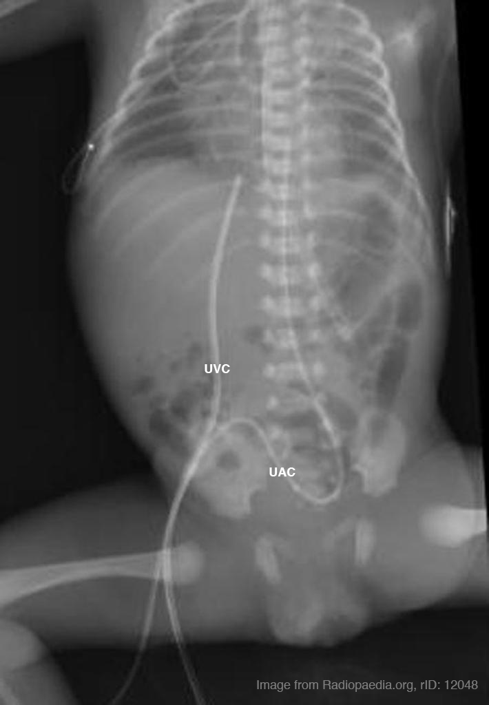
  • CXR: Evaluate for cardiomegaly, pulmonary vascular congestion
  • ECG: Evaluate for axis deviation (right axis deviation is normal for neonate)
  • ABG with co-oximetry

References

  1. Special thanks to Dr. Kelly Young, MD, MS, FAAP. Director, Pediatric Emergency Medicine Fellowship. Harbor-UCLA Medical Center Department of Emergency Medicine.
  2. Association AAOPAAH. Textbook of Neonatal Resuscitation. 2016.
  3. Lissauer T, Fanaroff AA, Miall L, Fanaroff J. Neonatology at a Glance. John Wiley & Sons; 2015.
  4. Steinhorn RH. Evaluation and Management of the Cyanotic Neonate. Clinical Pediatric Emergency Medicine. 2008;9(3):169-175. doi:10.1016/j.cpem.2008.06.006.
  5. MD MR. Chapter 7 – Cardiology. Twenty First Edition. Elsevier Inc.; 2018:156-202. doi:10.1016/B978-0-323-39955-5.00007-7.
  6. Gomella T, Cunningham M. Neonatology 7/E. McGraw-Hill Prof Med/Tech; 2013.
  7. Yee L. Cardiac emergencies in the first year of life. Emergency Medicine Clinics of NA. 2007;25(4):981–1008–vi. doi:10.1016/j.emc.2007.08.001.
  8. Yates MC, Rao PS. Pediatric cardiac emergencies. Emerg Med. 2013. doi:10.4172/2165-7548.1000164.
  9. Silberbach M, Hannon D. Presentation of congenital heart disease in the neonate and young infant. Pediatr Rev. 2007;28(4):123-131.
  10. Mastropietro CW, Tourner SP, Sarnaik AP. Emergency presentation of congenital heart disease in children. Pediatric Emergency …. 2008.
  11. Brousseau T, Sharieff GQ. Newborn Emergencies: The First 30 Days of Life. Pediatric Clinics of North America. 2006;53(1):69-84. doi:10.1016/j.pcl.2005.09.011.

Principles of Neonatal Resuscitation

The following resource for neonatal resuscitation and neonatal critical care was developed with the guidance of Dr. Agrawal (Neonatology) while on rotation at the White Memorial Medical Center Neonatal Intensive Care Unit.

Endotracheal Tube Size1-3

Simplified Formula
Estimated gestational age in weeks ÷ 10 = round to nearest half-size uncuffed tube

NRP Recommendation

Gestation age (weeks) Weight (kg) ETT Size (ID, mm) Depth (cm from lip)
<28 <1.0 2.5 6-7
28-34 1.0-2.0 3.0 7-8
34-38 2.0-3.0 3.5 8-9
>38 >3.0 3.5-4.0 9-10

Laryngoscope Blade Size

Age Blade
Preterm 0
Term 1

Umbilical Vein Catheter Placement4

ED Indications
Unstable neonate
Contraindications
Omphalocele
Gastroschisis
Necrotizing enterocolitis
Depth
4-5cm or until blood return (for emergent placement)

Umbilical artery/vein catheter position on plain radiograph.

Umbilical catheter size

Weight (kg) Size (F)
<1.5 3.5
1.5-3.5 5
>3.5 8

Umbilical catheter positioning on plain radiographs

Umbilical venous catheter position can be verified with a plain radiograph. Positioning within the umbilical vein can be confirmed by tracing a cephalad trajectory from the insertion point at the umbilicus. An umbilical artery catheter will first pass caudally into the internal iliac artery before travelling cephalad into a common iliac artery and the abdominal aorta.

Medications5

Medication Dose
Epinephrine 0.1mL/kg (1:10,000) IV, 0.01mg/kg
Volume Expansion 10mL/kg (normal saline, blood)
Naloxone 0.1-0.2mg/kg
Dopamine 5-20mcg/kg/min IV infusion

Neonatal Physiology and Transition to Extrauterine Life6

An important principle in neonatal resuscitation is supporting the appropriate transition from intra- to extra-uterine life which is dependent on several key anatomic and physiologic changes occurring in an optimal environment.

Anatomy7

Fetal Circulation
Neonatal Circulation

Fetal Circulation

In the fetal circulatory system, oxygenated blood is delivered via the umbilical vein, entering the inferior vena cava via the ductus venosus. The majority of this oxygenated blood passes through the right atrium and into the left atrium through the foramen ovale to enter the systemic circulation.

Meanwhile, high pulmonary pulmonary vascular resistance (due to hypoxic vasoconstriction in fluid-filled alveoli) means that most of the deoxygenated right ventricular output is routed through the ductus arteriosus and enters into the systemic circulation – mixing with oxygenated blood distal to the highest priority end-organs (brain and heart), to be reoxygenated at the placenta.

Post-transition Circulation

The transition to extra-uterine life involves several key steps detailed below and is supported by appropriate ventilation, oxygenation and temperature regulation.

  1. Alveolar Fluid Clearance
    Catecholamine and hormone changes (predominantly corticosteroids) during the process of labor induce changes in enzymatic expression that result in the resorption of alveolar fluid into the interstitial space. At the time of delivery, negative intra-thoracic pressure from inspiration further promotes the resorption of alveolar fluid. Mechanical thoracic compression from delivery may also contribute.
  2. Respiration and Breathing
    Disconnection from the placenta ceases the transfer of placenta-derived factors including prostaglandins. The withdrawal of tonic inhibition of central respiratory drive from prostaglandins with cord clamping stimulates rhythmic breathing. The infant’s initial breaths and resultant lung expansion promotes alveolar expansion and stimulates surfactant production – this decreases alveolar surface tension, increases lung compliance and further facilitates breathing.
  3. Circulatory Changes
    At delivery, clamping the umbilical cord removes a large bed of low-resistance circulation, increasing systemic vascular resistance and systemic blood pressure. At the same time, lung expansion and alveolar aeration decreases pulmonary vascular resistance and pulmonary arterial pressures. At the ductus arteriosus, increased systemic vascular resistance combined with decreased pulmonary vascular resistance decreases shunting and contributes to closure. Similarly, as left atrial pressure approaches and exceeds right atrial pressure, right-to-left flow across the foramen ovale ceases. Collectively, these changes serve to effectively separate the left- and right-sided circulations.

NRP Resuscitation Algorithm5,8

Neonatal Resuscitation Algorithm

References

  1. Luten R, Kahn N, Wears R, Kissoon N. Predicting Endotracheal Tube Size by Length in Newborns. J Emerg Med. 2007;32(4):343-347. doi:10.1016/j.jemermed.2007.02.035.
  2. Peterson J, Johnson N, Deakins K, Wilson-Costello D, Jelovsek JE, Chatburn R. Accuracy of the 7-8-9 Rule for endotracheal tube placement in the neonate. J Perinatol. 2006;26(6):333-336. doi:10.1038/sj.jp.7211503.
  3. Kempley ST, Moreiras JW, Petrone FL. Endotracheal tube length for neonatal intubation. Resuscitation. 2008;77(3):369-373. doi:10.1016/j.resuscitation.2008.02.002.
  4. Anderson J, Leonard D, Braner DAV, Lai S, Tegtmeyer K. Videos in Clinical Medicine. Umbilical Vascular Catheterization. Vol 359. 2008:e18. doi:10.1056/NEJMvcm0800666.
  5. Association AAOPAAH. Textbook of Neonatal Resuscitation. 2016.
  6. Caraciolo J Fernandes MD. Physiologic transition from intrauterine to extrauterine life. UpToDate.
  7. Sadler TW. Langman’s Medical Embryology. Lippincott Williams & Wilkins; 2011.
  8. Perlman JM, Wyllie J, Kattwinkel J, et al. Part 7: Neonatal Resuscitation: 2015 International Consensus on Cardiopulmonary Resuscitation and Emergency Cardiovascular Care Science With Treatment Recommendations. In: Vol 132. American Heart Association, Inc.; 2015:S204-S241. doi:10.1161/CIR.0000000000000276.