Ultrasound in Ectopic Pregnancy

Brief HPI:

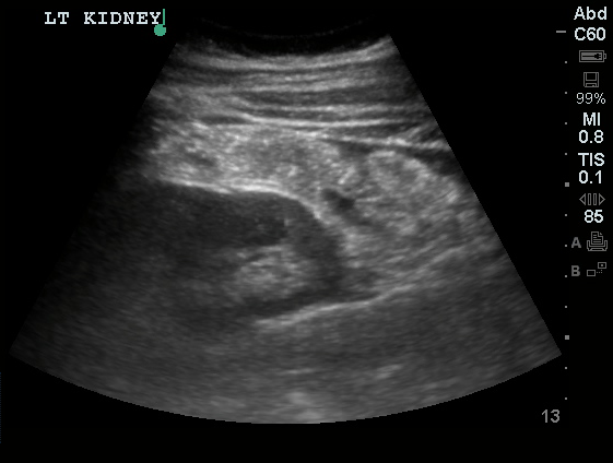
A 27 year-old female is brought in by ambulance with syncope. Pre-hospital providers report that the patient developed pelvic pain, vaginal bleeding and lost consciousness. On their arrival, her blood pressure was 80mmHg systolic, point-of-care glucose was normal – a peripheral IV was started, fluids were administered and the patient was transported to the emergency department. On arrival, vital signs were notable for tachycardia and hypotension. The patient was lethargic, maintaining arousal only with constant verbal or noxious stimulation. Her abdomen was markedly tender throughout with rebound and involuntary guarding. Her last menstrual period was 5 weeks ago and she suspected that she was pregnant. Peripheral venous access was expanded and uncrossmatched blood products were rapidly transfused. Whole blood on a point-of-care pregnancy test was positive1, and a bedside FAST demonstrated free intraperitoneal fluid in the hepatorenal recess with large free pelvic fluid. Gynecology was consulted for emergent operative management of suspected ruptured ectopic pregnancy with hemorrhagic shock and the patient was taken to the operating room.

Algorithm for the Evaluation of Suspected Ectopic Pregnancy

Algorithm for the evaluation of ectopic pregnancy

Gallery

The POCUS Atlas
The ultrasound images and videos used in this post come from The POCUS Atlas, a collaborative collection focusing on rare, exotic and perfectly captured ultrasound images.
The POCUS Atlas

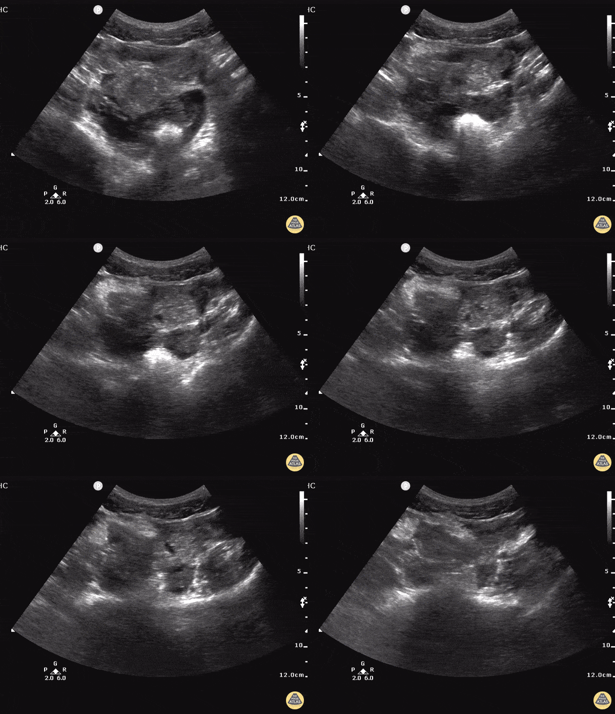
Ruptured Cornual Ectopic

Tubal Ectopic Pregnancy

Tubal Ectopic Pregnancy

Ectopic Pregnancy

Ectopic Pregnancy

Positive FAST in Ruptured Ectopic

Positive FAST in Ruptured Ectopic

The evaluation of suspected ectopic pregnancy, as with all complaints in the emergency department, begins with an assessment of patient stability: airway, breathing and circulation. The unstable patient requires immediate interventions to secure each critical component, all temporizing measures until the patient can be taken to the operating room for definitive management.

The evaluation and management algorithm for stable patients is dependent on findings of transabdominal & transvaginal ultrasonography, quantitative hCG level (relative to the institution-dependent discriminatory zone), and the identification of high risk historical and examination features that would prompt specialist consultation despite otherwise benign diagnostic tests.

If ultrasonography demonstrates a definite ectopic pregnancy (extrauterine live embryo,  adnexal mass containing yolk sac), gynecology consultation is warranted – the table below details candidates for attempts at pharmacologic therapy.

Requirements for methotrexate administration2,3

Absolute
Hemodynamic stability
Ultrasound findings consistent with an ectopic pregnancy
Willingness of the patient to adhere to close follow-up
No existing organ dysfunction: hepatic, renal, pulmonary, hematologic, immune
Relative
Unruptured ectopic mass <3.5cm
No fetal cardiac activity detected
hCG <5000 mIU/L

If an intrauterine pregnancy is identified such as a live embryo or yolk sac, barring the presence of risk factors for heterotopic pregnancy (namely, the use of assisted fertilization methods 2, 4-6), then an alternative cause for the patient’s symptoms should be sought.

If the ultrasound is non-diagnostic, patients should be stratified according to risk based on historical features, examination findings and quantitative hCG. If the hCG is above the institutional discriminatory zone, the absence of a definitive IUP is concerning, elevating suspicion for a non-visualized ectopic and warrants gynecology consultation. If the hCG is below the discriminatory zone, then certain features such as the presence of abdominal, adnexal or cervical motion tenderness, or high-risk ultrasonographic features including greater-than-moderate free pelvic fluid, complex fluid, or complex adnexal masses may be secondary features of ectopic pregnancy – again warranting consultation. If no high-risk features are present, close follow-up with repeat hCG and ultrasonography is reasonable.

Risk factors for ectopic pregnancy3

Risk factor OR
Previous tubal surgery 21
Sterilization 9.3
Previous ectopic 8.3
In utero exposure to diethylstilbestrol 5.6
Current IUD 5.0
History of PID 3.4
Infertility 2.7
Advanced maternal age 1.4-2.9
Smoking 1.5-3.9

Examination Findings in Ectopic Pregnancy6

Finding LR+
Cervical motion tenderness 4.9
Peritoneal irritation 4.2
Adnexal mass 2.4
Adnexal tenderness 1.9

Ultrasound Findings in Ectopic Pregnancy 7

Finding LR+
Ectopic cardiac activity >100
Ectopic gestational sac 23
Ectopic mass and fluid in Pouch of Douglas 9.9
Fluid in Pouch of Douglas 4.4
Ectopic mass 3.6
No IUP 2.2
Normal adnexa 0.55

Algorithm for the Evaluation of Vaginal Bleeding

Algorithm for the evaluation of vaginal bleeding

References:

  1. Fromm C, Likourezos A, Haines L, Khan ANGA, Williams J, Berezow J. Substituting whole blood for urine in a bedside pregnancy test. J Emerg Med. 2012;43(3):478-482. doi:10.1016/j.jemermed.2011.05.028.
  2. Bhatt S, Ghazale H, Dogra VS. Sonographic Evaluation of Ectopic Pregnancy. Radiol Clin North Am. 2007;45(3):549-560. doi:10.1016/j.rcl.2007.04.009.
  3. Barash JH, Buchanan EM, Hillson C. Diagnosis and management of ectopic pregnancy. Am Fam Physician. 2014;90(1):34-40.
  4. Lin EP, Bhatt S, Dogra VS. Diagnostic Clues to Ectopic Pregnancy. Radiographics. 2008;28(6):1661-1671. doi:10.1148/rg.286085506.
  5. Winder S, Reid S, Condous G. Ultrasound diagnosis of ectopic pregnancy. Australas J Ultrasound Med. 2011;14(2):29-33. doi:10.1002/j.2205-0140.2011.tb00192.x.
  6. Crochet JR, Bastian LA, Chireau MV. Does this woman have an ectopic pregnancy?: the rational clinical examination systematic review. JAMA. 2013;309(16):1722-1729. doi:10.1001/jama.2013.3914.
  7. Mol BW, van Der Veen F, Bossuyt PM. Implementation of probabilistic decision rules improves the predictive values of algorithms in the diagnostic management of ectopic pregnancy. Hum Reprod. 1999;14(11):2855-2862.
  8. First-Trimester Emergencies: A Practical Approach To Abdominal Pain And Vaginal Bleeding In Early Pregnancy. October 2003:1-20.
  9. Paspulati RM, Bhatt S, Nour S. Sonographic evaluation of first-trimester bleeding. Radiol Clin North Am. 2004;42(2):297-314. doi:10.1016/j.rcl.2004.01.005.
  10. Anderson FWJ, Hogan JG, Ansbacher R. Sudden Death: Ectopic Pregnancy Mortality. Obstet Gynecol. 2004;103(6):1218-1223. doi:10.1097/01.AOG.0000127595.54974.0c.
  11. Lozeau A-M, Potter B. Diagnosis and management of ectopic pregnancy. Am Fam Physician. 2005;72(9):1707-1714.
  12. Stone MB. Emergency Ultrasound Diagnosis of Ruptured Ectopic Pregnancy. Academic Emergency Medicine. 2009;16(12):1378-1378. doi:10.1111/j.1553-2712.2009.00538.x.
  13. Stein JC, Wang R, Adler N, et al. Emergency Physician Ultrasonography for Evaluating Patients at Risk for Ectopic Pregnancy: A Meta-Analysis. Ann Emerg Med. 2010;56(6):674-683. doi:10.1016/j.annemergmed.2010.06.563.
  14. Fromm C, Likourezos A, Haines L, Khan ANGA, Williams J, Berezow J. Substituting whole blood for urine in a bedside pregnancy test. J Emerg Med. 2012;43(3):478-482. doi:10.1016/j.jemermed.2011.05.028.
  15. Alkatout I, Honemeyer U, Strauss A, et al. Clinical diagnosis and treatment of ectopic pregnancy. Obstet Gynecol Surv. 2013;68(8):571-581. doi:10.1097/OGX.0b013e31829cdbeb.
  16. Arleo EK, DeFilippis EM. Cornual, interstitial, and angular pregnancies: clarifying the terms and a review of the literature. Clinical Imaging. 2014;38(6):763-770. doi:10.1016/j.clinimag.2014.04.002.
  17. Rodgers SK, Chang C, DeBardeleben JT, Horrow MM. Normal and Abnormal US Findings in Early First-Trimester Pregnancy: Review of the Society of Radiologists in Ultrasound 2012 Consensus Panel Recommendations. Radiographics. 2015;35(7):2135-2148. doi:10.1148/rg.2015150092.
  18. Diagnosis and Management of Ectopic Pregnancy: Green-top Guideline No. 21. BJOG. 2016;123(13):e15-e55. doi:10.1111/1471-0528.14189.
  19. Hahn SA, Promes SB, Brown MD, et al. Clinical Policy: Critical Issues in the Initial Evaluation and Management of Patients Presenting to the Emergency Department in Early Pregnancy. Ann Emerg Med. 2017;69(2):241–250.e20. doi:10.1016/j.annemergmed.2016.11.002.
  20. Lee R, Dupuis C, Chen B, Smith A, Kim YH. Diagnosing ectopic pregnancy in the emergency setting. Ultrasonography. 2018;37(1):78-87. doi:10.14366/usg.17044.

Acute Pelvic Pain

Evaluation of Acute Pelvic Pain

Acute Pelvic Pain

Key Historical Findings

Location
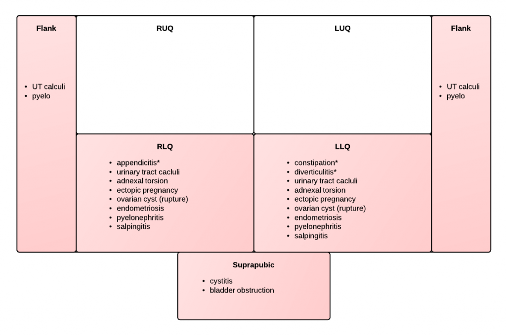
Lateralized: suggests process related to tube or ovary, consider unilateral urinary tract process. On right, add appendicitis to differential; on left, add diverticulitis (particularly if age >40.
Central: suggests process involving uterus, bladder or bilateral adnexa
Diffuse: suggests PID
Radiation
Radiation to rectum suggests pooling of fluid or blood in cul-de-sac
Onset
Abrupt: suggests acute intrapelvic hemorrhage (from ruptured ectopic or ovarian cyst), ovarian torsion, urolithiasis
Gradual: inflammatory process such as PID
Chronic/recurrent: suggests endometriosis, recurrent ovarian cyst, ovarian mass
Associated Symptoms
Fevers/chills: suggests infectious process
Nausea/vomiting: suggests process involving gastrointestinal tract, though may accompany pregnancy or severe pain associated with ovarian torsion, urolithiasis.
Dysuria: suggests process involving urinary tract, though may be associated with local vulvar/vaginal process
Urinary urgency: more specific for bladder or urethral irritation
Obstetric History
History of recurrent spontaneous abortions or prior ectopic pregnancy increases likelihood of recurrence.
Ongoing fertility treatments increase likelihood for ectopic/heterotopic (occurs in 1:100 with assisted reproduction compared to 1:8000 in general population)
Vaginal Bleeding
In non-pregnant: suggests PID, DUB, cervical or uterine cancer
In early pregnancy: may be associated with ectopic pregnancy, non-viable IUP, or subchorionic hemorrhage
In late pregnancy: may be associated with placental pathology (previa, abruption)

Key Physical Findings

  • Pelvic examination: assists with localization of lateralized process. Should be preceded by ultrasound if >20 weeks.
  • Abnormal vaginal discharge: suggests vaginitis, cervicitis, PID, or retained foreign body.
  • Cervical motion tenderness: suggests reproductive tract inflammation or irritation of adjacent structures (appendicitis, cystitis)
  • Unilateral adnexal mass/tenderness: associated with ovarian cyst/mass, TOA, ectopic, or ovarian torsion.

References:

  1. Hart, D., & Lipsky, A. (2013). Acute Pelvic Pain in Women. In Rosen's Emergency Medicine – Concepts and Clinical Practice (8th ed., Vol. 1, pp. 266-272). Elsevier Health Sciences.
  2. WikEM: Pelvic pain

Ectopic Pregnancy

HPI:

32F G8P7A2 at 5 weeks by LMP presenting with abdominal pain. The patient reports acute onset of sharp left lower abdominal pain 1.5 hours prior to presentation. The pain has been constant since onset, 10/10 in severity, radiating to lower back and exacerbated with movement. She denies vaginal bleeding or discharge, passage of clots or other products. She also denies trauma, lightheadedness/dizziness/syncope, shortness of breath, nausea/vomiting or changes in bowel or urinary habits.
Her pregnancy was detected 3 weeks ago with a home pregnancy test and was confirmed at her PCP one week later. She has not had an ultrasound during this pregnancy but has a history of uterine fibroids. She has no history of sexually transmitted infections, prior ectopic pregnancy, or use of assisted fertilization.

PMH:

  • HTN
  • Uterine fibroids

PSH:

None

FH:

Non-contributory

SHx:

  • Denies tobacco, alcohol or drug use.
  • Sexually active with husband only, no history of STI.

Meds:

None

Allergies:

NKDA

Physical Exam:

VS: T 37.4 HR 108 RR 36 BP 148/104 O2 99% RA
Gen: Alert and oriented female, appears uncomfortable due to pain.
HEENT: PERRL, EOMI, MMM.
CV: Tachycardia, regular rhythm, no murmurs.
Lungs: CTAB, no crackles.
Abd: Normoactive bowel sounds, tenderness to palpation in LLQ and suprapubic area, with guarding but no rebound tenderness. No CVAT.
GU: No external lesions. Closed cervical os, no blood or discharge, +CMT.
Ext: Warm, well-perfused with strong peripheral pulses.

Labs/Studies:

  • POC Hemoglobin: 11.8
  • POC ICON: positive

Imaging:

Bedside Ultrasound

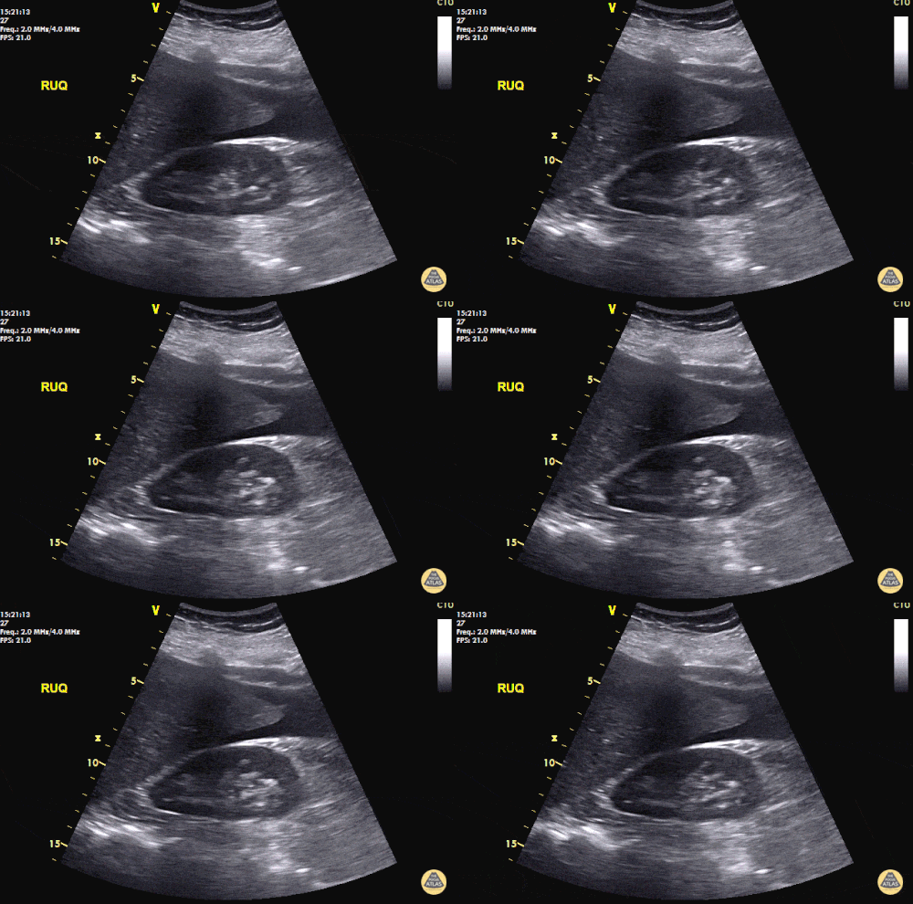
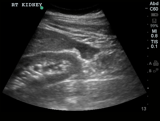
  1. Transabdominal: Free fluid in hepatorenal and splenorenal recesses
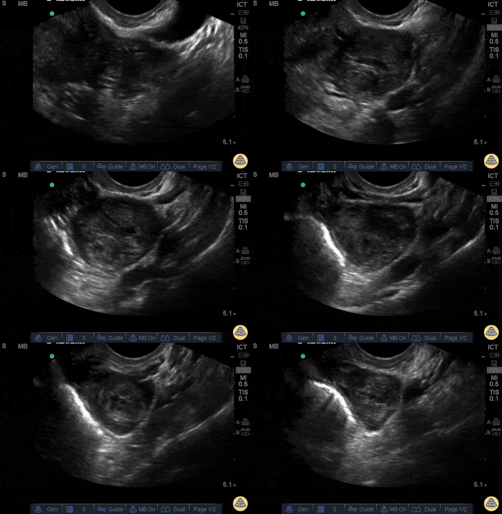
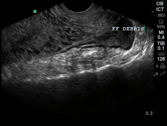
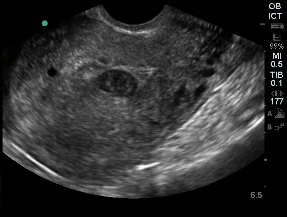
  2. Transvaginal: Free fluid and debris in posterior cul-de-sac, likely pseudogestational sac in endometrial cavity, no IUP identified. Formal ultrasound revealed fetus with cardiac activity in left adnexa.
Hepatorenal free fluid

Hepatorenal free fluid

Free fluid in the hepatorenal recess (Morison's Pouch)

Splenorenal Free Fluid

Splenorenal Free Fluid

Free fluid in the splenorenal recess.

Pelvic Free Fluid

Pelvic Free Fluid

Free fluid and debris in the posterior cul-de-sac.

Pseudogestational Sac

Pseudogestational Sac

No obvious yolk sac or fetal pole.

Assessment/Plan:

32 year-old ICON positive female with acute-onset pelvic pain. The patient remained hemodynamically stable and absence of definitive IUP on bedside ultrasound was confirmed with presence of fetal cardiac activity in left adnexa indicative of ectopic pregnancy. OB-Gyn was consulted and the patient was taken emergently to the OR.

Differential Diagnosis of First Trimester Abdominal Pain: 1

Differential Diagnosis of 1st Trimester Abdominal Pain

Initial Evaluation of First Trimester Abdominal Pain: 1

  • 2 large-bore IV’s, begin fluid resuscitation
  • POC testing: hemoglobin, urine pregnancy
  • CBC, type and cross (Rh), serum B-hCG
  • Emergent bedside ultrasound

Features Associated with Ectopic Pregnancy: 1

  • History
    • PID
    • Tubal ligation
    • Prior ectopic
    • IUD
    • Assisted fertilization
  • Physical
    • CMT
    • Peritoneal irritation
  • Ultrasound
    • Empty uterus
    • Adnexal mass
    • Free fluid
  • Ultrasonographic Findings in the Evaluation of Ectopic Pregnancy: 2

    • Discriminatory hCG (1500-3000 mIU/mL): absence of IUP suggests ectopic or abnormal gestation
    • Normal IUP
      • 4-5wks: gestational sac (0.2-0.5cm)
      • 5wks: two echogenic rings
      • 5.5wks: yolk sac
      • 6wk: embryonic pole
      • 6.5wk: fetal cardiac activity
    • Abnormal IUP
      • >2cm gestational sac without fetal pole
      • CRL >0.5cm without cardiac activity
    • Ectopic
      • Extrauterine gestational sac with or without cardiac activity
      • Extrauterine ring sign
      • Non-homogenous adnexal mass

    Contraindications to Medical Management: 2,3

    • Absolute
      • Breast-feeding
      • Immunodeficiency
      • PUD
      • Pulmonary, hepatic or renal dysfunction
    • Relative
      • Ectopic mass > 3.5cm
      • Fetal cardiac activity

    References:

    1. Dart, R. (2003). First Trimester Emergencies A Practical Approach To Abdominal Pain And Vaginal Bleeding In Early Pregnancy. EB Medicine, 5(11), 1–20.
    2. Barnhart, K. T. (2009). Clinical practice. Ectopic pregnancy. The New England journal of medicine, 361(4), 379–387. doi:10.1056/NEJMcp0810384
    3. Jurkovic, D., & Wilkinson, H. (2011). Diagnosis and management of ectopic pregnancy. BMJ (Clinical research ed.), 342(jun10 1), d3397–d3397. doi:10.1136/bmj.d3397

    Acute Pelvic Pain

    Pelvic US - free fluidID:

    19yo G0, hCG negative, presenting with lower abdominal pain for 3 weeks.

    HPI:

    The patient states that she has had progressively worsening lower abdominal pain for the past three weeks. She describes the pain as constant, cramping, currently 8/10 in severity with radiation to the right flank. The pain is improved somewhat with ibuprofen and worsened with movement. She reports subjective F/C, some vaginal bleeding, but no other discharge and no dysuria. She is sexually active with one partner, using condoms occasionally. She has no significant PMH, no history of STI and a PSH of appendectomy. She denies any current or prior T/E/D use.

    PE:

    • VS: 110/60mmHg, 60bpm, 99.5°, 16/min
    • HEENT: NC/AT, PERRL, EOMI, MMM w/o lesions
    • CV: RRR, no M/G/R
    • Lungs: CTAB
    • Abd: +BS, soft, non-distended, TTP RLQ > LLQ, no rebound/guarding
    • Back: no CVAT
    • Pelvic: external genitalia normal, scant blood in vault, os closed, no discharge, + CMT, + uterine tenderness, + adnexal tenderness

    Labs/Imaging:

    • TVUS: normal appearing uterus/adnexa, possible free fluid (hemorrhagic vs. inflammatory) in pelvis
    • Wet mount: negative
    • Urine dip: -LE/nitrites, -protein, +blood

    Assessment:

    19yo G0, hCG negative, with pelvic pain and vaginal bleeding. Findings of lower abdominal tenderness, cervical motion/uterine/adnexal tenderness on examination, and low-grade fever are suggestive of PID. Other considerations include UTI, however, absence of dysuria, CVAT, and negative urine dip do not support this diagnosis. Will evaluate further with GC/CT, and treat empirically with ceftriaxone 250mg IM x1 and doxycycline 100mg PO BID x14d with follow-up in 48h.

    Differential Diagnosis of Acute Pelvic Pain:

    Common causes:

    • Gynecologic
      • PID, TOA
      • Neoplasm (torsion, rupture)
      • Leiomyoma (torsion, degeneration)
      • Endometriosis
      • Endometritis
      • Ectopic pregnancy
      • SAB
    • Obstetric
      • Labor
      • Uterine rupture
      • Abruptio placentae
      • Diastasis symphesis pubis
    • Non-gynecologic
      • Appendicitis
      • Cystitis (UTI)
      • Diverticulitis
      • Urinary tract calculi
      • Abdominal wall trauma

     Location of pain:

    LocationOfPain

    ROS:

    • discharge + dyspareunia: PID
    • missed menses + cramping/bleeding: SAB, ectopic
    • anorexia, N/V: appendicitis, torsion

    A System for the Evaluation and Management of PID:

    • Pathogens: GC, CT, gardnerella, haemophilus
    • Evaluation:
      • Pelvic exam: CMT, uterine tenderness, adnexal tenderness, abnormal discharge, wet mount WBC’s
      • Labs: + GC/CT, ↑ ESR/CRP
      • VS: T > 101°
      • Imaging: thickened tubes, free pelvic fluid, TOA
    • Indications for admission:
      • Acute abdomen, toxic appearance, unstable VS
      • Pregnancy
      • Failed outpatient, can’t tolerate PO
      • TOA
    • Outpatient management:
      • Ceftriaxone 250mg IM x1
      • Doxycycline 100mg PO BID x14d
      • ± Metronidazole 500mg PO BID x14d

    References:

    1. Hacker and Moore’s essentials of obstetrics and gynecology. Philadelphia, PA: Saunders/Elsevier, 2010.
    2. CDC – Pelvic Inflammatory Disease – 2010 STD Treatment Guidelines: http://www.cdc.gov/std/treatment/2010/pid.htm