Seizure

Brief HPI

A 72 year-old male with a history of hypertension and hiatal hernia presents to the emergency department with one week of generalized weakness. His family report decreased oral intake with frequent emesis over the past four days. He denies chest pain, shortness of breath, abdominal pain, or other complaints. During the interview he has a generalized tonic-clonic seizure which persists for five minutes despite the administration of 4mg of lorazepam.

An Algorithm for the Management of Seizures

The management of active seizures is algorithmic, starting with a rapid assessment of airway patency, supporting ventilation (with appropriate positioning, nasopharyngeal airway adjuncts and bag-valve mask if needed) and ensuring adequate perfusion. Patients should have continuous vital sign monitoring, supplemental oxygen to maintain oxygen saturation >92% and intravenous access1.

Pharmacologic treatment follows a stepwise approach, detailed in the algorithm below. The focus is on immediate stabilization and progressively escalating anti-epileptic drugs eventually requiring endotracheal intubation and continuous infusions of sedatives2-4.

An Algorithm for the Management of Seizures

Pathophysiology

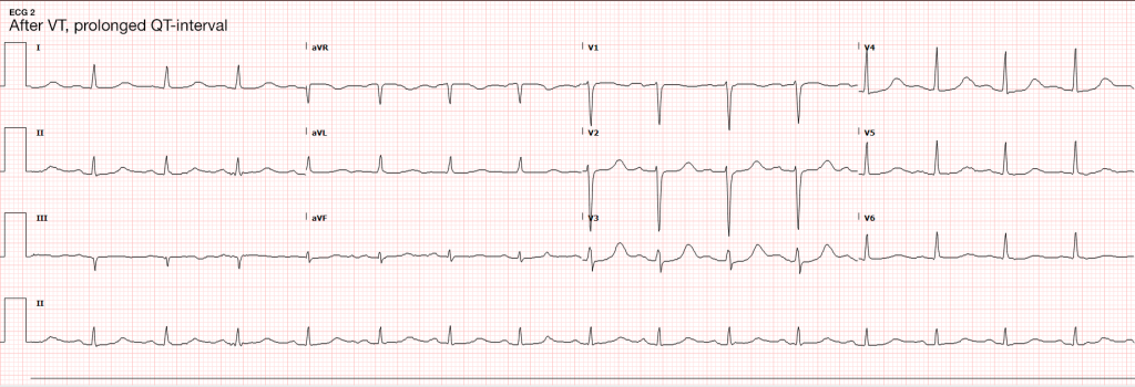
Seizures are caused by excessive and disorganized neuronal activation, typically induced by global alterations in the production and transmission of impulses (electrolyte derangements, drugs/toxins, infection), or foci of increased irritability (hemorrhage, stroke, mass) – a pathophysiologic motif that mimics cardiac tachyarrhythmias (sympathomimetic toxicity or scarred myocardium for example)1. Status epilepticus, defined as a seizure lasting greater than five minutes or recurrent seizures without a return to normal baseline, shares an equally high short-term mortality – greater than 20%5.
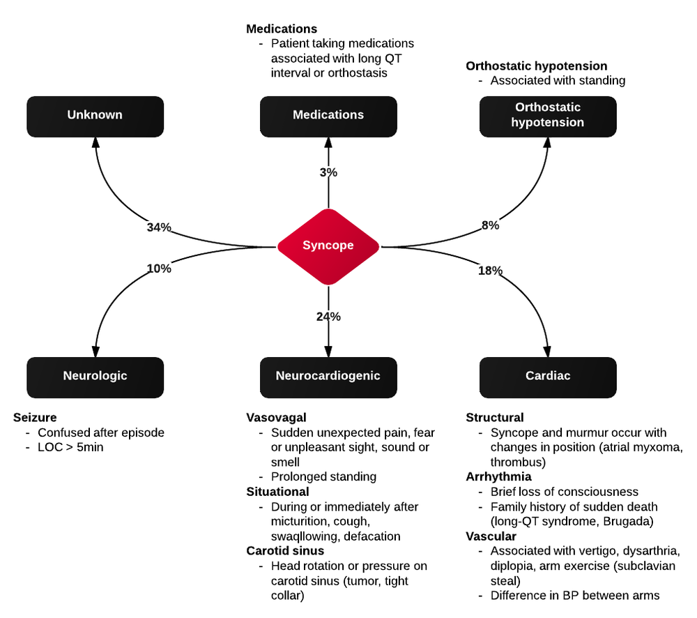
Syncope vs. Seizure

The algorithm below details historical and examination features that may assist with distinguishing epileptic seizure from non-epileptic activity6,7.

Clinical Features Distinguishing Seizure from Syncope

Case Conclusion

The patient continued to seize and a point-of-care chemistry panel revealed a serum sodium of 108mEq/L. Seizures abate after the infusion of hypertonic saline (100mL of 3% saline over 10 minutes, repeated until cessation of seizures). While hyponatremia is generally corrected slowly – owing to the risk of osmotic demyelination – immediate correction in this setting is critical8.


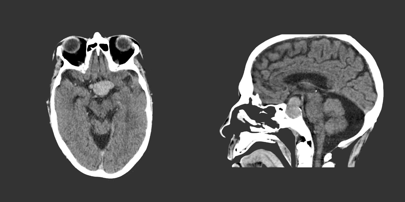
The remainder of the patient’s evaluation demonstrated urine osmolarity is 389mOsm/kg and urine sodium is 53mmol/L, in the setting of relative euvolemia on examination these findings were consistent with SIADH. Head computed tomography is obtained and reveals a sellar mass.

View Hyponatremia Algorithm

References

  1. McMullan JT, Davitt AM, Pollack CV Jr. Seizures. In: Rosen’s Emergency Medicine. Mosby Incorporated; 2002:2808. doi:10.1016/S1474-4422(06)70350-7.
  2. Billington M, Kandalaft OR, Aisiku IP. Adult Status Epilepticus: A Review of the Prehospital and Emergency Department Management. J Clin Med. 2016;5(9):74. doi:10.3390/jcm5090074.
  3. Huff JS, Morris DL, Kothari RU, Gibbs MA, Emergency Medicine Seizure Study Group. Emergency department management of patients with seizures: a multicenter study. Academic Emergency Medicine. 2001;8(6):622-628.
  4. Prasad M, Krishnan PR, Sequeira R, Al-Roomi K. Anticonvulsant therapy for status epilepticus. Prasad M, ed. Cochrane Database Syst Rev. 2014;16(9):CD003723. doi:10.1002/14651858.CD003723.pub3.
  5. Logroscino G, Hesdorffer DC, Cascino G, Annegers JF, Hauser WA. Short-term mortality after a first episode of status epilepticus. Epilepsia. 1997;38(12):1344-1349.
  6. Sheldon R, Rose S, Ritchie D, et al. Historical criteria that distinguish syncope from seizures. J Am Coll Cardiol. 2002;40(1):142-148.
  7. McKeon A, Vaughan C, Delanty N. Seizure versus syncope. Lancet Neurol. 2006;5(2):171-180. doi:10.1016/S1474-4422(06)70350-7.
  8. Goh KP. Management of hyponatremia. Am Fam Physician. 2004;69(10):2387-2394.

ECG Guide: Pediatrics

ECG Standard

  • Full standard: no adjustment
  • Half-standard: commensurate reduction in amplitude (usually 50%)
  • Mixed: reduction in amplitude of precordial leads

Atrial Abnormalities

Right Atrial Abnormality (P pulmonale)
Peaked P-wave in II (>3mm from 0-6mo or >2.5mm >6mo)
Causes: right atrial volume overload, ASD, Ebstein, Fontan
Left Atrial Abnormality (P mitrale)
Wide, notched P-wave in II or biphasic in V1
Causes: MS, MR

Axis

  • Anatomical dominance of right ventricle until approximately 6mo
  • RAD normal
  • eRAD suggests AV canal defect

T-waves

  • 1st week of life: Upright
  • Adolescent: Inverted
  • Adult: Upright

Ventricular Hypertrophy

Right Ventricular Hypertrophy
R-wave height >98% for age in lead V1
S-wave depth >98% for age in lead V6
T-wave abnormality (ex. upright in childhood)
Causes: pHTN, PS, ToF
Left Ventricular Hypertrophy
R-wave height >98% for age in lead V6
S-wave depth >98% for age in lead V1
Adult-pattern R-wave progression in newborn (no large R-waves and small S-waves in right precordial leads)
Left-axis deviation
Causes: AS, coarctation, VSD, PDA

Examples


Normal Neonatal ECG

  • 2mo old
  • RAD
  • Inverted T-waves (normal)
  • Tall R-waves in V1-V3


Extreme Axis Deviation

  • Neonate with Down syndrome
  • Isoelectric in I, Negative in aVF negative in II  mean QRS vector -87°
  • Extreme RAD suggestive of AV canal defect


LVH:

  • Unrepaired Coarctation
  • Deep S-wave in V1 (>98%)
  • Tall R-wave in V6 (>98%)


RVH:

  • 10 year-old boy with pulmonary Hypertension
  • RAD after expected age for normal RAD
  • Tall R-waves in V1 (>98%)
  • Deep S-wave in V6 (>98%)


STEMI

  • ALCAPA (anomalous origin of the left coronary artery from the pulmonary artery): coronary artery arises anomalously from the pulmonary artery; as pulmonary arterial pressure falls during the first 6 months of infancy, prograde flow through the left coronary artery ceases and may even reverse.
  • HLHS (hypoplastic left heart syndrome): coronary arteries are perfused from a hypoplastic, narrow aorta that is susceptible to flow disruption
  • Orthotopic heart transplant with allograft vasculopathy
  • Kawasaki: coronary artery aneurysm with subsequent thrombosis


Benign early repolarization

  • 14 year-old male
  • Concave ST-segment elevation


Left Atrial Abnormality:

  • 9mo female with mitral insufficiency
  • Broad biphasic P-wave in V1
  • Tall, notched P-wave in II


Prolonged QT interval

  • 18-year-old female
  • Familial long QT syndrome and a history of cardiac arrest


WPW:

  • Delta wave, shortened PR interval

References

  1. O’Connor M, McDaniel N, Brady WJ. The pediatric electrocardiogram. Part I: Age-related interpretation. Am J Emerg Med. 2008;26(2):221-228. doi:10.1016/j.ajem.2007.08.003.
  2. Goodacre S, McLeod K. ABC of clinical electrocardiography: Paediatric electrocardiography. BMJ. 2002;324(7350):1382-1385.
  3. O’Connor M, McDaniel N, Brady WJ. The pediatric electrocardiogram Part II: Dysrhythmias. Am J Emerg Med. 2008;26(3):348-358. doi:10.1016/j.ajem.2007.07.034.
  4. O’Connor M, McDaniel N, Brady WJ. The pediatric electrocardiogram Part III: Congenital heart disease and other cardiac syndromes. Am J Emerg Med. 2008;26(4):497-503. doi:10.1016/j.ajem.2007.08.004.
  5. Schwartz P. Guidelines for the interpretation of the neonatal electrocardiogram. Eur Heart J. 2002;23(17):1329-1344. doi:10.1053/euhj.2002.3274.

Nonsustained Ventricular Tachycardia

Case 1

64M with a history of HFrEF (LVEF 20-25%), CAD, AICD (unknown indication), COPD, CKD III presenting with gradual onset shortness of breath, progressive bilateral lower extremity edema.
Examination consistent with severe acute decompensated heart failure presumed secondary to left ventricular dysfunction.
Telemetry monitoring with multiple episodes of nonsustained ventricular tachycardia.

In the ED, the patient developed worsening respiratory failure despite initiation of therapy, requiring endotracheal intubation. Continuous cardiac monitoring revealed persistent salvos of NSVT, progressing to slow ventricular tachycardia without device intervention.
Device interrogation revealed multiple events, 3 shocks, several ATP’s over the recorded period.

Evaluation and Management:

  • NSVT with known (severe) ischemic heart disease
  • For repetitive monomorphic ventricular tachycardia: amiodarone, beta-blockade (if tolerated), procainamide (IIA, C)1

ECG’s

ECG 1
ECG 1

ECG 1

Non-specific IVCD, LAA, VPC

ECG 2
ECG 2

ECG 2

VT initiated by fusion complex

Case 2

31F with autoimmune polyglandular syndrome (adrenal, thyroid and endocrine pancreatic insufficiency), presenting with fever and cough.
Evaluation consistent with sepsis presumed secondary to pulmonary source.
Telemetry monitoring initially with ventricular bigeminy, then nonsustained ventricular tachycardia.

In the ED, the patient developed pulseless ventricular tachycardia – apparently polymorphic. Chest compressions and epinephrine produced return of spontaneous circulation with recovery to baseline neurologic function.
ECG revealed prolonged QTc and chemistry panel notable for critical hypokalemia/hypomagnesemia.

Evaluation and Management:

  • NSVT progressing to VT
  • Initially attributed to electrolyte disturbances. However, serial ECG’s continued to show prolonged QTc (possibly acquired, home medications included metoclopramide and erythromycin). Early echocardiography demonstrated global hypokinesis with EF 30-35% attributed to severe sepsis and recurrent defibrillation. Cardiac CT after resolution of acute illness showed persistently depressed ejection fraction without coronary atherosclerosis. The presence of NICM associated with malignant dysrhythmias warranted ICD placement.
  • Cardioversion for hemodynamic compromise (I, B), B-blockade (I, B), amiodarone if no LQTS (I, C), urgent angiography if ischemia not excluded (I, C)1
  • Correction of electrolyte abnormalities (specifically hypokalemia) may decrease progression to VF.2

ECG’s

ECG 1
ECG 1

ECG 1

Ventricular bigeminy

ECG 2
ECG 2

ECG 2

Long-QT

VT on Telemetry
VT on Telemetry

VT on Telemetry

Non-sustained ventricular tachycardia noted on telemetry monitoring

Definition3,4

  • > 3-5 consecutive beats originating below the AV node
  • Rate > 100bpm
  • Duration <30s

Epidemiology3,5

  • Occurs in 0-4% of ambulatory patients
  • Increased frequency in males and with increasing age
  • In some patients, NSVT is associated with an increased risk of sustained tachyarrhythmias and sudden cardiac death. In others it is of little prognostic significance.6,7,8

Evaluation

In all patients:
History: including arrhythmogenic medications/substances, pertinent family history
Physical examination
ECG/CXR
TTE
In selected patients:
Exercise testing
Advanced imaging (CT/C-MR)
Electrophysiologic studies
Genetic testing

NSVT in the absence of structural heart disease

NSVT in Idiopathic Ventricular Tachycardia

Ventricular outflow arrhythmias:
RVOT: 70-80%, LBBB pattern
LVOT: 20-30%, RBBB pattern
Mechanism:
Adrenergically mediated
Occur during exercise, resolve as heart-rate increases, recur during recovery
Management:
Exclude arrhythmogenic right ventricular cardiomyopathy (imaging, myocardial biopsy)
If symptomatic, beta-blockade, ± IC anti-arrhythmic, CCB (verapamil) for ILVT
Prognosis:
Good, rare tachycardia-induced cardiomyopathy, rare SCD

NSVT in Polymorphic Ventricular Tachycardia

Mechanism
LQTS (acquired or inherited)
Familial catecholaminergic polymorphic VT
Management
Symptomatic (ex. syncope, cardiac arrest): ICD
Asymptomatic QTc > 550ms: consider ICD
Prognosis
Increased risk SCD

Arrhythmogenic Right Ventricular Cardiomyopathy

Mechanism
Fibrosis, fibro-fatty replacement of myocardium in RVIT/RVOT/RV apex
May occur with only subtle structural abnormalities of the right ventricle
LBBB morphology
Management
Anti-arrhythmics of limited utility
Catheter ablation, ICD backup
Prognosis
Increased risk SCD

NSVT with apparent structural heart disease1

Hypertension and LVH

Mechanism
Stretch-induced abnormal automaticity
Fibrotic tissue
Presence of NSVT correlates with degree of hypertrophy and subendocardial fibrosis
Management
Evaluation for ischemic heart disease
Aggressive medical management of hypertension (including beta-blockade)
Prognosis
Unclear

Valvular Disease

Mechanism
High incidence in AS, severe MR (25%)
Mechanical stress from dysfunctional valvular apparatus
Management
Beta-blockade if symptomatic
Prognosis
No evidence that NSVT is an independent predictor of SCD.

Ischemic Heart Disease9-14

Mechanism
Monomorphic VT associated with re-entry at the borders of ventricular scars
Ischemia induces polymorphic NSVT/VF
Management
Revascularization, beta-blockade, statin, ACE/ARB
MADIT I, MUSTT: ICD for ICM LVEF <40%, NSVT, EPS inducible VT
MADIT II, SCD-HeFT: ICD for moderate-to-severe LV dysfunction irrespective of NSVT or EPS findings
Prognosis
NSTEMI with NSVT >48h after admission 2x risk SCD (MERLIN-TIMI 36)
STEMI with NSVT common, not as predictive of ACM or SCD as LVEF (CARISMA)
NSVT <24h after admission for NSTEMI/STEMI not of prognostic significance.

Hypertrophic Cardiomyopathy

Mechanism
Genetic myocardial disease
Myocyte disarray, fibrosis, ischemia result in arrhythmogenic substrate
Management
Restriction of physical activity
ICD (NSVT, LV thickness, FH SCD, syncope, abnormal BP response to exercise)
Beta-blockade, anti-arrhythmic for symptoms
Prognosis
Increased risk SCD (1% annual)

Other Conditions

  • Non-ischemic dilated cardiomyopathy
  • Giant-cell myocarditis
  • Repaired TOF
  • Amyloidosis
  • Sarcoidosis
  • Chagas cardiomyopathy

Algorithm for the Evaluation of NSVT1

Algorithm for the Evaluation of Nonsustained Ventricular Tachycardia

References

  1. Zipes DP, Camm AJ, Borggrefe M, et al. ACC/AHA/ESC 2006 guidelines for management of patients with ventricular arrhythmias and the prevention of sudden cardiac death–executive summary: A report of the American College of Cardiology/American Heart Association Task Force and the European Society of Cardiology Committee for Practice Guidelines (Writing Committee to Develop Guidelines for Management of Patients with Ventricular Arrhythmias and the Prevention of Sudden Cardiac Death) Developed in collaboration with the European Heart Rhythm Association and the Heart Rhythm Society. Eur Heart J. 2006;27(17):2099–2140. doi:10.1093/eurheartj/ehl199.
  2. Higham PD, Adams PC, Murray A, Campbell RW. Plasma potassium, serum magnesium and ventricular fibrillation: a prospective study. Q J Med. 1993;86(9):609–617.
  3. Katritsis DG, Zareba W, Camm AJ. Nonsustained ventricular tachycardia. J Am Coll Cardiol. 2012;60(20):1993–2004. doi:10.1016/j.jacc.2011.12.063.
  4. Katritsis DG, Camm AJ. Nonsustained ventricular tachycardia: where do we stand? Eur Heart J. 2004;25(13):1093–1099. doi:10.1016/j.ehj.2004.03.022.
  5. Wellens HJ. Electrophysiology: Ventricular tachycardia: diagnosis of broad QRS complex tachycardia. Heart. 2001;86(5):579–585.
  6. Buxton AE, Lee KL, Fisher JD, Josephson ME, Prystowsky EN, Hafley G. A randomized study of the prevention of sudden death in patients with coronary artery disease. Multicenter Unsustained Tachycardia Trial Investigators. N Engl J Med. 1999;341(25):1882–1890. doi:10.1056/NEJM199912163412503.
  7. Jouven X, Zureik M, Desnos M, Courbon D, Ducimetière P. Long-term outcome in asymptomatic men with exercise-induced premature ventricular depolarizations. N Engl J Med. 2000;343(12):826–833. doi:10.1056/NEJM200009213431201.
  8. Udall JA, Ellestad MH. Predictive implications of ventricular premature contractions associated with treadmill stress testing. Circulation. 1977;56(6):985–989.
  9. Preliminary report: effect of encainide and flecainide on mortality in a randomized trial of arrhythmia suppression after myocardial infarction. The Cardiac Arrhythmia Suppression Trial (CAST) Investigators. N Engl J Med. 1989;321(6):406–412. doi:10.1056/NEJM198908103210629.
  10. Goldstein S. Propranolol therapy in patients with acute myocardial infarction: the Beta-Blocker Heart Attack Trial. Circulation. 1983;67(6 Pt 2):I53–7.
  11. Moss AJ. MADIT-I and MADIT-II. J Cardiovasc Electrophysiol. 2003;14(9 Suppl):S96–8.
  12. Moss AJ, Hall WJ, Cannom DS, et al. Improved survival with an implanted defibrillator in patients with coronary disease at high risk for ventricular arrhythmia. Multicenter Automatic Defibrillator Implantation Trial Investigators. N Engl J Med. 1996;335(26):1933–1940. doi:10.1056/NEJM199612263352601.
  13. Buxton AE, Lee KL, Fisher JD, Josephson ME, Prystowsky EN, Hafley G. A randomized study of the prevention of sudden death in patients with coronary artery disease. Multicenter Unsustained Tachycardia Trial Investigators. N Engl J Med. 1999;341(25):1882–1890. doi:10.1056/NEJM199912163412503.
  14. Bardy GH, Lee KL, Mark DB, et al. Amiodarone or an implantable cardioverter-defibrillator for congestive heart failure. N Engl J Med. 2005;352(3):225–237. doi:10.1056/NEJMoa043399.
  15. WikEM: Nonsustained Ventricular Tachycardia

Seizure

Definition

Seizure
Pathologic neuronal activation leading to abnormal function
Epilepsy
Recurrent unprovoked seizures

Classification

  • Cause
    • Primary: Unprovoked
    • Secondary: Provoked, caused by trauma, illness, intoxication, metabolic disturbances, etc.
  • Effect on mentation
    • Generalized: involvement of both hemispheres with associated loss of consciousness (tonic-clonic, absence, atonic, myoclonic)
    • Focal: Involving single hemisphere with preserved level of consciousness
  • Status epilepticus
    • Witnessed convulsions lasting >5min
    • Recurrent seizure without recovery from postictal period

Causes of Seizures

Causes of Seizures

Management of Seizures

Management of Seizures

Medications for Treatment of Seizures

Medication Dose (adult) Dose (peds) Comment
1st Line
Lorazepam 4mg IV <13kg: 0.1mg/kg (max 2mg)
13-39kg: 2mg

>39kg: 4mg
Repeat in 10min
Midazolam 10mg IM 0.2mg/kg IM (max 5mg) Repeat in 10min
Midazolam 10mg buccal 0.5mg/kg buccal (max 5mg) Repeat in 10min
2nd Line
Fosphenytoin 20mg PE/kg IV    
Phenytoin 20mg/kg IV   May cause hypotension
3rd Line
Midazolam 0.05-2mg/kg/hr    
Propofol 1-2mg/kg bolus then 20-200mcg/kg/min    
Pentobarbital 5-15mg/kg bolus then 0.5-5mg/kg/hr    
Special Conditions
Glucose 50mL D50/W   Hypoglycemia
MgSO4 6g IV over 15min   Eclampsia (20wks gestation to 6wks post-partum)
Pyridoxine 0.5g/min until seizures stop, max 5g   INH ingestion
3% saline 100-200mL over 1-2h   Confirmed hyponatremia

History

Points suggestive of seizure over alternative process
Abrupt onset
Duration < 120s
LOC
Purposeless activity: automatisms, tonic-clonic
Provocation: fever in children, substance withdrawal
Postictal state
Retrograde amnesia
Incontinence, oral trauma (buccal maceration, tongue laceration)
Rapidly resolving lactic acidosis
Important historical points for patients with seizure history
Recent illness
Medications (adherence, changes, interactions)
Substance use
Ictogenic factors
Recent/remote head trauma
Developmental abnormalities
Substance use
Sleep deprivation
Pregnancy

Key Physical Examination Findings

  • Vital sign abnormalities persisting beyond immediate postictal state (may suggest drug/toxin exposure, CNS lesion)
  • Nuchal rigidity
  • Signs of IVDA
  • Sequela

    • Head trauma
    • Tongue laceration
    • Shoulder dislocation (posterior)
  • Neurological exam

    • Stroke
    • Elevated ICP
    • Failure to note improvement in postictal confusion (encephalopathy, subclinical seizures)

Labs

  • Glucose
  • BMP (Na, Ca, Mg)
  • AED levels
  • CBC (leukocytosis and bandemia common post-seizure)
  • CSF
  • B-hCG
  • LFT (hepatic dysfunction, alcoholic hepatitis)
  • Lactate (rapidly resolves on repeat)

Indications for Imaging

  • New seizures
  • History of trauma
  • History of malignancy
  • Immunocompromised
  • Headache
  • Anti-coagulation
  • Focal neurological exam
  • Persistent AMS

References

  1. McMullan, J., Davitt, A., & Pollack, C. (2013). Seizures. In Rosen’s Emergency Medicine – Concepts and Clinical Practice (8th ed., Vol. 1, pp. 156-161). Elsevier Health Sciences
  2. WikEM: Seizure

Syncope

Causes of Syncope

Causes of Syncope

History

  • Rate of onset
  • Position at onset
  • Duration, rate of recovery
  • Preceding features

    • Obstruction: associated with exertion
    • Neurocardiogenic: associated with emotion, micturition, bowel movement, emesis, neck movement
  • Following features

    • Seizure: Postictal confusion
    • Hypotension: Initial VS
    • Associated trauma

Physical Examination

  • VS: rhythm, BP, temperature
  • HEENT: mucous membranes (laceration, dry), trauma, papilledema
  • CV: murmur (AS), rub (pericarditis), bruit (cerebrovascular disease), JVD (obstruction)
  • Lungs: crackles (CHF)
  • Abdomen: pulsatile mass (AAA)
  • Extremities: pulse discrepancy (dissection)
  • Neuro: focal findings (stroke, mass, seizure)

Evaluation

  • ECG: arrhythmia (PR, QT, Brugada, unanticipated hypertrophy, RV strain, pericarditis)
  • Orthostatic VS
  • CBC: anemia
  • BMP: electrolyte abnormalities (hyponatremia, hyper/hypokalemia)
  • Glucose: hypoglycemia
  • Troponin: ischemia
  • B-hCG: ectopic
  • Utox: drugs
  • CXR: dissection
  • CT head: focal neurological findings
  • CT PA: concern for PE
  • US abdomen: AAA

San Francisco Syncope Rules (CHESS)

  • CHF
  • Hematocrit <30%
  • ECG abnormality
  • SBP <90mmHg
  • SOB

References

  1. De Lorenzo, R. (2013). Syncope. In Rosen’s Emergency Medicine – Concepts and Clinical Practice (8th ed., Vol. 1, pp. 135-141). Elsevier Health Sciences.

Syncope

ID:

A 50 year-old male with a reported two-year history of infrequent spells, presenting with two spells in the past two days.

HPI:

The patient’s spells began two years ago, he recounts that he was watching television when he lost consciousness and a friend noted he started shaking; he does not recall the event, and awoke in the hospital. The next spell occurred one year later, though the patient is unable to recall much about this episode. The patient remained spell-free until yesterday when he was on a bus, lost consciousness and awoke in a hospital. He notes that he had bit his tongue and lost control of his bladder. He was discharged hours later with a prescription for an AED which he was unable to fill. This morning, the patient had another spell while in the bathroom. His roommate heard him fall, found him on the ground, and noted that his mouth was moving but did not see any other movements.

The patient’s episodes are all associated with loss of consciousness and are followed by 5-10 minutes of disorientation after which he recovers fully. The episodes are sometimes preceded by a feeling of “euphoria”, though this feeling sometimes occurs without subsequent LOC.

The patient denies any associated palpitations, dizziness/LH, chest pain or muscle pain.  He has not had any recent fevers/chills, dysuria, cough, headache, changes in vision, numbness/tingling, weakness, difficulty speaking or swallowing or weight loss. He also denies any history of head trauma.

Physical Examination:

  • VS: Stable and WNL
  • General: Well-appearing, pleasant, and in NAD.
  • HEENT: NC/AT. MMM. Small lesion on tongue.
  • Lungs: CTAB.
  • CV: RRR with occasional ectopic beats, no M/R/G.
  • Abdomen: S/NT/ND. Bowel sounds present.
  • Neurological exam: AAOx4, CN II-XII intact, motor/sensation/reflexes/coordination/gait WNL

Imaging/Studies:

  • EKG: Occasional PAC/PVC
  • CT Brain: Unremarkable except for mild age-related cerebral atrophy

Assessment & Plan:

50 year-old male with a history of HTN and a reported two-year history of infrequent spells presenting with two spells in the past two days. The description of the patient’s episodes could be consistent with seizures. Aspects supporting this notion include loss of consciousness and period of confusion following each episode. One of the recent episodes was also associated with tongue-biting and loss of bladder control. Additionally, some episodes are associated with a sensation of euphoria rising from the abdomen to the head which could be indicative of an aura. Characteristics that suggest other causes include the absence of noted convulsions and non-stereotyped nature of each episode which could be due to the patient’s poor recollection of these events and absence of reliable witnesses. In the case of true seizures, the possible etiologies in this patient include a mass, metabolic abnormalities, substance use, or concomitant infection exacerbating an existing propensity for seizure activity. Other, non-seizure causes warranting evaluation include cardiogenic syncope particularly given the evidence of ectopic beats on examination and electrocardiogram.

Differential Diagnosis of Syncope

First, is it syncope? History is very important for distinguishing syncope from other causes (seizure, dizziness, vertigo, presyncope). Ask about precipitating events, prodromal symptoms, post-ictal confusion. Common causes of syncope and their associated symptoms are detailed in the figure below.

References:

  1. Kapoor, W. N. (2000). Syncope. The New England journal of medicine, 343(25), 1856–1862. doi:10.1056/NEJM200012213432507