COVID-19

Brief HPI:

A 38 year-old male with a history of hypertension presents to the emergency department with fever, cough and shortness of breath. He notes 4 days of symptoms which have been gradually worsening despite over-the-counter treatments. He denies recent travel or sick contacts. While he attempted to remain isolated – his symptoms grew intolerable.

On arrival in the emergency department, vital signs were notable for tachycardia and hypoxia (SpO2 85%, improving to 92% on 4L by nasal cannula). Physical examination demonstrated tachypnea and accessory muscle use but clear lung fields, and no extremity edema nor jugular venous distension. A chest radiograph revealed patchy airspace opacities. A presumptive diagnosis of COVID-19 pneumonia was made.

While awaiting hospitalization, the patient’s hypoxia worsened though he remained otherwise alert and oriented. He was placed on 15L via non-rebreather and instructed regarding self-prone positioning. He was admitted to the intensive care unit.

An Algorithm for the Management of COVID-19 Hypoxic Respiratory Failure1-6

An algorithm for the management of COVID-19 respiratory failure

References

  1. Whittle, J., Pavlov, I., Sacchetti, A., Atwood, C., Rosenberg, M. (2020). Respiratory Support for Adult Patients with COVID‐19 Journal of the American College of Emergency Physicians Open https://dx.doi.org/10.1002/emp2.12071
  2. Hui, D., Chow, B., Chu, L., Ng, S., Lee, N., Gin, T., Chan, M. (2012). Exhaled Air Dispersion during Coughing with and without Wearing a Surgical or N95 Mask PLoS ONE  7(12), e50845. https://dx.doi.org/10.1371/journal.pone.0050845
  3. Hui, D., Chow, B., Lo, T., Ng, S., Ko, F., Gin, T., Chan, M. (2015). Exhaled Air Dispersion During Noninvasive Ventilation via Helmets and a Total Facemask Chest  147(5), 1336-1343. https://dx.doi.org/10.1378/chest.14-1934
  4. Hui, D., Chow, B., Lo, T., Tsang, O., Ko, F., Ng, S., Gin, T., Chan, M. (2019). Exhaled air dispersion during high-flow nasal cannula therapy versus CPAP via different masks European Respiratory Journal  53(4), 1802339. https://dx.doi.org/10.1183/13993003.02339-2018
  5. Sun, Q., Qiu, H., Huang, M., Yang, Y. (2020). Lower mortality of COVID-19 by early recognition and intervention: experience from Jiangsu Province Annals of Intensive Care  10(1), 33. https://dx.doi.org/10.1186/s13613-020-00650-2
  6. Roca, O., Caralt, B., Messika, J., Samper, M., Sztrymf, B., Hernández, G., García-de-Acilu, M., Frat, J., Masclans, J., Ricard, J. (2018). An Index Combining Respiratory Rate and Oxygenation to Predict Outcome of Nasal High-Flow Therapy American Journal of Respiratory and Critical Care Medicine  199(11), 1368-1376. https://dx.doi.org/10.1164/rccm.201803-0589oc

Simplified Airway Management Algorithm

An airway management algorithm, developed with Dr. Diane Birnbaumer, has been previously developed on ddxof: airway management algorithm. The algorithm provides detailed, step-by-step recommendations for specific airway classifications – divided into normal, anticipated difficult, crash, and failed airways.

While helpful as an educational tool, the algorithm is likely too complex for rapid review or bedside application. Admittedly sacrificing some detail, this simplified airway management algorithm highlights the critical steps, incorporates only the most commonly-used airway adjuncts, assumes imminent respiratory decompensation and failure of progressive intubation attempts.

Simplified Airway Management Algorithm

Simplified Airway Management Algorithm

Technique for Video Laryngoscopy

Overview

Laryngoscopy had until recently remained relatively unchanged since the introduction of the Macintosh and Miller blades in the 1940’s. Advancements in fiberoptic technology have led to the development of multiple novel devices obviating line-of-sight requirements for direct laryngoscopy which may aid with endotracheal intubation in some populations. While there is no evidence to suggest that video laryngoscopy improves patient-centered outcomes, familiarity with the technique of video laryngoscopy is a critical educational objective for trainees who should be comfortable in the use of this and other rescue airway management adjuncts.

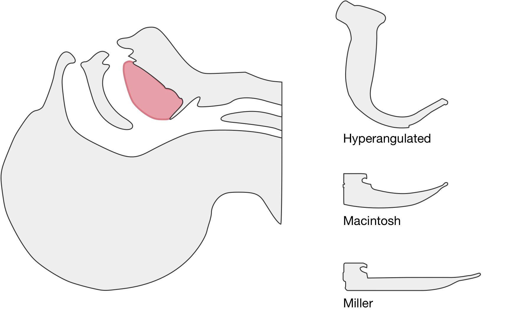
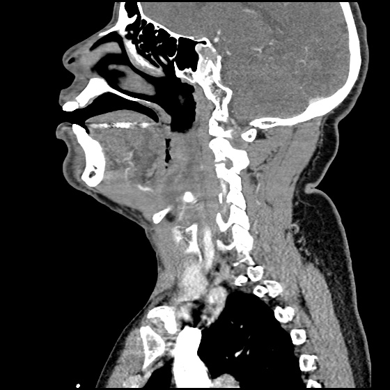
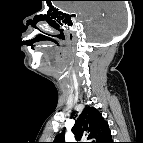
The image above shows a comparison of the curvature of straight (Miller), curved (Macintosh) and hyperangulated blades relative to the oral, pharyngeal and laryngeal axes. The hyperangulated blade easily positions a fiberoptic camera over the glottic opening, allowing easy visualization of the vocal cords. Passing the endotracheal tube without direct line-of-sight becomes the challenge of intubation.

The purpose of this guide is to explore the differences in technique for performing video laryngoscopy compared to direct laryngoscopy. Specifically, the guide is focused on video laryngoscopes with hyper-angulated blades (Verathon Glidescope, Storz C-MAC D-Blade) as these devices pose more challenges for appropriate use while potentially offering the greatest advantages for glottic visualization in challenging airway management.


Advantages and Disadvantages

Advantages1

  • Improved glottic visualization especially in scenarios with limited neck mobility as there is no need to align the oral-pharyngeal-laryngeal axes.
  • Allows others to view the screen and facilitate endotracheal intubation.

Disadvantages1

  • Difficulty passing endotracheal tube despite improved glottic visualization.
  • Obscured view by fogging or contaminated airway (blood, secretions, vomit).
  • Possibility for deterioration of skills in direct laryngoscopy.

Technique

Blade Insertion

Because of it’s unique shape, special care should be taken when inserting a hyperangulated blade to avoid dental/oropharyngeal injury. The handle of the blade may need to be angled towards the patient’s chest and turned slightly to facilitate atraumatic insertion.

Blade Advancement

Transition to viewing the screen once the tip of the blade is past the visualized posterior portion of the tongue. Advance the blade along the base of the tongue while monitoring the view on screen. Identify the epiglottis and guide the tip of the blade into the vallecula, the objective is to center the glottic aperture in the upper 2/3 of the screen.

Optimal View

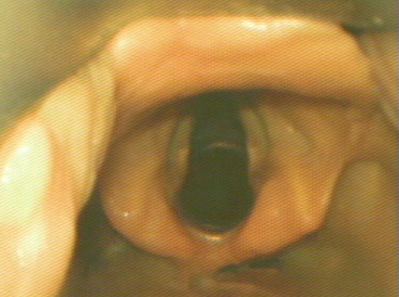
The optimal view shows the vocal cords centered in the upper 2/3 of the screen. This provides adequate screen “real estate” to show the endotracheal tube entering the screen and provide more visual feedback as the tube position is manipulated.

Blade Adjustment

The shape of the hyperangulated blade means that visualizing the airway is less dependent on the lifting motion applied with direct laryngoscopy. Instead, angulating the tip of the blade upward by gently rocking the handle back (taking great care to avoid dental/oropharyngeal trauma) will dramatically improve the view.

Endotracheal Tube Insertion

Directly visualize the insertion of the endotracheal tube into the oral cavity. The endotracheal tube should be loaded with an appropriately angulated rigid stylet. Follow the curvature of the laryngoscope blade entering from the right corner of the mouth. When the endotracheal tube is passed along the edge of the laryngoscope blade, few adjustments will be required for successful endotracheal intubation.

Endotracheal Tube Position Adjustments

If the view is optimized but the endotracheal tube is off-center and does not directly pass the vocal cords into the trachea, subtle adjustments to the endotracheal tube can help guide it into position. Hold the endotracheal tube like a pencil, turning it slightly clockwise or counter-clockwise between your index finger and thumb to manipulate the distal tip left or right above the laryngeal opening.

Stylet Removal

Once past the vocal cords, the hyperangulated rigid stylet will limit continued passage of the endotracheal tube into the trachea as it will get stuck on the anterior surface of the trachea. To facilitate easy passage into the trachea, the stylet should be pulled back slightly to allow passage into the trachea before being fully removed and the tube secured in position.


Review of Evidence

Emergency Department:

  • C-MAC associated with higher rate of successful intubation and greater proportion of grade I-II Cormack-Lehane views compared to direct laryngoscopy.5
  • Glidescope associated with higher rate of successful intubation (and fewer esophageal complications) compared to direct laryngoscopy. 6
  • Glidescope associated with higher rate of successful intubation compared to direct laryngoscopy. 7

Adjustments to Challenging Tube Placement:

  • Combination of flexible-tipped endotracheal tube with rigid stylet (Verathon GlideRight stylet) may increase intubation success.8
  • No apparent benefit to “reverse camber” loading of stylet into endotracheal tube. Reverse camber loading references placing the angled stylet 180° opposite to the natural curvature of the endotracheal tube.9
  • If a malleable stylet is used, 90° angulation was associated with higher rates of successful intubation. 10

Recent Data:

  • Propensity score-matched analysis of multicenter emergency department airway registry – no significant difference in rates of successful intubation comparing Glidescope to direct laryngoscopy. 11
  • Randomized trial comparing direct and video laryngoscopy using C-MAC showed no difference in rates of successful intubation, duration of intubation attempt, aspiration events or length of hospitalization. For interventions randomized to direct laryngoscopy, the video laryngoscope screen was covered. 12

Meta-Analyses:

  • Meta-analysis (predominantly for intubations in the operating room) suggests video laryngoscopy associated with fewer failed intubations, particularly among patients with anticipated difficult airway. Insufficient evidence to analyze effects on incidence of hypoxia, respiratory complications, time to intubation, or mortality.13

References

  1. Maldini B, Hodžović I, Goranović T, Mesarić J. CHALLENGES IN THE USE OF VIDEO LARYNGOSCOPES. Acta Clin Croat. 2016;55 Suppl 1:41-50. doi:10.20471/acc.2016.55.s1.05.
  2. Levitan RM, Heitz JW, Sweeney M, Cooper RM. The complexities of tracheal intubation with direct laryngoscopy and alternative intubation devices. Ann Emerg Med. 2011;57(3):240-247. doi:10.1016/j.annemergmed.2010.05.035.
  3. Hurford WE. The video revolution: a new view of laryngoscopy. Respir Care. 2010;55(8):1036-1045.
  4. Cheyne DR, Doyle P. Advances in laryngoscopy: rigid indirect laryngoscopy. F1000 Med Rep. 2010;2:61. doi:10.3410/M2-61.
  5. Sakles JC, Mosier J, Chiu S, Cosentino M, Kalin L. A Comparison of the C-MAC Video Laryngoscope to the Macintosh Direct Laryngoscope for Intubation in the Emergency Department. Ann Emerg Med. 2012;60(6):739-748. doi:10.1016/j.annemergmed.2012.03.031.
  6. Sakles JC, Mosier JM, Chiu S, Keim SM. Tracheal Intubation in the Emergency Department: A Comparison of GlideScope® Video Laryngoscopy to Direct Laryngoscopy in 822 Intubations. J Emerg Med. 2012;42(4):400-405. doi:10.1016/j.jemermed.2011.05.019.
  7. MD JMM, MPH USP, BA SC, MD JCS. Difficult Airway Management in the Emergency Department: GlideScope Videolaryngoscopy Compared to Direct Laryngoscopy. J Emerg Med. 2012;42(6):629-634. doi:10.1016/j.jemermed.2011.06.007.
  8. Radesic BP, Winkelman C, Einsporn R, Kless J. Ease of intubation with the Parker Flex-Tip or a standard Mallinckrodt endotracheal tube using a video laryngoscope (GlideScope). AANA J. 2012;80(5):363-372.
  9. Jones PM, Turkstra TP, Armstrong KP, et al. Effect of stylet angulation and endotracheal tube camber on time to intubation with the GlideScope. Can J Anesth/J Can Anesth. 2007;54(1):21-27.
  10. Turkstra TP, Harle CC, Armstrong KP, et al. The GlideScope-specific rigid stylet and standard malleable stylet are equally effective for GlideScope use. Can J Anesth/J Can Anesth. 2007;54(11):891-896.
  11. Choi HJ, Kim Y-M, Oh YM, et al. GlideScope video laryngoscopy versus direct laryngoscopy in the emergency department: a propensity score-matched analysis. BMJ Open. 2015;5(5):e007884-e007884. doi:10.1136/bmjopen-2015-007884.
  12. Driver BE, Prekker ME, Moore JC, Schick AL, Reardon RF, Miner JR. Direct Versus Video Laryngoscopy Using the C-MAC for Tracheal Intubation in the Emergency Department, a Randomized Controlled Trial. Acad Emerg Med. 2016;23(4):433-439. doi:10.1111/acem.12933.
  13. Lewis SR, Butler AR, Parker J, Cook TM, Smith AF. Videolaryngoscopy versus direct laryngoscopy for adult patients requiring tracheal intubation. Cochrane Database Syst Rev. 2016;11:CD011136. doi:10.1002/14651858.CD011136.pub2.

Penetrating Neck Trauma

Brief H&P

A young male presents to the emergency department after a self-inflicted stab wound to the neck. Examination revealed a knife handle protruding from the left lateral neck. A plain radiograph is shown below.

CXR: Radiopaque foreign body in left neck.

The patient was initially stable but developed shortness of breath upon attempting to lie flat for advanced imaging and was taken emergently to the operating room. Neck exploration showed no obvious neurovascular injuries, and the course of the 6cm blade was posterior to the trachea and esophagus. The knife was removed with “considerable force” as it was likely lodged within a portion of vertebral bone. The patient underwent esophagoscopy and bronchoscopy without identified tracheoesophageal injuries. The patient did well post-operatively and was discharged home.

Zones of Injury1-3

Previously, the evaluation and management of hemodynamically stable patients with penetrating neck injury was guided by the anatomic “zone” of injury. The affected zone guided the performance of additional diagnostic procedures including potentially morbid neck explorations.

Neck Zones of Injury

Understanding zone definitions remains important for the emergency physician to appreciate potentially implicated underlying structures. However, the advent of modern imaging modalities, specifically computed tomography with angiography, provides appropriate sensitivity for vascular and tracheoesophageal injuries when combined with detailed physical examination and maintenance of an appropriate threshold for the performance of additional studies if warranted by the clinical presentation (suboptimal imaging, concerning projectile trajectory, etc).

Zone Definition
I Clavicles/sternum to cricoid cartilage
II Cricoid cartilage to the angle of mandible
III Superior to the angle of mandible to the skull base


Algorithm for the Evaluation of Penetrating Neck Trauma


References

  1. Sperry JL, Moore EE, Coimbra R, et al. Western Trauma Association critical decisions in trauma: penetrating neck trauma. J Trauma Acute Care Surg. 2013;75(6):936-940. doi:10.1097/TA.0b013e31829e20e3.
  2. Brywczynski JJ, Barrett TW, Lyon JA, Cotton BA. Management of penetrating neck injury in the emergency department: a structured literature review. Emerg Med J. 2008;25(11):711-715. doi:10.1136/emj.2008.058792.
  3. Shiroff AM, Gale SC, Martin ND, et al. Penetrating neck trauma: a review of management strategies and discussion of the “No Zone” approach. Am Surg. 2013;79(1):23-29. doi:10.1007/978-3-662-49859-0_29.


Epiglottitis

Brief H&P:

30 year-old male with no significant medical history presenting with 24 hours of progressively worsening throat pain, difficulty swallowing and voice hoarseness. He reports subjective fevers and chills.
Vital signs notable for Tmax 38.4°C. On physical examination, the patient was sitting upright, unable to swallow secretions with faint inspiratory stridor and dysphonia (though he was able to speak in full sentences and without apparent respiratory distress). Oropharyngeal examination showed minimal right parapharyngeal edema without uvular or palatal deviation and there was exquisite right lateral neck tenderness to palpation.

Labs

  • CBC: 24.2/14.4/43.4/202
  • Wound culture: MSSA
IM-0001-0060
IM-0001-0062
IM-0001-0064
IM-0001-0066
IM-0001-0068
IM-0001-0070
IM-0001-0072
IM-0001-0074
IM-0001-0076
IM-0001-0078
IM-0001-0080

CT Neck/Soft Tissue with Contrast

Edema of the oropharynx/hypopharynx, consistent with epiglottitis and early abscess formation.

ED/Hospital Course

The patient acutely decompensated prior to fiberoptic laryngoscopy and proceeded emergently to the operating room for controlled intubation. The operative report described the following findings: “The patient had diffuse edema of the posterior oropharyngeal wall. The epiglottis was severely thickened, Omega shaped, soft to palpation and with moderate pressure, it appeared to come to a head and pus was expressed from the lingual side of the epiglottis.” The patient was extubated on hospital day three and discharged soon thereafter, he was doing well on follow-up.

Evaluation of Sore Throat – Applied

Evaluation of Sore Throat - Applied

Angioedema

AngioedemaHPI:

63-year old African American male with a history of HTN presenting with lip swelling x1 day. The patient states he was well until this morning when he noticed progressive swelling of his lips. The swelling is not associated with any difficulty speaking, swallowing or breathing and is not painful.

He denies new rashes or itching, and has no history of such swelling. He also denies any exposure to known allergens, recent insect bites or travel. He has been taking lisinopril for his blood pressure regularly for the past several months and denies any prior adverse effects (cough, rash).

PMH:

  • Parkinson Disease
  • HTN

PSH:

None

FH:

No family history of angioedema

SHx:

  • No t/e/d use
  • Lives at home with caretaker

Meds:

  • Lisinopril 20mg p.o. daily
  • Carbidopa/levodopa 50mg p.o. t.i.d.

Allergies:

NKDA

Physical Exam:

VS: T 37.8 HR 84 RR 14 BP 146/98 O2 99% RA
Gen: Well-appearing, no respiratory distress, speaking comfortably
HEENT: PERRL, significant external upper/lower lips swelling extending to lateral cheeks, non-tender, no fluctuance or overlying skin changes. No visible tongue swelling, floor of mouth swelling/tenderness, uvular/palatal deviation.
CV: RRR, no M/R/G
Lungs: CTAB, no crackles/wheezing, good air movement b/l
Abd: +BS, soft, NT/ND, no rebound/guarding
Ext: Warm, well-perfused, 2+ peripheral pulses
Skin: No visible skin lesions/rashes
Neuro: AAOx4, CN II-XII intact

Assessment/Plan:

63M with acute onset, progressive facial swelling. Currently restricted to external lips, with no evidence of airway compromise. Likely ACE inhibitor-induced angioedema given patient is on lisinopril and has no history of hereditary angioedema. Doubt anaphylaxis given no allergies, suspicious exposures or history of pruritus. Doubt infection given afebrile and painless swelling without e/o erythema.

Pathophysiology of ACE inhibitor-induced angioedema1

Pathophysiology of ACE-inhibitor induced angioedema

Angioedema is a vascular reaction associated with tissue (subcutaneous, submucosal) edema resulting from increased activity of vasoactive substances. The vasoactive substances in ACE inhibitor-induced angioedema are bradykinin and substance P. In the presence of ACE inhibition, these enzymes are inactivated through alternative pathways which, if disturbed, lead to angioedema.

Epidemiology of ACE inhibitor-induced angioedema

Angioedema occurs in 0.1-0.7% of patients taking ACE inhibitors, and 60% of cases occur within the first week of starting an ACE inhibitor (though it can occur as much as years later).2,3 ACE inhibitors are implicated as the cause of 20-40% of all ED visits for angioedema.4

Risk Factors2,5,6

  • Female
  • Age > 65yo
  • African American
  • Prior angioedema
  • Smoking
  • ACE inhibitor-associated cough

Clinical Features of ACE inhibitor-induced angioedema

Affected Sites:

  • Mucous membranes of the head and neck
    • Face
    • Tongue
    • Lips
    • Pharynx
    • Larynx
  • GI tract
    • Diffuse abdominal pain
    • Nausea/vomiting/diarrhea

Signs/Symptoms at initial presentation:4

  • SOB (89%)
  • Lip swelling (70%)
  • Tongue swelling (52%)
  • Voice change/hoarseness (29%)
  • Stridor (11%)

Key Clinical Features:

  • Onset in minutes with resolution in 24-72 hours
  • Absence of itching/urticaria7

Staging and Disposition:8

Stage Affected Site Outpatient (%) Floor (%) ICU (%) Intervention (%)
I Face, lip 48 52 0 0
II Soft palate 60 40 0 0
III Tongue 26 7 67 7
IV Larynx 0 0 100 24

Management of ACE inhibitor-induced angioedema

  • Proven benefit
    • Airway management
    • Withdrawal of ACE inhibitor
  • Unclear benefit
    • Epinephrine 0.3mg IM q15min
    • Diphenhydramine 50mg IV
    • Famotidine 20mg IV
    • Solumedrol 125mg IV
  • Future treatment options
    • FFP: contains ACE9
    • Icatibant: bradykinin B2 receptor antagonist10,11

References:

  1. Vleeming, W., van Amsterdam, J. G., Stricker, B. H. C., & de Wildt, D. J. (1998). ACE inhibitor-induced angioedema. Drug Safety, 18(3), 171–188. doi:10.2165/00002018-199818030-00003
  2. Grant, N. N., Deeb, Z. E., & Chia, S. H. (2007). Clinical experience with angiotensin-converting enzyme inhibitor-induced angioedema. Otolaryngology – head and neck surgery, 137(6), 931–935. doi:10.1016/j.otohns.2007.08.012
  3. Slater, E. E., Merrill, D. D., Guess, H. A., Roylance, P. J., Cooper, W. D., Inman, W. H., & Ewan, P. W. (1988). Clinical profile of angioedema associated with angiotensin converting-enzyme inhibition. JAMA : the journal of the American Medical Association, 260(7), 967–970.
  4. Banerji, A., Clark, S., Blanda, M., LoVecchio, F., Snyder, B., & Camargo, C. A. (2008). Multicenter study of patients with angiotensin-converting enzyme inhibitor-induced angioedema who present to the emergency department. Annals of allergy, asthma & immunology, 100(4), 327–332. doi:10.1016/S1081-1206(10)60594-7
  5. Gibbs, C. R., Lip, G. Y., & Beevers, D. G. (1999). Angioedema due to ACE inhibitors: increased risk in patients of African origin. British journal of clinical pharmacology, 48(6), 861–865.
  6. Morimoto, T., Gandhi, T. K., Fiskio, J. M., Seger, A. C., So, J. W., Cook, E. F., Fukui, T., et al. (2004). An evaluation of risk factors for adverse drug events associated with angiotensin-converting enzyme inhibitors. Journal of evaluation in clinical practice, 10(4), 499–509. doi:10.1111/j.1365-2753.2003.00484.x
  7. Kanani, A., Schellenberg, R., & Warrington, R. (2011). Urticaria and angioedema. Allergy, Asthma & Clinical Immunology, 7(Suppl 1), S9. doi:10.1186/1710-1492-7-S1-S9
  8. Ishoo, E., Shah, U. K., Grillone, G. A., Stram, J. R., & Fuleihan, N. S. (1999). Predicting airway risk in angioedema: staging system based on presentation. Otolaryngology – head and neck surgery, 121(3), 263–268.
  9. Hassen, G. W., Kalantari, H., Parraga, M., Chirurgi, R., Meletiche, C., Chan, C., Ciarlo, J., et al. (2013). Fresh frozen plasma for progressive and refractory angiotensin-converting enzyme inhibitor-induced angioedema. The Journal of emergency medicine, 44(4), 764–772. doi:10.1016/j.jemermed.2012.07.055
  10. Bas, M., Greve, J., Stelter, K., Bier, H., Stark, T., Hoffmann, T. K., & Kojda, G. (2010). Therapeutic Efficacy of Icatibant in Angioedema Induced by Angiotensin-Converting Enzyme Inhibitors: A Case Series. Annals of emergency medicine, 56(3), 278–282. doi:10.1016/j.annemergmed.2010.03.032
  11. MD, M. G., & MD, M. A. (2012). Icatibant: a novel approach to the treatment of angioedema related to the use of angiotensin-converting enzyme inhibitors. American Journal of Emergency Medicine, 30(8), 1664.e1–1664.e2. doi:10.1016/j.ajem.2011.09.014