Ascitic Fluid

Brief HPI:

A 56 year-old male with a history of alcoholic cirrhosis complicated by esophageal varices presents to the emergency department with abdominal distension. He notes gradually worsening symptoms over the past 2 weeks – roughly correlating with the timing of his last paracentesis. He has limited access to medical care and typically presents to emergency departments for palliative paracenteses. He is otherwise in his usual state of health and denies fevers, chills, abdominal pain, vomiting blood, or dark/bloody stools.

Vital signs are notable for a heart rate of 97bpm and blood pressure of 110/65mmHg – otherwise normal. Examination demonstrates a distended abdomen which is non-tender, dull to percussion and with a palpable fluid wave. Bedside ultrasonography shows large, homogenous-appearing ascites with readily-accessible pockets for drainage in the bilateral lower quadrants. A palliative paracentesis is performed with uncomplicated extraction of 4 liters of translucent, straw-colored fluid. Ascitic fluid analysis shows 90 white blood cells of which 10% are polymorphonuclear. The patient is observed briefly in the emergency department, noted symptomatic improvement and was discharged with a plan for telephone follow-up of fluid culture results.

An Algorithm for the Analysis of Ascitic Fluid

Algorithm for the Analysis of Ascitic Fluid

References

  1. Runyon BA. Care of patients with ascites. N Engl J Med. 1994;330(5):337-342. doi:10.1056/NEJM199402033300508.
  2. Wong CL, Holroyd-Leduc J, Thorpe KE, Straus SE. Does this patient have bacterial peritonitis or portal hypertension? How do I perform a paracentesis and analyze the results? JAMA. 2008;299(10):1166-1178. doi:10.1001/jama.299.10.1166.
  3. Tarn AC, Lapworth R. Biochemical analysis of ascitic (peritoneal) fluid: what should we measure? Ann Clin Biochem. 2010;47(Pt 5):397-407. doi:10.1258/acb.2010.010048.
  4. Li PK-T, Szeto CC, Piraino B, et al. ISPD Peritonitis Recommendations: 2016 Update on Prevention and Treatment. Perit Dial Int. 2016;36(5):481-508. doi:10.3747/pdi.2016.00078.
  5. MacIntosh T. Emergency Management of Spontaneous Bacterial Peritonitis – A Clinical Review. Cureus. 2018;10(3):e2253. doi:10.7759/cureus.2253.

Lactic Acidosis

HPI:

59F with a reported history of congestive heart failure, presenting with intermittent chest discomfort for three days.

She characterized this discomfort as “heartburn”, describing a mid-epigastric burning sensation radiating up her neck, not associated with exertion, lasting 1-2 hours and resolving with antacids. The patient has poor exercise tolerance at baseline and for the past several years has been able to ambulate only short distances around her home, and states that these symptoms have been worsening in the past week. She denies chest pain on exertion, orthopnea or paroxysmal nocturnal dyspnea. She states that she was diagnosed with congestive heart failure five years ago, but was never prescribed medications.

On further questioning, the patient reports several weeks of mouth and lip pain which has limited oral intake, though no dysphagia to solids or liquids. She otherwise denies fevers/chills, abdominal pain, nausea/vomiting, cough, changes in urinary or bowel habits.

In the emergency department, the patient was noted to have an elevated serum troponin, though ECG showed no changes of acute ischemia/infarction.

PMH:

  • Congestive heart failure

PSH:

  • None

FH:

  • Mother with diabetes
  • Father with MI at age 65

SHx:

  • 4-5 drinks of alcohol/day
  • No tobacco or drug use

Meds:

  • None

Allergies:

NKDA

Physical Exam:

VS: T 37.4 HR 106 RR 18 BP 145/82 O2 100% RA
Gen: Morbidly obese female, lying in bed, in no acute respiratory distress, speaking in complete sentences.
HEENT: Dry, cracked lips, slightly erythematous, otherwise moist mucous membranes, poor dentition. Mild scleral icterus. No cervical lymphadenopathy.
CV: Rapid rate, regular rhythm, normal S1/S2, II/VI systolic ejection murmur at LUSB, no radiation appreciated. No jugular venous distension.
Lungs: Clear to auscultation in posterior lung fields bilaterally, no crackles appreciated.
Chest: Well-circumscribed erythematous patch in folds beneath left breast, no underlying fluctuance, no significant tenderness to palpation. On contralateral breast, some hyperpigmentation but no erythema.
Abdomen: Obese, non-tender, non-distended. Patch of erythema below pannus, mildly tender to palpation.
Ext: Bilateral lower extremities with marked edema and overlying scaly plaques, some slightly ulcerated weeping serous fluid. Peripheral pulses are difficult to palpate, capillary refill difficult to assess.

Labs/Studies:

  • CBC: 11.1/11.1/34.5/212 (MCV 114.2)
  • BMP: 140/4.5/97/20/10/1.14/64
  • Anion Gap: 23
  • LFT: AST: 73, ALT: 26, AP: 300, TB: 4.6, DB: 2.1, Alb: 3.0, INR 1.3
  • BNP: 158
  • Troponin: 1.284
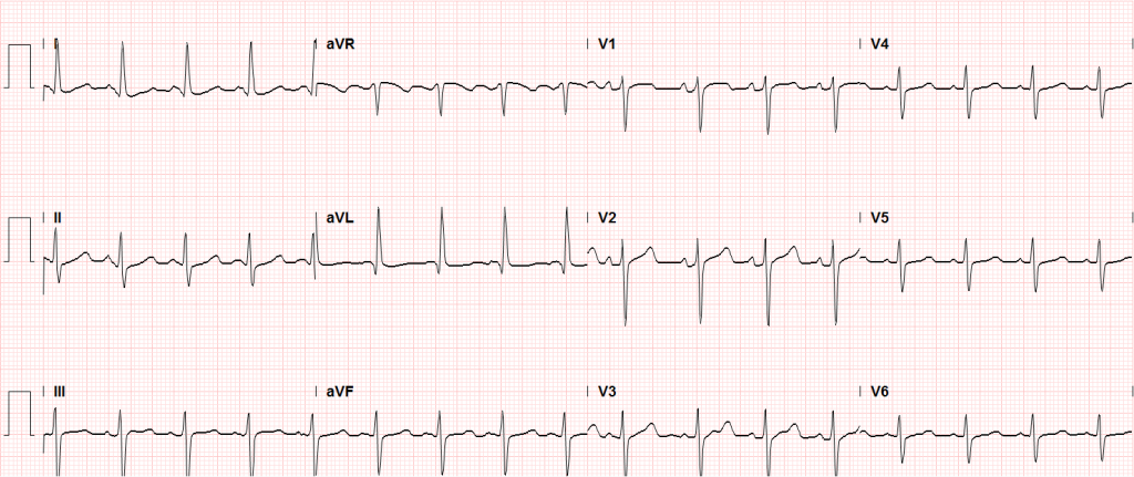
Sinus tachycardia, LVH, secondary repolarization abnormalities

Sinus tachycardia, LVH, secondary repolarization abnormalities

Imaging:

CT Pulmonary Angiography:
No evidence of central pulmonary embolism, thoracic aortic dissection, or thoracic aortic aneurysm. Evaluation of the peripheral vessels is limited due to motion artifact. No focal consolidation or pneumothorax.

CT Abdomen/Pelvis non-contrast:
No evidence of intra-abdominal abscess or definite source of infection. Marked hepatic steatosis.

CT Lower Extremity non-contrast:
Diffuse circumferential subcutaneous edema involving both lower extremities from the level of the mid thighs distally through the feet. There are bilateral subcutaneous calcifications which are likely venous calcifications in the setting of chronic venous stasis disease. There is some overlying skin thickening.

TTE:
There is moderate concentric left ventricular hypertrophy with hyperdynamic LV wall motion. The Ejection Fraction estimate is >70%. Grade I/IV (mild) LV diastolic dysfunction. No hemodynamically significant valve abnormalities.

US Abdomen:
Hepatomegaly, echogenic liver suggesting fatty infiltration. Moderately blunted hepatic vein waveforms suggesting decreased hepatic parenchymal compliance.

Assessment/Plan:

The patient was admitted to the cardiology service for management of NSTEMI and evaluation of undiagnosed CHF. She was started on a heparin continuous infusion. In addition, a CT pulmonary angiogram was obtained to evaluate for pulmonary embolism as an explanation of her progressive dyspnea on exertion. No PE, consolidation or effusion was identified.

Despite the patient’s reported history of congestive heart failure, there was no evidence that her symptoms were a result of an acute exacerbation with only a mildly elevated BNP but no jugular venous distension or evidence of pulmonary edema. The patient’s significant lower extremity edema was more suggestive of chronic venous stasis.

One notable laboratory abnormality that was explored was her elevated anion gap metabolic acidosis. Studies submitted included serum lactate, salicylates, osmolarity, CK, and urinalysis for ketonuria. This evaluation was notable for an elevated serum lactate of 13.2mmol/L and an arterial blood gas that showed adequate respiratory compensation (and no A-a gradient). Given the patient’s modest leukocytosis (with neutrophil predominance), and tachycardia, the concern for sepsis was increased though the source remained unclear. Prominent possibilities included a skin and soft-tissue infection vs. less likely intra-abdominal source though the patient’s physical examination was not suggestive of a process that would produce such a substantial lactic acidosis. Blood cultures were drawn and the patient was started on empiric antibiotics for the suspected sources. In addition, the patient was cautiously volume resuscitated given her reported history of CHF while pending a transthoracic echocardiogram to evaluate cardiac function. Additional imaging including CT abdomen/pelvis and lower extremities was obtained (though without contrast due to the patient’s recent exposure), and no obvious source was identified.

Over the next two days, the patient’s serum lactate downtrended to normal range, as did the serum troponin. A transthoracic echocardiogram showed an LVEF >70% with mild concentric hypertrophy and diastolic dysfunction. Blood and urine cultures were without growth.

Additional issues managed during the hospitalization included elevated serum transaminases (AST > ALT), conjugated hyperbilirubinemia and evidence of decreased hepatic synthetic function with hypoalbuminemia and elevated INR. Given the patient’s history of EtOH use, as well as other corroborating findings including macrocytic anemia, hypomagnesemia, folate and B12 deficiency, this was attributed to alcoholic hepatitis (discriminant function <32). Infectious hepatitis serologies were negative. The patient was started on nutritional supplements. Finally, the patient persistently complained of lip and oral mucosal pain. Examination was without discrete lesions but some mucosal redness was identified. Despite poor dentition, there was no evidence of abscess and HSV/HIV testing was negative. This was thought to be stomatitis caused by her identified nutritional deficiencies.

Differential Diagnosis of Elevated Serum Lactate 1,2

Differential Diagnosis of Elevated Serum Lactate

Algorithm for Evaluation of Acidemia 3,4

Algorithm for Evaluation of Acidemia

Algorithm for Evaluation of Alkalemia 3,4

Algorithm for Evaluation of Alkalemia

References:

  1. Fall, P. J., & Szerlip, H. M. (2005). Lactic acidosis: from sour milk to septic shock. Journal of intensive care medicine, 20(5), 255–271. doi:10.1177/0885066605278644
  2. Luft, F. C. (2001). Lactic acidosis update for critical care clinicians. Journal of the American Society of Nephrology : JASN, 12 Suppl 17, S15–9.
  3. Ingelfinger, J. R., Berend, K., de Vries, A. P. J., & Gans, R. O. B. (2014). Physiological Approach to Assessment of Acid–Base Disturbances. The New England journal of medicine, 371(15), 1434–1445. doi:10.1056/NEJMra1003327
  4. Ingelfinger, J. R., & Seifter, J. L. (2014). Integration of Acid–Base and Electrolyte Disorders. The New England journal of medicine, 371(19), 1821–1831. doi:10.1056/NEJMra1215672

Renal Failure in Cirrhosis

CC:

Consult for acute kidney injury

HPI:

63M with a history of liver cirrhosis of cryptogenic etiology, portal vein thrombosis, and esophageal varices s/p banding (2011) who was admitted to an OSH for altered mental status and hypotension requiring dopamine and was transferred to this facility for a higher level of care.

The nephrology service was consulted for elevated serum creatinine concerning for AKI. The patient has a baseline creatinine of 1.1 (3/2013), 1.9 on transfer and continued worsening to peak of 2.6 today.

PMH:

  • Asthma
  • COPD
  • Cirrhosis (PVT, encephalopathy)
  • Inguinal hernia (recurrent)

PSH:

  • Appendectomy
  • Bilateral inguinal hernia repair

FH:

  • Non-contributory

SHx:

  • Married
  • Denies t/e/d use

Meds:

  • albumin 25g i.v. q.6.h.
  • erythromycin 1,000mg p.o. q.1.h.
  • fluticasone-salmeterol 1 puff b.i.d.
  • lactulose 45g p.o. q.6.h.
  • neomycin 1,000mg p.o. q.1.h.
  • pantoprazole 40mg i.v. daily
  • rifaximin 550mg p.o. b.i.d.
  • sodium benzoate 5g p.o. b.i.d.

Allergies:

  • Sulfa

Physical Exam:

VS: T 37.4 HR 90 RR 15 BP 86/48 O2 97% RA
Gen: Chronically ill-appearing.
HEENT: PERRL, scleral icterus, MMM
CV: RRR
Lungs: CTAB
Abd: +BS, soft, non-tender, non-distended
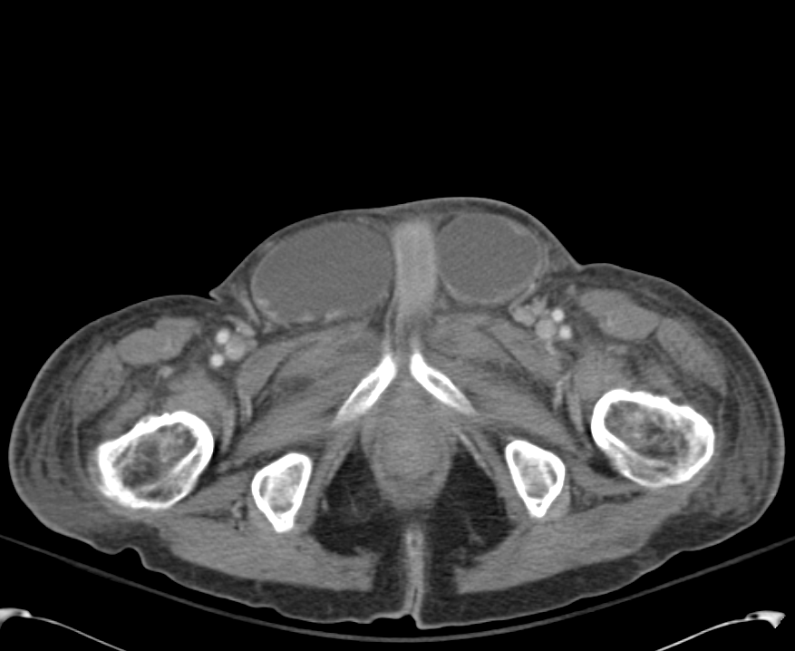
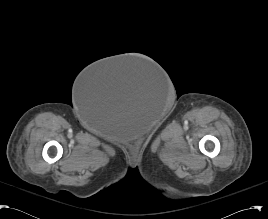
GU: Large ascites filled scrotum, testicles/inguinal canal not easily palpated
Ext: Warm, well-perfused
Skin: No palmar erythema, no vascular spiders
Neuro: AAOx4, CN II-XII grossly intact

Labs:

  • BMP: 134/4.5/103/20/41/3.0/106 (Ca 9.3, Mg 3.7, PO4 2.4)
  • LFT: AST 89, ALT 33, TB 26.6, CB 16.1, Alb 2.7
  • NH4 167

Imaging:

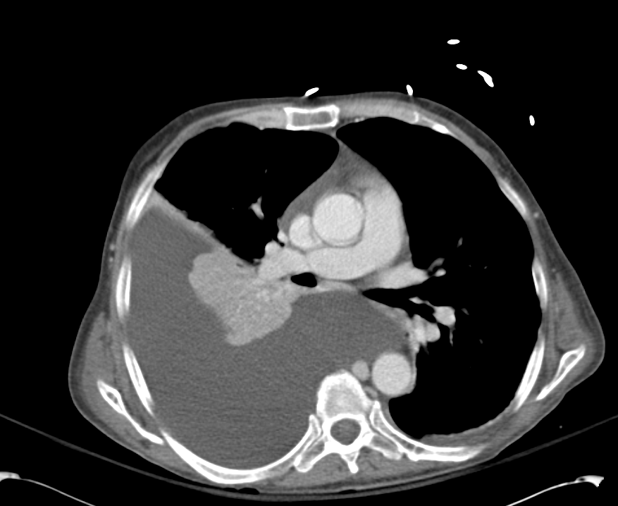
Pleural Effusion

Pleural Effusion

Large right pleural effusion with underlying compressive atelectasis.

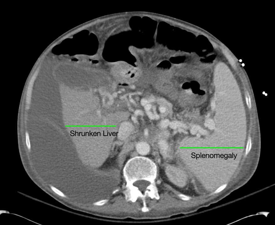
Cirrhosis and Portal Hypertension

Cirrhosis and Portal Hypertension

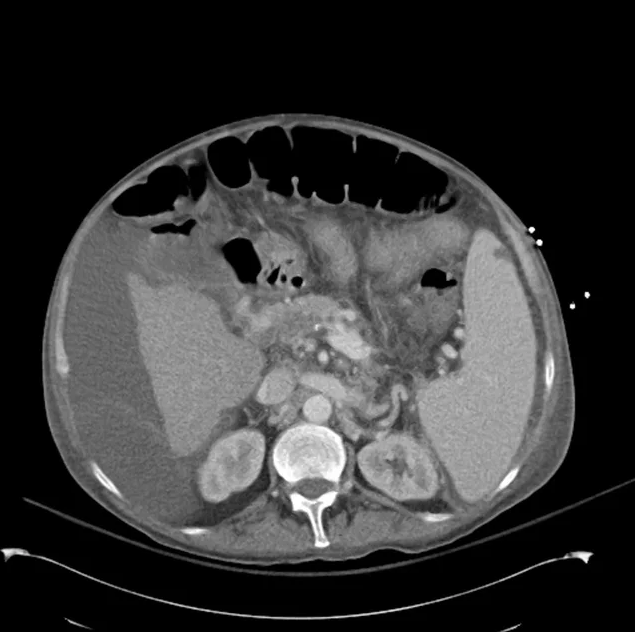
Shrunken/nodular liver with sequelae of portal hypertension including perisplenic collaterals, and splenomegaly.

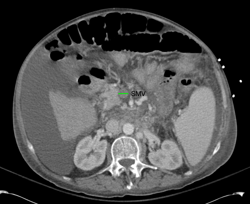
SMV Thrombosis

SMV Thrombosis

Near-total thrombosis of the portal vein extending down to superior mesenteric vein.

B/L Inguinal Hernias

B/L Inguinal Hernias

Large volume abdominal ascites with a large amount of fluid extending into the bilateral inguinal canals.

Large Right Inguinal Hernia

Large Right Inguinal Hernia

Large volume abdominal ascites with a large amount of fluid extending into the bilateral inguinal canals.

CT Abdomen/Pelvis (PVT)

CT Abdomen/Pelvis (PVT)

Assessment/Plan:

63M with a history of liver cirrhosis of cryptogenic etiology, recently with hypotension prior to transfer to this facility and increase in creatinine from 1.9-3.0 on current admission (from baseline 1.1).

These findings indicate acute kidney injury, likely hepatorenal syndrome vs. acute tubular necrosis 2/2 prolonged hypotension. Plan to discontinue diuretics and start albumin challenge (1g/kg/day divided q6h x2d). Will also check UA, urine Na/cr/urea/eos, renal US (evaluate obstruction, kidney size). Start midodrine/octreotide for underlying HRS.

  1. Neuro: Intermittent confusion. Lactulose, rifaximin, benzoate.
  2. Resp: 2L NC. ABG 7.36/51/87/27.7/+2. CXR: Large R effusion.
  3. CV: Levo 0.075. Midodrine 15 TID. MAPs 60, HR 80s.
  4. GI: NPO/NGT. TPN.
  5. Renal: See above.
  6. Heme: Coagulopathy, keep INR <2.5
  7. ID: Afebrile. No abx.
  8. Endo: Euglycemic

Renal Failure in Cirrhosis:

Renal failure in cirrhosis is associated with higher mortality both before and after transplant. The main causes of renal failure in cirrhosis are detailed below, with particular attention to an entity unique to cirrhosis: the hepatorenal syndrome.1

Disorder Pathogenesis Diagnosis Management
HRS Dilation of splanchnic arteries initially compensated by increased CO eventually decompensates with activation of mechanisms to preserve ECBV (RAAS, SNS, ADH) leading to fluid retention (ascites, edema) and renal failure due to intrarenal vasoconstriction.Bacterial translocation and the resulting inflammatory response may contribute to splanchnic vasodilation (through production of vasoactive factors like NO).
  • Serum creatinine > 1.5mg/dl-  Not reduced with 1g/kg albumin
  • No confounding factors (2d off diuretics, no nephrotoxic agents, no shock, no e/o intrinsic renal disease)
  • Type 1: doubling creatinine > 2.5mg/dL in <2wk
  • Type 2: stable, slower progression
  • Vasoconstrictor therapy-  Albumin
  • Portasystemic shunting
  • Renal replacement therapy
  • Prevention
    • Norfloxacin
    • Albumin
Intrinsic renal Some causes of liver disease are also associated with intrinsic renal pathology (ex. GN associated with HBV, HCV).
  • Proteinuria, hematuria
  • Renal bx
  • Active urinary sediment
  • Antiviral therapy if appropriate
Pre-renal AKI Hemorrhage (GIB), fluid losses (excess diuresis, diarrhea from lactulose).
  • Suspected from patient history
  • Low FENa, bland urine sediment
  • Hemorrhage: replace volume with fluids, blood products. Control bleeding.
  • Discontinue diuretics, administer fluids if tolerated
ATN Severe ischemic or toxic (NSAID’s, nephrotoxic medications)
  • Renal tubular epithelial cells favor ATN (granular casts common in ATN, HRS)
  • Withdraw therapy
  • Avoid nephrotoxic agents

Pathophysiology of Hepatorenal Syndrome:

Pathophysiology of Hepatorenal Syndrome

Evaluation:

The evaluation of suspected renal failure in patients with cirrhosis involves assessment of renal function for evidence of acute impairment, as well as analaysis of urine for protein or active sediment to suggest intrinsic renal disease (possibly warranting renal ultrasonography or biopsy). Additionally, patients should be evaluated for evidence of bacterial infection including assessment of ascites if present as SBP produces a more severe form of the inflammatory vasodilation mechanism suspected to play a role in HRS.

Treatment:

For renal failure not caused by the hepatorenal syndrome, identification and management of the underlying cause is critical (intrinsic renal disease, hypovolemia/hemorrhage, nephrotoxicity, infection). For suspected HRS, management is dependent on the acuity and setting. In the intensive care unit, vasoconstrictor therapy (norepinephrine, vasopressin) in association with albumin is effective in the treatment of HRS.2,3  In less acute settings, a combination of midodrine, octreotide and albumin improves renal function and is associated with lower short-term mortality.4 Alternatives for patients who do not respond to medical therapy include TIPS, dialysis and transplant.

Summary:

Renal failure in ESLD is due to the causes, complications or management of cirrhosis and has important implications, with HRS in particular offering the worst prognosis.5 Early recognition and management is critical to improving outcomes.

References:

  1. Ginès, P., & Schrier, R. W. (2009). Renal failure in cirrhosis. The New England journal of medicine, 361(13), 1279–1290. doi:10.1056/NEJMra0809139
  2. Singh, V., Ghosh, S., Singh, B., Kumar, P., Sharma, N., Bhalla, A., Sharma, A. K., et al. (2012). Noradrenaline vs. terlipressin in the treatment of hepatorenal syndrome: a randomized study. Journal of hepatology, 56(6), 1293–1298. doi:10.1016/j.jhep.2012.01.012
  3. Kiser, T. H., Fish, D. N., Obritsch, M. D., Jung, R., MacLaren, R., & Parikh, C. R. (2005). Vasopressin, not octreotide, may be beneficial in the treatment of hepatorenal syndrome: a retrospective study. Nephrology, dialysis, transplantation, 20(9), 1813–1820. doi:10.1093/ndt/gfh930
  4. Esrailian, E., Pantangco, E. R., Kyulo, N. L., Hu, K.-Q., & Runyon, B. A. (2007). Octreotide/Midodrine therapy significantly improves renal function and 30-day survival in patients with type 1 hepatorenal syndrome. Digestive diseases and sciences, 52(3), 742–748. doi:10.1007/s10620-006-9312-0
  5. Alessandria, C., Ozdogan, O., Guevara, M., Restuccia, T., Jiménez, W., Arroyo, V., Rodés, J., et al. (2005). MELD score and clinical type predict prognosis in hepatorenal syndrome: relevance to liver transplantation. Hepatology (Baltimore, Md.), 41(6), 1282–1289. doi:10.1002/hep.20687