Anticoagulant Reversal in Intracranial Hemorrhage

Brief HPI:

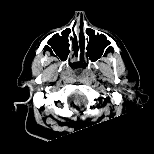
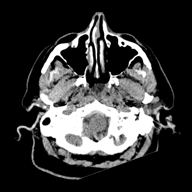
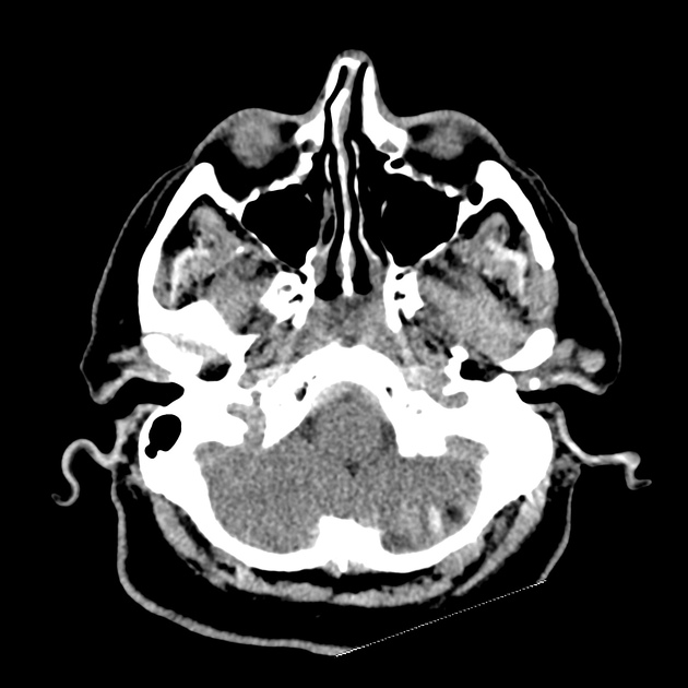
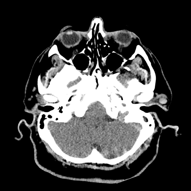
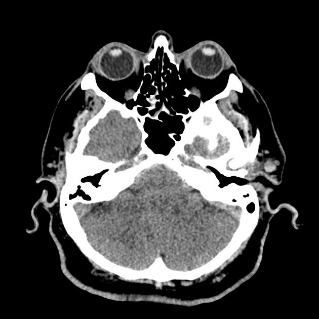
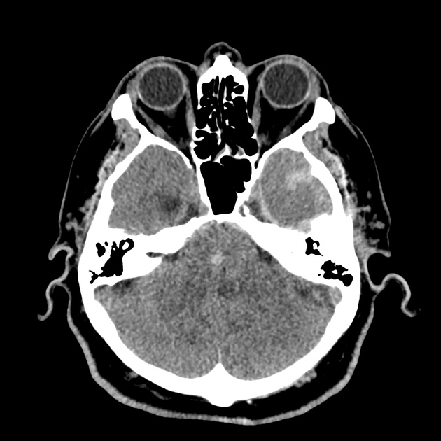
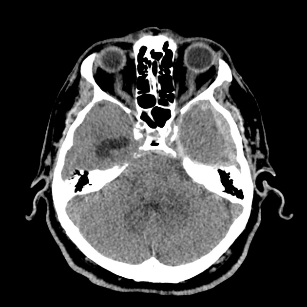
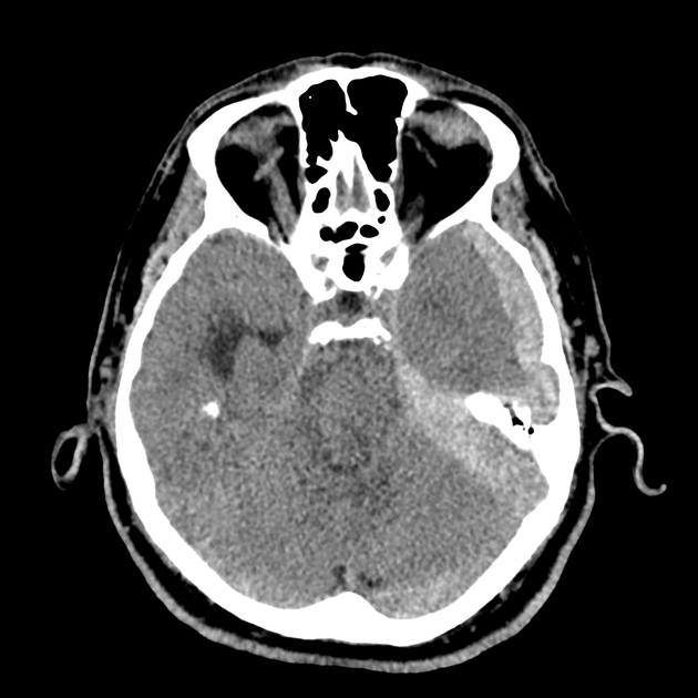
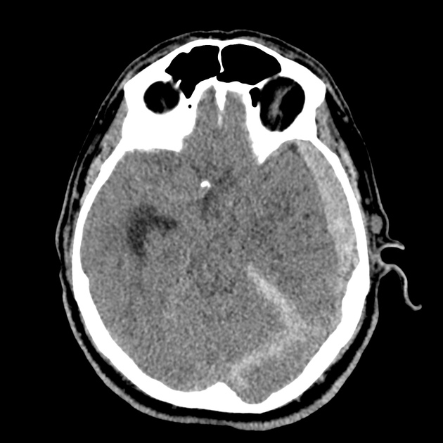
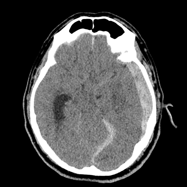
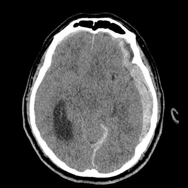
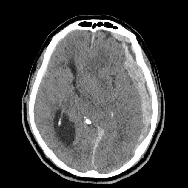
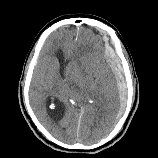
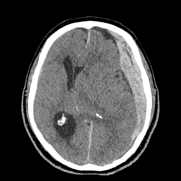
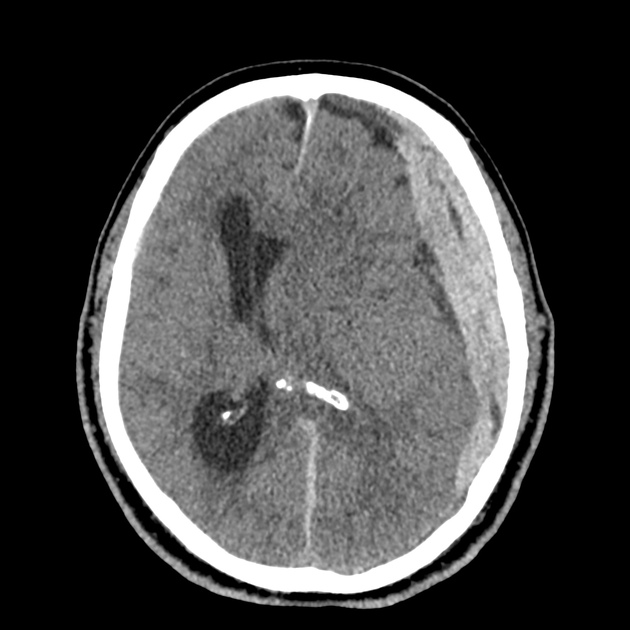
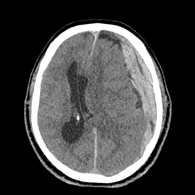
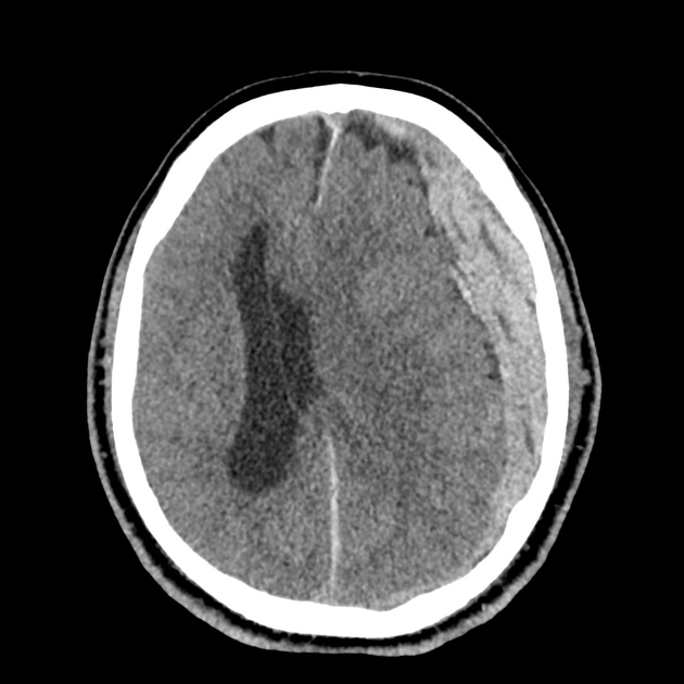
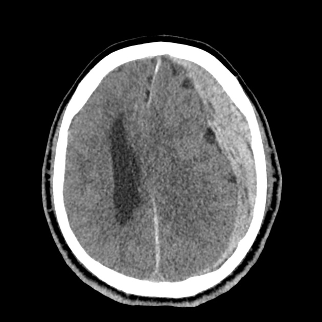
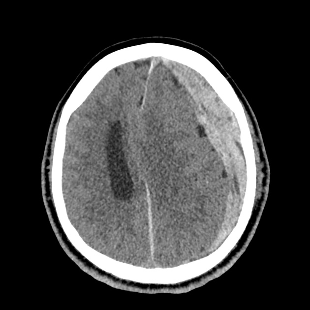
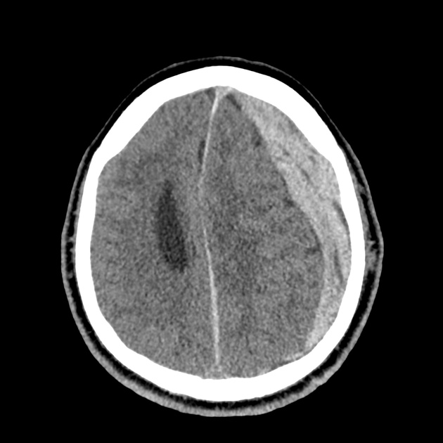
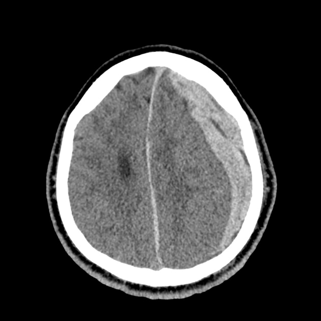
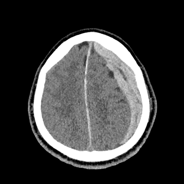
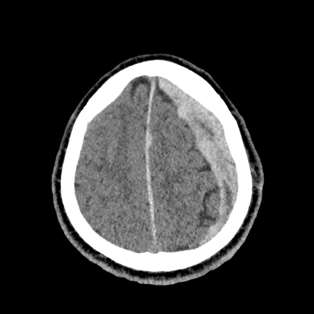
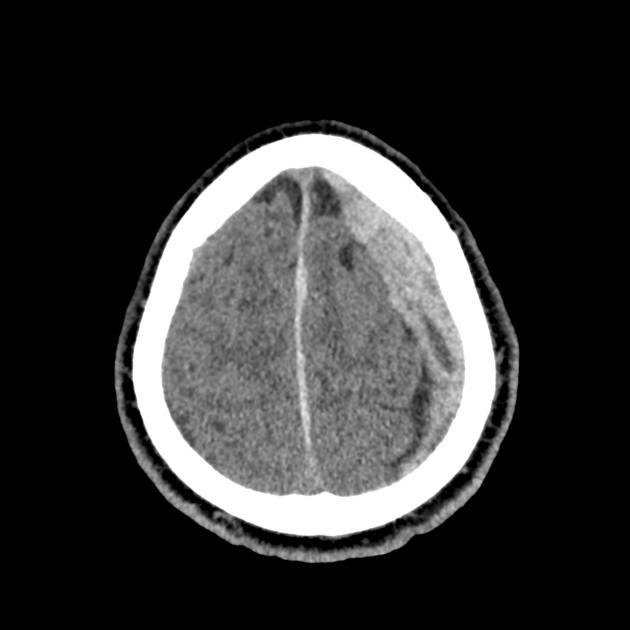
A 65-year-old male with a past medical history of hypertension, diabetes mellitus, and atrial fibrillation presents after a mechanical fall with a posterior scalp hematoma and altered mental status. The patient’s family reports that the patient is taking apixaban with his last dose 4 hours prior to arrival. Physical examination reveals a GCS of 13, blood pressure of 175/99, and asymmetric pupils. The patient is taken to CT where head imaging reveals left sided subdural hematoma with midline shift and developing uncal herniation.

00_subdural
02_subdural
04_subdural
06_subdural
08_subdural
10_subdural
12_subdural
14_subdural
16_subdural
18_subdural
20_subdural
22_subdural
24_subdural
26_subdural
28_subdural
30_subdural
32_subdural
34_subdural
36_subdural
38_subdural
40_subdural
42_subdural
44_subdural
46_subdural

CT Head:

Left cerebral convexity acute subdural hematoma producing substantial mass effect with midline shift and left uncal herniation.
Case courtesy of Dr Andrew Dixon, Radiopaedia.org. From the case rID: 32395

A nicardipine infusion is initiated and the head of the bed is elevated. Andexanet Alfa is not available, therefore an infusion of 4-Factor PCC is initiated. The patient is taken emergently to the operating room by neurosurgery for craniotomy and hematoma evacuation.

An Algorithm for the Reversal of Anticoagulation for Intracranial Hemorrhage 1-4

An Algorithm for Anticoagulant Reversal in Intracranial Hemorrhage


All Agents

For all agents, discontinue anticoagulation. Patients may require blood pressure control including anti-hypertensive infusions (goal SBP <140). Avoid reversal for intracranial hemorrhage associated with cerebral venous thrombosis. Use cautiously in patients with concomitant life-threatening ischemia, thrombosis, or severe DIC.

Vitamin K Antagonists (ex. warfarin)

Initial Dose

A fixed dose of 4F-PCC 1500 to 2000 units can be given as an initial dose with repeat dosing based on INR measurement 15 minutes after completion of infusion. Follow local institution guidelines if available.

Monitoring and Repeat Dosing

  • Vitamin K: if INR ≥1.4 at 12 hours 5
  • 4F-PCC: May consider repeat PCC dosing based on INR, though with increased DIC and thrombotic risk, it is recommended to correct further with FFP if INR remains ≥1.4 6

Direct Factor Xa Inhibitors (ex. rivaroxaban, apixaban)

Activated charcoal may be effective for up to six hours for apixaban 7 and eight hours for rivaroxaban 8.

*Andexanet alfa Regimens 9,10

  • Low-dose: rivaroxaban <10mg, apixaban <5mg, edoxaban <30mg or 8 or more hours since last dose
  • High-dose: If greater than above thresholds, or dose/timing unknown

Pentasaccharides (ex. fondaparinux)

Use high-dose Andexanet alfa regimen 12

Direct Thrombin inhibitors (ex. dabigatran)

Monitoring and Repeat Dosing

If ongoing significant bleeding after treatment, consider redosing idarucizumab and/or hemodialysis.

Alternative Regimens

If idarucizumab is not available, aPCC (50-80 units/kg) , 4F-PCC or 3F-PCC (50 units/kg) can be used in order of preference.

Unfractionated Heparin

Dosing

Determination of units of heparin is based on estimated active agent (half-life 1-2 hours)

  • Protamine sulfate 1mg/100 units IV, maximum dose 50mg
  • Alternatively, can give fixed dose of 25-50mg

Monitoring and Repeat Dosing

If aPTT is persistently elevated, repeat 0.5 mg/100 units

Low-Molecular Weight Heparin 13

Reversal is not indicated if more than 3-5 half-lives have passed since administration:

  • Enoxaparin mean half-life: 4-5 hours
  • Dalteparin mean half-life: 2.8 hours
  • Nadroparin mean half-life: 3.7 hours

If bleeding persists, or renal insufficiency, repeat dose .5 mg/1 mg enoxaparin or .5 mg/100 anti-Xa units.

This algorithm was developed by Dr. Taylor Martin. Taylor is an emergency medicine resident at McGovern Medical School at UTHealth Houston.

References

Guidelines & Reviews

  1. Greenberg SM, Ziai WC, Cordonnier C, et al. 2022 guideline for the management of patients with spontaneous intracerebral hemorrhage: a guideline from the american heart association/american stroke association. Stroke. Published online May 17, 2022:101161STR0000000000000407.
  2. Tomaselli GF, Mahaffey KW, Cuker A, et al. 2020 acc expert consensus decision pathway on management of bleeding in patients on oral anticoagulants: a report of the american college of cardiology solution set oversight committee. J Am Coll Cardiol. 2020;76(5):594-622.
  3. Frontera JA, Lewin JJ, Rabinstein AA, et al. Guideline for reversal of antithrombotics in intracranial hemorrhage: a statement for healthcare professionals from the neurocritical care society and society of critical care medicine. Neurocrit Care. 2016;24(1):6-46.
  4.  Freeman, W. David, Weitz, Jeffrey. “Reversal of anticoagulation in intracranial hemorrhage.” UpToDate. (2022) https://www.uptodate.com/contents/reversal-of-anticoagulation-in-intracranial-hemorrhage?search=anticoagulation%20reversal (Accessed on May 26, 2022)

Vitamin K Antagonists

  1. Ansell J, Hirsh J, Hylek E, Jacobson A, Crowther M, Palareti G. Pharmacology and management of the vitamin k antagonists: american college of chest physicians evidence-based clinical practice guidelines(8th edition). Chest. 2008;133(6 Suppl):160S-198S.
  2. Pabinger I, Brenner B, Kalina U, et al. Prothrombin complex concentrate (Beriplex p/n) for emergency anticoagulation reversal: a prospective multinational clinical trial. J Thromb Haemost. 2008;6(4):622-631.

Direct Factor Xa Inhibitors

  1. http://packageinserts.bms.com/pi/pi_eliquis.pdf
  2. https://www.bayer.com/sites/default/files/2020-11/xarelto-pm-en.pdf
  3. Demchuk AM, Yue P, Zotova E, et al. Hemostatic efficacy and anti-fxa (Factor xa) reversal with andexanet alfa in intracranial hemorrhage: annexa-4 substudy. Stroke. 2021;52(6):2096-2105.
  4. Cohen AT, Lewis M, Connor A, et al. Thirty-day mortality with andexanet alfa compared with prothrombin complex concentrate therapy for life-threatening direct oral anticoagulant-related bleeding. J Am Coll Emerg Physicians Open. 2022;3(2):e12655.
  5. Scaglione F. New oral anticoagulants: comparative pharmacology with vitamin K antagonists. Clin Pharmacokinet. 2013;52(2):69-82.

Pentasaccharides (ex. fondaparinux)

  1. Lu G, DeGuzman FR, Hollenbach SJ, et al. A specific antidote for reversal of anticoagulation by direct and indirect inhibitors of coagulation factor Xa. Nat Med. 2013;19(4):446-451.

Low-Molecular Weight Heparin

  1. Fareed J, Hoppensteadt D, Walenga J, et al. Pharmacodynamic and pharmacokinetic properties of enoxaparin : implications for clinical practice. Clin Pharmacokinet. 2003;42(12):1043-1057.

Epistaxis

Brief HPI:

A 63 year-old female with a history of hypertension, diabetes, and deep venous thrombosis on warfarin presents with epistaxis. She noted the spontaneous onset of nose bleeding 15 minutes prior to presentation. She had attempted compression but symptoms persisted so she was brought to the emergency department. On initial evaluation, she was in no acute distress and vital signs were normal. She was compressing her distal nares and was spitting up blood.

Oxymetolazone was administered and the patient was instructed regarding the appropriate position for compression, however bleeding continued when reassessed at 10- and then 30-minutes of compression. A bleeding focus could not be visualized on rhinoscopy so a nasal tampon was inserted with resolution of bleeding. Bleeding did not recur after two hours of observation in the emergency department. The patient’s INR was therapeutic two days prior to presentation and she was instructed to continue her usual regimen. At primary care follow-up two days later, the compression device was successfully removed.

Algorithm for the Management of Epistaxis1,2

Algorithm for the Management of Epistaxis

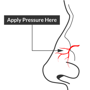
Epistaxis site of compression

Site of compression

External Compression

Begin with simple measures while preparing the necessary equipment and medications. Request that the patient gently blow their nose to clear clots, administer oxymetolazone 0.05% two sprays into the affected side. Apply firm pressure below the nasal bridge continuously for at least 10 minutes before reassessment. Commercial compression devices are available, or can be fashioned with tongue depressors3. Alternatively, the patient can apply pressure themselves.

Cautery

Again ask the patient to blow their nose to remove clots. Apply topical anesthetic for patient comfort prior to inspection with a nasal speculum. Additional suction (small tip, Frazier) may be required to improve visualization. If the bleeding site is identified, apply silver nitrate circumferentially around the source, then directly over the site. Avoid prolonged exposure or exposure to opposing sides of the nasal septum. If hemorrhage control is successful, patients may be discharged with a topical antimicrobial ointment such as polymixin-bacitracin-neomycin.

Packing 4,5

Multiple commercial anterior packing devices are available. Placement technique is similar for most, generally involving lubrication of the device with antimicrobial ointment or sterile water, sliding the device along the floor of the nasal cavity, followed by injection or inflation of the device to support tamponade. The incorporation of tranexamic acid (500mg in 5mL) into any phase of anterior packing may be beneficial 6,7. Packing the contralateral side to further support tamponade may be required.

Commonly used commercial devices are:

  • Merocel: lubricate with antimicrobial ointment, once deployed can rehydrate with saline or topical vasoconstrictor
  • Rapid Rhino
  • Rhino Rocket

Packing material should remain for 48-72 hours, during which patients should be re-evaluated. Prophylactic systemic antibiotics for the prevention of sinusitis or toxic shock are likely not required8.

Thrombogenic materials such as Floseal or Surgicel can also be used and may be better tolerated than packing materials9.

Posterior Control

If bleeding persists despite the above measures, a posterior site should be considered. Dual-balloon commercial devices are available for the control of posterior epistaxis and are deployed in a similar fashion to anterior devices. Once inserted, the posterior balloon should be inflated with air – with the volume guided by tension of the pilot cuff. The anterior balloon can then be inflated in a similar fashion. The posterior balloon cuff should be reinspected after 5 minutes as additional inflation may be required.

Commonly used commercial devices are:

If a commercial device is unavailable, a Foley catheter may be used. The catheter is introduced into the affected side. Once the tip is visualized in the posterior oropharynx, the balloon is inflated with approximately 10mL of sterile water. The catheter is then withdrawn gently to seat the balloon posteriorly. The catheter is secured in position against the nares with a clamp (taking care to pad the nares with gauze to prevent trauma) 10,11.

Patients with posterior epistaxis should be admitted with otolaryngology consultation. If bleeding continues despite these measures, emergent otolaryngology consultation for operative management is warranted.

Causes of Epistaxis12

Causes of Epistaxis

References

  1. Leong SCL, Roe RJ, Karkanevatos A. No frills management of epistaxis. Emerg Med J. 2005;22(7):470-472. doi:10.1136/emj.2004.020602.
  2. Barnes ML, Spielmann PM, White PS. Epistaxis: a contemporary evidence based approach. Otolaryngol Clin North Am. 2012;45(5):1005-1017. doi:10.1016/j.otc.2012.06.018.
  3. Moxham V, Reid C. Controlling epistaxis with an improvised device. Emergency Medicine Journal. 2001;18(6):518. doi:10.1136/emj.18.6.518.
  4. Singer AJ, Blanda M, Cronin K, et al. Comparison of nasal tampons for the treatment of epistaxis in the emergency department: A randomized controlled trial. Ann Emerg Med. 2005;45(2):134-139. doi:10.1016/j.annemergmed.2004.10.002.
  5. Iqbal IZ, Jones GH, Dawe N, et al. Intranasal packs and haemostatic agents for the management of adult epistaxis: systematic review. J Laryngol Otol. 2017;131(12):1065-1092. doi:10.1017/S0022215117002055.
  6. MD RZ, MD PM, MD SA, PhD AG, MD MS. A new and rapid method for epistaxis treatment using injectable form of tranexamic acid topically: a randomized controlled trial. American Journal of Emergency Medicine. 2013;31(9):1389-1392. doi:10.1016/j.ajem.2013.06.043.
  7. Kamhieh Y, Fox H. Tranexamic acid in epistaxis: a systematic review. Clin Otolaryngol. 2016;41(6):771-776. doi:10.1111/coa.12645.
  8. MD BC. Are Prophylactic Antibiotics Necessary for Anterior Nasal Packing in Epistaxis? YMEM. 2015;65(1):109-111. doi:10.1016/j.annemergmed.2014.08.011.
  9. Mathiasen RA, Cruz RM. Prospective, Randomized, Controlled Clinical Trial of a Novel Matrix Hemostatic Sealant in Patients with Acute Anterior Epistaxis. The Laryngoscope. 2005;115(5):899-902. doi:10.1097/01.MLG.0000160528.50017.3C.
  10. Holland NJ, Sandhu GS, Ghufoor K, Frosh A. The Foley catheter in the management of epistaxis. Int J Clin Pract. 2001;55(1):14-15.
  11. Hartley C, Axon PR. The Foley catheter in epistaxis management–a scientific appraisal. J Laryngol Otol. 1994;108(5):399-402.
  12. Kucik CJ, Clenney T. Management of epistaxis. Am Fam Physician. 2005;71(2):305-311.

Thromboelastography

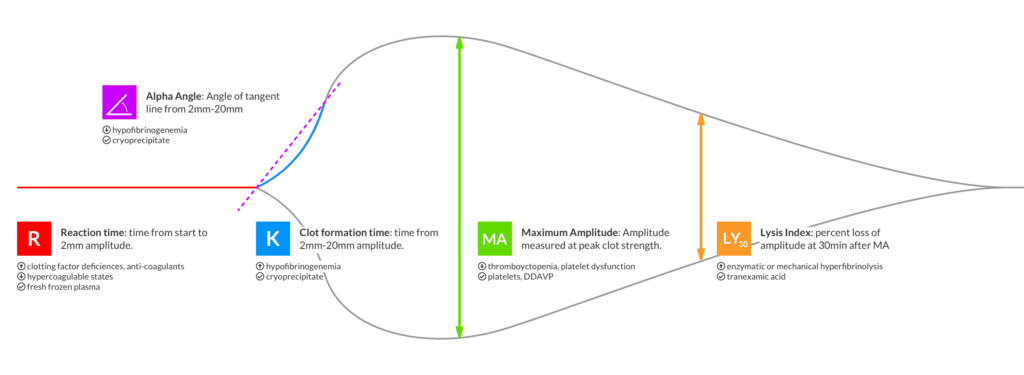
Thromboelastography (TEG) is an assessment of hemostatic function intended to evaluate in vivo coagulation parameters, guiding the targeted correction of coagulopathy1. TEG has predominantly been studied in cardiac surgery, though research has extended to other peri-operative and peri-procedural transfusion management2-5.

Recently, a randomized trial explored the use of TEG to guide transfusion in trauma patients requiring massive transfusion6. 111 patients meeting requirements for massive transfusion protocol activation were randomized to a conventional coagulation assay (CCA) or TEG-guided transfusion algorithm. Patients in the TEG group demonstrated significantly decreased mortality at 28 days and reductions in plasma and platelet transfusion requirements.

More research is needed before TEG can be recommended for use in trauma resuscitation or other common emergency department applications7,8, however it may be useful to prepare by becoming familiar with the most basic aspects of thromboelastography.

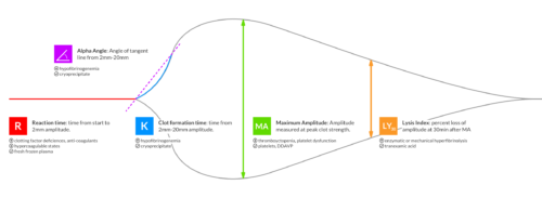
Thromboelastography Summary

Thromboelastography Summary

Examples

Normal

Normal

Anti-coagulants

Anti-coagulants

R,K: Increased
Angle: Decreased

Anti-Platelet

Anti-Platelet

R: Normal
K: Increased
MA: Decreased

Hypercoagulable

Hypercoagulable

R,K: Decreased
MA: Increased

FIbrinolysis

FIbrinolysis

MA: Decreasing
LY30: Increased

DIC (Phase 1)

DIC (Phase 1)

R,K: Decreased
MA: Increased
LY30: Increased

DIC (Phase 2)

DIC (Phase 2)

R,K: Increased
MA: Decreased

References

  1. Bolliger D, Seeberger MD, Tanaka KA. Principles and Practice of Thromboelastography in Clinical Coagulation Management and Transfusion Practice. Transfusion Medicine Reviews. 2012;26(1):1-13. doi:10.1016/j.tmrv.2011.07.005.
  2. Porte RJ, Bontempo FA, Knot EA, Lewis JH, Kang YG, Starzl TE. Systemic effects of tissue plasminogen activator-associated fibrinolysis and its relation to thrombin generation in orthotopic liver transplantation. Transplantation. 1989;47(6):978-984.
  3. Rahe-Meyer N, Solomon C, Hanke A, et al. Effects of fibrinogen concentrate as first-line therapy during major aortic replacement surgery: a randomized, placebo-controlled trial. Anesthesiology. 2013;118(1):40-50. doi:10.1097/ALN.0b013e3182715d4d.
  4. Weber CF, Klages M, Zacharowski K. Perioperative coagulation management during cardiac surgery. Current Opinion in Anaesthesiology. 2013;26(1):60-64. doi:10.1097/ACO.0b013e32835afd28.
  5. De Pietri L, Bianchini M, Montalti R, et al. Thrombelastography-guided blood product use before invasive procedures in cirrhosis with severe coagulopathy: A randomized, controlled trial. Hepatology. 2016;63(2):566-573. doi:10.1002/hep.28148.
  6. Gonzalez E, Moore EE, Moore HB, et al. Goal-directed Hemostatic Resuscitation of Trauma-induced Coagulopathy. Ann Surg. 2016;263(6):1051-1059. doi:10.1097/SLA.0000000000001608.
  7. Afshari A, Wikkelsø A, Brok J, Møller AM, Wetterslev J. Thrombelastography (TEG) or Thromboelastometry (ROTEM) to Monitor Haemotherapy Versus Usual Care in Patients with Massive Transfusion. Vol 24. (Afshari A, ed.). Chichester, UK: John Wiley & Sons, Ltd; 1996:404–3. doi:10.1002/14651858.CD007871.pub2.
  8. da Luz LT, Nascimento B, Rizoli S. Thrombelastography (TEG®): practical considerations on its clinical use in trauma resuscitation. Scand J Trauma Resusc Emerg Med. 2013;21(1):29. doi:10.1186/1757-7241-21-29.