Seizure

Brief HPI

A 72 year-old male with a history of hypertension and hiatal hernia presents to the emergency department with one week of generalized weakness. His family report decreased oral intake with frequent emesis over the past four days. He denies chest pain, shortness of breath, abdominal pain, or other complaints. During the interview he has a generalized tonic-clonic seizure which persists for five minutes despite the administration of 4mg of lorazepam.

An Algorithm for the Management of Seizures

The management of active seizures is algorithmic, starting with a rapid assessment of airway patency, supporting ventilation (with appropriate positioning, nasopharyngeal airway adjuncts and bag-valve mask if needed) and ensuring adequate perfusion. Patients should have continuous vital sign monitoring, supplemental oxygen to maintain oxygen saturation >92% and intravenous access1.

Pharmacologic treatment follows a stepwise approach, detailed in the algorithm below. The focus is on immediate stabilization and progressively escalating anti-epileptic drugs eventually requiring endotracheal intubation and continuous infusions of sedatives2-4.

An Algorithm for the Management of Seizures

Pathophysiology

Seizures are caused by excessive and disorganized neuronal activation, typically induced by global alterations in the production and transmission of impulses (electrolyte derangements, drugs/toxins, infection), or foci of increased irritability (hemorrhage, stroke, mass) – a pathophysiologic motif that mimics cardiac tachyarrhythmias (sympathomimetic toxicity or scarred myocardium for example)1. Status epilepticus, defined as a seizure lasting greater than five minutes or recurrent seizures without a return to normal baseline, shares an equally high short-term mortality – greater than 20%5.

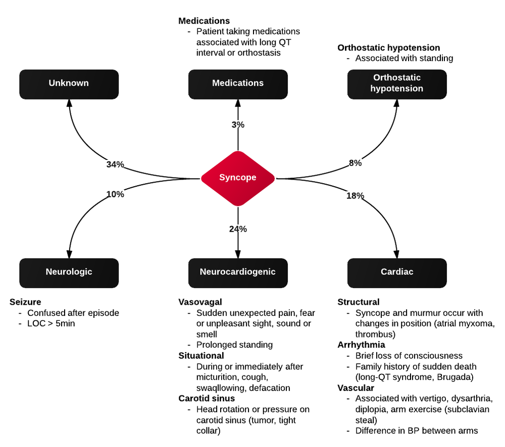
Syncope vs. Seizure

The algorithm below details historical and examination features that may assist with distinguishing epileptic seizure from non-epileptic activity6,7.

Clinical Features Distinguishing Seizure from Syncope

Case Conclusion

The patient continued to seize and a point-of-care chemistry panel revealed a serum sodium of 108mEq/L. Seizures abate after the infusion of hypertonic saline (100mL of 3% saline over 10 minutes, repeated until cessation of seizures). While hyponatremia is generally corrected slowly – owing to the risk of osmotic demyelination – immediate correction in this setting is critical8.
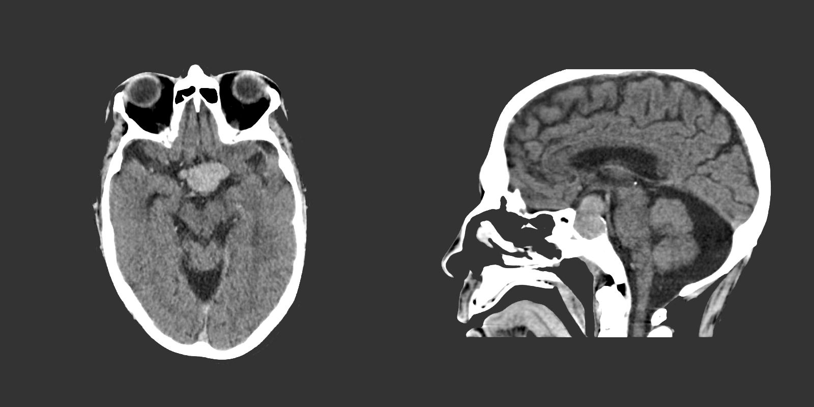

The remainder of the patient’s evaluation demonstrated urine osmolarity is 389mOsm/kg and urine sodium is 53mmol/L, in the setting of relative euvolemia on examination these findings were consistent with SIADH. Head computed tomography is obtained and reveals a sellar mass.

View Hyponatremia Algorithm

References

  1. McMullan JT, Davitt AM, Pollack CV Jr. Seizures. In: Rosen’s Emergency Medicine. Mosby Incorporated; 2002:2808. doi:10.1016/S1474-4422(06)70350-7.
  2. Billington M, Kandalaft OR, Aisiku IP. Adult Status Epilepticus: A Review of the Prehospital and Emergency Department Management. J Clin Med. 2016;5(9):74. doi:10.3390/jcm5090074.
  3. Huff JS, Morris DL, Kothari RU, Gibbs MA, Emergency Medicine Seizure Study Group. Emergency department management of patients with seizures: a multicenter study. Academic Emergency Medicine. 2001;8(6):622-628.
  4. Prasad M, Krishnan PR, Sequeira R, Al-Roomi K. Anticonvulsant therapy for status epilepticus. Prasad M, ed. Cochrane Database Syst Rev. 2014;16(9):CD003723. doi:10.1002/14651858.CD003723.pub3.
  5. Logroscino G, Hesdorffer DC, Cascino G, Annegers JF, Hauser WA. Short-term mortality after a first episode of status epilepticus. Epilepsia. 1997;38(12):1344-1349.
  6. Sheldon R, Rose S, Ritchie D, et al. Historical criteria that distinguish syncope from seizures. J Am Coll Cardiol. 2002;40(1):142-148.
  7. McKeon A, Vaughan C, Delanty N. Seizure versus syncope. Lancet Neurol. 2006;5(2):171-180. doi:10.1016/S1474-4422(06)70350-7.
  8. Goh KP. Management of hyponatremia. Am Fam Physician. 2004;69(10):2387-2394.

Febrile Seizure

Brief HPI:

An 8-month old female, fully-immunized, otherwise healthy is brought in by paramedics after 1 minute of witnessed generalized tonic-clonic shaking. The patient had otherwise been well, eating and behaving normally earlier that day. On EMS arrival, the patient was post-ictal but grew increasingly responsive en-route and upon presentation to the pediatric emergency department she was crying and appeared normal to her parents. Capillary glucose was 118g/dL. On examination the patient was noted to be febrile with a rectal temperature of 39.4°C. The remainder of the physical examination was normal.

ED Course:

The patient received anti-pyretics and a urinalysis was obtained which was not suggestive of urinary tract infection. During the 3-hour period of observation in the emergency department the patient remained at her normal baseline, had no further seizure activity, and tolerated oral intake with difficulty. The patient was suspected to have a simple febrile seizure and was discharged home.

Algorithm for the Diagnosis of Febrile Seizure

Algorithm for the Evaluation of Febrile Seizure

References

  1. Syndi Seinfeld DO, Pellock JM. Recent Research on Febrile Seizures: A Review. J Neurol Neurophysiol. 2013;4(165). doi:10.4172/2155-9562.1000165.
  2. Whelan H, Harmelink M, Chou E, et al. Complex febrile seizures-A systematic review. Dis Mon. 2017;63(1):5-23. doi:10.1016/j.disamonth.2016.12.001.
  3. Millichap JJ, Gordon Millichap J. Methods of investigation and management of infections causing febrile seizures. Pediatr Neurol. 2008;39(6):381-386. doi:10.1016/j.pediatrneurol.2008.07.017.
  4. Subcommittee on Febrile Seizures, American Academy of Pediatrics. Neurodiagnostic evaluation of the child with a simple febrile seizure. Pediatrics. 2011;127(2):389-394. doi:10.1542/peds.2010-3318.

Seizure

Definition

Seizure
Pathologic neuronal activation leading to abnormal function
Epilepsy
Recurrent unprovoked seizures

Classification

  • Cause
    • Primary: Unprovoked
    • Secondary: Provoked, caused by trauma, illness, intoxication, metabolic disturbances, etc.
  • Effect on mentation
    • Generalized: involvement of both hemispheres with associated loss of consciousness (tonic-clonic, absence, atonic, myoclonic)
    • Focal: Involving single hemisphere with preserved level of consciousness
  • Status epilepticus
    • Witnessed convulsions lasting >5min
    • Recurrent seizure without recovery from postictal period

Causes of Seizures

Causes of Seizures

Management of Seizures

Management of Seizures

Medications for Treatment of Seizures

Medication Dose (adult) Dose (peds) Comment
1st Line
Lorazepam 4mg IV <13kg: 0.1mg/kg (max 2mg)
13-39kg: 2mg

>39kg: 4mg
Repeat in 10min
Midazolam 10mg IM 0.2mg/kg IM (max 5mg) Repeat in 10min
Midazolam 10mg buccal 0.5mg/kg buccal (max 5mg) Repeat in 10min
2nd Line
Fosphenytoin 20mg PE/kg IV    
Phenytoin 20mg/kg IV   May cause hypotension
3rd Line
Midazolam 0.05-2mg/kg/hr    
Propofol 1-2mg/kg bolus then 20-200mcg/kg/min    
Pentobarbital 5-15mg/kg bolus then 0.5-5mg/kg/hr    
Special Conditions
Glucose 50mL D50/W   Hypoglycemia
MgSO4 6g IV over 15min   Eclampsia (20wks gestation to 6wks post-partum)
Pyridoxine 0.5g/min until seizures stop, max 5g   INH ingestion
3% saline 100-200mL over 1-2h   Confirmed hyponatremia

History

Points suggestive of seizure over alternative process
Abrupt onset
Duration < 120s
LOC
Purposeless activity: automatisms, tonic-clonic
Provocation: fever in children, substance withdrawal
Postictal state
Retrograde amnesia
Incontinence, oral trauma (buccal maceration, tongue laceration)
Rapidly resolving lactic acidosis
Important historical points for patients with seizure history
Recent illness
Medications (adherence, changes, interactions)
Substance use
Ictogenic factors
Recent/remote head trauma
Developmental abnormalities
Substance use
Sleep deprivation
Pregnancy

Key Physical Examination Findings

  • Vital sign abnormalities persisting beyond immediate postictal state (may suggest drug/toxin exposure, CNS lesion)
  • Nuchal rigidity
  • Signs of IVDA
  • Sequela

    • Head trauma
    • Tongue laceration
    • Shoulder dislocation (posterior)
  • Neurological exam

    • Stroke
    • Elevated ICP
    • Failure to note improvement in postictal confusion (encephalopathy, subclinical seizures)

Labs

  • Glucose
  • BMP (Na, Ca, Mg)
  • AED levels
  • CBC (leukocytosis and bandemia common post-seizure)
  • CSF
  • B-hCG
  • LFT (hepatic dysfunction, alcoholic hepatitis)
  • Lactate (rapidly resolves on repeat)

Indications for Imaging

  • New seizures
  • History of trauma
  • History of malignancy
  • Immunocompromised
  • Headache
  • Anti-coagulation
  • Focal neurological exam
  • Persistent AMS

References

  1. McMullan, J., Davitt, A., & Pollack, C. (2013). Seizures. In Rosen’s Emergency Medicine – Concepts and Clinical Practice (8th ed., Vol. 1, pp. 156-161). Elsevier Health Sciences
  2. WikEM: Seizure

Syncope

ID:

A 50 year-old male with a reported two-year history of infrequent spells, presenting with two spells in the past two days.

HPI:

The patient’s spells began two years ago, he recounts that he was watching television when he lost consciousness and a friend noted he started shaking; he does not recall the event, and awoke in the hospital. The next spell occurred one year later, though the patient is unable to recall much about this episode. The patient remained spell-free until yesterday when he was on a bus, lost consciousness and awoke in a hospital. He notes that he had bit his tongue and lost control of his bladder. He was discharged hours later with a prescription for an AED which he was unable to fill. This morning, the patient had another spell while in the bathroom. His roommate heard him fall, found him on the ground, and noted that his mouth was moving but did not see any other movements.

The patient’s episodes are all associated with loss of consciousness and are followed by 5-10 minutes of disorientation after which he recovers fully. The episodes are sometimes preceded by a feeling of “euphoria”, though this feeling sometimes occurs without subsequent LOC.

The patient denies any associated palpitations, dizziness/LH, chest pain or muscle pain.  He has not had any recent fevers/chills, dysuria, cough, headache, changes in vision, numbness/tingling, weakness, difficulty speaking or swallowing or weight loss. He also denies any history of head trauma.

Physical Examination:

  • VS: Stable and WNL
  • General: Well-appearing, pleasant, and in NAD.
  • HEENT: NC/AT. MMM. Small lesion on tongue.
  • Lungs: CTAB.
  • CV: RRR with occasional ectopic beats, no M/R/G.
  • Abdomen: S/NT/ND. Bowel sounds present.
  • Neurological exam: AAOx4, CN II-XII intact, motor/sensation/reflexes/coordination/gait WNL

Imaging/Studies:

  • EKG: Occasional PAC/PVC
  • CT Brain: Unremarkable except for mild age-related cerebral atrophy

Assessment & Plan:

50 year-old male with a history of HTN and a reported two-year history of infrequent spells presenting with two spells in the past two days. The description of the patient’s episodes could be consistent with seizures. Aspects supporting this notion include loss of consciousness and period of confusion following each episode. One of the recent episodes was also associated with tongue-biting and loss of bladder control. Additionally, some episodes are associated with a sensation of euphoria rising from the abdomen to the head which could be indicative of an aura. Characteristics that suggest other causes include the absence of noted convulsions and non-stereotyped nature of each episode which could be due to the patient’s poor recollection of these events and absence of reliable witnesses. In the case of true seizures, the possible etiologies in this patient include a mass, metabolic abnormalities, substance use, or concomitant infection exacerbating an existing propensity for seizure activity. Other, non-seizure causes warranting evaluation include cardiogenic syncope particularly given the evidence of ectopic beats on examination and electrocardiogram.

Differential Diagnosis of Syncope

First, is it syncope? History is very important for distinguishing syncope from other causes (seizure, dizziness, vertigo, presyncope). Ask about precipitating events, prodromal symptoms, post-ictal confusion. Common causes of syncope and their associated symptoms are detailed in the figure below.

References:

  1. Kapoor, W. N. (2000). Syncope. The New England journal of medicine, 343(25), 1856–1862. doi:10.1056/NEJM200012213432507