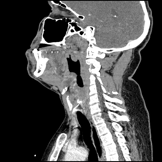
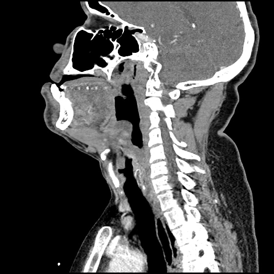
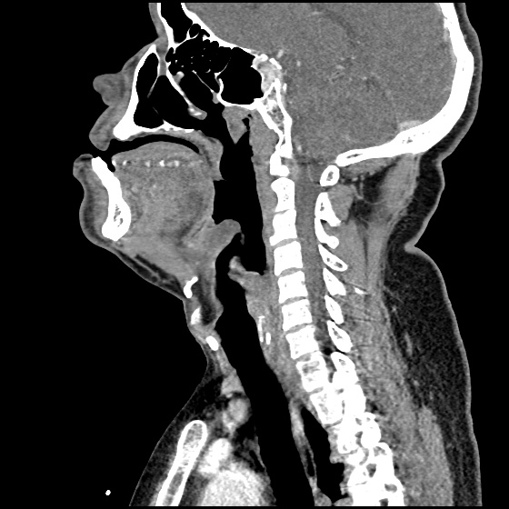
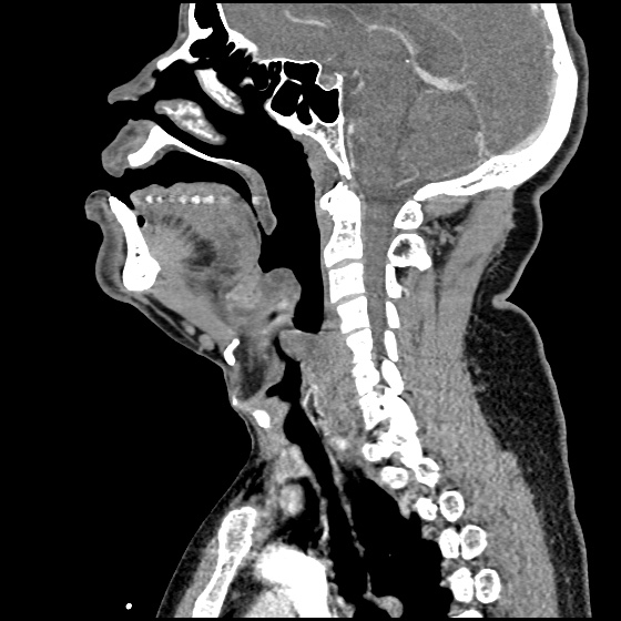
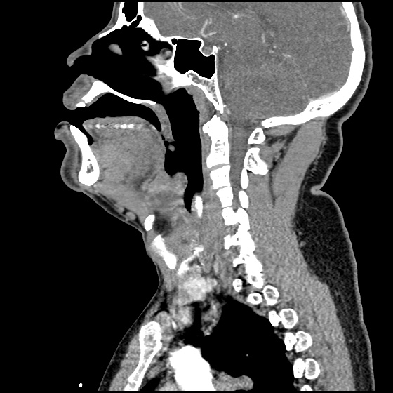
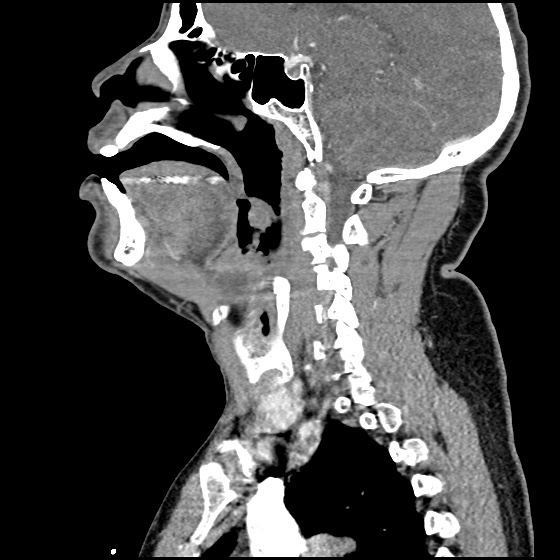
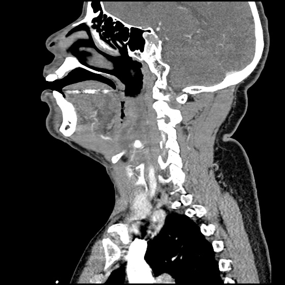
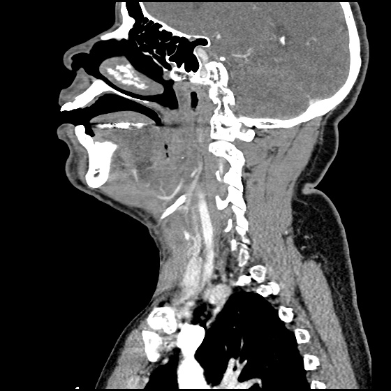
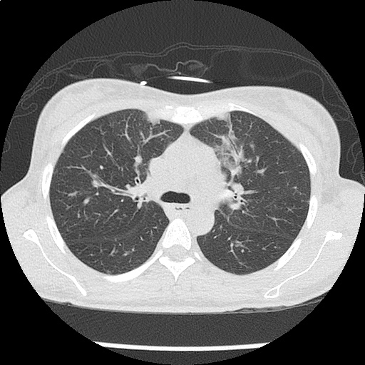
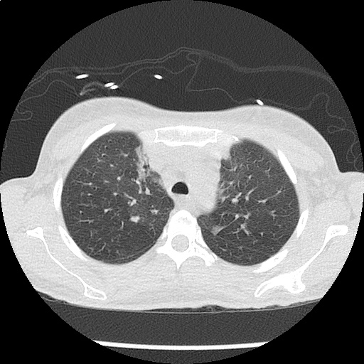
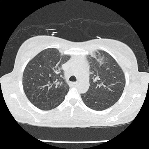
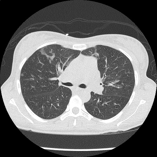
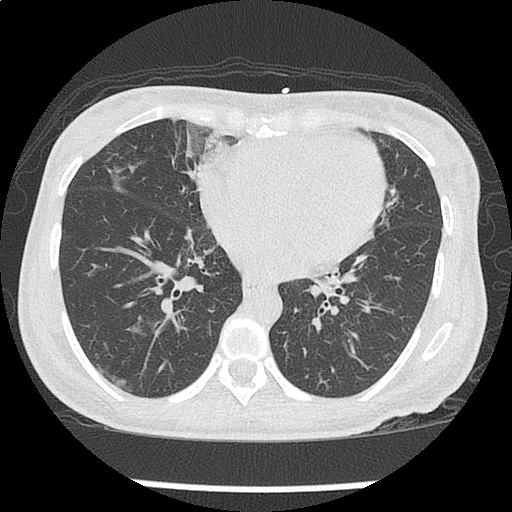
Neurosyphilis

Brief H&P

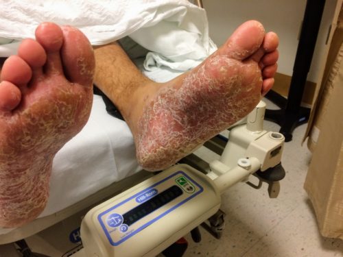
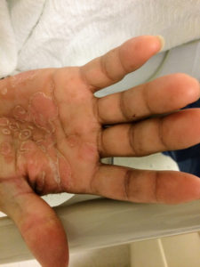
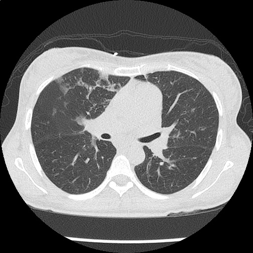
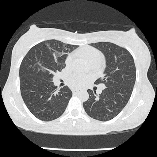
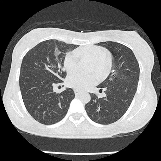
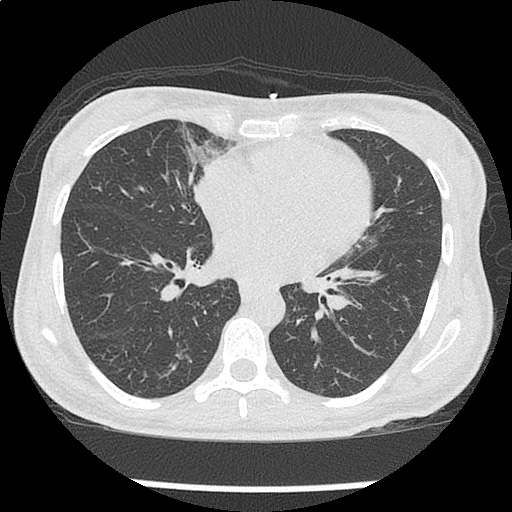
A young male with a history of HIV (untreated for the last year, with unknown CD4 count), and syphilis (reportedly treated with an intramuscular injection 1 year ago), presents with 4 months of a painful rash on the palms and soles and diplopia. Examination revealed the rash pictured below, ocular examination with minimal papilledema and anterior chamber inflammation.

Labs were unremarkable. CSF sampling was notable for 34 WBC’s with lymphocyte predominance (92%), and elevated protein (56mg/dL). The patient was admitted for syphilis with presumed neurosyphilis. Serum RPR titer was elevated at 1:64,  FTA-ABS and CSF VDRL were reactive. The patient was treated with intravenous penicillin and anti-retroviral therapy was reinitiated.

Epidemiology1

  • Transmission
    • Sexual contact (estimated transmission probability 60% per partner)
    • Trans-placental
  • Race/Sex
    •  African-American, Hispanic
    • Male > Female
    • Male (primary syphilis), female (secondary syphilis) – lesion visibility
    • Urban > rural

Natural History1

Stage Signs/Symptoms Incubation Period
Primary Chancre, reginal lymphadenopathy 3 weeks
Secondary Rash, fever, malaise, generalized lymphadenopathy, mucous membrane lesions, condyloma lata, headache, meningitis 2-12 weeks
Latent Asymptomatic Early (<1 year)

Late (>1 year)

Tertiary Cardiovascular:

Aortic aneurysm, aortic insufficiency, coronary artery ostial stenosis

<2 years
CNS:
Acute syphilitic meningitis: headache, confusion, meningeal irritation <2 years
Meningovascular: cranial nerve palsy 5-7 years
General paresis: headache, vertigo, personality changes, vascular event 5-7 years
Tabes dorsalis: dementia, ataxia, Argyl-Robertson, [arrow-down] proprioception 10-20 years
Gumma:

Local tissue destruction

1-46 years

Diagnosis1

  • Serologic
    • Non-treponemal (screening)
      • RPR, VDRL
      • Limitations:  sensitivity, false positive (age, pregnancy, drugs, malignancy, autoimmune, viral infections)
    • Treponemal (confirmatory)
      • FTA-ABS
    • Neurosyphilis
      • Indications for CSF sampling: neurologic/ophthalmologic symptoms, tertiary syphilis (aortitis, gumma, iritis), HIV coinfection with elevated RPR titer (> 1:32)
      • CSF: leukocytosis (predominantly lymphocytes),  protein
      • CSF VDRL reactive
      • Negative CSF FTA-ABS may rule out neurosyphilis

Syphilis in HIV-infected Individuals2

  • Primary: larger and more lesion, multiple ulcers
  • Secondary: genital ulcers more common, higher RPR/VDRL titers
  • Tertiary: possibly more rapid progression to neurosyphilis

References

  1. Singh AE, Romanowski B. Syphilis: review with emphasis on clinical, epidemiologic, and some biologic features. Clin Microbiol Rev. 1999;12(2):187-209.
  2. French P. Syphilis. BMJ. 2007;334(7585):143-147. doi:10.1136/bmj.39085.518148.BE.

Hematologic Emergencies

Sickle Cell Crises

  • Triggers: infection, acidosis, dehydration, cold-exposure, hypoxia, pregnancy
  • Presentation: exclude alternative more serious pathology prior to ascribing pain to vaso-occlusive crisis

Effects by Organ System

System Symptom
CNS Focal or generalized neurological symptoms, stroke, seizure
Pulmonary Acute chest syndrome (fever, chest pain, cough, hypoxia, pulmonary infiltrates), pulmonary embolism
GI Abdominal pain, nausea/vomiting
Renal Papillary necrosis
GU Priapism, testicular/ovarian ischemia
Muskuloskeletal Bone pain (back, proximal extremities), exclude osteomyelitis, avascular necrosis
ID Infection, functional asplenia (streptococcus, haemophilus)
OB Preterm labor, placental abruptions, SAB
Ophthalmology Acute retinal ischemia, hyphema (with intra-ocular hypertension)
Hematology
  • Sequestration crisis: acute anemia, often post-viral
  • Hemolytic crisis: acute anemia, reticulocytosis, hyperbilirubinemia
  • Megaloblastic crisis: folate deficiency
  • Aplastic crisis: inadequate reticulocytosis

Evaluation

  • CBC with reticulocyte count
    •  Hemoglobin: suggests sequestration or hemolytic crisis
    • Reticulocyte index: suggests aplastic or megaloblastic crisis
  • LDH/haptoglobin: evaluate for hemolysis
  • UA: evaluate for infection/infarction
  • CXR: evaluate for acute chest syndrome

Management

  • Rehydration (hypotonic fluids)
  • Analgesia
  • Supplemental oxygen if hypoxic
  • Exchange transfusion for priapism, neurologic symptoms, aplastic/sequestration/hemolytic crises

Transfusion Reactions

  • Epidemiology: overall 0.25%, 0.09% severe
  • Management: stop transfusion

Management by Presumed Etiology

Reaction Mechanism Signs/symptoms Management
Acute, Severe
Acute hemolysis Incompatibility Fevers, HR, BP, vomiting, back pain IVF, vasopressors if needed, furosemide
Anaphylaxis IgA-mediated 1min: flushing laryngospasm, bronchospasm, BP Epinephrine, steroids, diphenhydramine, IVF
Sepsis Bacterial contamination (Y. entercolitica), increased risk in platelet transfusion Fevers, BP IVF, vasopressors if needed, broad-spectrum antibiotics
TRALI (transfusion-related acute lung injury) Non-cardiogenic pulmonary edema, increased risk in FFP transfusion Hypoxia, respiratory distress, XR bilateral infiltrates Supplemental oxygen, PPV/ETT
TACO (transfusion-associated circulatory overload) Hypervolemia in patients with history of CHF Hypoxia, respiratory distress, heart failure Supplemental oxygen, PPV/ETT, furosemide
Acute, Minor
Simple febrile reaction Cytokine-mediated Isolated fever Acetaminophen
Minor allergic reaction Response to transfused plasma proteins Urticaria, pruritus, flushing Diphenhydramine
Delayed
Delayed hemolysis Minor RBC antigens 5-10d, low-grade hemolysis  
GVHD Immunocompromised host Fever, rash, N/V, transaminitis, pancytopenia  
Massive Transfusion
Massive transfusion Large-volume, refrigerated products Coagulopathy, hypothermia, hypocalcemia, hyperkalemia, lactic acidosis

Bleeding Disorders

Overview

  • Disorders of primary hemostasis
    • General: present with mucocutaneous, post-operative bleeding
    • vWD
    • Platelet disorders
      • Medication-induced: NSAID, valproate, B-lactam, SSRI
      • Systemic disease: hepatic, renal failure
    • ITP: antibody-mediated platelet destruction
  • Disorders of secondary hemostasis
    • General: present with bleeding into soft-tissue, joints
    • Hemophilia A (VIII)
    • Hemophilia B (IX)
  • Disorders of both primary and secondary hemostasis
    • DIC
    • Liver disease
    • Severe vWD
  • Evaluation
    • PT: VII, vitamin K
    • PTT: VIII, IX, XI, XIII, vWD, heparin
    • Increased PT/PTT: XI, V, vitamin K, heparin, DIC
    • CBC: degree of anemia, platelet count, differential (hematopoetic disorders)
  • Management
    • Thrombocytopenia
      • Prophylactic transfusion for avoidance of spontaneous hemorrhage for platelet count <10,000
      • Transfusion for active bleeding at platelet count <50,000
      • Dosing
        • Adults: one RDP increases platelet count by 7-10,000
        • Pediatrics: 5-10ml/kg
      • ITP
        • Transfuse platelets for active bleeding
        • High-dose steroids (prednisone 1mg/kg)
        • IVIG (1g/kg/d)
      • Uremia
        • Hemodialysis
        • DDAVP (0.3ug/kg IV)
      • vWD
        • DDAVP (0.3ug/kg IV)
        • Severe: VWF (Humate-P) 40-80IU/kg
        • Tranexamic acid
      • Hemophilia A
        • Minor: 20IU/kg
        • Major: 50IU/kg
      • Hemophilia B
        • Minor: 40IU/kg
        • Major: 100IU/kg

DIC/TTP/HUS

  • Disseminated Intravascular Coagulation
    • Etiology: severe systemic illness/injury
      • Trauma, burn, crush
      • Sepsis
      • Malignancy
      • Obstetric complication: abruption, amniotic fluid embolism
      • Hemolytic anemia
    • Exam: petechiae/purpura, hemorrhage (puncture site, GI, GU, pulmonary)
    • Labs:
      • PT/PTT
      • Fibrinogen
      • CBC: schistocytes, thrombocytopenia
      • FDP/D-Dimer
    • Management
      • Treat underlying illness
      • Transfuse (PRBC, FFP for INR > 2, cryoprecipitate for fibrinogen < 100)
      • Heparin if apparent embolic events
      • Consult hematology
  • TTP/HUS
    • Presentation
      • Thrombocytopenia
      • Altered mental status
      • Renal dysfunction
      • Fever
      • MAHA
    • TTP: more commonly associated with altered mental status
      • Etiology: drugs, pregnancy, infection (HIV)
      • Mechanism: ULvWF uncleaved by dysfunctional ADAMTS-13
    • HUS: more commonly associated with renal dysfunction
      • Mechanism: toxin from E. coli, Shigella
      • Timing: 1-2wks after diarrheal illness
    • Evaluation
      • CBC: anemia, schistocytes, thrombocytopenia
      • PT/PTT (normal)
      • BUN/Creatinine
      • LDH
    • Management
      • Platelets contraindicated except as stopgap measure in ICH (can worsen process)
      • Plasma exchange with FFP (replaces functional ADAMTS-13)
      • Steroids (prednisone 1mg/kg daily)
      • Hematology consultation

Complications of anti-thrombotic therapy

  • Agents
    • Anti-platelet
      • TXA: Aspirin
      • ADP: clopidogrel, ticagrelor, prasugrel
      • GPIIb/IIIa: abciximab, eptifibatide, tirofiban
    • Anti-coagulants
      • Anti-thrombin: heparin, LMWH (enoxaparin, dalteparin)
      • Vitamin K antagonist: warfarn (anti-II, VII, IX, X)
      • Direct thrombin inhibitor: bivalirudin, argatroban, dabigatran
      • Xa inhibitor: rivaroxaban, apixaban
    • Fibrinolytics
      • Alteplase, tenectaplase
  • Complications
    • HIT: platelet count decrease >50% at 5 days

Summary of Management

Agent Reversal
Aspirin, clopidogrel 5-10U platelets

DDAVP 0.3ug/kg

GPIIb/IIIa Abciximab: 5-10U platelets

Eptifibatide/tirofiban: none

Heparin Protamine 1mg/100mg heparin in last 2-3 hours
LMWH Enoxaparin: 1mg/1mg

Dalteparin: 1mg/100U

Warfarin See supratherapeutic INR algorithm
DTI Dabigatran: Praxbind, hemodialysis, consider Factor VIIa
Xa PCC
Fibrinolytics 10U cryoprecipitate, 2U FFP, consider platelets and aminocaproic acid (4-5g IV)

Oncologic Emergencies

Overview

  •  Complications
    • Airway obstruction
    • PNA
    • Pleural effusion
    • Pericardial effusion
    • VTE
    • SVC syndrome
      • Symptoms: dyspnea (airway edema), chest fullness, blurred vision, headache (increased ICP)
    • Massive hemoptysis
      • Management: ETT (large-bore for bronschoscopy), affected side down
  • Brain Metastases
    • Cancers: melanoma, lung, breast, colorectal
    • Management: dexamethasone 10mg IV load, elevated HOB, hypertonic saline or mannitol, prophylactic anti-eplipetics
  • Meningitis
    • Pathogens: Listeria (ampicillin), Cryptococcus (amphotericin)
    • Evaluation: CSF sampling with cytology (diagnose leptomeningeal metastases)

Metabolic Disturbances

  • Hypercalcemia
    • Cancers: MM, RCC, lymphoma, bone metastases (breast, lung, prostate)
    • Mechanism: metastatic destruction, PTH-RP, tumor calcitriol
    • Prognosis: 50% 30-day mortality
    • Symptoms
      • Chronic: anorexia, nausea/vomiting, constipation, fatigue, memory loss
      • Acute: CNS (lethargy, somnolence)
    • Findings
      • Calcium: >13.0mg/dL
      • ECG: QT shortening
    • Treatment
      • Mild: IVF
      • Severe: IVF, loop diuretics, bisophosphanate (pamidronate 90mg IV infused over 4 hours), consider calcitriol, consider hemodialysis if cannot tolerate fluids or unlikely to respond to diuretics
  • Hyponatremia
    • Cancers: lung (small-cell), pancreatic, ovarian, lymphoma, thymoma, CNS
    • Mechanism: SIADH
    • Symptoms: muscle twitching, seizure, coma
    • Management: fluid restriction, if seizing administer 3% hypertonic saline at 100cc/hr until resolution
  • Hypernatremia
    • Mechanism: decreased intake, increased GI losses from chemotherapy
    • Management: cautious fluid resuscitation
  • Tumor Lysis Syndrome (TLS)
    • Cancers: hematologic, rapid-growth solid tumors
    • Mechanism: release of intracellular contents (uric acid, K, PO4, Ca)
    • Timing: 1-4 days after therapy (chemo, radiation)
    • Diagnosis
      • Uric acid >8mg/dL
      • Potassium >6mEq/L
      • Calcium <7mg/dL
      • PO4 >4.5mg/dL
      • Acute kidney injury
    • Management
      • IVF, allopurinol, rasburicase, urinary alkalinization
      • Consider hemodialysis if volume overloaded

Localized Complications

  • Musculoskeletal Complications
    • Spinal cord compression
      • Cancers: prostate, breast, lung, RCC, non-Hodgkin lymphoma, MM (5-10% of all cancer patients)
      • Sites: thoracic (60%), lumbosacral (30%), cervical (10%)
      • Symptoms: pain (worse lying flat, cough/sneeze, heavy lifting)
      • Evaluation: MRI (se 93%, sp 97%)
      • Management: dexamethasone 10mg IV load, 4mg q6h, neurosurgical consultation, radiation oncology consultation
    • Pathologic fracture
      • Features: sudden onset, low-force mechanism
  • Therapy Complications
    • Neutropenic fever
      • Definition: ANC <500 or ANC <1000 with expected nadir <500 (nadir typically occurs 5-10d after chemotherapy) with Tmax >38.3°C or >38.0°C for >1h
      • Examination: subtle signs of infection, thorough examination is critical (skin, catheter, perineum)
      • Treatment: carbapenem monotherapy, vancomycin if indwelling catheter, oncology consultation for colony stimulating factors
    • Chemotherapy-induced vomiting
      • Management: ondansetron with dexamethasone, consider NK-1 antagonist (aprepitant)

Hematologic Malignancies

  • Acute leukemia
    • Signs/Symptoms: leukopenia (infection), anemia (weakness/fatigue), thrombocytopenia (bleeding)
    • Diagnosis: >5% blasts
  • Thrombocytopenia
    • Management
      • No bleeding, goal >10,000
      • Fever, coagulopathy, hyperleukoctosis, goal >20,000
      • One unit of platelets increases count by 5,000
  • Hyperleukocytosis
    • Definition: WBC > 50-100k
    • Complications: microvascular congestion (pulmonary, cerebral, coronary)
    • Symptoms
      • CNS: confusion, somnolence, coma
      • Pulmonary: dyspnea, respiratory alkalosis
    • Management: cytoreduction (induction chemotherapy, increased risk TLS)
  • Hyperviscosity
    • Cancer: macroglobulinemia, MM
    • Symptoms: epistaxis, purpura, GIB, neuro deficits
    • Diagnosis: serum viscosity > 1.4-1.8
    • Management: emergent plasmapheresis
  • Polycythemia
    • Diagnosis: Hb >17
    • Differential: dehydration, hypoxia, smoking, altitude
    • Symptoms: HA, vertigo, angina, claudication, pruritus (after showering)
    • Complications: thrombosis (stroke), bleeding
    • Management: emergent phlebotomy (500cc if otherwise healthy)
  • Thrombocytosis
    • Diagnosis: platelet >1,000,000
    • Symptoms: vasomotor (HA, lightheadedness, syncope, chest pain, paresthesias)
    • Management: low-dose aspirin

Adrenal/Pituitary Emergencies

Adrenal Emergencies

  • Hormones: aldosterone, cortisol, androgens, catecholamines
  • Adrenal insufficiency
    • Primary
      • Causes
        • Autoimmune (associated with other endocrinopathies, PTH, DM)
        • Infection (TB, viral, meningococcemia)
        • Infiltration (sarcoidosis, amyloidosis)
        • Hemorrhage (trauma, anti-coagulation)
        • Malignancy (primary, metastatic)
      • Signs/Symptoms
        • AMS
        • Hypotension (refractory)
        • GI: anorexia, nausea/vomiting, diarrhea
        • Hyperpigmentation
      • Labs
        • Hyponatremia
        • Hyperkalemia
        • Hypercalcemia
        • Mild metabolic acidosis
        • Hypoglycemia
    • Secondary
      • Causes
        • Iatrogenic (>5mg prednisone/day for > 2 weeks)
        • Pituitary/sellar tumors
        • Hemorrhage (Sheehan)
        • Cranial radiation
      • Signs/Symptoms
        • RAAS function maintained, hypotension rare
        • Features of pituitary/hypothalamic dysfunction: menstrual disturbances, headache, vision changes, galactorrhea, acromegaly
    • Adrenal Crisis
      • Precipitated by physiologic stressor: sepsis, MI, trauma, surgery
      • Diagnosis
        • AM cortisol <3
        • ACTH stimulation peak cortisol <15
        • ACTH level
      • Management
        • Glucose management
        • Fluid resuscitation
        • Dexamethasone 10mg IV
        • Identify and treat precipitant

Cushing syndrome

  • Causes
    • Pituitary adenoma (Cushing disease)
    • Malignancy (ACTH-producing): SCLC, pancreatic, carcinoid
    • Adrenal neoplasm
  • Signs/Symptoms
    • Obesity, fat deposition in face, neck
    • Skin atrophy with striae
    • Proximal myopathy
    • Hypertension

Pheochromocytoma

  • Familial: MEN 2A/2B, NF, Von Hippel-Lindau
  • Signs/Symptoms
    • Refractory hypertension (paroxysmal)
    • Heat intolerance, sweating, weight loss
  • Diagnosis
    • 24h urine metanephrine, catecholamine
    • CT/MRI

Hypopituitarism

  • Adenoma
    • Symtoms/Signs
      • Headache
      • Vision changes (bitemporal hemianopsia)
      • Cavernous sinus involvement (CN III, IV, V1, V2, VI)
  • Ischemic necrosis
    • Sickle cell disease, vasculitis, cavernous sinus thrombosis, infection, TBI, post-partum (Sheehan)
  • Pituitary apoplexy
    • Acute loss of pituitary function from infection/hemorrhage, rarely tumor
    • Symptoms/Signs
      • Abrupt onset headache
      • Vision changes
      • Meningismus
      • ALOC

Thyroid Emergencies

Hyperthyroidism

Symptoms

Constitutional Weight loss, heat intolerance, perspiration
Cardiopulmonary Palpitations, chest pain, dyspnea
Neuropsychiatric Tremor, anxiety, double vision, muscle weakness
Neck Fullness, dysphagia, dysphonia
Musculoskeletal Extremity swelling
Reproductive Irregular menses, decreased libido, gynecomastia

Signs

Vital signs Tachycardia, widened pulse pressure, fever
Cardiovascular Hyperdynamic precordium, CHF, atrial fibrillation, systolic flow murmur
Ophthalmologic Widened palpebral fissure, periorbital edema, proptosis, diplopia, restricted superior gaze
Neurologic Tremor, hyperreflexia, proximal muscle weakness
Dermatologic Palmar erythema, hyperpigmented plaques or non-pitting edema of tibia
Neck Enlarged or nodular thyroid

Causes

  • Grave disease
    • Mechanism: thyroid-stimulating antibodies
    • Female > Male (10x)
    • Findings: ophthalmopathy (lid lag), infiltrative dermopathy (pretibial)
  • Toxic adenoma, toxic multinodular goiter
    • Mechanism: Excess thyroid hormone production
  • Thyroiditis
    • Mechanism: inflammation results in increased thyroid hormone release, typically followed by depletion and TSH suppression resulting in hypothyroidism
    • Signs/symptoms: tachycardia, weight loss, irritability, sweating, anxiety, heat intolerance
    • Subacute thyroiditis
      • Post-viral
      • Symptoms: hoarseness, dysphagia, painful thyroid
    • Hashimoto
      • Typically hypothyroidism
    • Drug-induced: Lithium, amiodarone
    • Trauma: surgical, direct

Thyroid Storm

  • Essentially an exaggeration of thyrotoxicosis featuring marked hyperthermia (104-106°F), tachycardia (HR > 140bpm), and altered mental status (agitation, delirium, coma).
  • Precipitants
    • Medical: Sepsis, MI, CVA, CHF, PE, visceral ischemia
    • Trauma: Surgery, blunt, penetrating
    • Endocrine: DKA, HHS, hypoglycemia
    • Drugs: Iodine, amiodarone, inhaled anesthetics
    • Pregnancy: post-partum, hyperemesis gravidarum
  • Scoring (Burch, Wartofsky)
  • Management
    • Supportive measures
      • Volume resuscitation (with MVI, Thiamine) and cooling
      • Benzodiazepines for agitation
    • Beta-blockade
      • Propranolol 60-80mg PO q4h
      • Propranolol 0.5-1.0mg IV, repeat q15min then 1-2mg q3h
      • Esmolol continuous infusion
    • MTZ/PTU 1-hour prior to iodine
      • Methimazole 20mg (except pregnancy)
      • Propylthiouracil 600mg (hepatotoxic)
    • Steroids: dexamethasone
    • Iodine
    • Endocrinology consultation

Hypothyroidism

Symptoms

Constitutional Weight gain, cold intolerance, fatigue
Cardiopulmonary Dyspnea, decreased exercise capacity
Neuropsychiatric Impaired concentration and attention
Musculoskeletal Extremity swelling
Gastrointestinal Constipation
Reproductive Irregular menses, erectile dysfunction, decreased libido
Integumentary Coarse hair, dry skin, alopecia, thin nails

Signs

Vital signs Bradycardia, hypothermia
Cardiovascular Prolonged QT, increased ventricular arrhythmia, accelerated CAD, diastolic heart failure, peripheral edema
Neurologic Lethargy, slowed speech, agitation, seizures, ataxia/dysmetria, mononeuropathy, delayed relaxation of reflexes
Musculoskeletal Proximal myopathy, pseudohypertrophy, polyarthralgia
Gastrointestinal Ileus

Causes

  • Hashimoto: auto-antiboids
  • Thyroidectomy
  • Radiation, radioactive iodine ablation

Myxedema Coma

  • Precipitants
    • Critical illness: sepsis (especially PNA), CVA, MI, CHF, trauma, burns
    • Endocrine: DKA, hypoglycemia
    • Drugs: amiodarone, lithium, phenytoin, rifampin, medication non-adherence
    • Environmental: cold exposure
  • Recognition
    • History: hypothyroidism, thyroidectomy scar and acute precipitating illness
    • Hypothermia: temp <95.9°F (or normal in presence of infection)
    • AMS: lethargy, confusion, coma, agitation, psychosis, seizures
    • Hypotension: refractory to volume resuscitation and pressors
    • Bradypnea: with hypercapnia and hypoxia
    • Skin: non-pitting edema of face and hands
    • Hyponatremia
  • Management
    • Airway protection
    • Fluid resuscitation
    • Thyroid hormone replacement
      • Young, otherwise healthy patients: T3 10ug IV q4h
      • Elderly, cardiac compromise: 300ug IV x1
      • Steroids: dexamethasone 1h prior to thyroid hormone
    • Treat precipitating illness

Interpretation of Thyroid Function Tests

Condition TSH T4
None Normal Normal
Hyperthyroidism Low High
Hypothyroidism High Low
Subclinical hyperthyroidism Low Normal
Subclinical hypothyroidism High Normal
Sick euthyroid Low Low

Acid-Base Disturbances

Method

  • Primary disturbance (acidemia/alkalemia)
  • Primary process (metabolic/respiratory)
  • Presence of mixed disorder
    • Increase PCO2 of 10, increases HCO3 by 1 (acute) or 3 (chronic)
    • Decreased PCO2 of 10, decreases HCO3 by 2 (acute) or 5 (chronic)
    • Increase HCO3 of 1, increases PCO2 by 0.7
    • Decreased HCO3, add 15, result should equal PCO2 and number after decimal of pH
  • Anion gap

Causes

  • Anion Gap
    • Methanol
    • Uremia
    • DKA/AKA
    • Paraldehyde, propylene glycol
    • INH
    • Lactate
    • Ethylene glycol
    • Salicylate
  • Non-Anion Gap
    • Fistulae
    • Ureteral fistulae
    • Saline
    • Diarrhea
    • Carbonic anhydrase inhibitors
    • Spironolactone
    • RTA
  • Metabolic Alkalosis
    • Vomiting
    • Volume depletion
    • Diuretics
    • Steroids
  • Respiratory Acidosis
    • CNS lesion
    • Myopathies
    • Chest wall abnormalities
    • Obstructive lung disease
  • Respiratory Alkalosis
    • Anxiety
    • Fever
    • Hyperthyroidism
    • Hypoxia
    • Sympathomimetic

See Also

Portal Venous Gas

Brief HPI

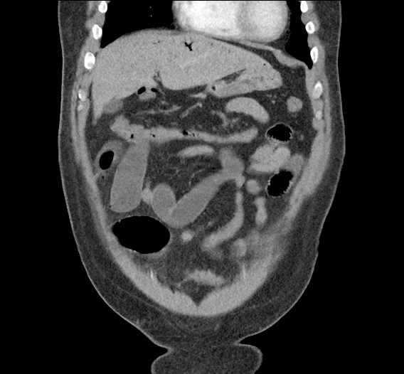
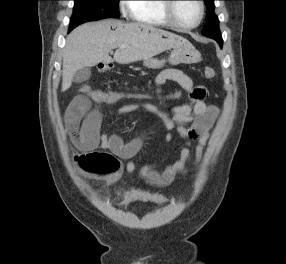
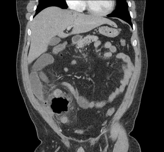
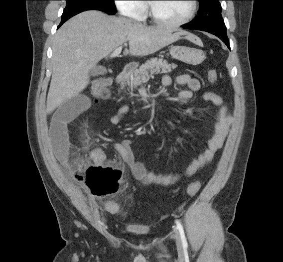
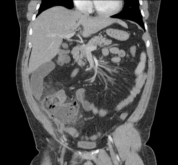
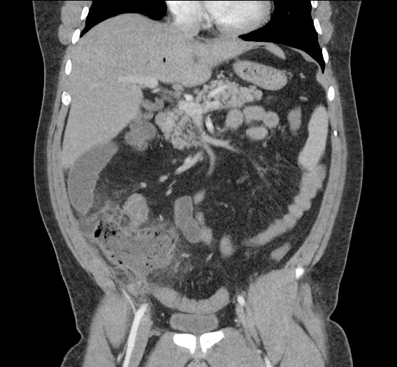
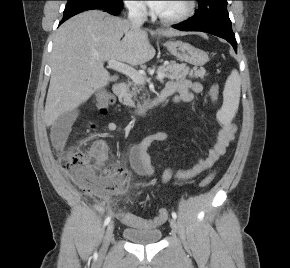
Young male with no significant medical history presenting with progressively worsening right lower quadrant abdominal pain with marked tenderness to palpation and involuntary guarding.

Imaging

portal_venous_gas_0000_Layer-Comp-1
portal_venous_gas_0001_Layer-Comp-2
portal_venous_gas_0002_Layer-Comp-3
portal_venous_gas_0003_Layer-Comp-4
portal_venous_gas_0004_Layer-Comp-5
portal_venous_gas_0005_Layer-Comp-6
portal_venous_gas_0006_Layer-Comp-7
portal_venous_gas_0007_Layer-Comp-8
portal_venous_gas_0008_Layer-Comp-9
portal_venous_gas_0009_Layer-Comp-10
portal_venous_gas_0010_Layer-Comp-11

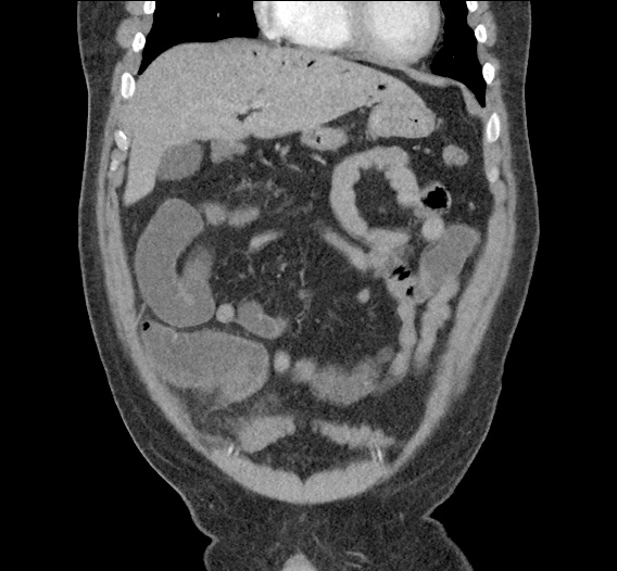
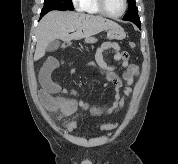
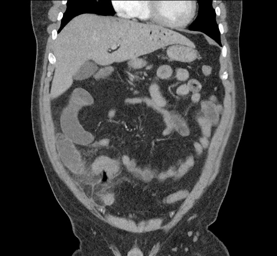
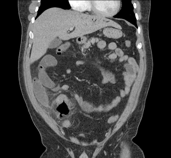
CT Abdomen/Pelvis with Contrast

Inflammatory changes in the right lower quadrant concerning for ruptured appendicitis with approximately 9 cm abscess.
Gas in the liver likely representing portal venous gas which can be seen in the setting of appendicitis vs less likely secondary to bowel ischemia.

Differentiation between Portal Venous Gas and Pneumobilia

Portal venous gas vs. Pneumobilia

References

  1. Rabou Ahmed A and Frank Gaillard. “Pneumobilia.” Radiopaedia. http://radiopaedia.org/articles/pneumobilia.
  2. Morgan Matt A and Donna D’Souza. “Portal venous gas.” Radiopaedia. http://radiopaedia.org/articles/portal-venous-gas
  3. Sebastià C, Quiroga S, Espin E, Boyé R, Alvarez-Castells A, Armengol M. Portomesenteric vein gas: pathologic mechanisms, CT findings, and prognosis. Radiographics. 2000;20(5):1213–24–discussion1224–6. doi:10.1148/radiographics.20.5.g00se011213.
  4. Sherman SC, Tran H. Pneumobilia: benign or life-threatening. J Emerg Med. 2006;30(2):147-153. doi:10.1016/j.jemermed.2005.05.016.

Epiglottitis

Brief H&P:

30 year-old male with no significant medical history presenting with 24 hours of progressively worsening throat pain, difficulty swallowing and voice hoarseness. He reports subjective fevers and chills.
Vital signs notable for Tmax 38.4°C. On physical examination, the patient was sitting upright, unable to swallow secretions with faint inspiratory stridor and dysphonia (though he was able to speak in full sentences and without apparent respiratory distress). Oropharyngeal examination showed minimal right parapharyngeal edema without uvular or palatal deviation and there was exquisite right lateral neck tenderness to palpation.

Labs

  • CBC: 24.2/14.4/43.4/202
  • Wound culture: MSSA
IM-0001-0060
IM-0001-0062
IM-0001-0064
IM-0001-0066
IM-0001-0068
IM-0001-0070
IM-0001-0072
IM-0001-0074
IM-0001-0076
IM-0001-0078
IM-0001-0080

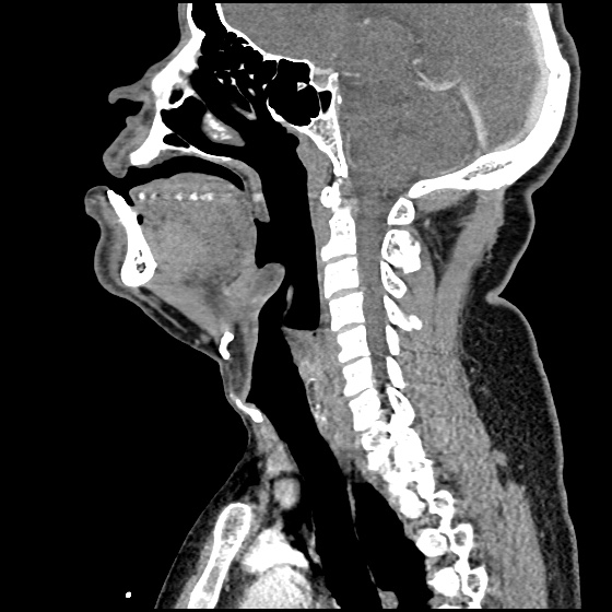
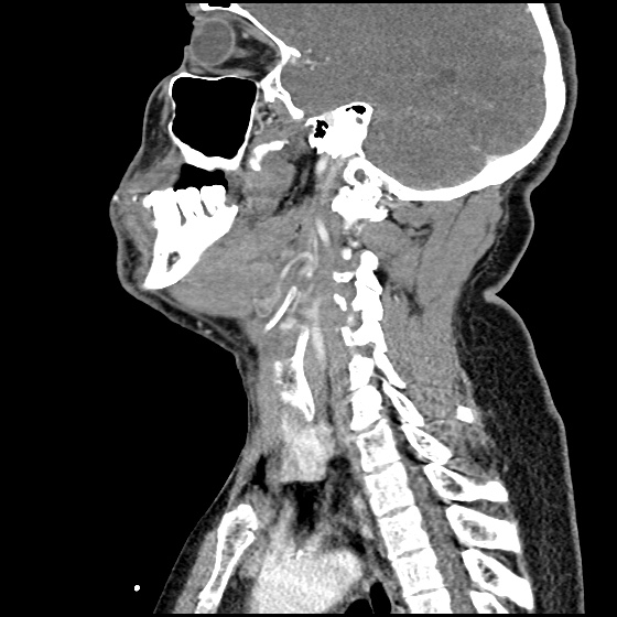
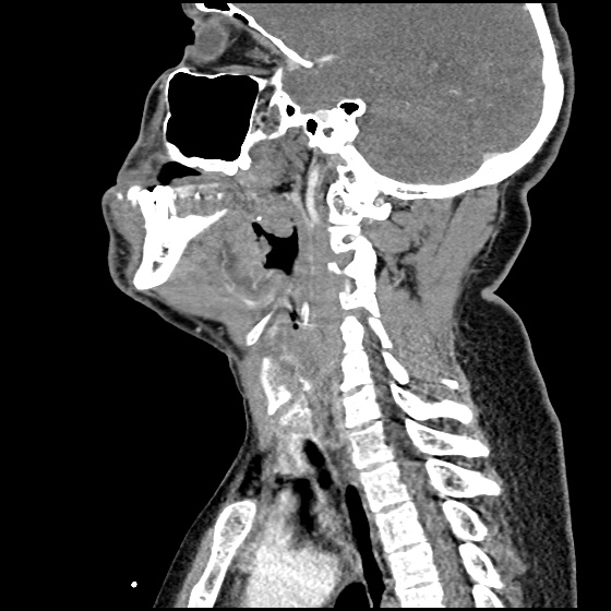
CT Neck/Soft Tissue with Contrast

Edema of the oropharynx/hypopharynx, consistent with epiglottitis and early abscess formation.

ED/Hospital Course

The patient acutely decompensated prior to fiberoptic laryngoscopy and proceeded emergently to the operating room for controlled intubation. The operative report described the following findings: “The patient had diffuse edema of the posterior oropharyngeal wall. The epiglottis was severely thickened, Omega shaped, soft to palpation and with moderate pressure, it appeared to come to a head and pus was expressed from the lingual side of the epiglottis.” The patient was extubated on hospital day three and discharged soon thereafter, he was doing well on follow-up.

Evaluation of Sore Throat – Applied

Evaluation of Sore Throat - Applied

Spinal Epidural Abscess

Case Presentation

HPI:

34M with no PMH presenting with joint pain and rash. The patient was in his usual state of good health until 1 week prior to presentation, noting bilateral shoulder pain. Diagnosed with musculoskeletal process at outside hospital and discharged with analgesics. Presented with partner due to worsening pain involving multiple joints, a non-painful, non-pruritic rash on bilateral lower extremities, and apparent confusion/hallucinations. Social history was non-contributory, no recent procedures or instrumentation.

Objectively, vital signs were notable for tachycardia and elevated core temperature. The patient was ill-appearing, disoriented and unable to provide detailed history. Skin examination was notable for non-blanching petechial rash with areas of confluence most dense in anterior distal lower extremities, rarer proximally, and otherwise without palm/sole involvement. Mucous membranes were dry, neck was supple. There was tenderness to palpation and manipulation of bilateral shoulders. No back tenderness to palpation or percussion was identified. Neurological examination notable for disorientation, intact cranial nerve function, pain-limited weakness in bilateral upper extremities particularly shoulder abduction, and 4/5 hip flexion, knee flexion/extension in bilateral lower extremities.

Labs:

  • CBC: 34.0/11.8/35.7/216
  • Differential: 31 bands
  • INR: 1.94
  • BMP: 131/5.3/102/17/88/2.55/215
  • LFT: AST 93, ALT 57, AP 237, TB 2.9, DB 1.9, Alb 1.4
  • Lactate: 3.3
  • UA: 47WBC, 5RBC
  • Utox: Negative
  • ESR: 83, CRP: 11.9
  • HIV: Nonreactive

Radiology

  • CT head: Negative
  • CXR: Negative
  • XR Shoulder: Negative
  • CT Chest/Abdomen/Pelvis non-contrast: Mild bilateral hydrouereter/hyndronephrosis, L4-L5 grade 2 anterolisthesis.
IM-0001-0005
IM-0001-0006
IM-0001-0007
IM-0001-0008
IM-0001-0009
IM-0001-0010
IM-0001-0011
IM-0001-0012
IM-0001-0013
IM-0001-0014

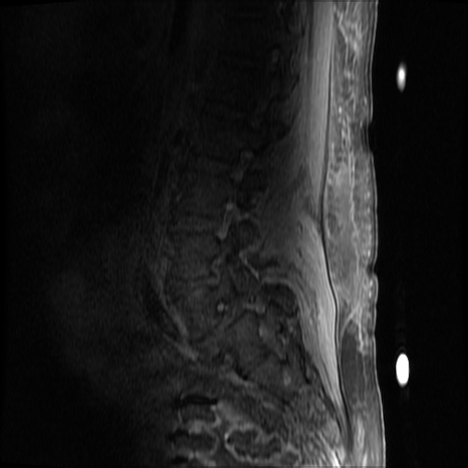
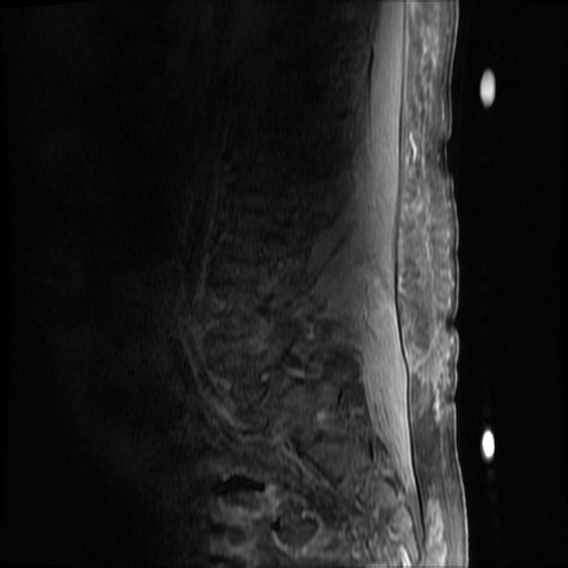
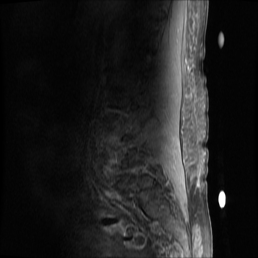
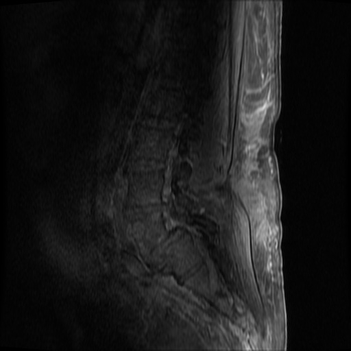
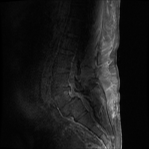
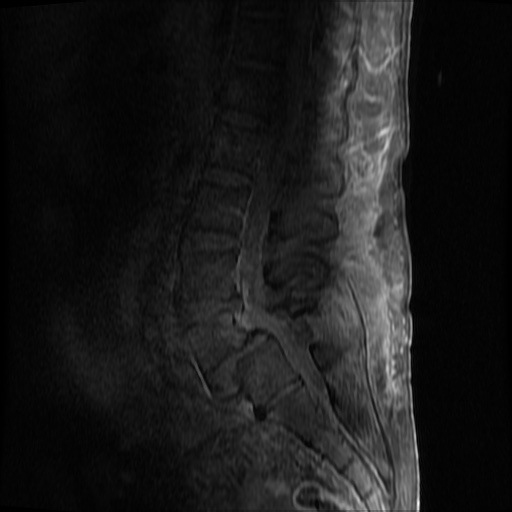
MRI Lumbar Spine w/contrast

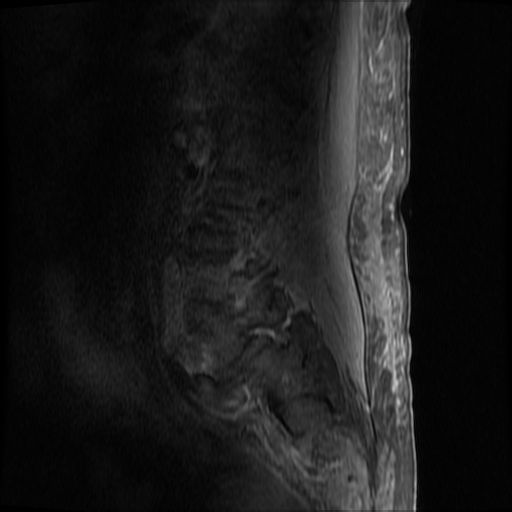
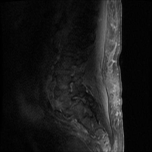
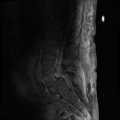
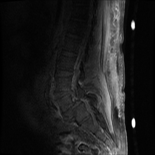
Diffuse epidural enhancement posterior to the L4 and L5 vertebral bodies compressing the thecal sac and resulting in moderate severe spinal canal stenosis. Rim enhancement of the 1.5 cm left paraspinal fluid that may be within the L4 tendon sheath or simply paraspinal abscess.

Assessment/Plan:

Severe sepsis with end-organ dysfunction, unclear source (urinary tract involvement unlikely to account for severity of illness). Covered empirically with broad-spectrum anti-microbials including CNS infection given component of encephalitis. Admitted to the intensive care unit.

Hospital Course:

On hospital day 1, the patient underwent non-contrast MRI of the entire neuraxis with findings concerning for L4-L5 and L5-S1 epidural and paraspinal infection resulting in moderate-severe spinal canal stenosis. Blood and urine cultures grew gram-positive cocci in clusters.

On hospital day 2, the patient became increasingly somnolent. Repeat examination by consulting neurology service was concerning for evidence of meningeal irritation. Cultures speciated as methicillin-sensitive staphylococcus aureus and oxacillin was added. MRI was repeated with gadolinium, findings concerning for L4 epidural vs. paraspinal abscess.

On hospital day 3, the patient’s mental status continued to worsen and he was intubated for airway protection. Neurosurgical intervention was deferred due to deteriorating clinical status. Shoulder synovial fluid aspirate culture positive for MSSA, orthopedic surgery consulted for washout/serial arthrocentesis. TTE performed without evidence of valvular vegetation.

On hospital day 4, additional warm joints were aspirated by orthopedic surgery including knee, bilateral ankles, and shoulder each of which ultimately grew MSSA.

On hospital day 6, the patient underwent OR washout of affected joints with intraoperative findings of purulent fluid. TEE performed without evidence of valvular vegetation. The following day, underwent fluoroscopically-guided lumbar puncture, CSF studies inconclusive. Rifampin added for high-grade bacteremia with multiple seeded sites.

The patient was extubated on hospital day 9 and transferred out of the intensive care unit. The following day, he became increasingly tachypneic with evidence of volume overload on examination and was intubated and returned to the intensive care unit. Sustained PEA arrest post-intubation with ROSC, possibly secondary to pneumothorax vs. hypoxia from extensive mucous plugging. Required increasing vasopressor support over the subsequent 12 hours, emergent CVVHD for worsening academia and hypervolemia. The patient sustained another arrest and ultimately expired.

The final impression was that of high-grade bacteremia from unclear source (vague history of proximate hand laceration/infection) with resultant seeding of epidural/paraspinal space, urinary tract, multiple joints, and likely CNS/meninges. Review of abdominal ultrasonography with evidence of cirrhosis, suggesting that some component of initial hepatic synthetic dysfunction may have been chronic and this may have increased the patient’s risk for disseminated infection and SEA. Neurosurgical intervention was not pursued due to unstable clinical status and as the patient’s neurological findings were not consistent with the location of the identified lesion.

Spinal Epidural Abscess (SEA)1

Risk factors:

  • Immunocompromise: diabetes, cirrhosis, CKD, HIV/AIDS
  • Anatomic: DJD, trauma, prior surgery
  • Introduction: IVDA, epidural anesthesia, tattoo

Organism:

  • S. aureus, 2/3
  • S. epidermidis (associated with device, instrumentation)
  • E. coli (urine spread)
  • P. aeruginosa (IVDA)
  • Rare: anaerobes, mycobacteria, fungi

Staging:

  1. Back pain at affected site
  2. Nerve root pain from affected level
  3. Weakness, sensory deficit, bladder/bowel dysfunction
  4. Paralysis

Clinical features:

  • Back pain (75%)
  • Fever (50%)
  • Neuro deficit (33%)

Diagnosis:

  • Labs: Leukocytosis, ESR/CRP, blood cultures
  • Imaging: MRI with gadolinium, 90% sensitivity
  • Clinical findings and laboratory studies are insensitive and non-specific, in one study, approximately ½ of patients had >2 visits.

Prevalence of abnormal physical findings 2

Finding Prevalence
Fever (T>38°C) 19-32%
Focal spinal TTP 52-62%
Diffuse spinal TTP 63-65%
Positive SLR 11-13%
Abnormal sensation 17-27%
Weakness 29-40%
Abnormal reflexes 8-17%
Abnormal rectal tone 5-10%
Saddle anesthesia 2%

Clinical Decision Guideline 3

Spinal Epidural Abscess Clinical Decision Guideline

Management:

  • Neurosurgical evacuation/fusion
  • Antibiotics (vancomycin, oxacillin, cefepime)
  • Neurosurgical intervention may not result in neurological recovery if symptoms present for > 24-36 hours and may be impractical in the setting of panspinal infection.

References:

  1. Darouiche RO. Spinal epidural abscess. N Engl J Med. 2006;355(19):2012–2020. doi:10.1056/NEJMra055111.
  2. Davis DP, Wold RM, Patel RJ, et al. The clinical presentation and impact of diagnostic delays on emergency department patients with spinal epidural abscess. J Emerg Med. 2004;26(3):285–291. doi:10.1016/j.jemermed.2003.11.013.
  3. Davis DP, Salazar A, Chan TC, Vilke GM. Prospective evaluation of a clinical decision guideline to diagnose spinal epidural abscess in patients who present to the emergency department with spine pain. J Neurosurg Spine. 2011;14(6):765–770. doi:10.3171/2011.1.SPINE1091.
  4. WikEM: Epidural abscess (spinal)

Nonsustained Ventricular Tachycardia

Case 1

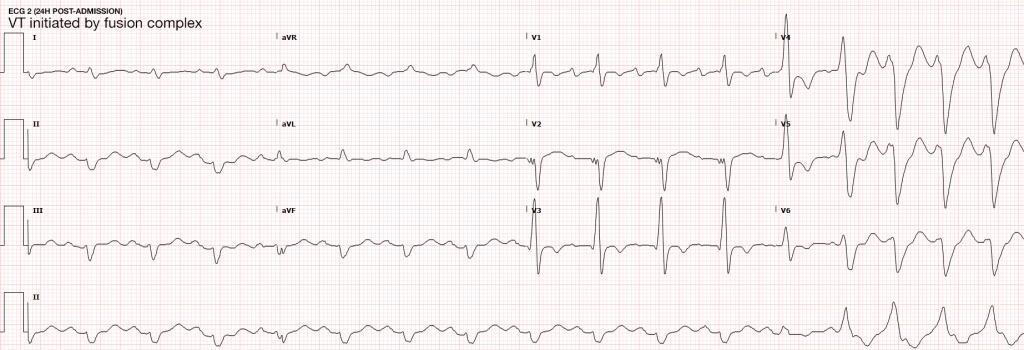
64M with a history of HFrEF (LVEF 20-25%), CAD, AICD (unknown indication), COPD, CKD III presenting with gradual onset shortness of breath, progressive bilateral lower extremity edema.
Examination consistent with severe acute decompensated heart failure presumed secondary to left ventricular dysfunction.
Telemetry monitoring with multiple episodes of nonsustained ventricular tachycardia.

In the ED, the patient developed worsening respiratory failure despite initiation of therapy, requiring endotracheal intubation. Continuous cardiac monitoring revealed persistent salvos of NSVT, progressing to slow ventricular tachycardia without device intervention.
Device interrogation revealed multiple events, 3 shocks, several ATP’s over the recorded period.

Evaluation and Management:

  • NSVT with known (severe) ischemic heart disease
  • For repetitive monomorphic ventricular tachycardia: amiodarone, beta-blockade (if tolerated), procainamide (IIA, C)1

ECG’s

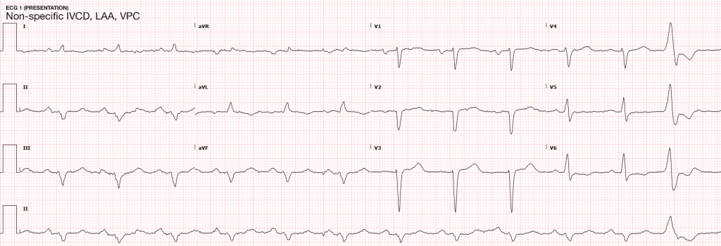
ECG 1
ECG 1

ECG 1

Non-specific IVCD, LAA, VPC

ECG 2
ECG 2

ECG 2

VT initiated by fusion complex

Case 2

31F with autoimmune polyglandular syndrome (adrenal, thyroid and endocrine pancreatic insufficiency), presenting with fever and cough.
Evaluation consistent with sepsis presumed secondary to pulmonary source.
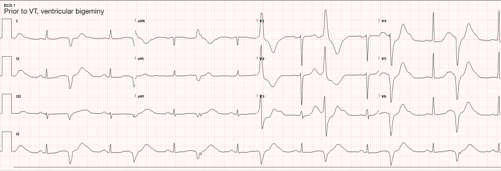
Telemetry monitoring initially with ventricular bigeminy, then nonsustained ventricular tachycardia.

In the ED, the patient developed pulseless ventricular tachycardia – apparently polymorphic. Chest compressions and epinephrine produced return of spontaneous circulation with recovery to baseline neurologic function.
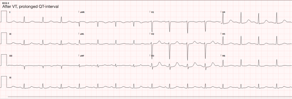
ECG revealed prolonged QTc and chemistry panel notable for critical hypokalemia/hypomagnesemia.

Evaluation and Management:

  • NSVT progressing to VT
  • Initially attributed to electrolyte disturbances. However, serial ECG’s continued to show prolonged QTc (possibly acquired, home medications included metoclopramide and erythromycin). Early echocardiography demonstrated global hypokinesis with EF 30-35% attributed to severe sepsis and recurrent defibrillation. Cardiac CT after resolution of acute illness showed persistently depressed ejection fraction without coronary atherosclerosis. The presence of NICM associated with malignant dysrhythmias warranted ICD placement.
  • Cardioversion for hemodynamic compromise (I, B), B-blockade (I, B), amiodarone if no LQTS (I, C), urgent angiography if ischemia not excluded (I, C)1
  • Correction of electrolyte abnormalities (specifically hypokalemia) may decrease progression to VF.2

ECG’s

ECG 1
ECG 1

ECG 1

Ventricular bigeminy

ECG 2
ECG 2

ECG 2

Long-QT

VT on Telemetry
VT on Telemetry

VT on Telemetry

Non-sustained ventricular tachycardia noted on telemetry monitoring

Definition3,4

  • > 3-5 consecutive beats originating below the AV node
  • Rate > 100bpm
  • Duration <30s

Epidemiology3,5

  • Occurs in 0-4% of ambulatory patients
  • Increased frequency in males and with increasing age
  • In some patients, NSVT is associated with an increased risk of sustained tachyarrhythmias and sudden cardiac death. In others it is of little prognostic significance.6,7,8

Evaluation

In all patients:
History: including arrhythmogenic medications/substances, pertinent family history
Physical examination
ECG/CXR
TTE
In selected patients:
Exercise testing
Advanced imaging (CT/C-MR)
Electrophysiologic studies
Genetic testing

NSVT in the absence of structural heart disease

NSVT in Idiopathic Ventricular Tachycardia

Ventricular outflow arrhythmias:
RVOT: 70-80%, LBBB pattern
LVOT: 20-30%, RBBB pattern
Mechanism:
Adrenergically mediated
Occur during exercise, resolve as heart-rate increases, recur during recovery
Management:
Exclude arrhythmogenic right ventricular cardiomyopathy (imaging, myocardial biopsy)
If symptomatic, beta-blockade, ± IC anti-arrhythmic, CCB (verapamil) for ILVT
Prognosis:
Good, rare tachycardia-induced cardiomyopathy, rare SCD

NSVT in Polymorphic Ventricular Tachycardia

Mechanism
LQTS (acquired or inherited)
Familial catecholaminergic polymorphic VT
Management
Symptomatic (ex. syncope, cardiac arrest): ICD
Asymptomatic QTc > 550ms: consider ICD
Prognosis
Increased risk SCD

Arrhythmogenic Right Ventricular Cardiomyopathy

Mechanism
Fibrosis, fibro-fatty replacement of myocardium in RVIT/RVOT/RV apex
May occur with only subtle structural abnormalities of the right ventricle
LBBB morphology
Management
Anti-arrhythmics of limited utility
Catheter ablation, ICD backup
Prognosis
Increased risk SCD

NSVT with apparent structural heart disease1

Hypertension and LVH

Mechanism
Stretch-induced abnormal automaticity
Fibrotic tissue
Presence of NSVT correlates with degree of hypertrophy and subendocardial fibrosis
Management
Evaluation for ischemic heart disease
Aggressive medical management of hypertension (including beta-blockade)
Prognosis
Unclear

Valvular Disease

Mechanism
High incidence in AS, severe MR (25%)
Mechanical stress from dysfunctional valvular apparatus
Management
Beta-blockade if symptomatic
Prognosis
No evidence that NSVT is an independent predictor of SCD.

Ischemic Heart Disease9-14

Mechanism
Monomorphic VT associated with re-entry at the borders of ventricular scars
Ischemia induces polymorphic NSVT/VF
Management
Revascularization, beta-blockade, statin, ACE/ARB
MADIT I, MUSTT: ICD for ICM LVEF <40%, NSVT, EPS inducible VT
MADIT II, SCD-HeFT: ICD for moderate-to-severe LV dysfunction irrespective of NSVT or EPS findings
Prognosis
NSTEMI with NSVT >48h after admission 2x risk SCD (MERLIN-TIMI 36)
STEMI with NSVT common, not as predictive of ACM or SCD as LVEF (CARISMA)
NSVT <24h after admission for NSTEMI/STEMI not of prognostic significance.

Hypertrophic Cardiomyopathy

Mechanism
Genetic myocardial disease
Myocyte disarray, fibrosis, ischemia result in arrhythmogenic substrate
Management
Restriction of physical activity
ICD (NSVT, LV thickness, FH SCD, syncope, abnormal BP response to exercise)
Beta-blockade, anti-arrhythmic for symptoms
Prognosis
Increased risk SCD (1% annual)
Other Conditions

  • Non-ischemic dilated cardiomyopathy
  • Giant-cell myocarditis
  • Repaired TOF
  • Amyloidosis
  • Sarcoidosis
  • Chagas cardiomyopathy

Algorithm for the Evaluation of NSVT1

Algorithm for the Evaluation of Nonsustained Ventricular Tachycardia

References

  1. Zipes DP, Camm AJ, Borggrefe M, et al. ACC/AHA/ESC 2006 guidelines for management of patients with ventricular arrhythmias and the prevention of sudden cardiac death–executive summary: A report of the American College of Cardiology/American Heart Association Task Force and the European Society of Cardiology Committee for Practice Guidelines (Writing Committee to Develop Guidelines for Management of Patients with Ventricular Arrhythmias and the Prevention of Sudden Cardiac Death) Developed in collaboration with the European Heart Rhythm Association and the Heart Rhythm Society. Eur Heart J. 2006;27(17):2099–2140. doi:10.1093/eurheartj/ehl199.
  2. Higham PD, Adams PC, Murray A, Campbell RW. Plasma potassium, serum magnesium and ventricular fibrillation: a prospective study. Q J Med. 1993;86(9):609–617.
  3. Katritsis DG, Zareba W, Camm AJ. Nonsustained ventricular tachycardia. J Am Coll Cardiol. 2012;60(20):1993–2004. doi:10.1016/j.jacc.2011.12.063.
  4. Katritsis DG, Camm AJ. Nonsustained ventricular tachycardia: where do we stand? Eur Heart J. 2004;25(13):1093–1099. doi:10.1016/j.ehj.2004.03.022.
  5. Wellens HJ. Electrophysiology: Ventricular tachycardia: diagnosis of broad QRS complex tachycardia. Heart. 2001;86(5):579–585.
  6. Buxton AE, Lee KL, Fisher JD, Josephson ME, Prystowsky EN, Hafley G. A randomized study of the prevention of sudden death in patients with coronary artery disease. Multicenter Unsustained Tachycardia Trial Investigators. N Engl J Med. 1999;341(25):1882–1890. doi:10.1056/NEJM199912163412503.
  7. Jouven X, Zureik M, Desnos M, Courbon D, Ducimetière P. Long-term outcome in asymptomatic men with exercise-induced premature ventricular depolarizations. N Engl J Med. 2000;343(12):826–833. doi:10.1056/NEJM200009213431201.
  8. Udall JA, Ellestad MH. Predictive implications of ventricular premature contractions associated with treadmill stress testing. Circulation. 1977;56(6):985–989.
  9. Preliminary report: effect of encainide and flecainide on mortality in a randomized trial of arrhythmia suppression after myocardial infarction. The Cardiac Arrhythmia Suppression Trial (CAST) Investigators. N Engl J Med. 1989;321(6):406–412. doi:10.1056/NEJM198908103210629.
  10. Goldstein S. Propranolol therapy in patients with acute myocardial infarction: the Beta-Blocker Heart Attack Trial. Circulation. 1983;67(6 Pt 2):I53–7.
  11. Moss AJ. MADIT-I and MADIT-II. J Cardiovasc Electrophysiol. 2003;14(9 Suppl):S96–8.
  12. Moss AJ, Hall WJ, Cannom DS, et al. Improved survival with an implanted defibrillator in patients with coronary disease at high risk for ventricular arrhythmia. Multicenter Automatic Defibrillator Implantation Trial Investigators. N Engl J Med. 1996;335(26):1933–1940. doi:10.1056/NEJM199612263352601.
  13. Buxton AE, Lee KL, Fisher JD, Josephson ME, Prystowsky EN, Hafley G. A randomized study of the prevention of sudden death in patients with coronary artery disease. Multicenter Unsustained Tachycardia Trial Investigators. N Engl J Med. 1999;341(25):1882–1890. doi:10.1056/NEJM199912163412503.
  14. Bardy GH, Lee KL, Mark DB, et al. Amiodarone or an implantable cardioverter-defibrillator for congestive heart failure. N Engl J Med. 2005;352(3):225–237. doi:10.1056/NEJMoa043399.
  15. WikEM: Nonsustained Ventricular Tachycardia

Gastrointestinal Bleeding

Evaluation and Management of Gastrointestinal Bleeding

Evaluation and Management of Gastrointestinal Bleeding

Key Historical Features

Quantity
Patient’s estimate
Symptoms suggestive of anemia/volume depletion: (pre)syncope, dyspnea
Appearance/Location
Distinguish upper from lower GI bleding
PMH
Prior episodes and source
History of aortic aneurysm graft
Comorbidities: presence of CAD, CHF, liver disease or diabetes increases mortality
Medications/substance use
Gastrotoxic, anti-coagulants, anti-platelet agents
Alcohol abuse

Key Physical Findings

Vital signs
Tachycardia or hypotension
Eyes
Conjuntival pallor suggests anemia
Scleral icterus suggests liver disease
Abdomen
Hyperactive bowel sounds may be present in UGIB (blood is cathartic)
Epigastric tenderness to palpation suggests PUD
Diffuse tenderness suggests bowel ischemia, obstruction/ileus, or perforation
Rectal (digital, anoscopy)
May reveal fissures, hemorrhoids or polyps

Labs/Diagnostic Tests

  • CBC: consider transfusion for Hb <8-10g/dL particularly in elderly or those with CAD
  • BMP: BUN:creatinine > 36 in the absence of renal failure suggests UGIB
  • PT/PTT/INR: coagulopathy
  • Lactate: elevated in bowel ischemia or systemic hypoperfusion
  • T&S or T&C
  • ECG: screen for myocardial ischemia

Blatchford Scoring System

Item Value Points
BUN 18-22 2
22-28 3
28-70 4
>70 6
Hb (male) 12-13 1
10-12 3
<10 6
Hb (female) 10-12 1
<10 6
SBP 100-109 1
90-99 2
<90 3
Other HR > 100 1
Melena 1
Syncope 2
Liver disease 2
Heart failure 2

References:

  1. Goralnick, E., & Meguerdichian, D. (2013). Gastrointestinal Bleeding. In Rosen's Emergency Medicine – Concepts and Clinical Practice (8th ed., Vol. 1, pp. 248-253). Elsevier Health Sciences.

Nausea and Vomiting

Pathophysiology of Nausea and Vomiting

Pathophysiology of Nausea and Vomiting

Complications of Nausea and Vomiting

  • Hypovolemia: activates RAAS
  • Metabolic alkalosis: loss of hydrogen ions in vomitus
  • Hypokalemia: RAAS promotes sodium retention and potassium excretion
  • Esophageal injury: Mallory-Weiss tear, Boerhaave syndrome
  • Aspiration

Key Historical Findings

Duration of vomiting
Acute: Episodic and occurring for <1 week. Suggestive of obstructive, toxic/metabolic, infectious, ischemic or neurologic process.
Chronic: Episodic and occurring for >1 month. Suggestive of partial obstruction, motility disorder or neurologic process.
Onset
Acute onset: suggests pancreatitis, gastroenteritis, or cholecystitis.
Timing
Post prandial: delayed >1 hour suggests gastric outlet obstruction or gastroparesis.
Contents
Bile: presence of bile suggests patent connection between duodenum and stomach (no GOO)
Undigested food: suggests upper GI tract process (achalasia, esophageal stricture, Zenker)
Feculent: suggests distal bowel obstruction
Associated symptoms
Headache: suggests elevated ICP

Causes of Nausea and Vomiting

Causes of Nausea and Vomiting

References

  1. Zun, L. (2013). Nausea and Vomiting. In Rosen’s Emergency Medicine – Concepts and Clinical Practice (8th ed., Vol. 1, pp. 238-247). Elsevier Health Sciences.

Abdominal Pain

Pathophysiology of Abdominal Pain

  1. Visceral: distension of hollow organs or capsular stretch of solid organs.
  2. Somatic: parietal peritoneal irritation
  3. Referred

    • Extra-abdominopelvic

      • Epigastric: inferior MI
      • Pelvic: hip
      • Abdominal: lower lobe pneumonia/infarction
    • Abdominopelvic

      • Shoulder: diaphragmatic irritation (ex. perforated duodenal ulcer, splenic pathology)
      • Mid-back: aortopathy, pancreatitis
      • Flank: renal pathology
      • Low back: uterus, rectum

Concerning Historical Features

  • Elderly: increased probability for severe disease with poor clinical diagnostic accuracy
  • Immunocompromised: HIV/AIDS, uncontrolled diabetes, chronic liver disease, chemotherapy, other immunosuppression
  • Pain preceding nausea/vomiting: increased likelihood of surgical process
  • Abrupt onset, duration <48h, constant timing
  • Prior abdominal surgical history: consider bowel obstruction
  • No prior episodes of similar pain
  • Recent antibiotic or steroid use: may mask signs of infection
  • Cardiac risk factors (HTN, vascular disease, atrial fibrillation: increased risk for mesenteric ischemia or aortic aneurysm
  • Heavy NSAID use or anticoagulation: increase concern for gastrointestinal bleeding

Imaging

  • Plain film reserved for those who would otherwise not undergo CT. XR abdomen for bowel obstruction or radiopaque foreign body.
  • CT abdomen/pelvis with IV contrast, particularly if elderly or immunocompromised.
  • Ultrasound preferred for hepatobiliary pathology
  • Bedside ultrasound for identification of IUP, free intraperitoneal fluid, cholecystitis, CBD dilation, ascites, hydronephrosis, aortopathy, volume status.

Causes of Abdominal Pain

Causes of Abdominal Pain

References

  1. Budhram, G., & Bengiamin, R. (2013). Abdominal Pain. In Rosen’s Emergency Medicine – Concepts and Clinical Practice (8th ed., Vol. 1, pp. 223-231). Elsevier Health Sciences.

Chest Pain

An Algorithm for the Evaluation of Chest Pain

Algorithm for the Evaluation of Chest Pain

NOTE: Algorithm revised in November, 2017. The prior version is no longer supported but remains available here.

Guided Lecture

EM Ed
Watch “Chest Pain: It’s Giving Me Angina” from EM Ed. In this lecture Dr. Celedon reviews the critical differential diagnosis for chest pain and how to safely and effectively work up patient’s with this challenging chief complaint.

References

  1. Brown, J. (2013). Chest Pain. In Rosen’s Emergency Medicine – Concepts and Clinical Practice (8th ed., Vol. 1, pp. 214-222). Elsevier Health Sciences.

Endocrine Emergencies

HPI

30 year-old female with a history of autoimmune polyglandular syndrome (adrenal, thyroid and endocrine pancreatic insufficiency), polysubstance use, brought to the emergency department by ambulance with reported chief complaint of fever. On presentation, the patient reported fever for one day, associated with cough. She was lethargic and confused, answering yes/no questions but unable to provide detailed history. She states that she has been taking her home medications as prescribed, which include hydrocortisone, fludrocortisone, synthroid and insulin. No collateral information was immediately available.

Additional history was obtained from chart review upon discharge. The patient was hospitalized two weeks prior with pneumonia and discharged after two days. For 2-3 days prior to presentation, she reported the following symptoms to family members: nausea/vomiting, cough, decreased oral intake, fevers, and palpitations – she did not take her home medications during this time.

Physical Exam

VS: T 38.6 HR 112 RR 18 BP 149/82 O2 90% RA
Gen: Alert, fatigued, slow responses.
HEENT: No meningeal irritation, dry mucous membranes.
Pulmonary: Tachypnea, inspiratory wheezing and faint crackles at left and right inferior lung fields, appreciated anteriorly as well.
Neuro: Alert, oriented to self, situation, not month/year. PERRL, EOMI, facial muscles symmetric, tongue protrudes midline without fasciculation. Peripheral sensation grossly intact to light touch and moves all extremities on command.

Labs

  • VBG: alkalemia, primary respiratory
  • CBC: no leukocytosis, normal differential, normocytic anemia
  • BMP: 131, 2.5 , 94, 28, 11, 1.6, 115
  • Mg: 1.3
  • Lactate: 1.0
  • TSH: 17 , T4: 1.03
  • Troponin: 0.129

ECG

ECG 1
ECG 2

Imaging

  • CXR: Negative acute.
  • CT Head: Negative acute.
  • CT Cardiac: NICM, EF 35%.
IM-0001-0026
IM-0001-0030
IM-0001-0034
IM-0001-0038
IM-0001-0042
IM-0001-0046
IM-0001-0050
IM-0001-0054
IM-0001-0058
IM-0001-0062
IM-0001-0066
IM-0001-0070

CT Chest non-contrast

  • Diffuse patchy GGO (pulmonary edema, atypical pneumonia, alveolar hemorrhage, others).
  • Multiple bilateral pulmonary nodules.
  • Possible pulmonary arterial hypertension.

Hospital Course

The patient’s evaluation in the emergency department was concerning for severe sepsis secondary to suspected pulmonary source (given association of fever with cough, hypoxia and abnormal chest imaging findings). The patient had persistent alteration in mental status concerning for CNS infection. While preparing for lumbar puncture, cardiac monitoring revealed sustained polymorphic ventricular tachycardia without appreciable pulse. CPR was initiated, amiodarone 150mg IV push administered and at first pulse check a perfusing sinus rhythm was noted with immediate recovery of prior baseline mental status. Amiodarone load was continued and additional potassium sulfate (PO and IV) was administered. Review of telemetry monitoring revealed preceding 30-45 minutes of non-sustained ventricular tachycardia. The patient had two more episodes of sustained ventricular tachycardia requiring defibrillation. The patient was admitted to the medical intensive care unit for continued management.

#Sustained Ventricular Tachycardia
Initially attributed to critical hypokalemia and hypomagnesemia. However, after appropriate repletion serial ECG’s continued to demonstrate prolonged QT interval (possibly acquired secondary to medications, later review revealed multiple promotility agents for treatment of gastroparesis which could contribute to QT-prolongation including erythromycin and metoclopramide, also associated with endocrinopathies). Early echocardiography demonstrated global hypokinesis with estimated EF 30-35%. This was initially attributed to severe sepsis, as well as recurrent defibrillation. However, cardiac CT after resolution of acute illness showed persistent depressed ejection fraction, no evidence of coronary atherosclerosis. The presence of non-ischemic cardiomyopathy (may be attributable to chronic endocrine dysfunction or prior history of methamphetamine abuse) associated with malignant dysrhythmias warranted ICD placement for secondary prevention which the patient was scheduled to receive.

#Severe Sepsis
Attributed to pulmonary source given CT findings, healthcare associated and covered broadly. Mental status gradually improved and returned to baseline. CT head was negative, lumbar puncture deferred.

#Hypokalemia
Unclear etiology. Adrenal insufficiency commonly associated with hyperkalemia and no history of surreptitious fludrocortisone use. Possibly secondary to GI losses. Improved with repletion.

#Autoimmune Polyglandular Syndrome
Started on stress-dose steroids in emergency department. Transiently developed DKA which was reversed appropriately and hydrocortisone was tapered to home regimen. Home levothyroxine was resumed.

Endocrine Emergencies: Hyperthyroidism

Symptoms

Constitutional Weight loss, heat intolerance, perspiration
Cardiopulmonary Palpitations, chest pain, dyspnea
Neuropsychiatric Tremor, anxiety, double vision, muscle weakness
Neck Fullness, dysphagia, dysphonia
Musculoskeletal Extremity swelling
Reproductive Irregular menses, decreased libido, gynecomastia

Signs

Vital signs Tachycardia, widened pulse pressure, fever
Cardiovascular Hyperdynamic precordium, CHF, atrial fibrillation, systolic flow murmur
Ophthalmologic Widened palpebral fissure, periorbital edema, proptosis, diplopia, restricted superior gaze
Neurologic Tremor, hyperreflexia, proximal muscle weakness
Dermatologic Palmar erythema, hyperpigmented plaques or non-pitting edema of tibia
Neck Enlarged or nodular thyroid

Thyroid Storm

Essentially an exaggeration of thyrotoxicosis featuring marked hyperthermia (104-106°F), tachycardia (HR > 140bpm), and altered mental status (agitation, delirium, coma).

Precipitants
Medical: Sepsis, MI, CVA, CHF, PE, visceral ischemia
Trauma: Surgery, blunt, penetrating
Endocrine: DKA, HHS, hypoglycemia
Drugs: Iodine, amiodarone, inhaled anesthetics
Pregnancy: post-partum, hyperemesis gravidarum

Scoring (Burch, Wartofsky)

Fever
99-100 5
100-101 10
101-102 15
102-103 20
103-104 25
>104 30
Tachycardia
90-110 5
110-120 10
120-130 15
130-140 20
>140 25
Mental Status
Normal 0
Mild agitation 10
Extreme lethargy 20
Coma, seizure 30
CHF
Absent 0
Mild (edema) 5
Moderate (rales, atrial fibrillation) 10
Pulmonary edema 15
GI
None 0
Nausea/vomiting, abdominal pain 10
Jaundice 20
Precipitating Event
None 0
Present 10
  • >45: thyroid storm
  • 25-44: impending thyroid storm
  • <25: unlikely thyroid storm

Management

Supportive measures
Volume resuscitation and cooling
Benzodiazepines for agitation
Beta-blockade
Propranolol 60-80mg PO q4h
Propranolol 0.5-1.0mg IV, repeat q15min then 1-2mg q3h
Esmolol continuous infusion
Endocrinology consultation
PTU, SSKI

Endocrine Emergencies: Hypothyroidism

Symptoms

Constitutional Weight gain, cold intolerance, fatigue
Cardiopulmonary Dyspnea, decreased exercise capacity
Neuropsychiatric Impaired concentration and attention
Musculoskeletal Extremity swelling
Gastrointestinal Constipation
Reproductive Irregular menses, erectile dysfunction, decreased libido
Integumentary Coarse hair, dry skin, alopecia, thin nails

Signs

Vital signs Bradycardia, hypothermia
Cardiovascular Prolonged QT, increased ventricular arrhythmia, accelerated CAD, diastolic heart failure, peripheral edema
Neurologic Lethargy, slowed speech, agitation, seizures, ataxia/dysmetria, mononeuropathy, delayed relaxation of reflexes
Musculoskeletal Proximal myopathy, pseudohypertrophy, polyarthralgia
Gastrointestinal Ileus

Myxedema Coma

Precipitants
Critical illness: sepsis (especially PNA), CVA, MI, CHF, trauma, burns
Endocrine: DKA, hypoglycemia
Drugs: amiodarone, lithium, phenytoin, rifampin, medication non-adherence
Environmental: cold exposure
Recognition
History: hypothyroidism, thyroidectomy scar and acute precipitating illness
Hypothermia: temp <95.9°F (or normal in presence of infection)
AMS: lethargy, confusion, coma, agitation, psychosis, seizures
Hypotension: refractory to volume resuscitation and pressors
Bradypnea: with hypercapnia and hypoxia
Hyponatremia

Management

  • Airway protection
  • Fluid resuscitation
  • Thyroid hormone replacement
    • Young, otherwise healthy patients: T3 10ug IV q4h
    • Elderly, cardiac compromise: 300ug IV x1
  • Hydrocortisone: 50-100mg IV q6-8h
  • Treat precipitating illness

Interpretation of Thyroid Function Tests

Condition TSH T4
None Normal Normal
Hyperthyroidism Low High
Hypothyroidism High Low
Subclinical hyperthyroidism Low Normal
Subclinical hypothyroidism High Normal
Sick euthyroid Low Low

Endocrine Emergencies: Adrenal Insufficiency

Either primary due to adrenal gland failure (often secondary to autoimmune destruction), or secondary most often due to exogenous glucocorticoid administration (usually requiring more than 30mg/day for > 3wks).

Symptoms

Constitutional Weakness, fatigue
Gastrointestinal Anorexia, nausea, cramping
Neuropsychiatric Depression, apathy
Reproductive Amenorrhea, decreased libido
Musculoskeletal Myalgia, arthralgia

Signs

General Hyponatremia, orthostatic hypotension, low-grade fever
Primary Hyperpigmentation, hyperkalemia, hyperchloremia, acidosis
Secondary Hypoglycemia

Management

Maintenance
Hydrocortisone 20mg qAM, 10mg qPM
Fludrocortisone 50-100ug daily
Minor illness (x2)
Hydrocortisone 40mg qAM, 20mg qPM
Fludrocortisone 50-200ug daily
Adrenal Crisis
Dexamethasone 4mg IV or hydrocortisone 100mg IV
2-3L 0.9% NaCl
Treat precipitating illness

Life-Threatening Electrolyte Abnormalities3

Critical Hypokalemia

Causes
GI losses (diarrhea, laxative use)
Renal losses (hyperaldosteronism, diuretics)
Cellular shifts (alkalosis)
ECG changes
U-waves 4
T-wave flattening
Ventricular arrhythmias (exacerbated with digoxin use)
Treatment
Maximum rate 10-20mEq/h with ECG monitoring
If malignant ventricular arrhythmias or arrest imminent, consider more rapid administration (10mEq over 5 minutes)


Critical Hypomagnesemia

Causes
GI, renal losses
Thyroid dysfunction
Treatment
1-2g IV over 5-60 minutes or IVP for Torsades

Conclusion

Unfortunately, this patient’s comprehensive clinical picture does not fit neatly into a particular category of endocrinologic pathology. Her underlying autoimmune disorder manifests both primary adrenal and thyroid dysfunction. Components of the patient’s presentation are suggestive of critical hypothyroidism (myxedema coma) including alteration in mental status, QT-prolongation and hyponatremia as well as possible precipitant of pneumonia. However, despite elevated TSH, the patient’s free T4 level was within normal range. Also absent was hypoventilation (the patient was appropriately tachypneic for degree of hypoxia and with resultant respiratory alkalosis) or bradycardia/hypothermia.
Similarly, adrenal insufficiency is typically associated with hyperkalemia, whereas our patient had critical hypokalemia that was determined to be at least a contributory factor to her ventricular dysrhythmia. The etiology of the patient’s hypokalemia remained unexplained.

References:

  1. Sharma, A., & Levy, D. (2009). Thyroid and Adrenal Disorders. In Rosen’s Emergency Medicine (8th ed., Vol. 2, pp. 1676-1692). Elsevier Health Sciences.
  2. Savage MW, Mah PM, Weetman AP, Newell-Price J. Endocrine emergencies. Postgrad Med J. 2004;80(947):506–515. doi:10.1136/pgmj.2003.013474.
  3. ECC Committee, Subcommittees and Task Forces of the American Heart Association. 2005 American Heart Association Guidelines for Cardiopulmonary Resuscitation and Emergency Cardiovascular Care. Circulation. 2005;112(24 Suppl):IV1–203. doi:10.1161/CIRCULATIONAHA.105.166550.
  4. Levis JT. ECG diagnosis: hypokalemia. Perm J. 2012;16(2):57.

Dyspnea

Causes of Dyspnea

Causes of Dyspnea

Findings in Selected Causes of Dyspnea

Condition History Symptoms Findings Evaluation
Anaphylaxis Exposure to allergen Abrupt onset, facial swelling Stridor, wheezing, hives  
PE Immobilization, malignancy, prior DVT/PE, surgery, OCP Abrupt onset, pleuritic chest pain Tachycardia, hypoxia ECG (RV strain)
CT PA, D-dimer
LE US (DVT)
Pneumonia Exposure, tobacco use Fever, productive cough Focal rales CXR
CBC
Blood/respiratory cultures
Pneumothorax Trauma, thin male Abrupt onset, chest pain Decreased BS, subQ emphysema, JVD and tracheal deviation if tension CXR
US
Fluid overload Dietary indiscretion, medication non-adherence Orthopnea, PND JVD, S3/S4, peripheral edema CXR
US
ECG
BNP
COPD/Asthma Tobacco use, personal/family history Progressive Retractions, accessory muscle use, wheezing CXR
US (distinguish from fluid overload)
Malignancy Tobacco use, weight loss Hemoptysis   CXR
CT Chest

References

  1. Braithwaite, S., & Perina, D. (2013). Dyspnea. In Rosen’s Emergency Medicine – Concepts and Clinical Practice (8th ed., Vol. 1, pp. 206-213). Elsevier Health Sciences.

Altered Mental Status

Components of Consciousness

Components of Consciousness

Causes of Altered Mental Status

Causes of Altered Mental Status

History

Rate of onset
Abrupt: CNS
Gradual: Systemic

Physical Examination

  • Vital Signs

    • Blood Pressure: low (shock), high (SAH, stroke, ICP)
    • Heart Rate: low (medication overdose, conduction block), high (hypovolemia, infection, anemia, thyrotoxicosis, drug/toxin)
    • Temperature: low/high (infection, drug/toxin, environmental)
    • Respiratory Rate: low/high (CNS, drug/toxin, metabolic derangement)
  • Eyes

    • Unilateral dilation: CNS/structural cause
    • Papilledema: ICP
    • EOM: cranial nerve dysfunction
    • Oculocephalic: brainstem function
  • Head: trauma
  • Mucous membranes: hydration, laceration
  • Neck: meningeal irritation
  • Pulmonary: respiratory effort
  • CV: murmur, arrhythmia, CO
  • Abdomen: pulsatile mass, sequelae of liver failure
  • Skin: rash, needle tracks

Labs

  • Glucose
  • ECG: arrhythmia, ischemia, electrolyte abnormalities
  • BMP: electrolytes, renal failure, anion gap
  • ABG: hypoxemia, hypercarbia
  • Urinalysis: infection, SG
  • Utox
  • CBC: leukocytosis, leukopenia, severe anemia, thrombocytopenia
  • Ammonia: hepatic encephalopathy
  • TFT: thyrotoxicosis, myxedema coma
  • CSF: meningitis, encephalitis

Imaging

  • CT head: Non-contrast sufficient to identify ICH. Use contrast if mass/infection suspected
  • CTA head/neck: If aneurysm, AVM, venous sinus thrombosis or vertebrobasilar insufficiency suspected
  • CXR: PNA

References

  1. Bassin, B., & Cooke, J. (2013). Depressed Consciousness and Coma. In Rosen’s Emergency Medicine – Concepts and Clinical Practice (8th ed., Vol. 1, pp. 142-150). Elsevier Health Sciences.