STEMI
STEMI
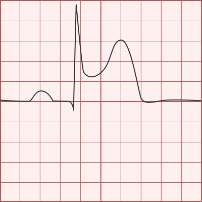
- ST-segment elevation ≥ 1mm in two contiguous leads
- : ≥ 2mm V2-V3
- : ≥ 1.5mm V2-V3
Posterior STEMI
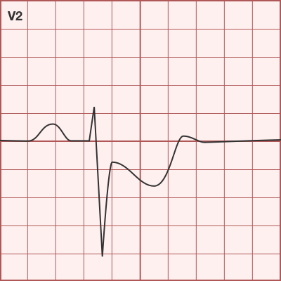
- ST-segment depression V1-V3 Posterior ECG
- ST-segment elevation ≥ 0.5mm in V7-V9
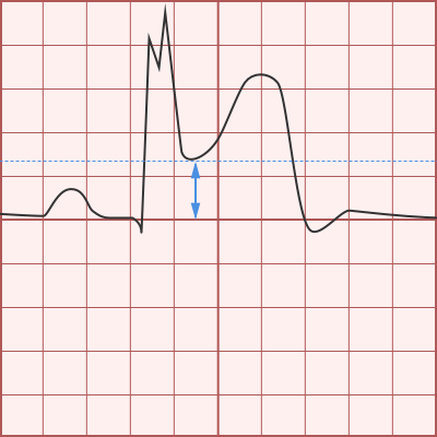
Sgarbossa Criteria
- Evaluation for STEMI in LBBB or paced rhythm
-
Normal: ST-segment discordant with QRS
- QRS associated with ST-segment depression
- QRS associated with (commensurate) ST-segment elevation
- Score ≥ 3 98% specific for MI
Elevation
- Concordant ST-segment elevation ≥ 1mm in any lead (5 points)
Depression

- Concordant ST-segment depression ≥ 1mm in V1-V3 (3 points)
Discordant Elevation
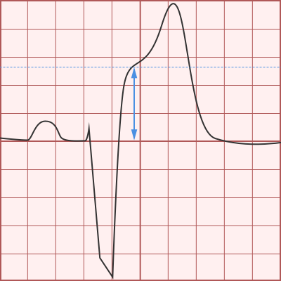
- Discordant ST-segment elevation ≥ 5mm in any lead (2 points)
Modified Sgarbossa Criteria
- ST:S ratio ≥ 0.25 in any lead
- Presence of any criterion is positive


Other Causes of ST-segment Elevation
Benign Early Repolarization

- Concave ST-segment elevation
- Notch at J-point
- Asymmetric T-waves (steeper descent)
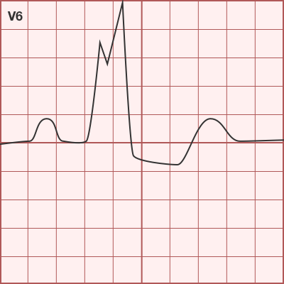
Pericarditis

- Diffuse ST-segment elevation (except aVR)
- PR-segment depression
- Ratio: ST-elevation to T-wave amplitude ≥ 0.25 in V6 suggests pericarditis
LVH Strain
- ST-segment elevation in V1-V3 in the setting of LVH
LV Aneurysm
- Q-waves with ST-segment elevation in precordial leads
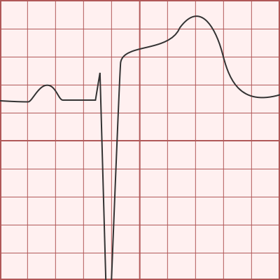
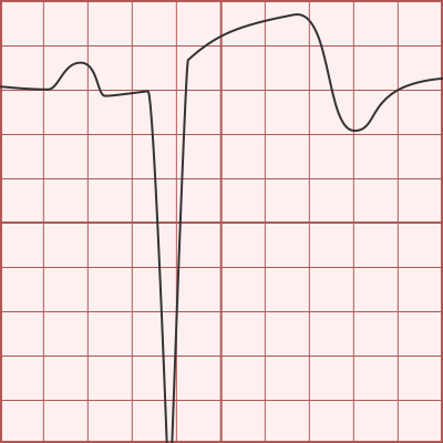
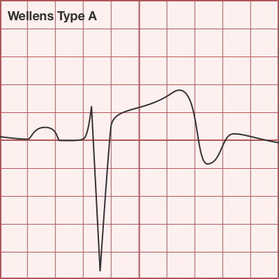
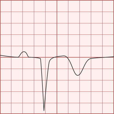
Ischemia and Prior Infarcts
Wellens: Type B

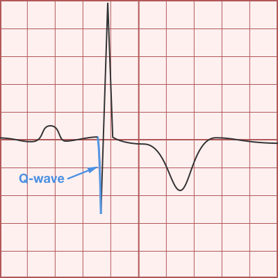
Q-waves
- ≥ 40ms duration
- Depth ≥ 25% of R-wave height
Syncope
ARVD

- Epsilon wave
Brugada Syndrome: Type 1
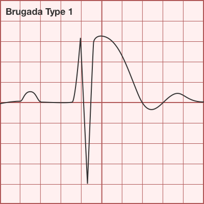
- Type 1: Coved ST-segment elevation
Brugada Syndrome: Type 2
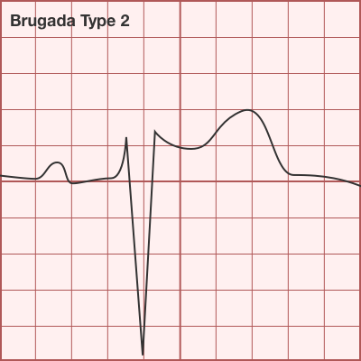
- Type 2: Saddle-back ST-segment elevation
HCM
- Deep, narrow Q-waves
Wolff-Parkinson-White
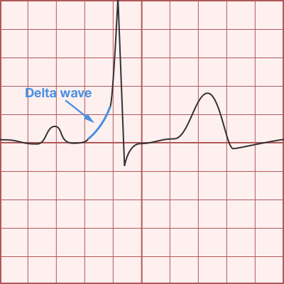
- Shortened PR-interval
- Delta-wave
Other
Atrial Abnormalities
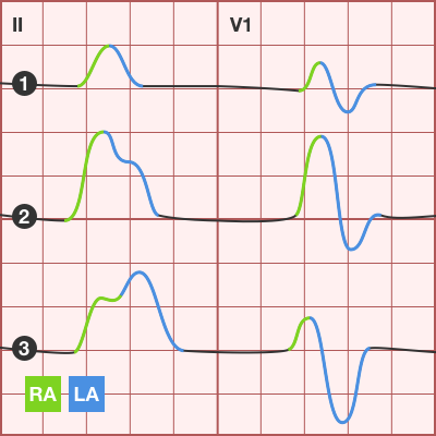
- Normal
- RAA: P-wave amplitude > 2.5mm in inferior leads
- LAA: P-wave duration increased (terminal negative portion >0.04s), amplitude of terminal negative component >1mm below isoelectric line in V1
Left Bundle Branch Block
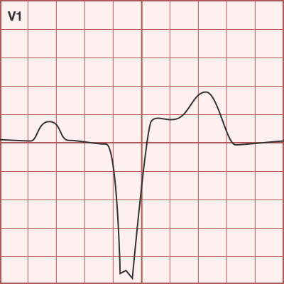
- QRS duration > 0.12s (3 boxes)
- Broad or notched R-wave with prolonged upstroke in I, aVL, V5, V6
- Associated ST-segment depression and T-wave inversion
- Reciprocal changes in V1, V2 (deep S-wave)
- Possible LAD
Right Bundle Branch Block
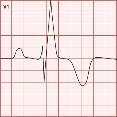

- QRS duration > 0.12s (3 boxes)
- RSR’ in V1, V2
- Reciprocal changes in I, aVL, V5, V6 (deep S-wave)
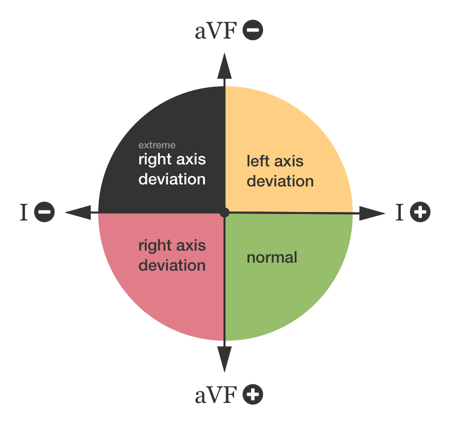
Axes
All illustrations are available for free, licensed (along with all content on this site) under Creative Commons Attribution-ShareAlike 4.0 International Public License.