Anticoagulant Reversal in Intracranial Hemorrhage

Brief HPI:

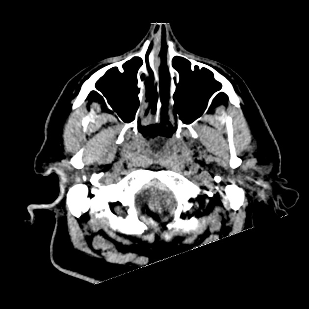
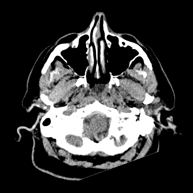
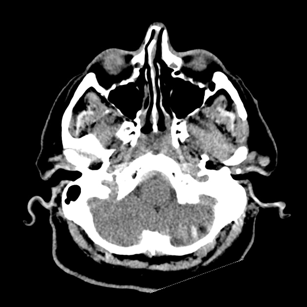
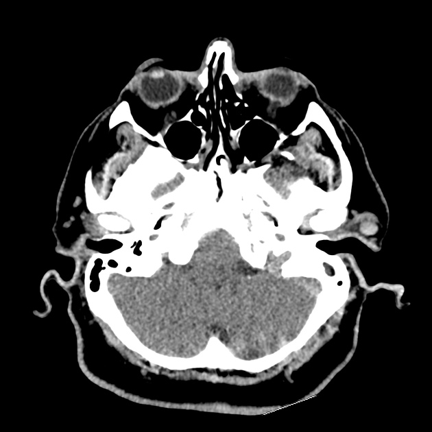
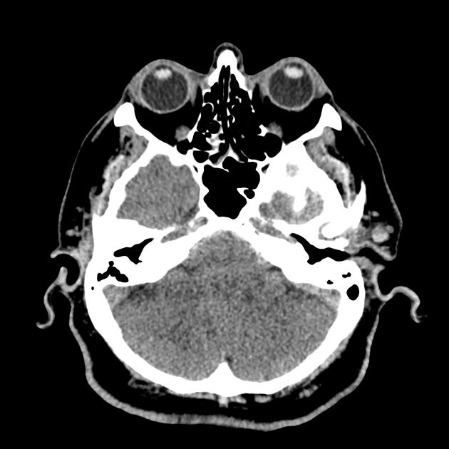
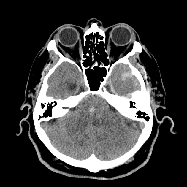
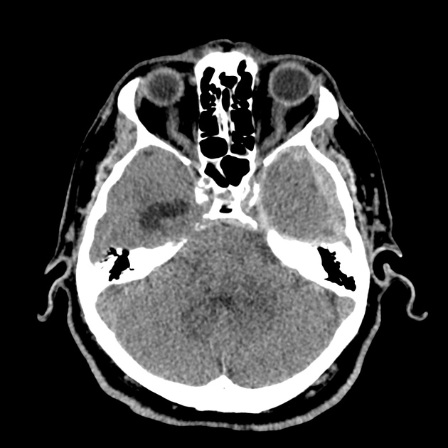
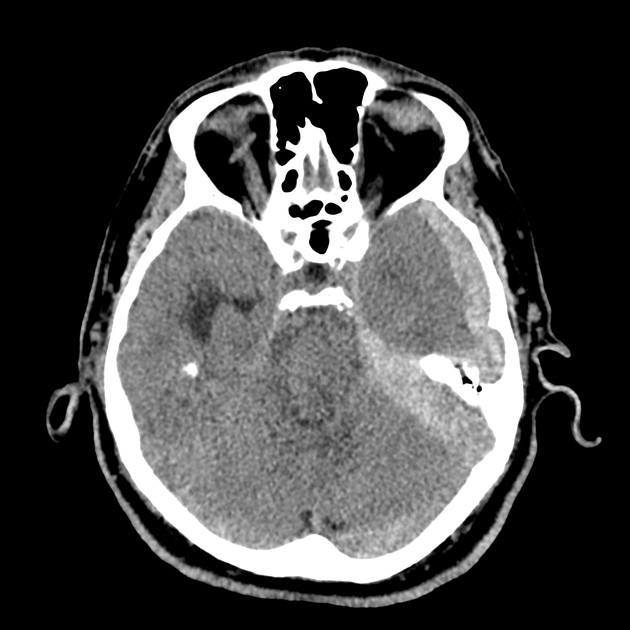
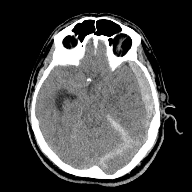
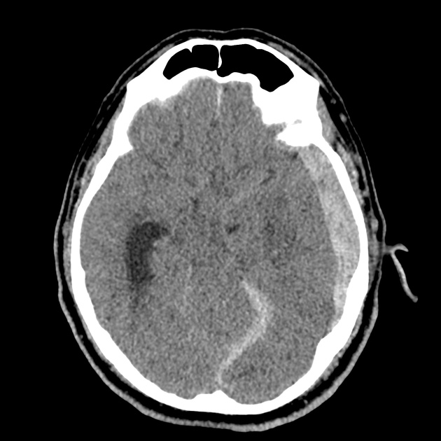
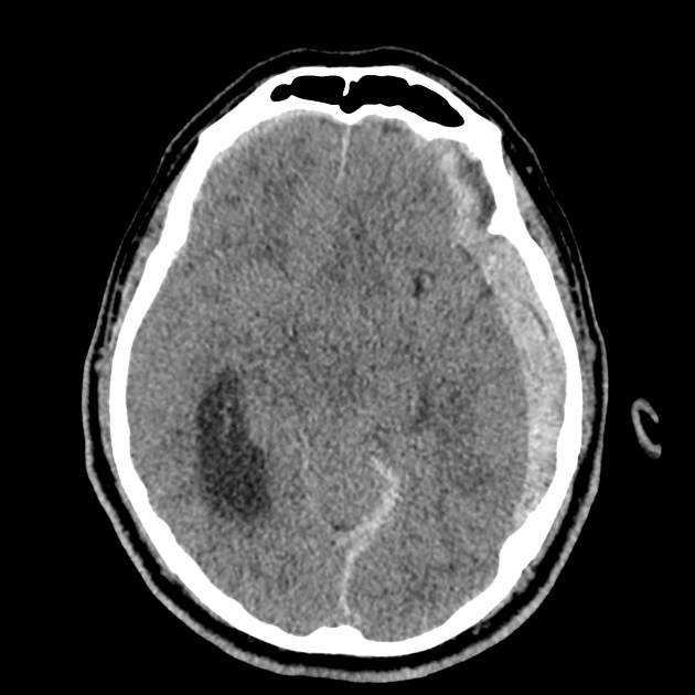
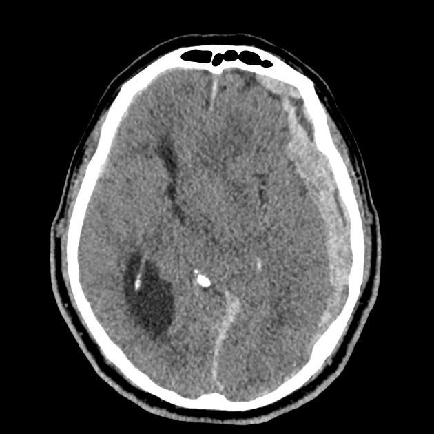
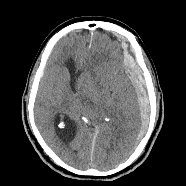
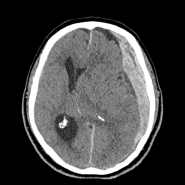
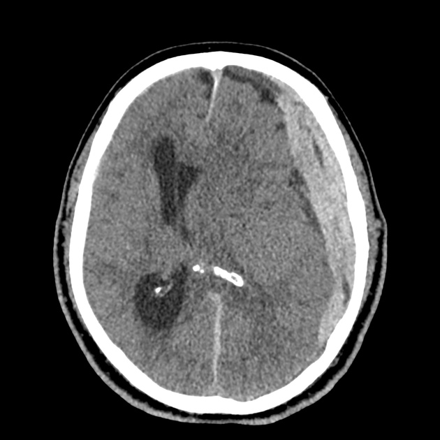
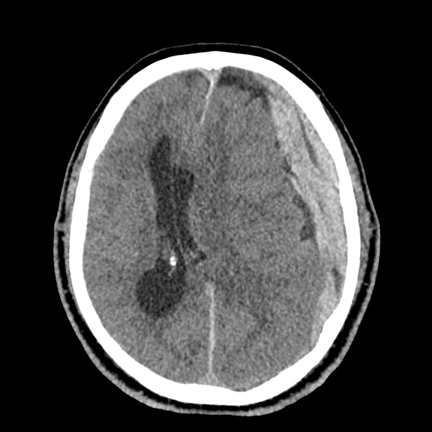
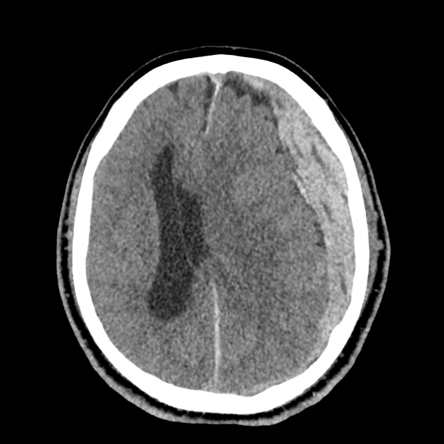
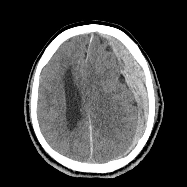
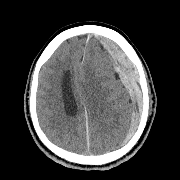
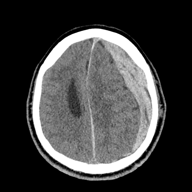
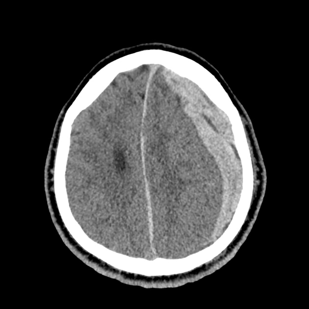
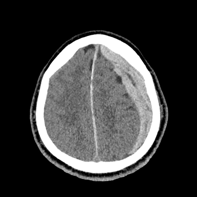
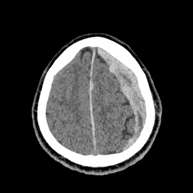
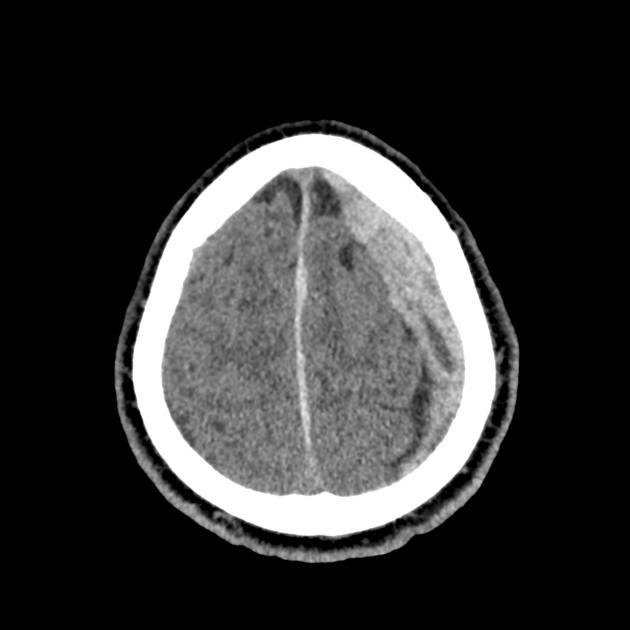
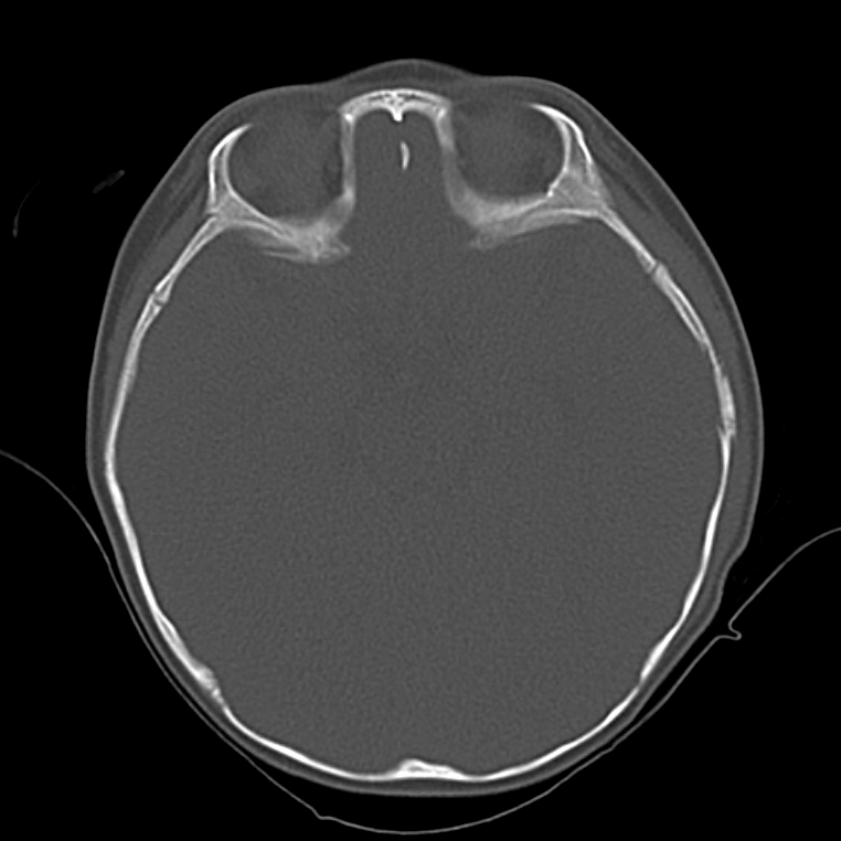
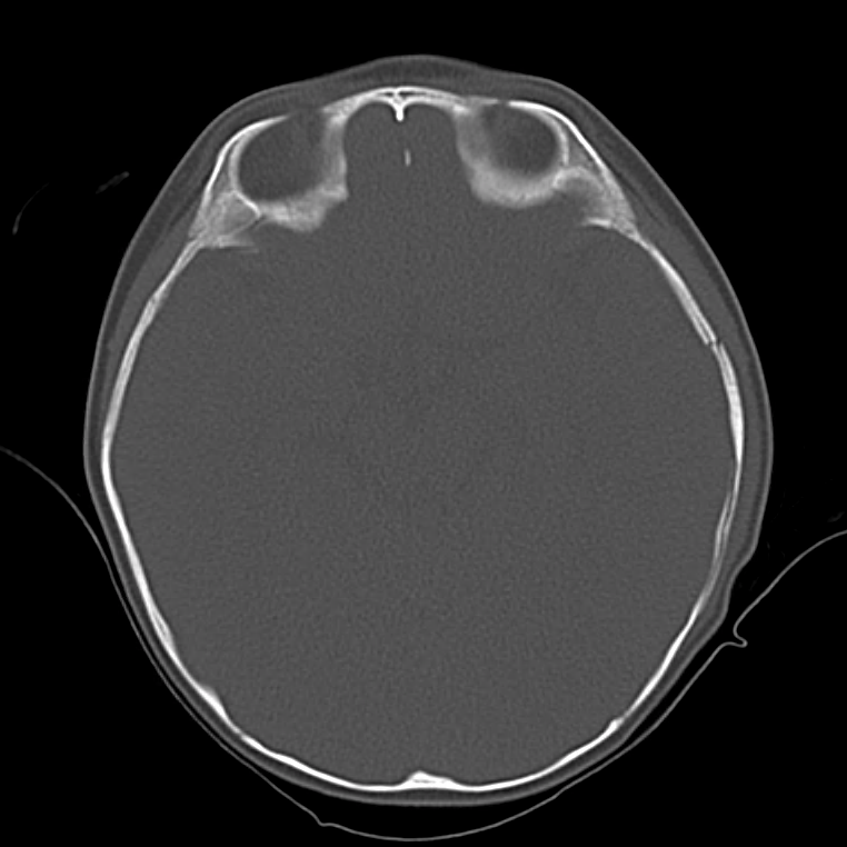
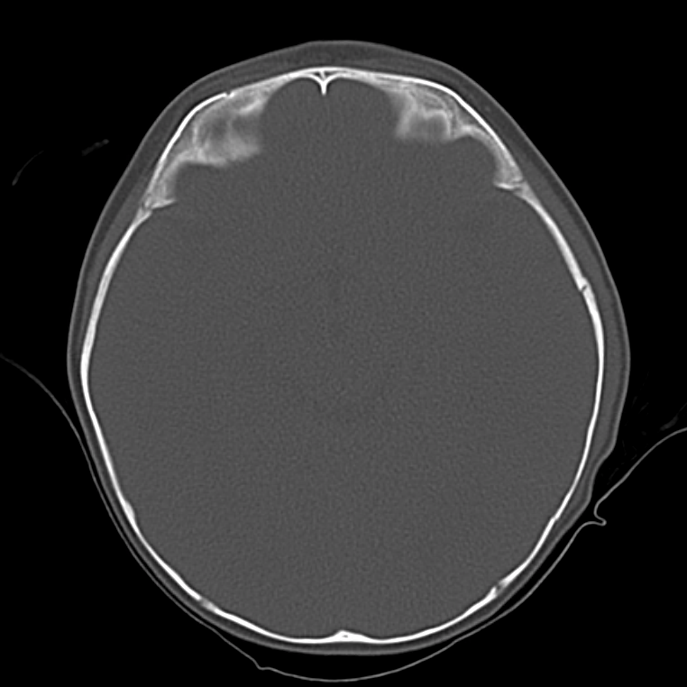
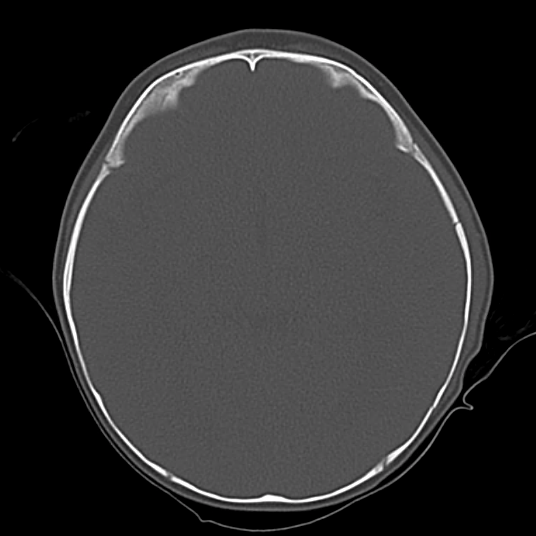
A 65-year-old male with a past medical history of hypertension, diabetes mellitus, and atrial fibrillation presents after a mechanical fall with a posterior scalp hematoma and altered mental status. The patient’s family reports that the patient is taking apixaban with his last dose 4 hours prior to arrival. Physical examination reveals a GCS of 13, blood pressure of 175/99, and asymmetric pupils. The patient is taken to CT where head imaging reveals left sided subdural hematoma with midline shift and developing uncal herniation.

00_subdural
02_subdural
04_subdural
06_subdural
08_subdural
10_subdural
12_subdural
14_subdural
16_subdural
18_subdural
20_subdural
22_subdural
24_subdural
26_subdural
28_subdural
30_subdural
32_subdural
34_subdural
36_subdural
38_subdural
40_subdural
42_subdural
44_subdural
46_subdural

CT Head:

Left cerebral convexity acute subdural hematoma producing substantial mass effect with midline shift and left uncal herniation.
Case courtesy of Dr Andrew Dixon, Radiopaedia.org. From the case rID: 32395

A nicardipine infusion is initiated and the head of the bed is elevated. Andexanet Alfa is not available, therefore an infusion of 4-Factor PCC is initiated. The patient is taken emergently to the operating room by neurosurgery for craniotomy and hematoma evacuation.

An Algorithm for the Reversal of Anticoagulation for Intracranial Hemorrhage 1-4

An Algorithm for Anticoagulant Reversal in Intracranial Hemorrhage


All Agents

For all agents, discontinue anticoagulation. Patients may require blood pressure control including anti-hypertensive infusions (goal SBP <140). Avoid reversal for intracranial hemorrhage associated with cerebral venous thrombosis. Use cautiously in patients with concomitant life-threatening ischemia, thrombosis, or severe DIC.

Vitamin K Antagonists (ex. warfarin)

Initial Dose

A fixed dose of 4F-PCC 1500 to 2000 units can be given as an initial dose with repeat dosing based on INR measurement 15 minutes after completion of infusion. Follow local institution guidelines if available.

Monitoring and Repeat Dosing

  • Vitamin K: if INR ≥1.4 at 12 hours 5
  • 4F-PCC: May consider repeat PCC dosing based on INR, though with increased DIC and thrombotic risk, it is recommended to correct further with FFP if INR remains ≥1.4 6

Direct Factor Xa Inhibitors (ex. rivaroxaban, apixaban)

Activated charcoal may be effective for up to six hours for apixaban 7 and eight hours for rivaroxaban 8.

*Andexanet alfa Regimens 9,10

  • Low-dose: rivaroxaban <10mg, apixaban <5mg, edoxaban <30mg or 8 or more hours since last dose
  • High-dose: If greater than above thresholds, or dose/timing unknown

Pentasaccharides (ex. fondaparinux)

Use high-dose Andexanet alfa regimen 12

Direct Thrombin inhibitors (ex. dabigatran)

Monitoring and Repeat Dosing

If ongoing significant bleeding after treatment, consider redosing idarucizumab and/or hemodialysis.

Alternative Regimens

If idarucizumab is not available, aPCC (50-80 units/kg) , 4F-PCC or 3F-PCC (50 units/kg) can be used in order of preference.

Unfractionated Heparin

Dosing

Determination of units of heparin is based on estimated active agent (half-life 1-2 hours)

  • Protamine sulfate 1mg/100 units IV, maximum dose 50mg
  • Alternatively, can give fixed dose of 25-50mg

Monitoring and Repeat Dosing

If aPTT is persistently elevated, repeat 0.5 mg/100 units

Low-Molecular Weight Heparin 13

Reversal is not indicated if more than 3-5 half-lives have passed since administration:

  • Enoxaparin mean half-life: 4-5 hours
  • Dalteparin mean half-life: 2.8 hours
  • Nadroparin mean half-life: 3.7 hours

If bleeding persists, or renal insufficiency, repeat dose .5 mg/1 mg enoxaparin or .5 mg/100 anti-Xa units.

This algorithm was developed by Dr. Taylor Martin. Taylor is an emergency medicine resident at McGovern Medical School at UTHealth Houston.

References

Guidelines & Reviews

  1. Greenberg SM, Ziai WC, Cordonnier C, et al. 2022 guideline for the management of patients with spontaneous intracerebral hemorrhage: a guideline from the american heart association/american stroke association. Stroke. Published online May 17, 2022:101161STR0000000000000407.
  2. Tomaselli GF, Mahaffey KW, Cuker A, et al. 2020 acc expert consensus decision pathway on management of bleeding in patients on oral anticoagulants: a report of the american college of cardiology solution set oversight committee. J Am Coll Cardiol. 2020;76(5):594-622.
  3. Frontera JA, Lewin JJ, Rabinstein AA, et al. Guideline for reversal of antithrombotics in intracranial hemorrhage: a statement for healthcare professionals from the neurocritical care society and society of critical care medicine. Neurocrit Care. 2016;24(1):6-46.
  4.  Freeman, W. David, Weitz, Jeffrey. “Reversal of anticoagulation in intracranial hemorrhage.” UpToDate. (2022) https://www.uptodate.com/contents/reversal-of-anticoagulation-in-intracranial-hemorrhage?search=anticoagulation%20reversal (Accessed on May 26, 2022)

Vitamin K Antagonists

  1. Ansell J, Hirsh J, Hylek E, Jacobson A, Crowther M, Palareti G. Pharmacology and management of the vitamin k antagonists: american college of chest physicians evidence-based clinical practice guidelines(8th edition). Chest. 2008;133(6 Suppl):160S-198S.
  2. Pabinger I, Brenner B, Kalina U, et al. Prothrombin complex concentrate (Beriplex p/n) for emergency anticoagulation reversal: a prospective multinational clinical trial. J Thromb Haemost. 2008;6(4):622-631.

Direct Factor Xa Inhibitors

  1. http://packageinserts.bms.com/pi/pi_eliquis.pdf
  2. https://www.bayer.com/sites/default/files/2020-11/xarelto-pm-en.pdf
  3. Demchuk AM, Yue P, Zotova E, et al. Hemostatic efficacy and anti-fxa (Factor xa) reversal with andexanet alfa in intracranial hemorrhage: annexa-4 substudy. Stroke. 2021;52(6):2096-2105.
  4. Cohen AT, Lewis M, Connor A, et al. Thirty-day mortality with andexanet alfa compared with prothrombin complex concentrate therapy for life-threatening direct oral anticoagulant-related bleeding. J Am Coll Emerg Physicians Open. 2022;3(2):e12655.
  5. Scaglione F. New oral anticoagulants: comparative pharmacology with vitamin K antagonists. Clin Pharmacokinet. 2013;52(2):69-82.

Pentasaccharides (ex. fondaparinux)

  1. Lu G, DeGuzman FR, Hollenbach SJ, et al. A specific antidote for reversal of anticoagulation by direct and indirect inhibitors of coagulation factor Xa. Nat Med. 2013;19(4):446-451.

Low-Molecular Weight Heparin

  1. Fareed J, Hoppensteadt D, Walenga J, et al. Pharmacodynamic and pharmacokinetic properties of enoxaparin : implications for clinical practice. Clin Pharmacokinet. 2003;42(12):1043-1057.

BRUE

Brief H&P:

A 4 month-old male with no past medical history and fully-immunized is brought to the emergency department by her mother after an episode of breathing difficulty. She describes that just prior to presentation she noted her child had stopped breathing. She lifted him from the bed and noted some blue discoloration to the mouth with limp extremities. She began to stimulate him by rubbing his chest and abdomen and he began crying after approximately 30 seconds.

She states that he has since returned to normal and she was able to feed him upon arrival to the emergency department without apparent difficulty or vomiting. Prior to the episode, the patient had been in his usual state of health (normal oral intake, urine/stool). No family history of sudden death.

On physical examination, vital signs are normal. The child appears comfortable. Head is normocephalic and atraumatic with normal anterior fontanelle. Mucous membranes are moist, heart sounds are normal and lungs are clear. The abdomen is soft and without organomegaly. The remainder of a detailed physical examination is unremarkable.

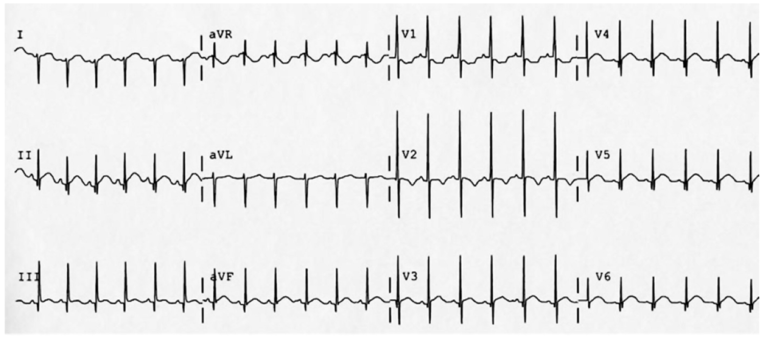
The patient was placed on continuous pulse oximetry, remained well-appearing on serial reassessments and had no further episodes while continuing to feed normally. An ECG was obtained:

The patient’s mother was counseled regarding the diagnosis of low-risk BRUE and the reassuring evaluation and ED observation period. She states that she is able to present to her pediatrician the subsequent morning for evaluation. She was counseled regarding return precautions prior to discharge.

An Algorithm for the Evaluation and Management of Brief Resolved Unexplained Events (BRUE)1,2

An Algorithm for the Evaluation and Management of Brief Resolved Unexplained Events (BRUE)

Differential Diagnosis for BRUE3

Category Causes
Environmental Abuse/trauma
Toxicological
CNS Seizure
Intracranial mass
Cardiovascular Congenital heart disease
Arrhythmia
Pulmonary Airway obstruction
Central apnea
Apnea of prematurity
Pneumonia
Bronchiolitis
Pertussis
Gastrointestinal GERD
IEM Glycogen storage disease
Hyperinsulinism
Fatty acid oxidation defects
This algorithm was developed by Dr. Ali Sina Mirab. Dr. Mirab is a PGY-3 emergency medicine resident at the McGovern Medical School at the University of Texas Health Science Center at Houston (UTHealth).

Special thanks to Dr. Thomas McCarty, Assistant Professor and Pediatric Emergency Medicine Fellowship Director in the Department of Emergency Medicine at McGovern Med EM for his review of the algorithm.

References:

  1. Tieder JS, Bonkowsky JL, Etzel RA, et al. Clinical Practice Guideline: Brief Resolved Unexplained Events (Formerly Apparent Life-Threatening Events) and Evaluation of Lower-Risk Infants: Executive Summary. Pediatrics. 2016:137(5):e20160591.
  2. Merritt JL, Quinonez RA, Bonkowsky JL, et al. A framework for evaluation of the higher-risk infant after a brief resolved unexplained event. Pediatrics. 2019;144(2):e20184101.
  3. McGovern MC, Smith MBH. Causes of apparent life threatening events in infants: a systematic review. Arch Dis Child. 2004;89(11):1043-1048.

Hyperammonemia

Brief H&P

A 38 year-old male with unknown medical history is brought to the emergency department by EMS with agitation and bizarre behavior. According to prehospital report, the patient was acting erratically – shouting incomprehensibly in the middle of a busy street with possible associated seizure activity.

On evaluation, the patient was found to be tachycardic, hypertensive, and markedly agitated. Physical examination with a focus on toxidromes was notable for the presence of rotary nystagmus suggestive of hallucinogen including phencyclidine toxicity. The patient required pharmacologic sedation to allow for a broad evaluation of altered mental status.

ED Course

The patient’s workup including core temperature, head imaging and laboratory tests (including AST/ALT, albumin, INR) were unremarkable with the exception of an ammonia level of 142 umol/L (normal range 16-53), slightly elevated CK, and urine toxicology screen with multiple positive agents. Over the course of several hours in the emergency department, the patient’s mental status gradually improved reaching normal level of alertness and orientation with normal neurological examination. He acknowledged PCP use as well as a prior history of seizures (possibly related to ethanol withdrawal) without routine anti-epileptic drug use. He denied known history of liver disease.

The patient’s hyperammonemia was attributed to a hypercatabolic state secondary to phencyclidine-induced agitation with possible seizure. He was discharged with resources for assistance with substance cessation.

An Algorithm for the Differential Diagnosis of Hyperammonemia:

Algorithm for the Differential Diagnosis of Hyperammonemia

References:

  1. Kalra A, Norvell JP. Cause for Confusion: Noncirrhotic Hyperammonemic Encephalopathy. Clin Liver Dis. 2020;15(6):223-227. doi:10.1002/cld.929
  2. Mallet M, Weiss N, Thabut D, Rudler M. Why and when to measure ammonemia in cirrhosis? Clin Res Hepatol Gas. 2018;42(6):505-511. doi:10.1016/j.clinre.2018.01.004
  3. Hassan AAI, Ibrahim W, Subahi A, Mohamed A. ‘All that glitters is not gold’: when hyperammonaemia is not from hepatic aetiology. Bmj Case Reports. 2017;2017:bcr-2017-219441. doi:10.1136/bcr-2017-219441
  4. Odigwe CC, Khatiwada B, Holbrook C, et al. Noncirrhotic Hyperammonemia Causing Relapsing Altered Mental Status. Bayl Univ Medical Cent Proc. 2017;28(4):472-474. doi:10.1080/08998280.2015.11929312
  5. Upadhyay R, Bleck TP, Busl KM. Hyperammonemia: What Urea-lly Need to Know: Case Report of Severe Noncirrhotic Hyperammonemic Encephalopathy and Review of the Literature. Case Reports Medicine. 2016;2016:1-10. doi:10.1155/2016/8512721
  6. Walker V. Severe hyperammonaemia in adults not explained by liver disease. Ann Clin Biochem. 2011;49(3):214-228. doi:10.1258/acb.2011.011206
  7. Laish I, Ari ZB. Noncirrhotic hyperammonaemic encephalopathy. Liver Int. 2011;31(9):1259-1270. doi:10.1111/j.1478-3231.2011.02550.x
  8. LaBuzetta JN, Yao JZ, Bourque DL, Zivin J. Adult Nonhepatic Hyperammonemia: A Case Report and Differential Diagnosis. Am J Medicine. 2010;123(10):885-891. doi:10.1016/j.amjmed.2010.02.029
  9. Clay AS, Hainline BE. Hyperammonemia in the ICU. Chest. 2007;132(4):1368-1378. doi:10.1378/chest.06-2940
  10. Weng T-I, Shih FF-Y, Chen W-J. Unusual causes of hyperammonemia in the ED. Am J Emerg Medicine. 2004;22(2):105-107. doi:10.1016/j.ajem.2003.12.011
  11. Hawkes ND, Thomas GAO, Jurewicz A, et al. Non-hepatic hyperammonaemia: an important, potentially reversible cause of encephalopathy. Postgrad Med J. 2001;77(913):717. doi:10.1136/pmj.77.913.717

Hypertensive Emergency

Brief HPI:

A 62 year-old female with a history of hypertension, diabetes and coronary artery disease is brought to the emergency department with altered mental status. The patient is confused and unable to provide history. Her family note that symptoms have been gradually worsening for the past one day and she had previously been in her usual state of good health. There was no history of recent illness, medication changes, recreational substance use, sick contacts, or travel.

On evaluation, vital signs were notable for hypertension (224/120mmHg, comparable on all extremities) though otherwise normal including afebrile core temperature – capillary glucose was 114mg/dL. On examination, the patient was awake and alert, making coordinated movements symmetrically in all four extremities without hyperreflexia or increased tone. Speech was unintelligible and the patient was unable to follow simple commands.

Labs/Imaging

Laboratory tests were notable for a serum creatinine of 1.2mg/dL (baseline unknown) but otherwise normal including CBC, troponin, TSH, and UA. ECG demonstrated left ventricular hypertrophy without acute ischemic changes. Imaging including chest radiograph and CT head non-contrast and CTA brain/neck were normal. Lumbar puncture was performed and CSF was normal.

Hospital Course

The patient was initiated on a continuous infusion of nicardipine for presumed hypertensive encephalopathy and admitted to the medical intensive care unit. An MRI was performed on hospital day 1 and demonstrated chronic microvascular ischemic changes. The patient’s mental status gradually improved over the course of her hospitalization and she was discharged home on hospital day 4.

An Algorithm for the Evaluation and Management of Hypertensive Emergencies

An Algorithm for the Evaluation and Management of Hypertensive Emergencies

References

General

  1. Lloyd-Jones DM, Morris PB, Ballantyne CM, et al. 2017 Focused Update of the 2016 ACC Expert Consensus Decision Pathway on the Role of Non-Statin Therapies for LDL-Cholesterol Lowering in the Management of Atherosclerotic Cardiovascular Disease Risk: A Report of the American College of Cardiology Task Force on Expert Consensus Decision Pathways. In: Vol 70. 2017:1785-1822. doi:10.1016/j.jacc.2017.07.745.
  2. Janke AT, McNaughton CD, Brody AM, Welch RD, Levy PD. Trends in the Incidence of Hypertensive Emergencies in US Emergency Departments From 2006 to 2013. J Am Heart Assoc. 2016;5(12). doi:10.1161/JAHA.116.004511.
  3. Rodriguez MA, Kumar SK, De Caro M. Hypertensive crisis. Cardiology in Review. 2010;18(2):102-107. doi:10.1097/CRD.0b013e3181c307b7.
  4. Katz JN, Gore JM, Amin A, et al. Practice patterns, outcomes, and end-organ dysfunction for patients with acute severe hypertension: the Studying the Treatment of Acute hyperTension (STAT) registry. Am Heart J. 2009;158(4):599–606.e1. doi:10.1016/j.ahj.2009.07.020.
  5. Elliott WJ. Clinical features in the management of selected hypertensive emergencies. Prog Cardiovasc Dis. 2006;48(5):316-325. doi:10.1016/j.pcad.2006.02.004.
  6. Aggarwal M, Khan IA. Hypertensive crisis: hypertensive emergencies and urgencies. Cardiol Clin. 2006;24(1):135-146. doi:10.1016/j.ccl.2005.09.002.
  7. Varon J, Marik PE. Clinical review: the management of hypertensive crises. Crit Care. 2003;7(5):374-384. doi:10.1186/cc2351.
  8. Shayne PH, Pitts SR. Severely increased blood pressure in the emergency department. YMEM. 2003;41(4):513-529. doi:10.1067/mem.2003.114.
  9. Vaughan CJ, Delanty N. Hypertensive emergencies. The Lancet. 2000;356(9227):411-417. doi:10.1016/S0140-6736(00)02539-3.

Ischemic Stroke

  1. Powers WJ, Rabinstein AA, Ackerson T, et al. 2018 Guidelines for the Early Management of Patients With Acute Ischemic Stroke: A Guideline for Healthcare Professionals From the American Heart Association/American Stroke Association. Stroke. 2018;49(3):e46-e110. doi:10.1161/STR.0000000000000158.

Hemorrhagic Stroke

  1. Hemphill JC, Greenberg SM, Anderson CS, et al. Guidelines for the Management of Spontaneous Intracerebral Hemorrhage: A Guideline for Healthcare Professionals From the American Heart Association/American Stroke Association. Stroke. 2015;46(7):2032-2060. doi:10.1161/STR.0000000000000069.

Subarachnoid Hemorrhage

  1. Connolly ES, Rabinstein AA, Carhuapoma JR, et al. Guidelines for the management of aneurysmal subarachnoid hemorrhage: a guideline for healthcare professionals from the American Heart Association/american Stroke Association. Stroke. 2012;43(6):1711-1737. doi:10.1161/STR.0b013e3182587839.

Renal

  1. Gillies MA, Kakar V, Parker RJ, Honoré PM, Ostermann M. Fenoldopam to prevent acute kidney injury after major surgery-a systematic review and meta-analysis. Crit Care. 2015;19(1):449. doi:10.1186/s13054-015-1166-4.
  2. Tumlin JA, Dunbar LM, Oparil S, et al. Fenoldopam, a dopamine agonist, for hypertensive emergency: a multicenter randomized trial. Fenoldopam Study Group. Academic Emergency Medicine. 2000;7(6):653-662.
  3. Shusterman NH, Elliott WJ, White WB. Fenoldopam, but not nitroprusside, improves renal function in severely hypertensive patients with impaired renal function. Am J Med. 1993;95(2):161-168.

Aortic Disease

  1. Hiratzka LF, Bakris GL, Beckman JA, et al. 2010 ACCF/AHA/AATS/ACR/ASA/SCA/SCAI/SIR/STS/SVM guidelines for the diagnosis and management of patients with Thoracic Aortic Disease: a report of the American College of Cardiology Foundation/American Heart Association Task Force on Practice Guidelines, American Association for Thoracic Surgery, American College of Radiology, American Stroke Association, Society of Cardiovascular Anesthesiologists, Society for Cardiovascular Angiography and Interventions, Society of Interventional Radiology, Society of Thoracic Surgeons, and Society for Vascular Medicine. Circulation. 2010;121(13):e266-e369. doi:10.1161/CIR.0b013e3181d4739e.

Pregnancy

  1. Townsend R, O’Brien P, Khalil A. Current best practice in the management of hypertensive disorders in pregnancy. Integr Blood Press Control. 2016;9:79-94. doi:10.2147/IBPC.S77344.
  2. Al-Safi Z, Imudia AN, Filetti LC, Hobson DT, Bahado-Singh RO, Awonuga AO. Delayed Postpartum Preeclampsia and Eclampsia. Obstet Gynecol. 2011;118(5):1102-1107. doi:10.1097/AOG.0b013e318231934c.
  3. Hypertension in pregnancy: diagnosis and management. National Institute for Health and Care Excellence. https://www.nice.org.uk/guidance/cg107. Published August 1, 2010. Accessed May 20, 2019.

Cerebrospinal Fluid

Brief HPI:

An approximately 70 year-old male with unknown medical history is brought to the emergency department with altered mental status. A community member contacted police after not seeing the patient for the past three days which was unusual. Upon entering the patient’s home, EMS found the patient on the ground, unresponsive. Capillary glucose was normal and naloxone was administered without appreciable effect.

On arrival in the emergency department, the patient remained unresponsive to verbal and noxious stimulation and was intubated for airway protection. Vital signs were notable for hypotension (BP 88/45mmHg) and a core temperature of 96.5°F. Physical examination demonstrated cool extremities and ecchymosis and edema involving the right upper and lower extremities. The patient’s blood pressure improved with fluid resuscitation and empiric broad-spectrum antibiotics were administered due to concern for infection in the setting of hypothermia.

Laboratory/Imaging Results

Laboratory tests were notable for leukocytosis and creatine kinase above the threshold for detection. Radiology preliminary interpretation of non-contrast head imaging was normal. A lumbar puncture was performed with grossly purulent cerebrospinal fluid.

00_ventriculitis
01_ventriculitis
02_ventriculitis
03_ventriculitis
04_ventriculitis
05_ventriculitis
06_ventriculitis
07_ventriculitis
08_ventriculitis
09_ventriculitis

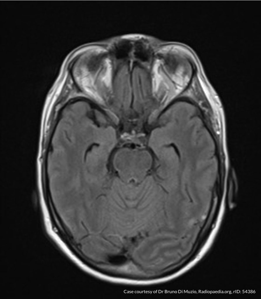
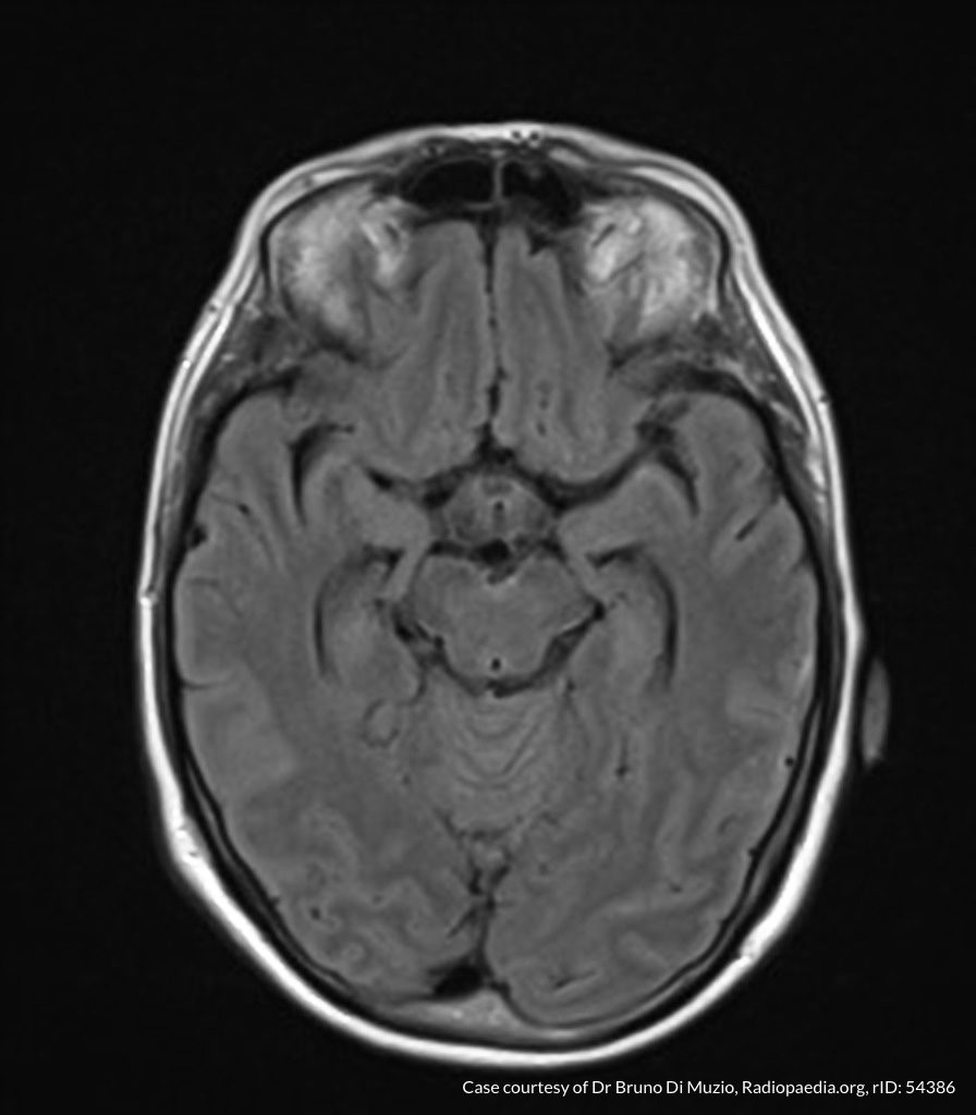
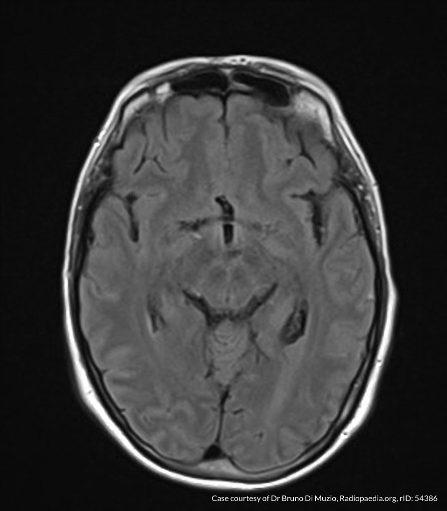
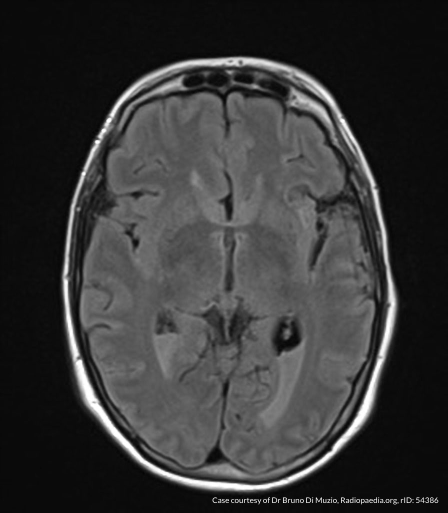
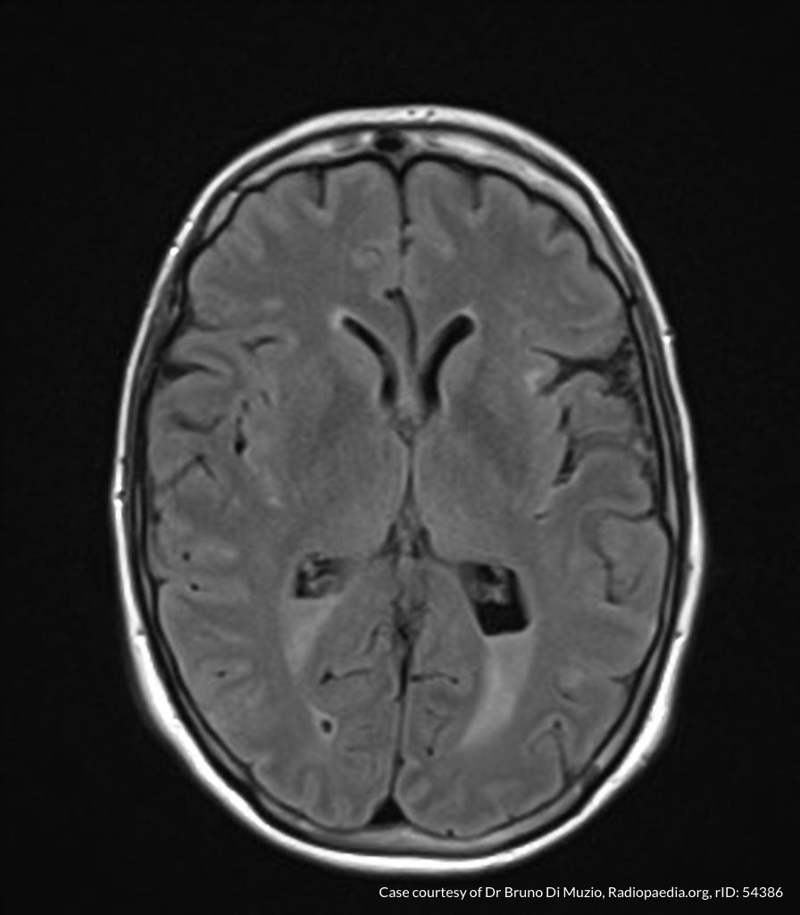
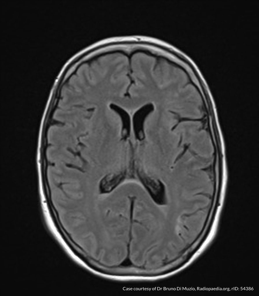
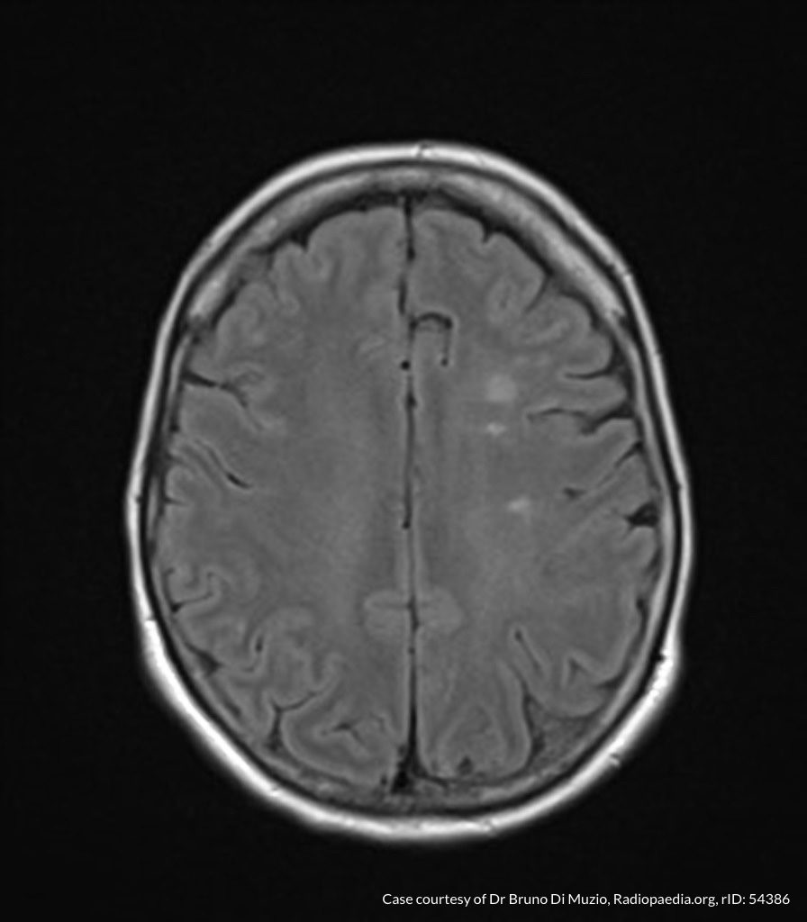
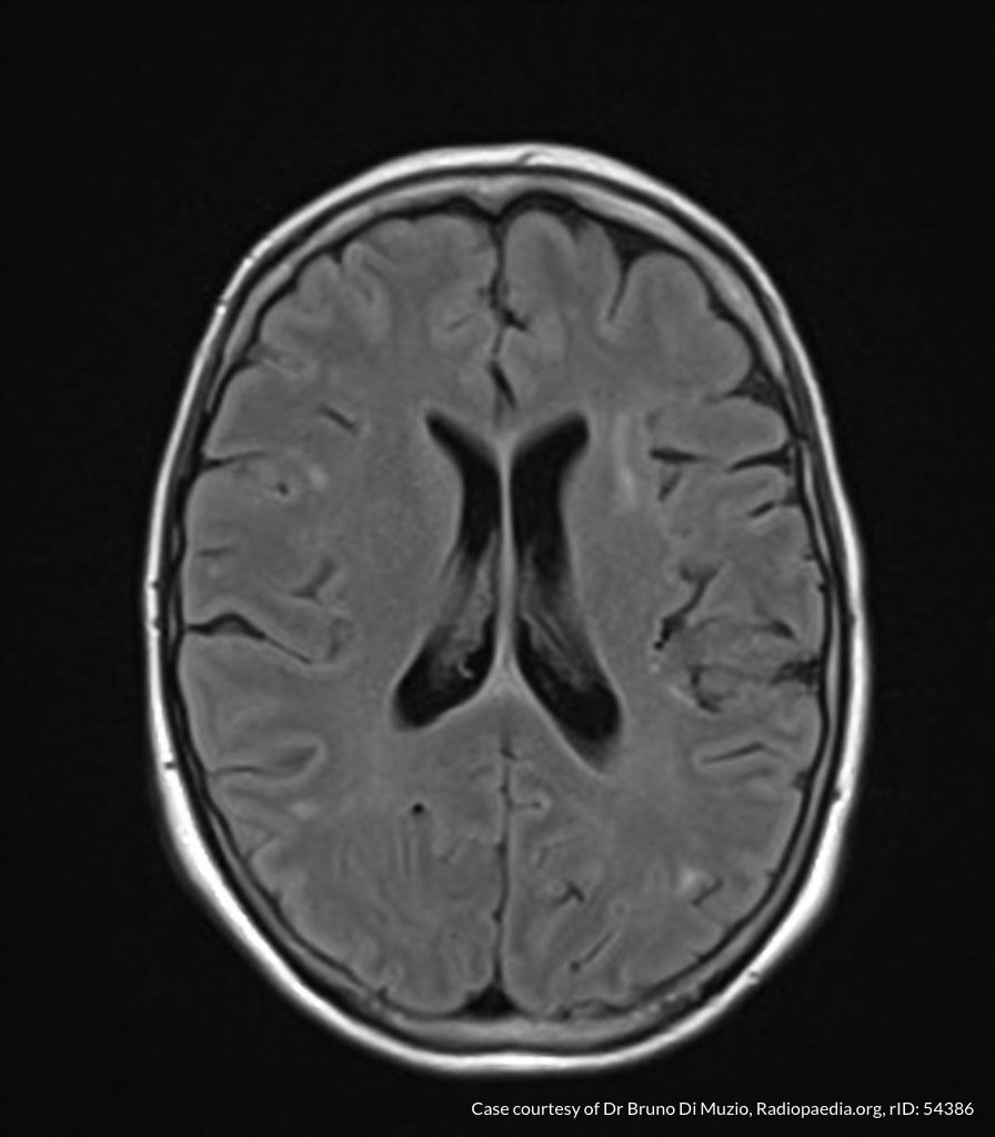
MRI Brain

Dependent material within the occipital horns of the lateral ventricles consistent with ventriculitis.

Hospital Course

The patient was admitted for the treatment of presumed meningitis. Radiology final interpretation of non-contrast head computed tomography commented on ventricular debris suggestive of ventriculitis which was later confirmed on magnetic resonance imaging1,2. Due to poor response to systemic antibiotics, neurosurgery was consulted, a ventricular drain was placed with administration of intrathecal antibiotics. The patient’s condition continued to deteriorate and family members elected to allow his natural death.

An Algorithm for the Analysis of Cerebrospinal Fluid (CSF)3-14

An Algorithm for the Analysis of Cerebrospinal Fluid (CSF)

References

  1. Lesourd A, Magne N, Soares A, et al. Primary bacterial ventriculitis in adults, an emergent diagnosis challenge: report of a meningoccal case and review of the literature. BMC Infect Dis. 2018;18(1):226. doi:10.1186/s12879-018-3119-4.
  2. Gofman N, To K, Whitman M, Garcia-Morales E. Successful treatment of ventriculitis caused by Pseudomonas aeruginosa and carbapenem-resistant Klebsiella pneumoniae with i.v. ceftazidime-avibactam and intrathecal amikacin. Am J Health Syst Pharm. 2018;75(13):953-957. doi:10.2146/ajhp170632.
  3. Dorsett M, Liang SY. Diagnosis and Treatment of Central Nervous System Infections in the Emergency Department. Emerg Med Clin North Am. 2016;34(4):917-942. doi:10.1016/j.emc.2016.06.013.
  4. Perry JJ, Alyahya B, Sivilotti MLA, et al. Differentiation between traumatic tap and aneurysmal subarachnoid hemorrhage: prospective cohort study. BMJ. 2015;350:h568. doi:10.1136/bmj.h568.
  5. Lee SCM, Lueck CJ. Cerebrospinal fluid pressure in adults. J Neuroophthalmol. 2014;34(3):278-283. doi:10.1097/WNO.0000000000000155.
  6. Brouwer MC, Thwaites GE, Tunkel AR, van de Beek D. Dilemmas in the diagnosis of acute community-acquired bacterial meningitis. Lancet. 2012;380(9854):1684-1692. doi:10.1016/S0140-6736(12)61185-4.
  7. Wright BLC, Lai JTF, Sinclair AJ. Cerebrospinal fluid and lumbar puncture: a practical review. J Neurol. 2012;259(8):1530-1545. doi:10.1007/s00415-012-6413-x.
  8. Gorchynski J, Oman J, Newton T. Interpretation of traumatic lumbar punctures in the setting of possible subarachnoid hemorrhage: who can be safely discharged? Cal J Emerg Med. 2007;8(1):3-7.
  9. Deisenhammer F, Bartos A, Egg R, et al. Guidelines on routine cerebrospinal fluid analysis. Report from an EFNS task force. Eur J Neurol. 2006;13(9):913-922. doi:10.1111/j.1468-1331.2006.01493.x.
  10. Seehusen DA, Reeves MM, Fomin DA. Cerebrospinal fluid analysis. Am Fam Physician. 2003;68(6):1103-1108.
  11. Shah KH, Edlow JA. Distinguishing traumatic lumbar puncture from true subarachnoid hemorrhage. J Emerg Med. 2002;23(1):67-74.
  12. Walker HK, Hall WD, Hurst JW. Clinical Methods: The History, Physical, and Laboratory Examinations. 1990.
  13. Mayefsky JH, Roghmann KJ. Determination of leukocytosis in traumatic spinal tap specimens. Am J Med. 1987;82(6):1175-1181.
  14. Geiseler PJ, Nelson KE, Levin S, Reddi KT, Moses VK. Community-acquired purulent meningitis: a review of 1,316 cases during the antibiotic era, 1954-1976. Rev Infect Dis. 1980;2(5):725-745.

Thrombocytopenia

Brief HPI:

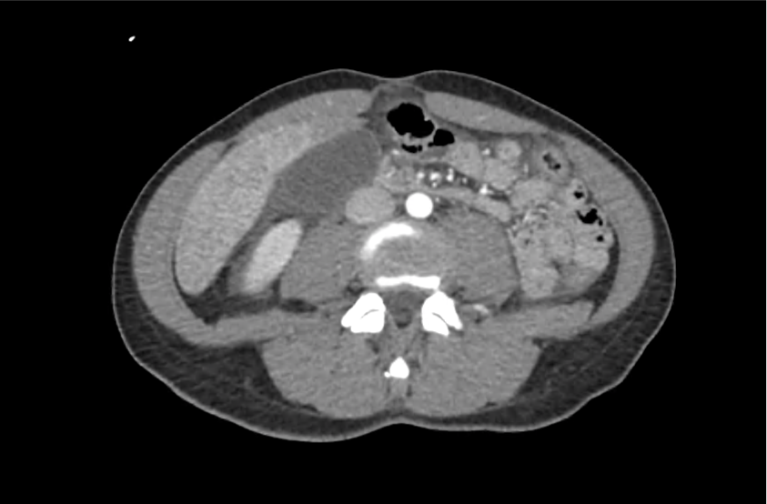
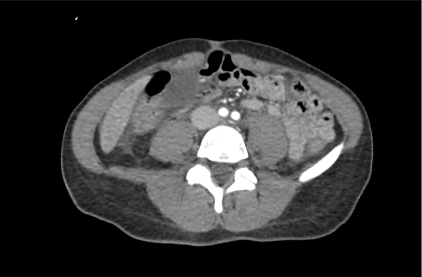
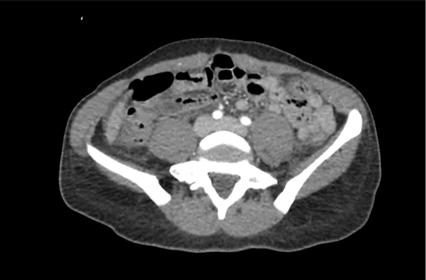
A middle-aged female with no known medical history is brought to the emergency department with altered mental status. Her family notes worsening confusion over the past 2-3 days associated with vomiting and yellow discoloration of skin and eyes.

Initial vital signs were normal, though with borderline hypotension (99/64mmHg). Examination demonstrated an alert, but lethargic patient with jaundice and scleral icterus, no skin lesions were appreciated. Laboratory studies were obtained:

CBC

  • WBC: 21.3 (N: 83%, Bands: 11%)
  • Hb: 5.5
  • Plt: 6k
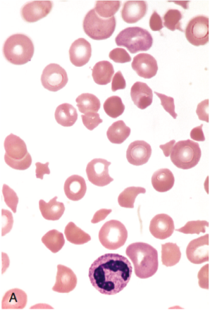
  • Marked schistocytes

Coagulation Panel

  • INR: 1.26
  • PTT: Normal
  • Fibrinogen: Normal
  • FDP: Normal
  • D-dimer: >9,000 (normal 250)
  • Haptoglobin: Undetectable
  • LDH: 1493

CMP

  • Creatinine: 1.1
  • AST/ALT: Normal
  • TB: 4.3, DB: 0.8

Imaging:

CT Head: No acute intracranial process.

ttp_ct_01
ttp_ct_02
ttp_ct_03
ttp_ct_04
ttp_ct_05
ttp_ct_06
ttp_ct_07
ttp_ct_08
ttp_ct_09
ttp_ct_10
ttp_ct_11
ttp_ct_12
ttp_ct_13
ttp_ct_14
ttp_ct_15
ttp_ct_16
ttp_ct_17
ttp_ct_18
ttp_ct_19

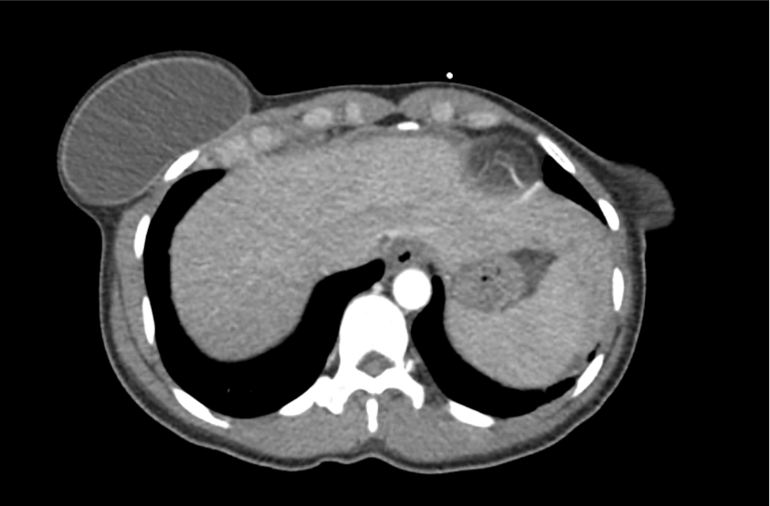
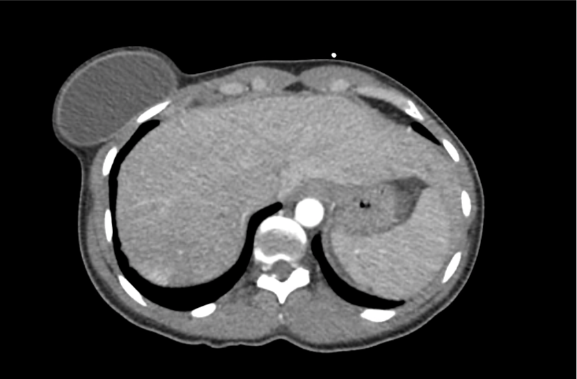
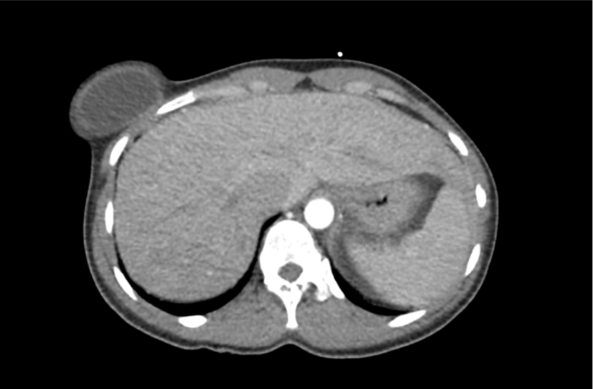
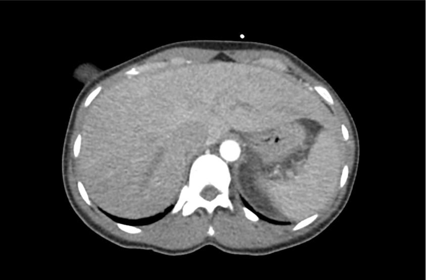
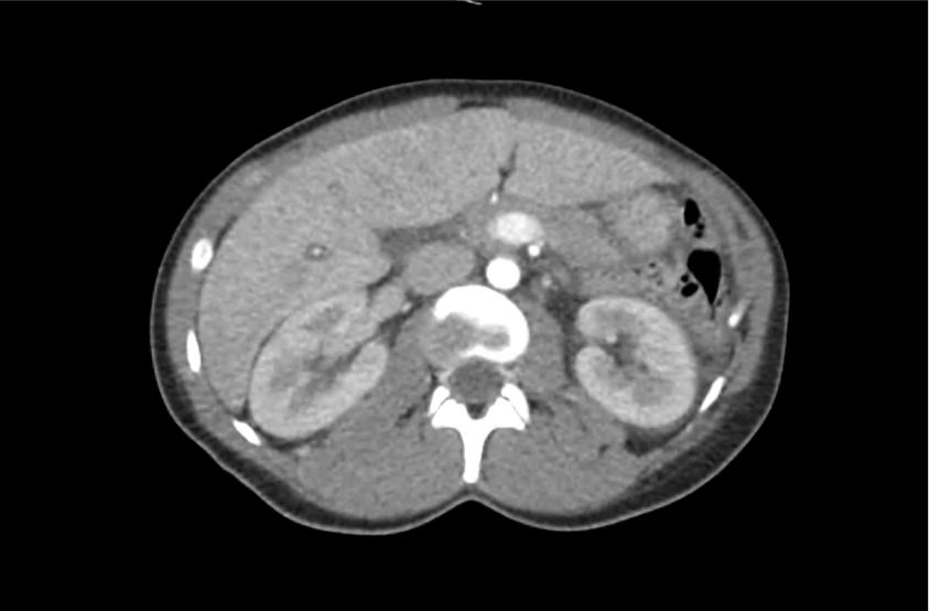
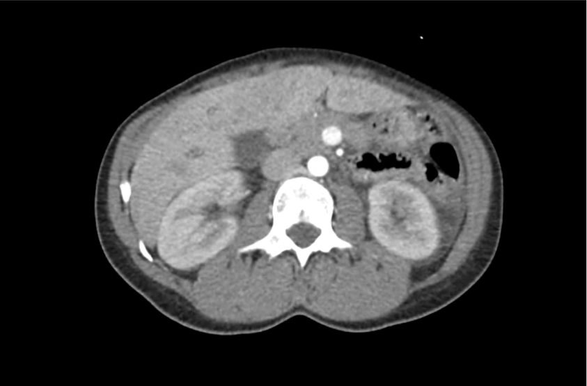
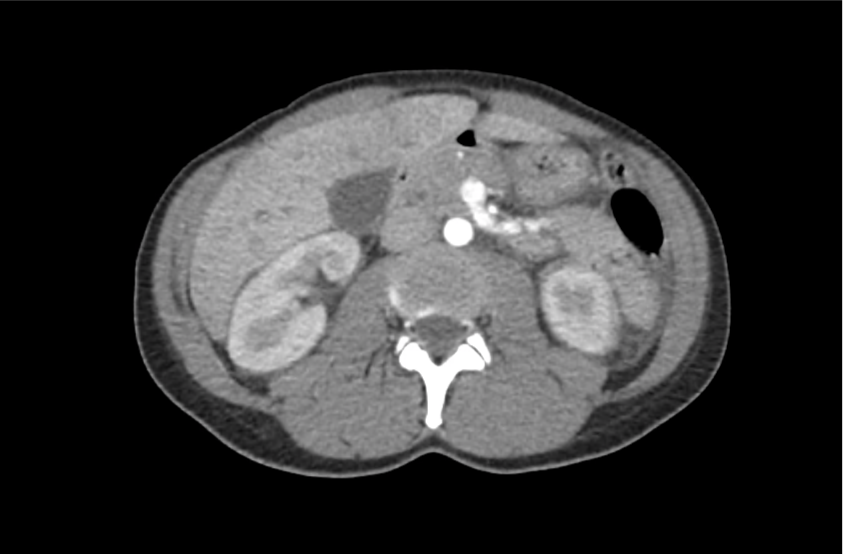
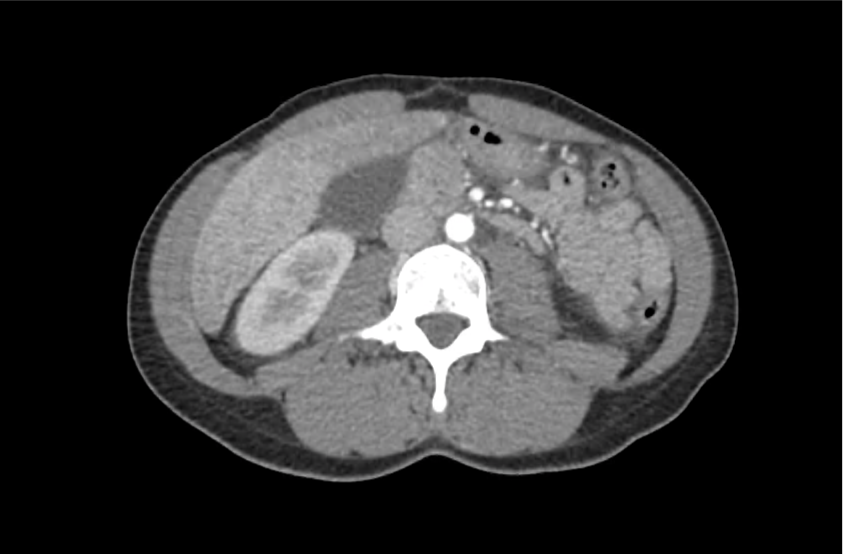
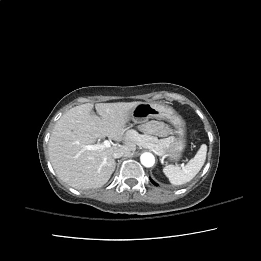
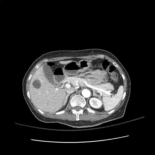
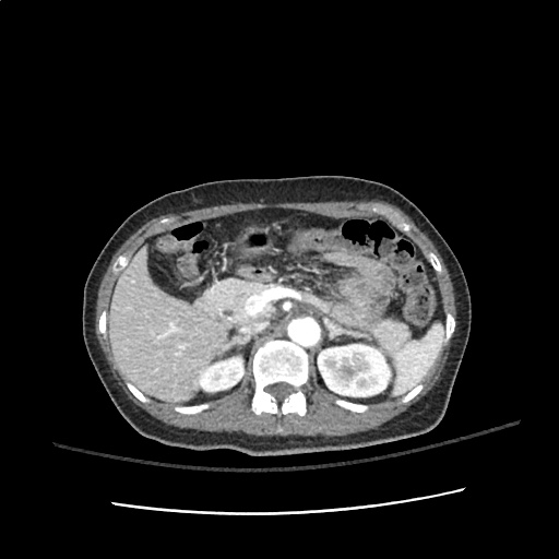
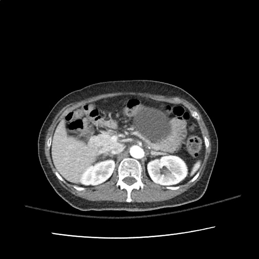
CT Abdomen/Pelvis with Contrast

Moderate free intra-abdominal fluid, heterogeneous liver with periportal edema, dense right middle lobe consolidation.

ED Course:

The patient developed worsening respiratory failure with hypoxia and tachypnea requiring endotracheal intubation. Thrombotic thrombocytopenic purpura was suspected and while awaiting emergent plasma exchange transfusion, the patient arrested and resuscitation efforts were unsuccessful.

The patient’s ADAMTS13 activity level was <3%. Autopsy demonstrated consolidation of the right middle lobe with possible lymphoproliferative mass, and lung petechial hemorrhages from microvascular thrombi.

Differential Diagnosis of Thrombocytopenia 1-7

Differential Diagnosis of Thrombocytopenia

Algorithm for the Evaluation of Thrombocytopenia 8

Algorithm for the Evaluation of Thrombocytopenia

Definition 9

  • Mild: <150k
  • Moderate: 100-150k
  • Severe: <50k
    • 10-30k: bleeding with minimal trauma
    • <10k: increased risk spontaneous bleeding

History 9,10

  • Prior platelet count
  • Family history bleeding disorders
  • Medications
    • Heparin
    • Quinine, quinidine
    • Rifampin
    • Trimethoprim-sulfamethoxazole
    • Vancomycin
  • Alcohol use
  • Travel-related infections

Physical Examination 9,10

  • Splenomegaly (liver disease)
  • Lymphadenopathy (infection, malignancy)

Workup 10,11

Schistocytes

Red blood cell fragments (schistocytes) 11

  • hCG
  • Repeat CBC
    • Detect spurious measure
    • Neutrophil-predominant leukocytosis: bacterial infection
    • Immature leukocytes (blasts): leukemia, myelodysplasia
  • Peripheral smear
    • Schistocytes: microangiopathic process (DIC, TTP, HUS)
    • Atypical lymphocytes: viral infection
    • Intracellular parasites: malaria
    • Hypersegmented neutrophils: nutritional deficiency
  • Infectious features: HIV, HCV, EBV, H.pylori, blood cultures
  • Autoimmune features: ANA, APL-Ab
  • Suspected occult liver disease: LFT, PT/PTT/INR
  • Suspected thrombotic microangiopathy: PT/PTT/INR, haptoglobin, LDH, fibrinogen, FDP, d-dimer

Specific Conditions 2-6,9,12-20

Disease Cause Presentation Laboratory Findings Treatment
DIC Sepsis
Trauma
Burn
Malignancy
Bleeding
Multi-organ failure
Shock
INR
Fibrinogen
FDP
D-dimer
Directed at underlying cause
Transfusion thresholds for hemorrhage:
FFP for INR >1.5
Platelets if <50k
Cryoprecipitate of fibrinogen <100mg/dL
TTP Insufficient ADAMTS-13 activity Non-specific constitutional symptoms (ex. weakness)
Neuro: headache, AMS, focal neuro deficit
GI: abdominal pain, nausea/vomiting
LDH
Reticulocyte
Unconjugated bilirubin
Haptoglobin
Plasma exchange
HUS Shiga-toxin-producing bacteria, E. coli O157:H7 Bloody diarrhea, anuria, oliguria, and hypertension Aggressive supportive care
HELLP Spectrum of eclampsia Hypertension
Visual symptoms
Headache
RUQ abdominal pain
AST/ALT
Uric acid
Unconjugated bilirubin
LDH
Reticulocyte
Haptoglobin
Delivery, MgSO4
ITP Primary ITP

Secondary ITP
– Drug
– Autoimmune
– Infection
– Malignancy

Usually asymptomatic, may have petechiae or easy bruising Isolated thrombocytopenia Steroids
HIT Exposure to heparin or LMWH Thrombocytopenia or a 50 percent reduction in platelet count between 5-10d exposure
New thrombosis or skin necrosis
4 T’s score
Platelet factor 4 antibodies Withdraw heparin

References

  1. Greinacher A, Selleng S. How I evaluate and treat thrombocytopenia in the intensive care unit patient. Blood. 2016;128(26):3032-3042. doi:10.1182/blood-2016-09-693655.
  2. Joly BS, Coppo P, Veyradier A. Thrombotic thrombocytopenic purpura. Blood. 2017;129(21):2836-2846. doi:10.1182/blood-2016-10-709857.
  3. Leslie SD, Toy PT. Laboratory hemostatic abnormalities in massively transfused patients given red blood cells and crystalloid. Am J Clin Pathol. 1991;96(6):770-773.
  4. Neunert C, Lim W, Crowther M, et al. The American Society of Hematology 2011 evidence-based practice guideline for immune thrombocytopenia. Blood. 2011;117(16):4190-4207. doi:10.1182/blood-2010-08-302984.
  5. Kappler S, Ronan-Bentle S, Graham A. Thrombotic microangiopathies (TTP, HUS, HELLP). Emerg Med Clin North Am. 2014;32(3):649-671. doi:10.1016/j.emc.2014.04.008.
  6. Greinacher A. Heparin-Induced Thrombocytopenia. Solomon CG, ed. N Engl J Med. 2015;373(3):252-261. doi:10.1056/NEJMcp1411910.
  7. Reardon JE Jr., Marques MB. Evaluation of Thrombocytopenia. Lab Med. 2006;37(4):248-250. doi:10.1309/R7P79KERAJHPRHLT.
  8. Stasi R. How to approach thrombocytopenia. Hematology Am Soc Hematol Educ Program. 2012;2012:191-197. doi:10.1182/asheducation-2012.1.191.
  9. Gauer RL, Braun MM. Thrombocytopenia. Am Fam Physician. 2012;85(6):612-622.
  10. Abrams CS. 172 – Thrombocytopenia. Twenty Fifth Edition. Elsevier Inc.; 2016:1159–1167.e2. doi:10.1016/B978-1-4557-5017-7.00172-0.
  11. Wilson CS, Vergara-Lluri ME, Brynes RK. Chapter 11 – Evaluation of Anemia, Leukopenia, and Thrombocytopenia. Second Edition. Elsevier Inc.; 2017:195-234.e195. doi:10.1016/B978-0-323-29613-7.00011-9.
  12. Hui P, Cook DJ, Lim W, Fraser GA, Arnold DM. The frequency and clinical significance of thrombocytopenia complicating critical illness: a systematic review. Chest. 2011;139(2):271-278. doi:10.1378/chest.10-2243.
  13. Jokiranta TS. HUS and atypical HUS. Blood. 2017;129(21):2847-2856. doi:10.1182/blood-2016-11-709865.
  14. Neunert CE. Management of newly diagnosed immune thrombocytopenia: can we change outcomes? Hematology Am Soc Hematol Educ Program. 2017;2017(1):400-405. doi:10.1182/asheducation-2017.1.400.
  15. Lambert MP, Gernsheimer TB. Clinical updates in adult immune thrombocytopenia. Blood. 2017;129(21):2829-2835. doi:10.1182/blood-2017-03-754119.
  16. Arepally GM. Heparin-induced thrombocytopenia. Blood. 2017;129(21):2864-2872. doi:10.1182/blood-2016-11-709873.
  17. Aster RH, Bougie DW. Drug-induced immune thrombocytopenia. N Engl J Med. 2007;357(6):580-587. doi:10.1056/NEJMra066469.
  18. Boral BM, Williams DJ, Boral LI. Disseminated Intravascular Coagulation. Am J Clin Pathol. 2016;146(6):670-680. doi:10.1093/ajcp/aqw195.
  19. Scully M, Hunt BJ, Benjamin S, et al. Guidelines on the diagnosis and management of thrombotic thrombocytopenic purpura and other thrombotic microangiopathies. Br J Haematol. 2012;158(3):323-335. doi:10.1111/j.1365-2141.2012.09167.x.
  20. Levine RL, Hursting MJ, Drexler A, Lewis BE, Francis JL. Heparin-induced thrombocytopenia in the emergency department. Ann Emerg Med. 2004;44(5):511-515. doi:10.1016/j.annemergmed.2004.06.004.

Hyperthermia

Brief H&P

A young male with unknown medical history is brought in by ambulance with altered mental status. EMS reports that the patient was agitated, requiring restraints for transportation. On arrival, the patient is agitated, uncooperative and unable to provide history. Vital signs are notable for tachycardia, tachypnea and hypertension. Physical examination demonstrates diaphoresis and mydriasis, as well as increased muscle tone – particularly in the lower extremities with ankle clonus. A core temperature is obtained and noted to be elevated at 41.5°C. Point-of-care glucose is normal.

Rapid external cooling measures were instituted and several doses of intravenous benzodiazepines were administered with improvement in agitation. Laboratory studies were notable for a modest leukocytosis (WBC 18.4 without immature forms), serum sodium was 135 without osmolar gap, creatine kinase was slightly elevated without renal dysfunction, and thyroid function tests were normal. Toxicology screen was negative. ECG revealed sinus tachycardia but was otherwise normal and non-contrast computed tomography of the head was normal.

After a brief admission in the intensive care unit, the patient’s mental status improved and he reported MDMA use on the evening of presentation, he also described a history of major depression and was taking paroxetine.

Evaluation of Elevated Temperature

The designation of 38°C as “suspicious” for fever dates to 1868 and the analysis of over one million (axillary) temperature measurements by Carl Wunderlich1. Any cutoff is arbitrary and requires recognition of the clinical context and normal daily variations (with nadir in the morning and peak in evening) 2,3. What is clear is that peripheral thermometry (unless demonstrating fever) is unreliable and a core temperature should be sought4.

Thermoregulation

Temperature homeostasis is a balance between heat production and dissipation maintained by the anterior hypothalamus. Heat production is a byproduct of normal metabolic processes and skeletal muscle activity. Conservation, maintenance or dissipation of heat is aided by cutaneous vasodilation, sweating, or behavioral responses.

Fever is caused by endogenous or exogenous pyrogens which alter the homeostatic set-point, inducing thermogenesis and elevating the body temperature. Precipitants of fever are usually infectious, however non-infectious processes (ex. malignancy, tissue ischemia/infarction, auto-immune disease) resulting in inflammation can provoke a similar response 5-7.

There is no explicit temperature distinction to diagnose hyperthermia, instead the physiologic mechanism is different. In hyperthermia, the body’s homeostatic mechanisms are dysfunctional or overwhelmed due to heat exposure, excess production, ineffective dissipation or hypothalamic malfunction 8.

Algorithm for the Evaluation of Hyperthermia 8-15

Algorithm for the Evaluation of Hyperthermia

Implicated Agents in Drug-Induced Hyperthermic Syndromes 9,10

Serotonin Syndrome

Class Examples
SSRI sertraline, fluoxetine, paroxetine
Other anti-depressants trazodone, venlafaxine, lithium
MAOI phenelzine, isocarboxazid
Anti-epileptic drugs valproate
Analgesics meperidine, fentanyl, tramadol
Anti-emetic ondansetron, metoclopramide
Anti-migraine sumatriptan
Antimicrobial linezolid, ritonavir
Illicit substances MDMA, LSD

Neuroleptic Malignant Syndrome (NMS)

Class Examples
Typical anti-psychotic haloperidol, prochlorperazine
Atypical anti-psychotic risperidone, olanzapine, quetiapine, aripiprazole
Anti-dopaminergic metoclopramide, droperidol

References:

  1. Wunderlich CA. Das Verhalten Der Eigenwärme in Krankheiten. 1870.
  2. Mackowiak PA, Wasserman SS, Levine MM. A critical appraisal of 98.6 degrees F, the upper limit of the normal body temperature, and other legacies of Carl Reinhold August Wunderlich. JAMA. 1992;268(12):1578-1580.
  3. Lee-Chiong TL, Stitt JT. Disorders of temperature regulation. Compr Ther. 1995;21(12):697-704.
  4. Niven DJ, Gaudet JE, Laupland KB, Mrklas KJ, Roberts DJ, Stelfox HT. Accuracy of peripheral thermometers for estimating temperature: a systematic review and meta-analysis. Ann Intern Med. 2015;163(10):768-777. doi:10.7326/M15-1150.
  5. Dinarello CA. Infection, fever, and exogenous and endogenous pyrogens: some concepts have changed. J Endotoxin Res. 2004;10(4):201-222. doi:10.1179/096805104225006129.
  6. Greisman LA, Mackowiak PA. Fever: beneficial and detrimental effects of antipyretics. Curr Opin Infect Dis. 2002;15(3):241-245.
  7. Dinarello CA. Thermoregulation and the pathogenesis of fever. Infect Dis Clin North Am. 1996;10(2):433-449.
  8. Simon HB. Hyperthermia. N Engl J Med. 1993;329(7):483-487. doi:10.1056/NEJM199308123290708.
  9. Boyer EW, Shannon M. The serotonin syndrome. N Engl J Med. 2005;352(11):1112-1120. doi:10.1056/NEJMra041867.
  10. Berman BD. Neuroleptic malignant syndrome: a review for neurohospitalists. Neurohospitalist. 2011;1(1):41-47. doi:10.1177/1941875210386491.
  11. Hayes BD, Martinez JP, Barrueto F. Drug-induced hyperthermic syndromes: part I. Hyperthermia in overdose. Emerg Med Clin North Am. 2013;31(4):1019-1033. doi:10.1016/j.emc.2013.07.004.
  12. Oruch R, Pryme IF, Engelsen BA, Lund A. Neuroleptic malignant syndrome: an easily overlooked neurologic emergency. Neuropsychiatr Dis Treat. 2017;13:161-175. doi:10.2147/NDT.S118438.
  13. Musselman ME, Saely S. Diagnosis and treatment of drug-induced hyperthermia. Am J Health Syst Pharm. 2013;70(1):34-42. doi:10.2146/ajhp110543.
  14. Ahuja N, Cole AJ. Hyperthermia syndromes in psychiatry. Adv psychiatr treat (Print). 2018;15(03):181-191. doi:10.1192/apt.bp.107.005090.
  15. Tomarken JL, Britt BA. Malignant hyperthermia. Ann Emerg Med. 1987;16(11):1253-1265. doi:10.1016/S0196-0644(87)80235-4.

Pediatric Head Trauma

Brief H&P:

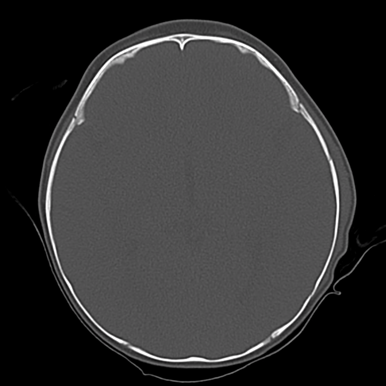
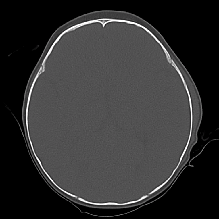
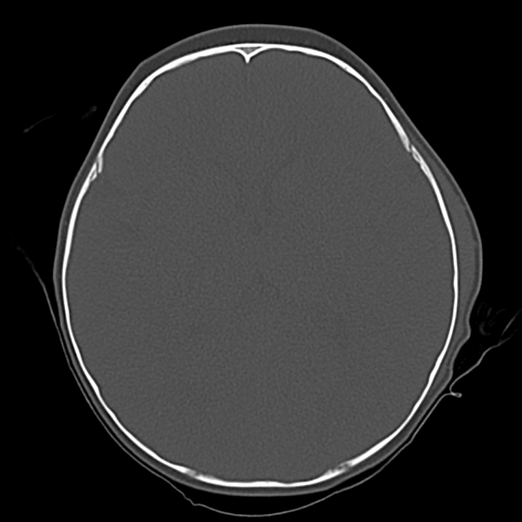
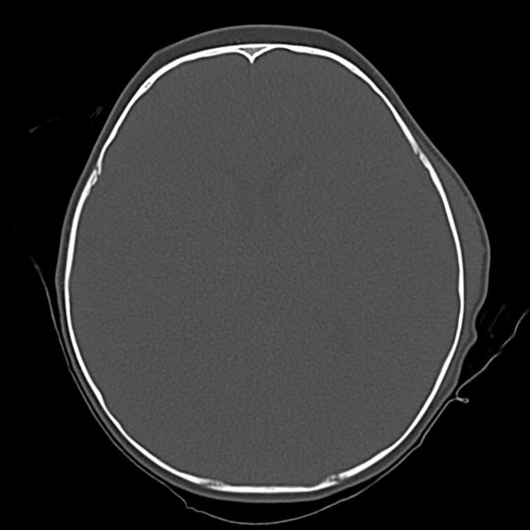
A young child, otherwise healthy, is brought to the pediatric emergency department after a fall. The parents report a fall from approximately 2 feet after which the patient cried immediately and without apparent loss of consciousness. Over the course of the day, the patient developed an enlarging area of swelling over the left head. The parents were concerned about a progressive decrease in activity and interest in oral intake by the child, and they were brought to the emergency department for evaluation. Examination demonstrated a well-appearing and interactive child – appropriate for age. Head examination was notable for a 5x5cm hematoma over the left temporoparietal skull with an underlying palpable skull irregularity not present on the contralateral side. Non-contrast head computed tomography was obtained.

Imaging

ct_head_0007_Screen-Shot-2017-06-05-at-4.18.59-PM
ct_head_0006_Screen-Shot-2017-06-05-at-4.19.02-PM
ct_head_0005_Screen-Shot-2017-06-05-at-4.19.04-PM
ct_head_0004_Screen-Shot-2017-06-05-at-4.19.08-PM
ct_head_0003_Screen-Shot-2017-06-05-at-4.19.10-PM
ct_head_0002_Screen-Shot-2017-06-05-at-4.19.15-PM
ct_head_0001_Screen-Shot-2017-06-05-at-4.19.18-PM
ct_head_0000_Screen-Shot-2017-06-05-at-4.19.22-PM

CT Head

Fracture of the left temporal and parietal bone with overlying scalp hematoma.

Algorithm for the Evaluation of Pediatric Head Trauma (PECARN)1,2,3

Algorithm for the evaluation of pediatric head trauma

References

  1. Kuppermann N, Holmes JF, Dayan PS, et al. Identification of children at very low risk of clinically-important brain injuries after head trauma: a prospective cohort study. Lancet. 2009;374(9696):1160-1170. doi:10.1016/S0140-6736(09)61558-0.
  2. Brenner D, Elliston C, Hall E, Berdon W. Estimated risks of radiation-induced fatal cancer from pediatric CT. American Journal of Roentgenology. 2001;176(2):289-296. doi:10.2214/ajr.176.2.1760289.
  3. Schonfeld D, Bressan S, Da Dalt L, Henien MN, Winnett JA, Nigrovic LE. Pediatric Emergency Care Applied Research Network head injury clinical prediction rules are reliable in practice. Archives of Disease in Childhood. 2014;99(5):427-431. doi:10.1136/archdischild-2013-305004.

Rapid Pediatric Assessment

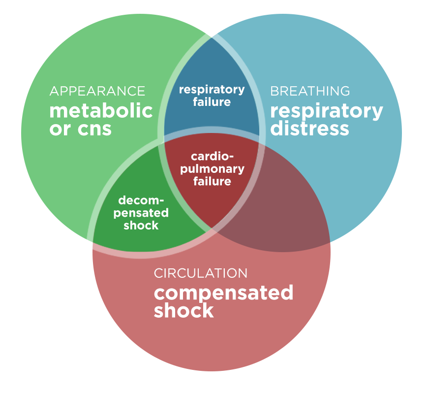
This post presents a tool for the rapid assessment of the cardiopulmonary status and cerebral/metabolic function of critically ill pediatric patients. The purpose is not to establish a diagnosis, rather to identify the particular physiological derangements to prioritize initial interventions. The tool was initially designed as a “triangle” – it has been adapted here (with permission) as a Venn diagram.1

Pediatric Assessment Diagram

Pediatric Assessment Diagram

Assessment of Appearance

  • Tone: Moves spontaneously, resists examination
  • Interactivity: Interacts with environment, reaches for items
  • Consolability: Comforted by caregiver
  • Gaze: Makes eye contact

Assessment of Work of Breathing

  • Airway Sounds: Stridor, grunting, wheezing
  • Position: Tripod
  • Retractions

Assessment of Circulation

  • Pallor
  • Mottling
  • Cyanosis

Management

Impression Interventions
Respiratory distress
  • Position of comfort
  • Oxygen, suction
  • Therapy as appropriate (albuterol, epinephrine, etc)
  • Labs/radiographs as indicated
Respiratory failure
  • Head/airway positioning
  • 100% oxygen
  • Ventilation support (BVM)
  • Advanced airway
Shock (compensated and decompensated)
  • Oxygen
  • Access
  • Fluid resuscitation
  • Specific therapy (antibiotics, surgery)
  • Labs/radiographs as indicated
CNS/Metabolic
  • Pulse oximetry
  • Rapid glucose
  • Labs/radiographs as indicated
Cardiopulmonary Failure
  • Head/airway positioning
  • 100% oxygen
  • Ventilation support (BVM)
  • Chest compressions as needed
  • Specific therapy (defibrillation, epinephrine, amiodarone)
  • Labs/radiographs as indicated

References:

  1. The pediatric assessment triangle: a novel approach for the rapid evaluation of children. Pediatr Emerg Care. 2010;26(4):312-315. doi:10.1097/PEC.0b013e3181d6db37.

Altered Mental Status Applied

H&P

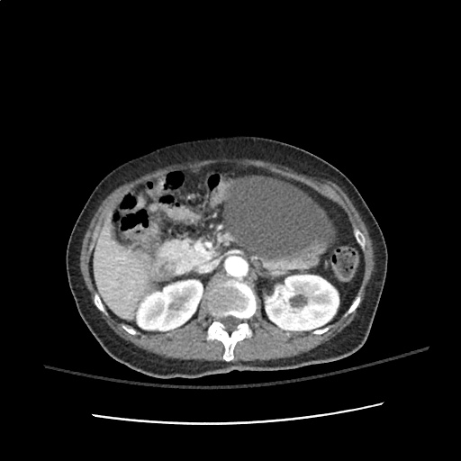
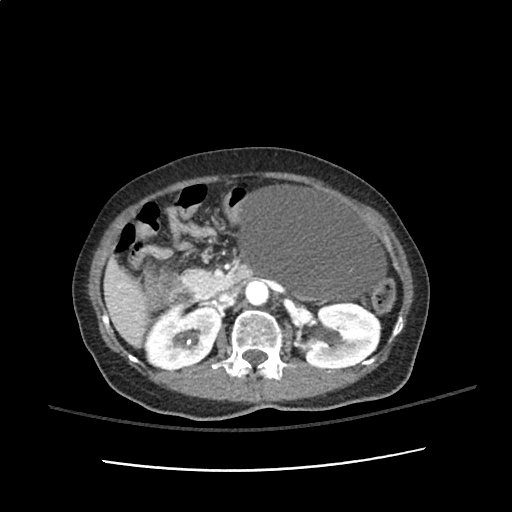
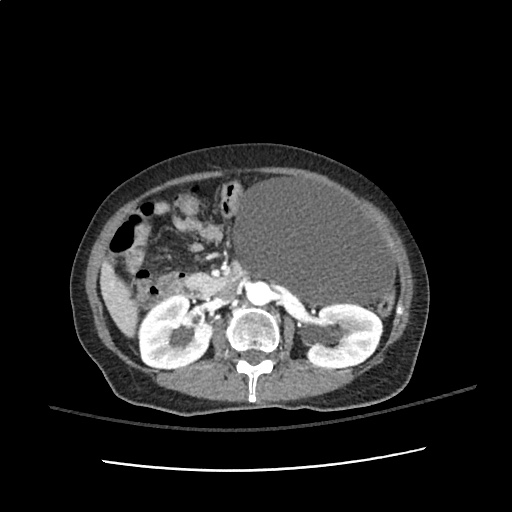
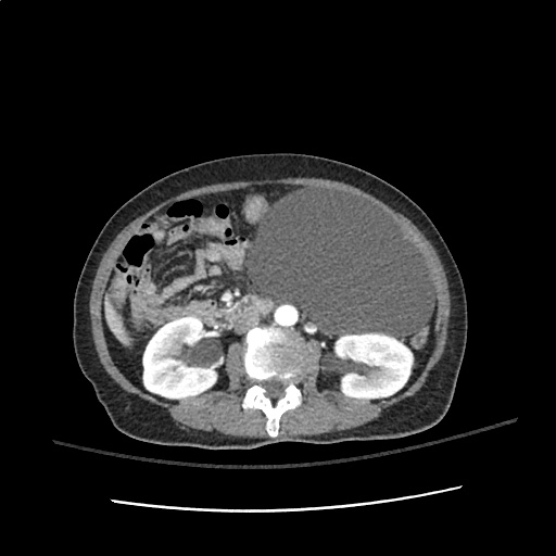
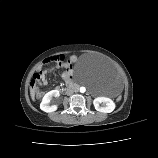
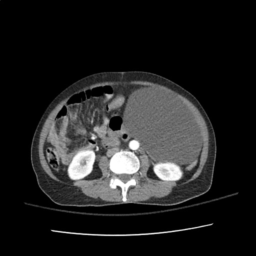
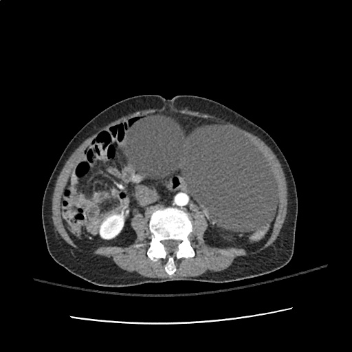
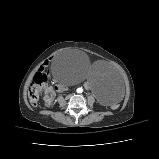
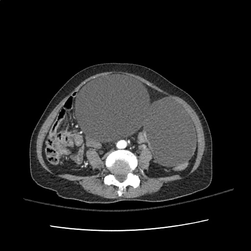
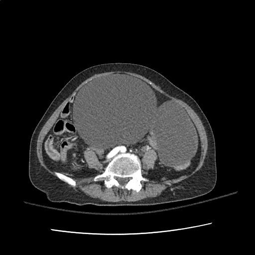
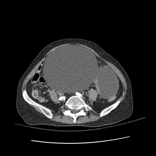
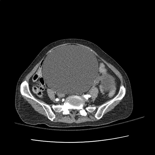
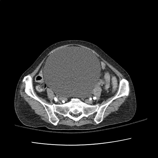
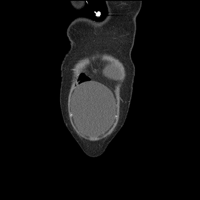
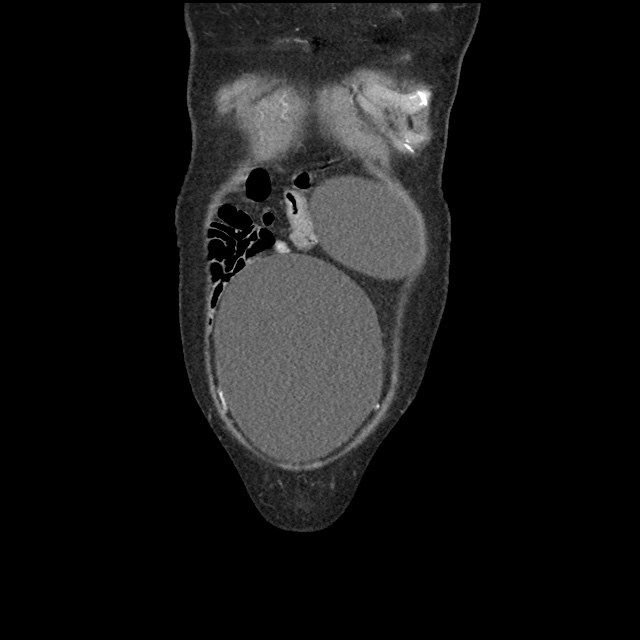
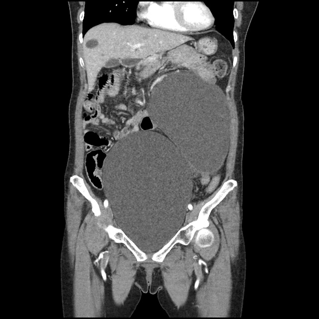
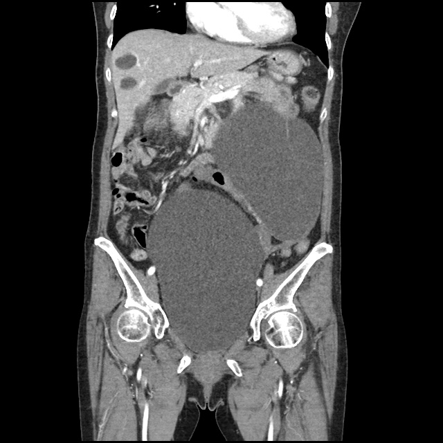
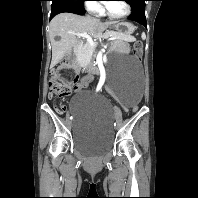
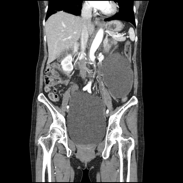
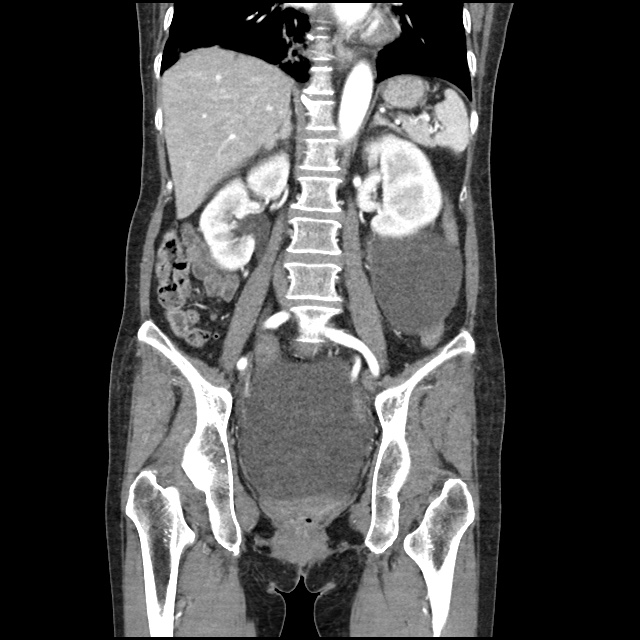
58 year-old female with no known past medical history, brought to emergency department by husband due to fatigue and weakness. The patient does not speak and cannot provide history. Her husband describes a progressive decline from normal baseline two weeks ago, noting lethargy/fatigue. Noted decreased speech and attention one week ago, and absent speech and requiring assistance with ambulation for the past two days. Thorough review of systems unremarkable excepting vomiting with decreased oral intake (tolerating fluids) and prior headache which resolved.

On examination, vital signs were normal, the patient was lying in bed and in no acute distress. The patient was non-verbal and did not follow commands (GCS E4-M5-V2). She was unable to comply with a thorough neurological examination, however pupils were equal and reactive, eyes tracked without nystagmus, no facial asymmetry noted, reflexes 1+ and symmetric in UE/LE, cannot participate in strength/sensory testing. Abdominal examination notable for infraumbilical and left-sided mass which elicits groans with palpation, though no rigidity or guarding. Mucous membranes moist, no skin tenting.

Labs

  • CBC: 13.5 (97% neutrophils) , 12.9, 38.2, 240
  • BMP: 107, 2.4, 70, 28, 9, 10, 0.44, 102
  • Serum osmolarity: 224
  • Urine osmolarity: 239
  • UNa: 20

Imaging

IM-0001-0022
IM-0001-0024
IM-0001-0026
IM-0001-0028
IM-0001-0032
IM-0001-0036
IM-0001-0040
IM-0001-0044
IM-0001-0048
IM-0001-0052
IM-0001-0056
IM-0001-0060
IM-0001-0064
IM-0001-0068
IM-0001-0072
IM-0001-0076
IM-0001-0080
IM-0001-0084
IM-0001-0088
IM-0001-0092
IM-0001-0010
IM-0001-0014
IM-0001-0018
IM-0001-0022
IM-0001-0026
IM-0001-0030
IM-0001-0034
IM-0001-0038
IM-0001-0042
IM-0001-0046
IM-0001-0050

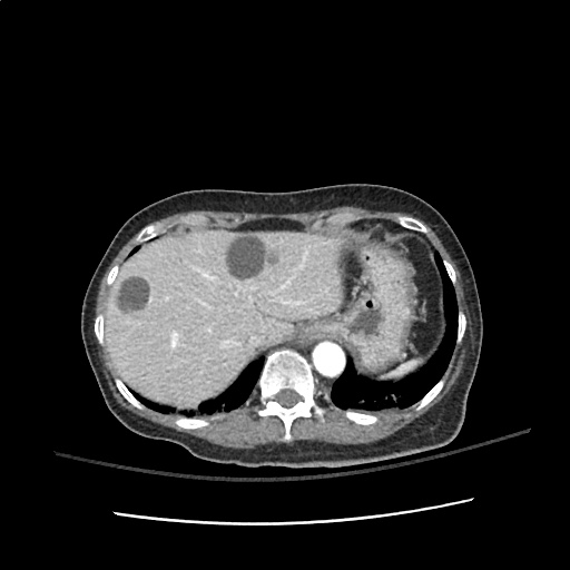
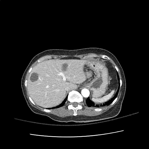
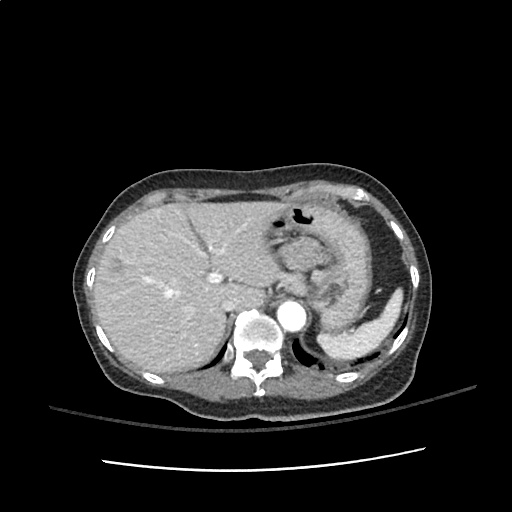
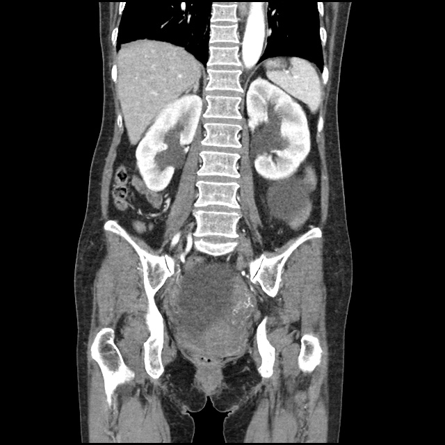
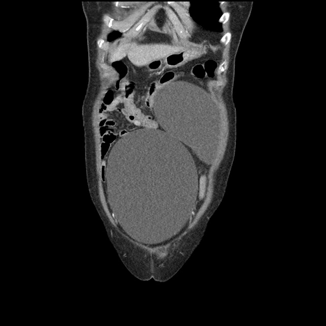
CT abdomen/pelvis with intravenous contrast

  • Large, 15 cm cystic mass in the left abdomen, which likely arises from the mesentery. This mass is suspicious for neoplasm.
  • Multiple low-density cystic lesions in the liver, which measure up to 4.5 cm in diameter and are concerning for metastatic disease. Alternatively, these may represent benign hepatic cysts which are unrelated to the mesenteric mass.
  • Massively distended bladder, with moderate bilateral hydronephrosis and mild hydroureter.

Hospital Course

The patient was admitted to the medical intensive care unit. The following problem list details findings from the extensive inpatient evaluation.

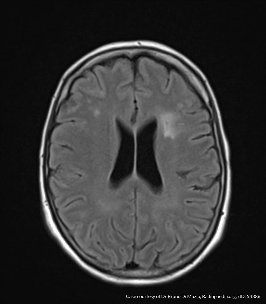
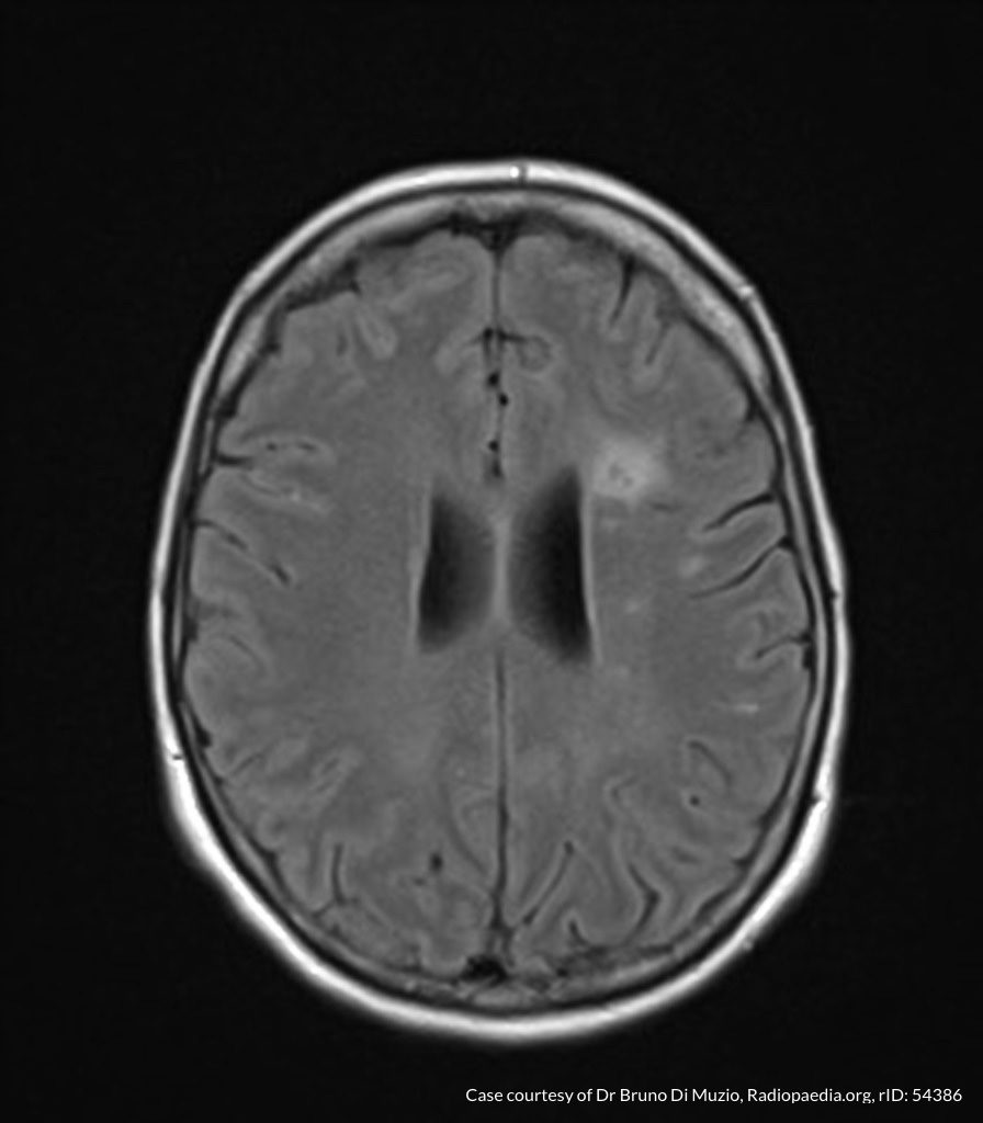
#Altered Mental Status: The patient’s dramatically depressed level of consciousness improved gradually with correction of hyponatremia and the patient was alert, oriented and at baseline at the time of discharge. Evaluation included MRI brain which showed only chronic microvascular changes. A lumbar puncture was notable for isolated elevation of CSF protein. The patient was treated empirically for HSV encephalitis until CSF HSV PCR resulted negative. Neurology was consulted and identified increased CSF oligoclonal bands of unclear significance.

#Hyponatremia: Nephrology consulted, presumed SIADH based on urine studies (secondary to infection or malignancy). Corrected upon discharge.

#Pelvic Mass: Initially thought to arise from small bowel on CT abdomen/pelvis, after bladder decompression and transvaginal ultrasound, thought to arise from adnexa. Gynecology consulted, cyst characteristics (homogenous, fluid-filled) suggest benign process and tumor markers within normal limits. No acute intervention, drainage or biopsy warranted.

#Bladder distension: Unclear etiology, associated with mild/moderate hydronephrosis. Thought to be secondary to bladder outlet obstruction secondary to pelvic mass. Indwelling catheter placed, discontinued prior to discharge with successful spontaneous voiding trial and normal post-void residual.

Hyponatremia Applied

Hyponatremia Applied

Altered Mental Status Applied

Altered Mental Status Applied

Altered Mental Status

Components of Consciousness

Components of Consciousness

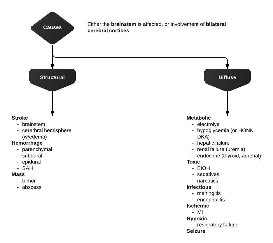
Causes of Altered Mental Status

Causes of Altered Mental Status

History

Rate of onset
Abrupt: CNS
Gradual: Systemic

Physical Examination

  • Vital Signs

    • Blood Pressure: low (shock), high (SAH, stroke, ICP)
    • Heart Rate: low (medication overdose, conduction block), high (hypovolemia, infection, anemia, thyrotoxicosis, drug/toxin)
    • Temperature: low/high (infection, drug/toxin, environmental)
    • Respiratory Rate: low/high (CNS, drug/toxin, metabolic derangement)
  • Eyes

    • Unilateral dilation: CNS/structural cause
    • Papilledema: ICP
    • EOM: cranial nerve dysfunction
    • Oculocephalic: brainstem function
  • Head: trauma
  • Mucous membranes: hydration, laceration
  • Neck: meningeal irritation
  • Pulmonary: respiratory effort
  • CV: murmur, arrhythmia, CO
  • Abdomen: pulsatile mass, sequelae of liver failure
  • Skin: rash, needle tracks

Labs

  • Glucose
  • ECG: arrhythmia, ischemia, electrolyte abnormalities
  • BMP: electrolytes, renal failure, anion gap
  • ABG: hypoxemia, hypercarbia
  • Urinalysis: infection, SG
  • Utox
  • CBC: leukocytosis, leukopenia, severe anemia, thrombocytopenia
  • Ammonia: hepatic encephalopathy
  • TFT: thyrotoxicosis, myxedema coma
  • CSF: meningitis, encephalitis

Imaging

  • CT head: Non-contrast sufficient to identify ICH. Use contrast if mass/infection suspected
  • CTA head/neck: If aneurysm, AVM, venous sinus thrombosis or vertebrobasilar insufficiency suspected
  • CXR: PNA

References

  1. Bassin, B., & Cooke, J. (2013). Depressed Consciousness and Coma. In Rosen’s Emergency Medicine – Concepts and Clinical Practice (8th ed., Vol. 1, pp. 142-150). Elsevier Health Sciences.

Delirium

ID:

A 70 year-old female with a PMH of HTN, DM, hyperlipidemia and stage I breast cancer s/p lumpectomy with sentinel LN biopsy several years ago presented for elective surgery complicated by post-operative bleeding. She is now 4 days post-op and was found to be confused, somnolent and occasionally agitated.

HPI:

The patient could not be interviewed.

PE:

  • VS: Stable and within normal limits
  • General: unremarkable except for crackles in bilateral lung bases
  • MSE: only arouses to sternal rub and becomes agitated, moving all four extremities spontaneously and symmetrically.
  • Reflexes: corneal and gag reflexes present, suppresses eye movements with head turn, deep tendon reflexes 3+ throughout UE/LE bilaterally.

Assessment:

70 year-old woman with a history of HTN, DM, hyperlipidemia and breast cancer presents with worsening confusion, somnolence and occasional agitation four days after surgery. The combination of significantly altered consciousness and absence of focal neurological findings, all in the setting of a complicated surgical course suggest delirium.

Differential Diagnosis of Altered Mental Status:

Levels of consciousness

There are different levels of consciousness, they are named in the diagram below but are better described by the characteristics observed.

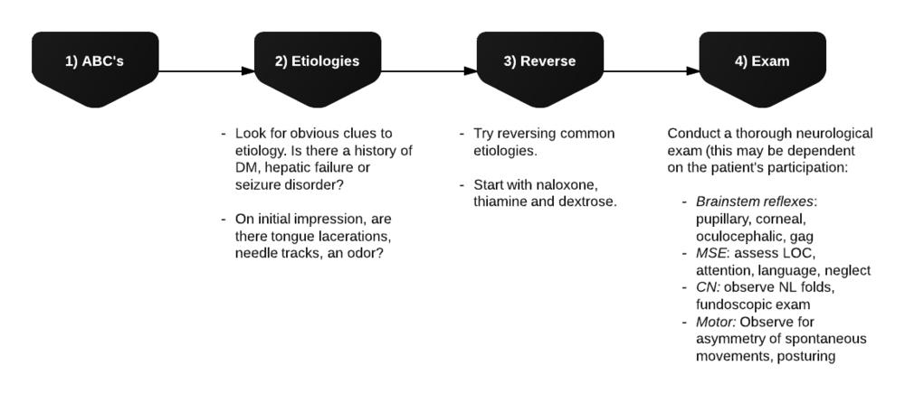
Initial assessment

Differential Diagnosis for Altered Mental Status

References:

  1. Inouye, S. K. (2006). Delirium in Older Persons. The New England journal of medicine, 354(11), 1157–1165. doi:10.1056/NEJMra052321
  2. Blueprints neurology. Philadelphia: Wolters Kluwer Health/Lippincott William & Wilkins, 2009.
  3. Tindall SC. Level of Consciousness. In: Walker HK, Hall WD, Hurst JW, editors. Clinical Methods: The History, Physical, and Laboratory Examinations. 3rd edition. Boston: Butterworths; 1990. Chapter 57. Available from: http://www.ncbi.nlm.nih.gov/books/NBK380/