Isolation Precautions

An Algorithm for the Determination of Isolation Precautions

An Algorithm for the Determination of Isolation Precautions

References

  1. Siegel, J., Rhinehart, E., Jackson, M., Chiarello, L., Committee, H. (2007). 2007 Guideline for Isolation Precautions: Preventing Transmission of Infectious Agents in Health Care Settings American Journal of Infection Control 35(10), S65-S164. https://dx.doi.org/10.1016/j.ajic.2007.10.007
  2. Liang, S., Theodoro, D., Schuur, J., Marschall, J. (2014). Infection Prevention in the Emergency Department Annals of Emergency Medicine 64(3), 299-313. https://dx.doi.org/10.1016/j.annemergmed.2014.02.024
  3. Liang, S., Riethman, M., Fox, J. (2018). Infection Prevention for the Emergency Department: Out of Reach or Standard of Care? Emergency medicine clinics of North America 36(4), 873-887. https://dx.doi.org/10.1016/j.emc.2018.06.013
  4. Gottenborg, E., Barron, M. (2016). Isolation Precautions in the Inpatient Setting Hospital Medicine Clinics 5(1), 30-42. https://dx.doi.org/10.1016/j.ehmc.2015.08.004
  5. Harding, A., Almquist, L., Hashemi, S. (2011). The use and need for standard precautions and transmission-based precautions in the emergency department. Journal of emergency nursing: JEN : official publication of the Emergency Department Nurses Association 37(4), 367-73; quiz 424-5. https://dx.doi.org/10.1016/j.jen.2010.11.017

COVID-19

Brief HPI:

A 38 year-old male with a history of hypertension presents to the emergency department with fever, cough and shortness of breath. He notes 4 days of symptoms which have been gradually worsening despite over-the-counter treatments. He denies recent travel or sick contacts. While he attempted to remain isolated – his symptoms grew intolerable.

On arrival in the emergency department, vital signs were notable for tachycardia and hypoxia (SpO2 85%, improving to 92% on 4L by nasal cannula). Physical examination demonstrated tachypnea and accessory muscle use but clear lung fields, and no extremity edema nor jugular venous distension. A chest radiograph revealed patchy airspace opacities. A presumptive diagnosis of COVID-19 pneumonia was made.

While awaiting hospitalization, the patient’s hypoxia worsened though he remained otherwise alert and oriented. He was placed on 15L via non-rebreather and instructed regarding self-prone positioning. He was admitted to the intensive care unit.

An Algorithm for the Management of COVID-19 Hypoxic Respiratory Failure1-6

An algorithm for the management of COVID-19 respiratory failure

References

  1. Whittle, J., Pavlov, I., Sacchetti, A., Atwood, C., Rosenberg, M. (2020). Respiratory Support for Adult Patients with COVID‐19 Journal of the American College of Emergency Physicians Open https://dx.doi.org/10.1002/emp2.12071
  2. Hui, D., Chow, B., Chu, L., Ng, S., Lee, N., Gin, T., Chan, M. (2012). Exhaled Air Dispersion during Coughing with and without Wearing a Surgical or N95 Mask PLoS ONE  7(12), e50845. https://dx.doi.org/10.1371/journal.pone.0050845
  3. Hui, D., Chow, B., Lo, T., Ng, S., Ko, F., Gin, T., Chan, M. (2015). Exhaled Air Dispersion During Noninvasive Ventilation via Helmets and a Total Facemask Chest  147(5), 1336-1343. https://dx.doi.org/10.1378/chest.14-1934
  4. Hui, D., Chow, B., Lo, T., Tsang, O., Ko, F., Ng, S., Gin, T., Chan, M. (2019). Exhaled air dispersion during high-flow nasal cannula therapy versus CPAP via different masks European Respiratory Journal  53(4), 1802339. https://dx.doi.org/10.1183/13993003.02339-2018
  5. Sun, Q., Qiu, H., Huang, M., Yang, Y. (2020). Lower mortality of COVID-19 by early recognition and intervention: experience from Jiangsu Province Annals of Intensive Care  10(1), 33. https://dx.doi.org/10.1186/s13613-020-00650-2
  6. Roca, O., Caralt, B., Messika, J., Samper, M., Sztrymf, B., Hernández, G., García-de-Acilu, M., Frat, J., Masclans, J., Ricard, J. (2018). An Index Combining Respiratory Rate and Oxygenation to Predict Outcome of Nasal High-Flow Therapy American Journal of Respiratory and Critical Care Medicine  199(11), 1368-1376. https://dx.doi.org/10.1164/rccm.201803-0589oc

Cerebrospinal Fluid

Brief HPI:

An approximately 70 year-old male with unknown medical history is brought to the emergency department with altered mental status. A community member contacted police after not seeing the patient for the past three days which was unusual. Upon entering the patient’s home, EMS found the patient on the ground, unresponsive. Capillary glucose was normal and naloxone was administered without appreciable effect.

On arrival in the emergency department, the patient remained unresponsive to verbal and noxious stimulation and was intubated for airway protection. Vital signs were notable for hypotension (BP 88/45mmHg) and a core temperature of 96.5°F. Physical examination demonstrated cool extremities and ecchymosis and edema involving the right upper and lower extremities. The patient’s blood pressure improved with fluid resuscitation and empiric broad-spectrum antibiotics were administered due to concern for infection in the setting of hypothermia.

Laboratory/Imaging Results

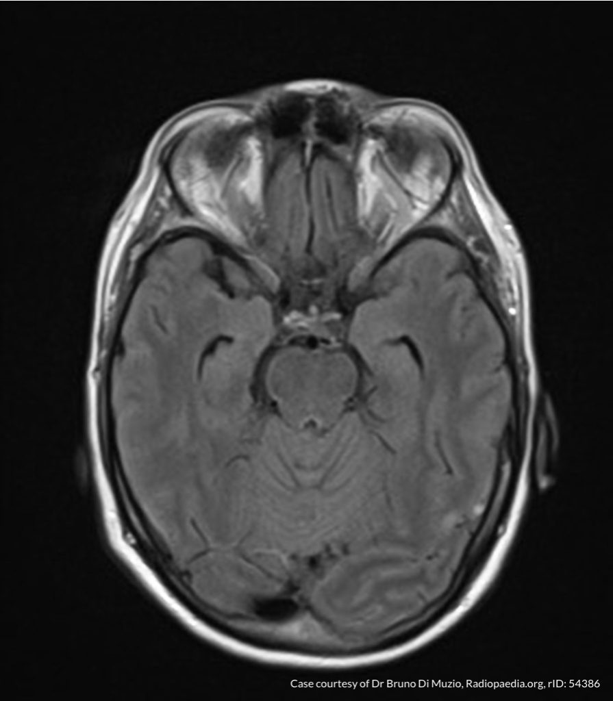
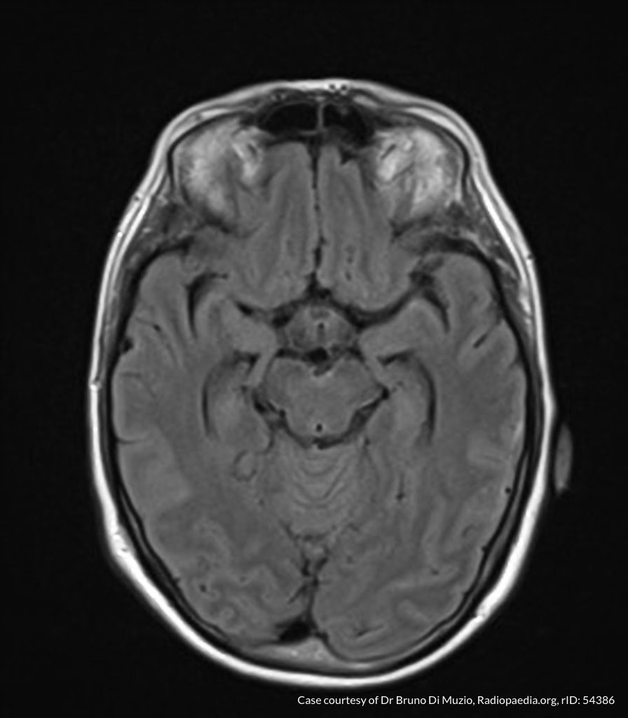
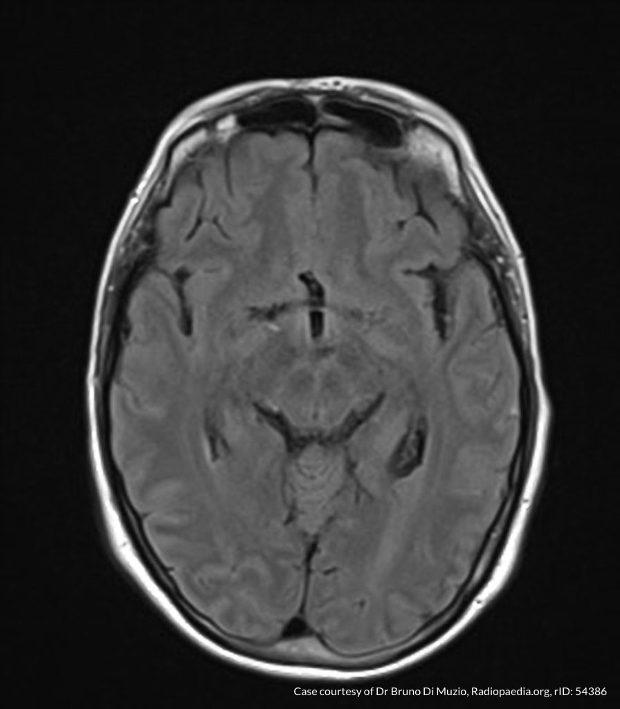
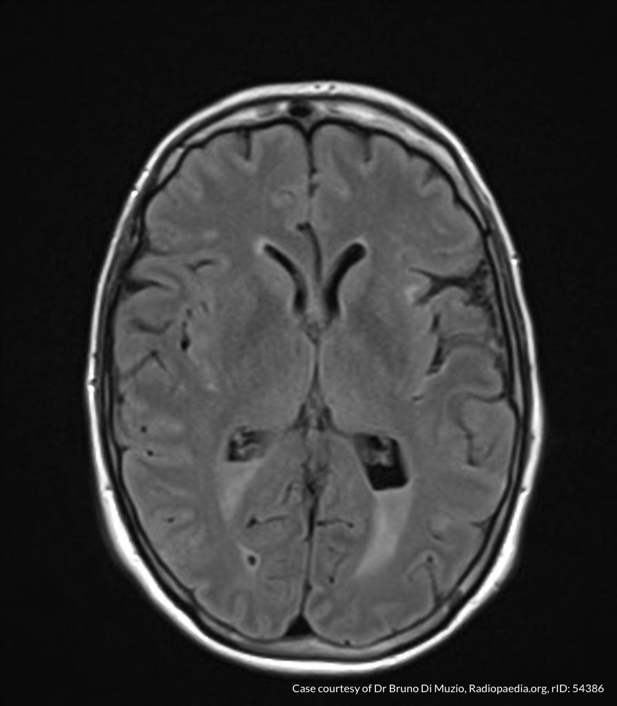
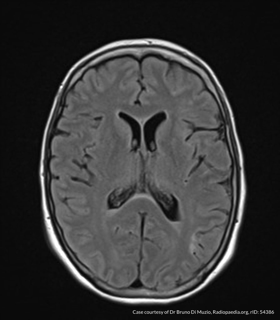
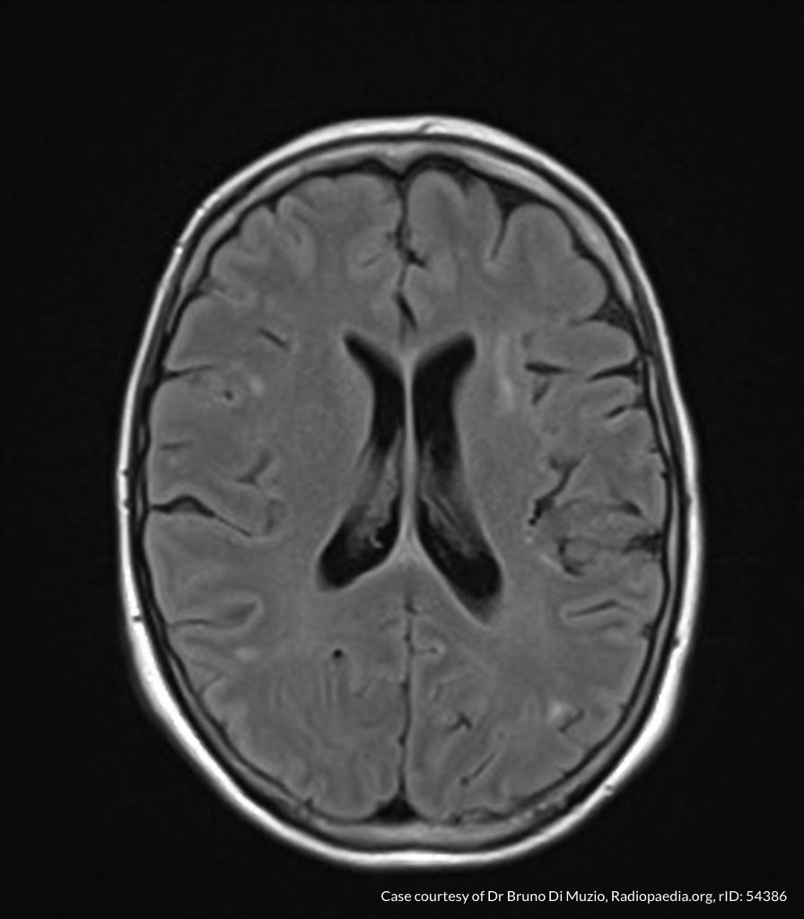
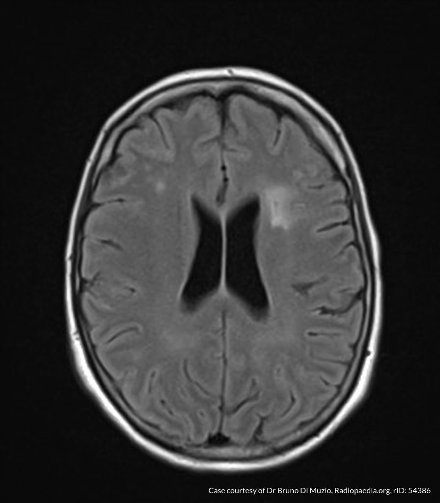
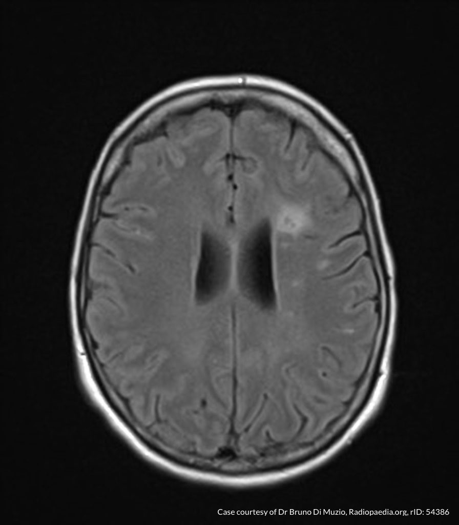
Laboratory tests were notable for leukocytosis and creatine kinase above the threshold for detection. Radiology preliminary interpretation of non-contrast head imaging was normal. A lumbar puncture was performed with grossly purulent cerebrospinal fluid.

00_ventriculitis
01_ventriculitis
02_ventriculitis
03_ventriculitis
04_ventriculitis
05_ventriculitis
06_ventriculitis
07_ventriculitis
08_ventriculitis
09_ventriculitis

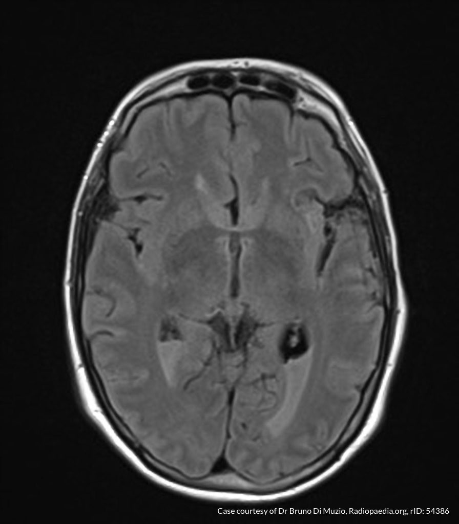
MRI Brain

Dependent material within the occipital horns of the lateral ventricles consistent with ventriculitis.

Hospital Course

The patient was admitted for the treatment of presumed meningitis. Radiology final interpretation of non-contrast head computed tomography commented on ventricular debris suggestive of ventriculitis which was later confirmed on magnetic resonance imaging1,2. Due to poor response to systemic antibiotics, neurosurgery was consulted, a ventricular drain was placed with administration of intrathecal antibiotics. The patient’s condition continued to deteriorate and family members elected to allow his natural death.

An Algorithm for the Analysis of Cerebrospinal Fluid (CSF)3-14

An Algorithm for the Analysis of Cerebrospinal Fluid (CSF)

References

  1. Lesourd A, Magne N, Soares A, et al. Primary bacterial ventriculitis in adults, an emergent diagnosis challenge: report of a meningoccal case and review of the literature. BMC Infect Dis. 2018;18(1):226. doi:10.1186/s12879-018-3119-4.
  2. Gofman N, To K, Whitman M, Garcia-Morales E. Successful treatment of ventriculitis caused by Pseudomonas aeruginosa and carbapenem-resistant Klebsiella pneumoniae with i.v. ceftazidime-avibactam and intrathecal amikacin. Am J Health Syst Pharm. 2018;75(13):953-957. doi:10.2146/ajhp170632.
  3. Dorsett M, Liang SY. Diagnosis and Treatment of Central Nervous System Infections in the Emergency Department. Emerg Med Clin North Am. 2016;34(4):917-942. doi:10.1016/j.emc.2016.06.013.
  4. Perry JJ, Alyahya B, Sivilotti MLA, et al. Differentiation between traumatic tap and aneurysmal subarachnoid hemorrhage: prospective cohort study. BMJ. 2015;350:h568. doi:10.1136/bmj.h568.
  5. Lee SCM, Lueck CJ. Cerebrospinal fluid pressure in adults. J Neuroophthalmol. 2014;34(3):278-283. doi:10.1097/WNO.0000000000000155.
  6. Brouwer MC, Thwaites GE, Tunkel AR, van de Beek D. Dilemmas in the diagnosis of acute community-acquired bacterial meningitis. Lancet. 2012;380(9854):1684-1692. doi:10.1016/S0140-6736(12)61185-4.
  7. Wright BLC, Lai JTF, Sinclair AJ. Cerebrospinal fluid and lumbar puncture: a practical review. J Neurol. 2012;259(8):1530-1545. doi:10.1007/s00415-012-6413-x.
  8. Gorchynski J, Oman J, Newton T. Interpretation of traumatic lumbar punctures in the setting of possible subarachnoid hemorrhage: who can be safely discharged? Cal J Emerg Med. 2007;8(1):3-7.
  9. Deisenhammer F, Bartos A, Egg R, et al. Guidelines on routine cerebrospinal fluid analysis. Report from an EFNS task force. Eur J Neurol. 2006;13(9):913-922. doi:10.1111/j.1468-1331.2006.01493.x.
  10. Seehusen DA, Reeves MM, Fomin DA. Cerebrospinal fluid analysis. Am Fam Physician. 2003;68(6):1103-1108.
  11. Shah KH, Edlow JA. Distinguishing traumatic lumbar puncture from true subarachnoid hemorrhage. J Emerg Med. 2002;23(1):67-74.
  12. Walker HK, Hall WD, Hurst JW. Clinical Methods: The History, Physical, and Laboratory Examinations. 1990.
  13. Mayefsky JH, Roghmann KJ. Determination of leukocytosis in traumatic spinal tap specimens. Am J Med. 1987;82(6):1175-1181.
  14. Geiseler PJ, Nelson KE, Levin S, Reddi KT, Moses VK. Community-acquired purulent meningitis: a review of 1,316 cases during the antibiotic era, 1954-1976. Rev Infect Dis. 1980;2(5):725-745.

Febrile Seizure

Brief HPI:

An 8-month old female, fully-immunized, otherwise healthy is brought in by paramedics after 1 minute of witnessed generalized tonic-clonic shaking. The patient had otherwise been well, eating and behaving normally earlier that day. On EMS arrival, the patient was post-ictal but grew increasingly responsive en-route and upon presentation to the pediatric emergency department she was crying and appeared normal to her parents. Capillary glucose was 118g/dL. On examination the patient was noted to be febrile with a rectal temperature of 39.4°C. The remainder of the physical examination was normal.

ED Course:

The patient received anti-pyretics and a urinalysis was obtained which was not suggestive of urinary tract infection. During the 3-hour period of observation in the emergency department the patient remained at her normal baseline, had no further seizure activity, and tolerated oral intake with difficulty. The patient was suspected to have a simple febrile seizure and was discharged home.

Algorithm for the Diagnosis of Febrile Seizure

Algorithm for the Evaluation of Febrile Seizure

References

  1. Syndi Seinfeld DO, Pellock JM. Recent Research on Febrile Seizures: A Review. J Neurol Neurophysiol. 2013;4(165). doi:10.4172/2155-9562.1000165.
  2. Whelan H, Harmelink M, Chou E, et al. Complex febrile seizures-A systematic review. Dis Mon. 2017;63(1):5-23. doi:10.1016/j.disamonth.2016.12.001.
  3. Millichap JJ, Gordon Millichap J. Methods of investigation and management of infections causing febrile seizures. Pediatr Neurol. 2008;39(6):381-386. doi:10.1016/j.pediatrneurol.2008.07.017.
  4. Subcommittee on Febrile Seizures, American Academy of Pediatrics. Neurodiagnostic evaluation of the child with a simple febrile seizure. Pediatrics. 2011;127(2):389-394. doi:10.1542/peds.2010-3318.

Tetanus Prophylaxis

An Algorithm for Tetanus Prophylaxis in Adults1

Algorithm for Tetanus Prophylaxis in Adults

References:

  1. Diphtheria, tetanus, and pertussis: recommendations for vaccine use and other preventive measures. Recommendations of the Immunization Practices Advisory committee (ACIP). MMWR Recomm Rep. 1991;40(RR-10):1-28.

Neurosyphilis

Brief H&P

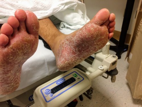
A young male with a history of HIV (untreated for the last year, with unknown CD4 count), and syphilis (reportedly treated with an intramuscular injection 1 year ago), presents with 4 months of a painful rash on the palms and soles and diplopia. Examination revealed the rash pictured below, ocular examination with minimal papilledema and anterior chamber inflammation.

Labs were unremarkable. CSF sampling was notable for 34 WBC’s with lymphocyte predominance (92%), and elevated protein (56mg/dL). The patient was admitted for syphilis with presumed neurosyphilis. Serum RPR titer was elevated at 1:64,  FTA-ABS and CSF VDRL were reactive. The patient was treated with intravenous penicillin and anti-retroviral therapy was reinitiated.

Epidemiology1

  • Transmission
    • Sexual contact (estimated transmission probability 60% per partner)
    • Trans-placental
  • Race/Sex
    •  African-American, Hispanic
    • Male > Female
    • Male (primary syphilis), female (secondary syphilis) – lesion visibility
    • Urban > rural

Natural History1

Stage Signs/Symptoms Incubation Period
Primary Chancre, reginal lymphadenopathy 3 weeks
Secondary Rash, fever, malaise, generalized lymphadenopathy, mucous membrane lesions, condyloma lata, headache, meningitis 2-12 weeks
Latent Asymptomatic Early (<1 year)

Late (>1 year)

Tertiary Cardiovascular:

Aortic aneurysm, aortic insufficiency, coronary artery ostial stenosis

<2 years
CNS:
Acute syphilitic meningitis: headache, confusion, meningeal irritation <2 years
Meningovascular: cranial nerve palsy 5-7 years
General paresis: headache, vertigo, personality changes, vascular event 5-7 years
Tabes dorsalis: dementia, ataxia, Argyl-Robertson, [arrow-down] proprioception 10-20 years
Gumma:

Local tissue destruction

1-46 years

Diagnosis1

  • Serologic
    • Non-treponemal (screening)
      • RPR, VDRL
      • Limitations:  sensitivity, false positive (age, pregnancy, drugs, malignancy, autoimmune, viral infections)
    • Treponemal (confirmatory)
      • FTA-ABS
    • Neurosyphilis
      • Indications for CSF sampling: neurologic/ophthalmologic symptoms, tertiary syphilis (aortitis, gumma, iritis), HIV coinfection with elevated RPR titer (> 1:32)
      • CSF: leukocytosis (predominantly lymphocytes),  protein
      • CSF VDRL reactive
      • Negative CSF FTA-ABS may rule out neurosyphilis

Syphilis in HIV-infected Individuals2

  • Primary: larger and more lesion, multiple ulcers
  • Secondary: genital ulcers more common, higher RPR/VDRL titers
  • Tertiary: possibly more rapid progression to neurosyphilis

References

  1. Singh AE, Romanowski B. Syphilis: review with emphasis on clinical, epidemiologic, and some biologic features. Clin Microbiol Rev. 1999;12(2):187-209.
  2. French P. Syphilis. BMJ. 2007;334(7585):143-147. doi:10.1136/bmj.39085.518148.BE.

Epiglottitis

Brief H&P:

30 year-old male with no significant medical history presenting with 24 hours of progressively worsening throat pain, difficulty swallowing and voice hoarseness. He reports subjective fevers and chills.
Vital signs notable for Tmax 38.4°C. On physical examination, the patient was sitting upright, unable to swallow secretions with faint inspiratory stridor and dysphonia (though he was able to speak in full sentences and without apparent respiratory distress). Oropharyngeal examination showed minimal right parapharyngeal edema without uvular or palatal deviation and there was exquisite right lateral neck tenderness to palpation.

Labs

  • CBC: 24.2/14.4/43.4/202
  • Wound culture: MSSA
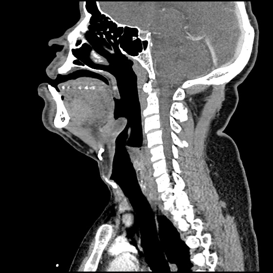
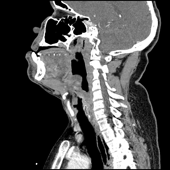
IM-0001-0060
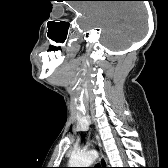
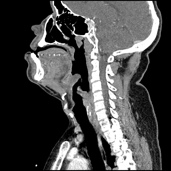
IM-0001-0062
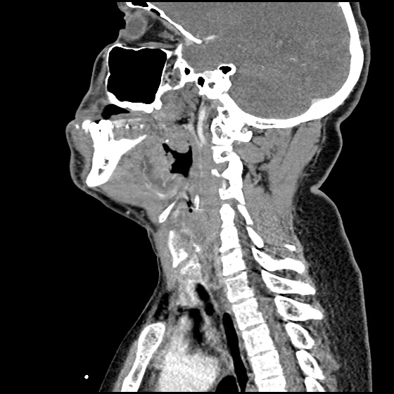
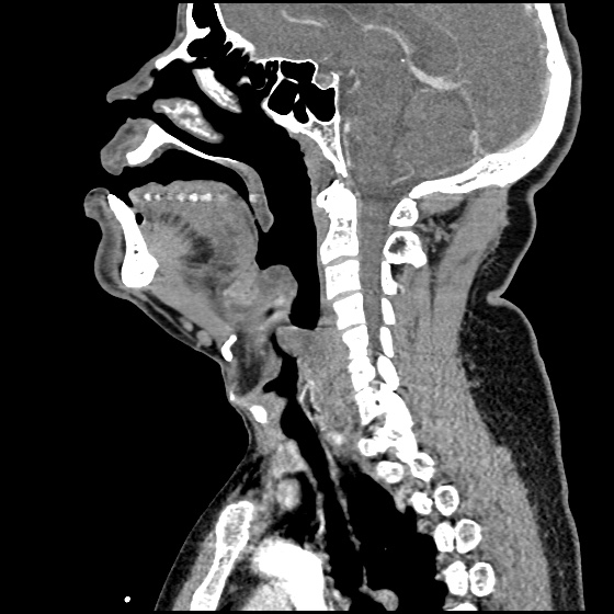
IM-0001-0064
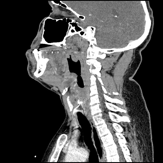
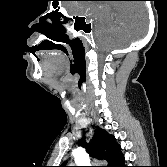
IM-0001-0066
IM-0001-0068
IM-0001-0070
IM-0001-0072
IM-0001-0074
IM-0001-0076
IM-0001-0078
IM-0001-0080

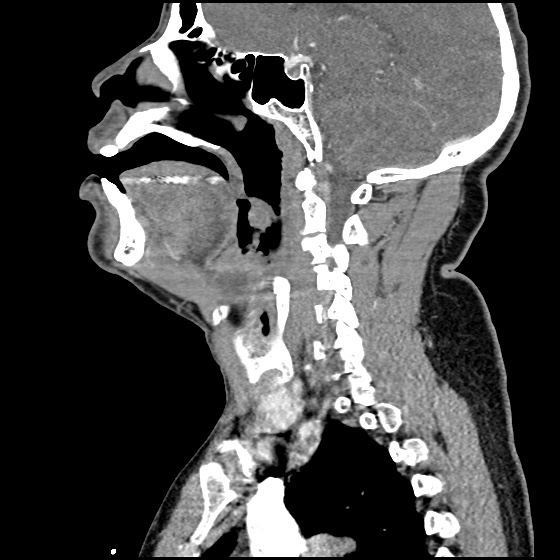
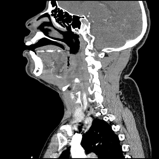
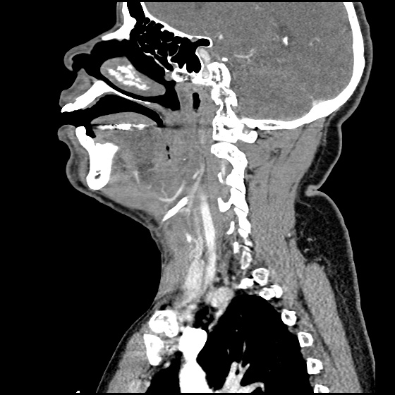
CT Neck/Soft Tissue with Contrast

Edema of the oropharynx/hypopharynx, consistent with epiglottitis and early abscess formation.

ED/Hospital Course

The patient acutely decompensated prior to fiberoptic laryngoscopy and proceeded emergently to the operating room for controlled intubation. The operative report described the following findings: “The patient had diffuse edema of the posterior oropharyngeal wall. The epiglottis was severely thickened, Omega shaped, soft to palpation and with moderate pressure, it appeared to come to a head and pus was expressed from the lingual side of the epiglottis.” The patient was extubated on hospital day three and discharged soon thereafter, he was doing well on follow-up.

Evaluation of Sore Throat – Applied

Evaluation of Sore Throat - Applied

Spinal Epidural Abscess

Case Presentation

HPI:

34M with no PMH presenting with joint pain and rash. The patient was in his usual state of good health until 1 week prior to presentation, noting bilateral shoulder pain. Diagnosed with musculoskeletal process at outside hospital and discharged with analgesics. Presented with partner due to worsening pain involving multiple joints, a non-painful, non-pruritic rash on bilateral lower extremities, and apparent confusion/hallucinations. Social history was non-contributory, no recent procedures or instrumentation.

Objectively, vital signs were notable for tachycardia and elevated core temperature. The patient was ill-appearing, disoriented and unable to provide detailed history. Skin examination was notable for non-blanching petechial rash with areas of confluence most dense in anterior distal lower extremities, rarer proximally, and otherwise without palm/sole involvement. Mucous membranes were dry, neck was supple. There was tenderness to palpation and manipulation of bilateral shoulders. No back tenderness to palpation or percussion was identified. Neurological examination notable for disorientation, intact cranial nerve function, pain-limited weakness in bilateral upper extremities particularly shoulder abduction, and 4/5 hip flexion, knee flexion/extension in bilateral lower extremities.

Labs:

  • CBC: 34.0/11.8/35.7/216
  • Differential: 31 bands
  • INR: 1.94
  • BMP: 131/5.3/102/17/88/2.55/215
  • LFT: AST 93, ALT 57, AP 237, TB 2.9, DB 1.9, Alb 1.4
  • Lactate: 3.3
  • UA: 47WBC, 5RBC
  • Utox: Negative
  • ESR: 83, CRP: 11.9
  • HIV: Nonreactive

Radiology

  • CT head: Negative
  • CXR: Negative
  • XR Shoulder: Negative
  • CT Chest/Abdomen/Pelvis non-contrast: Mild bilateral hydrouereter/hyndronephrosis, L4-L5 grade 2 anterolisthesis.
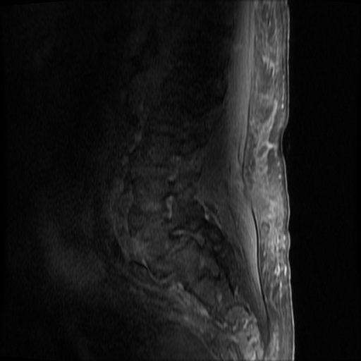
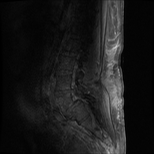
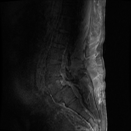
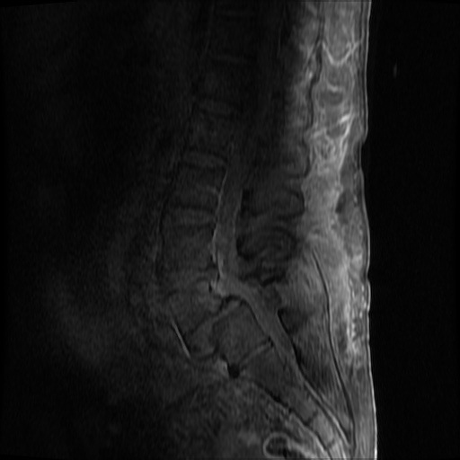
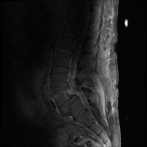
IM-0001-0005
IM-0001-0006
IM-0001-0007
IM-0001-0008
IM-0001-0009
IM-0001-0010
IM-0001-0011
IM-0001-0012
IM-0001-0013
IM-0001-0014

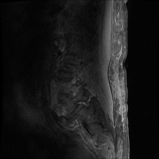
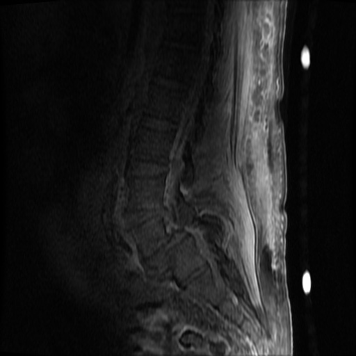
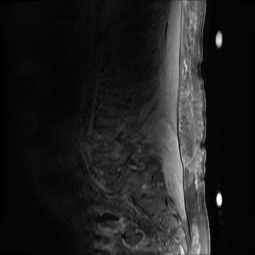
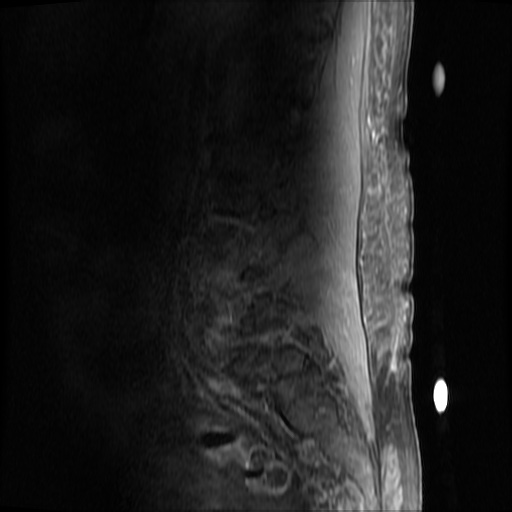
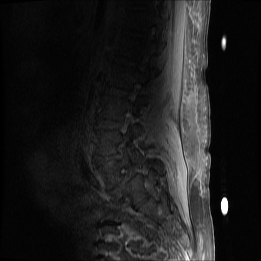
MRI Lumbar Spine w/contrast

Diffuse epidural enhancement posterior to the L4 and L5 vertebral bodies compressing the thecal sac and resulting in moderate severe spinal canal stenosis. Rim enhancement of the 1.5 cm left paraspinal fluid that may be within the L4 tendon sheath or simply paraspinal abscess.

Assessment/Plan:

Severe sepsis with end-organ dysfunction, unclear source (urinary tract involvement unlikely to account for severity of illness). Covered empirically with broad-spectrum anti-microbials including CNS infection given component of encephalitis. Admitted to the intensive care unit.

Hospital Course:

On hospital day 1, the patient underwent non-contrast MRI of the entire neuraxis with findings concerning for L4-L5 and L5-S1 epidural and paraspinal infection resulting in moderate-severe spinal canal stenosis. Blood and urine cultures grew gram-positive cocci in clusters.

On hospital day 2, the patient became increasingly somnolent. Repeat examination by consulting neurology service was concerning for evidence of meningeal irritation. Cultures speciated as methicillin-sensitive staphylococcus aureus and oxacillin was added. MRI was repeated with gadolinium, findings concerning for L4 epidural vs. paraspinal abscess.

On hospital day 3, the patient’s mental status continued to worsen and he was intubated for airway protection. Neurosurgical intervention was deferred due to deteriorating clinical status. Shoulder synovial fluid aspirate culture positive for MSSA, orthopedic surgery consulted for washout/serial arthrocentesis. TTE performed without evidence of valvular vegetation.

On hospital day 4, additional warm joints were aspirated by orthopedic surgery including knee, bilateral ankles, and shoulder each of which ultimately grew MSSA.

On hospital day 6, the patient underwent OR washout of affected joints with intraoperative findings of purulent fluid. TEE performed without evidence of valvular vegetation. The following day, underwent fluoroscopically-guided lumbar puncture, CSF studies inconclusive. Rifampin added for high-grade bacteremia with multiple seeded sites.

The patient was extubated on hospital day 9 and transferred out of the intensive care unit. The following day, he became increasingly tachypneic with evidence of volume overload on examination and was intubated and returned to the intensive care unit. Sustained PEA arrest post-intubation with ROSC, possibly secondary to pneumothorax vs. hypoxia from extensive mucous plugging. Required increasing vasopressor support over the subsequent 12 hours, emergent CVVHD for worsening academia and hypervolemia. The patient sustained another arrest and ultimately expired.

The final impression was that of high-grade bacteremia from unclear source (vague history of proximate hand laceration/infection) with resultant seeding of epidural/paraspinal space, urinary tract, multiple joints, and likely CNS/meninges. Review of abdominal ultrasonography with evidence of cirrhosis, suggesting that some component of initial hepatic synthetic dysfunction may have been chronic and this may have increased the patient’s risk for disseminated infection and SEA. Neurosurgical intervention was not pursued due to unstable clinical status and as the patient’s neurological findings were not consistent with the location of the identified lesion.

Spinal Epidural Abscess (SEA)1

Risk factors:

  • Immunocompromise: diabetes, cirrhosis, CKD, HIV/AIDS
  • Anatomic: DJD, trauma, prior surgery
  • Introduction: IVDA, epidural anesthesia, tattoo

Organism:

  • S. aureus, 2/3
  • S. epidermidis (associated with device, instrumentation)
  • E. coli (urine spread)
  • P. aeruginosa (IVDA)
  • Rare: anaerobes, mycobacteria, fungi

Staging:

  1. Back pain at affected site
  2. Nerve root pain from affected level
  3. Weakness, sensory deficit, bladder/bowel dysfunction
  4. Paralysis

Clinical features:

  • Back pain (75%)
  • Fever (50%)
  • Neuro deficit (33%)

Diagnosis:

  • Labs: Leukocytosis, ESR/CRP, blood cultures
  • Imaging: MRI with gadolinium, 90% sensitivity
  • Clinical findings and laboratory studies are insensitive and non-specific, in one study, approximately ½ of patients had >2 visits.

Prevalence of abnormal physical findings 2

Finding Prevalence
Fever (T>38°C) 19-32%
Focal spinal TTP 52-62%
Diffuse spinal TTP 63-65%
Positive SLR 11-13%
Abnormal sensation 17-27%
Weakness 29-40%
Abnormal reflexes 8-17%
Abnormal rectal tone 5-10%
Saddle anesthesia 2%

Clinical Decision Guideline 3

Spinal Epidural Abscess Clinical Decision Guideline

Management:

  • Neurosurgical evacuation/fusion
  • Antibiotics (vancomycin, oxacillin, cefepime)
  • Neurosurgical intervention may not result in neurological recovery if symptoms present for > 24-36 hours and may be impractical in the setting of panspinal infection.

References:

  1. Darouiche RO. Spinal epidural abscess. N Engl J Med. 2006;355(19):2012–2020. doi:10.1056/NEJMra055111.
  2. Davis DP, Wold RM, Patel RJ, et al. The clinical presentation and impact of diagnostic delays on emergency department patients with spinal epidural abscess. J Emerg Med. 2004;26(3):285–291. doi:10.1016/j.jemermed.2003.11.013.
  3. Davis DP, Salazar A, Chan TC, Vilke GM. Prospective evaluation of a clinical decision guideline to diagnose spinal epidural abscess in patients who present to the emergency department with spine pain. J Neurosurg Spine. 2011;14(6):765–770. doi:10.3171/2011.1.SPINE1091.
  4. WikEM: Epidural abscess (spinal)
Fever

Causes of Fever

Causes of Fever

Key Features

  • Morbidity and mortality increase with age and comborbidities
  • Most common sources in elderly: respiratory, genitourinary, skin/soft-tissue
  • Atypical presentations: functional decline, altered mental status

Immediate Evaluation and Management

  • Critical Findings

    • Altered mental status
    • Respiratory distress
    • Hemodynamic instability
  • Critical Interventions

    • Airway management, supplemental O2
    • Cardiac monitoring
    • Fluid resuscitation
    • Empiric antibiotics
    • Cooling measures (T>41.0°C)
Pathophysiology of Fever

Production of endogenous or exogenous pyrogens
Increase temperature set point in hypothalamus
Patient experiences chills when core temperature < set point
Vasoconstriction, shivering causes fever
Patient experiences euthermia, though may feel malaise, fatigue
Resolution
Patient experiences sweats until core temperature returns to normal set point

References

  1. Blum, F., & Biros, M. (2013). Fever in the Adult Patient. In Rosen’s Emergency Medicine – Concepts and Clinical Practice (8th ed., Vol. 1, pp. 119-123). Elsevier Health Sciences.

Necrotizing Soft-Tissue Infection (NSTI)

HPI:

40 year-old male with a history of diabetes presents with right foot pain and swelling. His symptoms began 3 days ago with pain on the lateral surface of his right foot, described as aching, non-radiating and exacerbated with walking. Yesterday, he noted more prominent swelling and redness involving 4th and 5th toes. He denies trauma, fevers, and discharge.

PMH:

  • Diabetes mellitus, diagnosed 8yrs ago

PSH:

  • None

FH:

  • Non-contributory

SHx:

  • Lives with wife and 2 children and works an office job.
  • Ten year history of tobacco use, quit 3 years ago.
  • No EtOH or drug abuse.

Meds:

  • Metformin 500mg p.o. b.i.d.
  • Ibuprofen p.r.n. joint pain

Allergies:

NKDA

Physical Exam:

VS: T 101.2 HR 88 RR 14 BP 147/71 O2 100% RA
Gen: Obese male, pleasant and in no acute distress, lying in bed with right foot raised.
HEENT: PERRL, EOMI, dry mucous membranes.
CV: RRR, normal S1/S2, no extra heart sounds, no murmurs.
Lungs: CTAB
Abd: +BS, non-tender.
Ext: Right lower extremity with 8x8cm area of erythema predominantly involving lateral aspect of foot, dorsum of foot and 3-5th digits. There is a shallow, 1x1cm ulcer on the plantar surface of foot near 5th MTP. Area is also notable for ecchymosis and palpable crepitus. There is minimal tenderness to palpation or with active/passive range of motion.
Skin: The remainder of the skin exam is unremarkable.
Neuro: AAOx3.

Labs/Studies:

  • BMP: 134/4.3/104/26/18/1.4/206
  • WBC: 27.3/13.1/40/189 (90% neutrophils)
  • Lactate: 1.2
  • CRP: [pending]

Imaging:

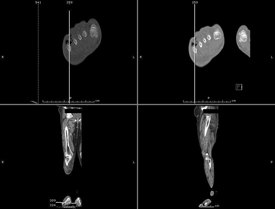
CT Lower Extremity

  1. Calf cellulitis and gas-producing cellulitis in the lateral foot and toes.
  2. Thigh and inguinal lymphadenopathy.
  3. Although gas is seen down to the level of the bone, no definite bony changes are identified to establish a diagnosis of osteomyelitis. Please note that MRI is more sensitive for detection of early osteomyelitis.

Assessment/Plan:

40M with DM and diabetic foot ulcer resulting in a necrotizing soft tissue infection as evidenced by gas on imaging. Recommended surgical debridement and started on broad-spectrum antibiotics including:

  • vancomycin 1g i.v. q.12.h.
  • cefepime 2g i.v. q.8.h.
  • metronidazole 500mg i.v. q.8.h.

The patient underwent amputation of 3-5th digits with good surgical margins and was discharged on post-operative day three in good condition.

Skin and soft-tissue layers and their infections: 1

Skin and soft-tissue layers and their infections

Necrotizing Soft-Tissue Infections (NSTI):2,3,4

Risk Factors

  • IVDA
  • Comorbid conditions
    • DM
    • Obesity
    • Immunosuppression

Physical Exam

  • Early (non-specific)
    • Swelling
    • Erythema
    • Pain
  • Late (non-sensitive)
    • Tense edema outside affected skin perimeter
    • Disproportionate pain
    • Ecchymosis
    • Bullae
    • Crepitus
    • Systemic signs (fever, tachycardia, hypotension)

Treatment

  • Surgical debridement
  • Antimicrobials
    • Carbapenem, combination B-lactam B-lactamase
    • Vancomycin, linezolid (MRSA coverage)
    • Clindamycin (inhibit protein synthesis)
  • Supportive therapy

LRINEC score 5

Name Value Score
CRP ≥150 4
WBC 15-25
>25
1
2
Hb 11-13.5
<11
1
2
Na <135 2
Creatinine >1.6 2
Glucose >180 1

<5 Low risk, 6-7 Intermediate risk, >8 High risk

References:

  1. Morchi, R. (2/18/14). Emergency Medicine Procedures Cadaver Lab. Clinical Clerkship at UCLA. Los Angeles, CA.
  2. Goldstein, E. J. C., Anaya, D. A., & Dellinger, E. P. (2007). Necrotizing Soft-Tissue Infection: Diagnosis and Management. Clinical infectious diseases, 44(5), 705–710. doi:10.1086/511638
  3. Headley, A. J. (2003). Necrotizing soft tissue infections: a primary care review. American family physician, 68(2), 323–328.
  4. McHenry, C. R., Piotrowski, J. J., Petrinic, D., & Malangoni, M. A. (1995). Determinants of mortality for necrotizing soft-tissue infections. Annals of surgery, 221(5), 558–63.
  5. Wong, C.-H., Khin, L.-W., Heng, K.-S., Tan, K.-C., & Low, C.-O. (2004). The LRINEC (Laboratory Risk Indicator for Necrotizing Fasciitis) score: A tool for distinguishing necrotizing fasciitis from other soft tissue infections. Critical Care Medicine, 32(7), 1535–1541. doi:10.1097/01.CCM.0000129486.35458.7D

Cervical Lymphadenopathy

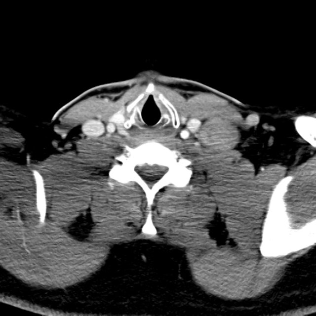
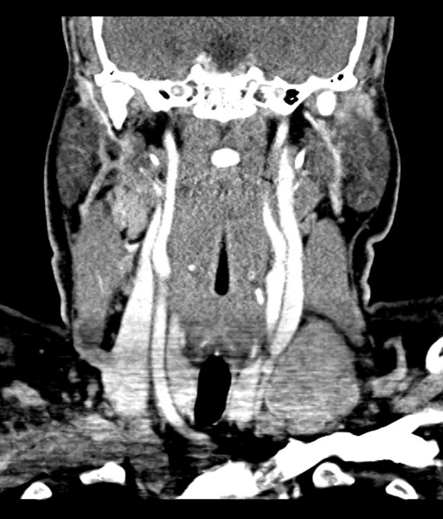
32 year-old male, previously healthy, with slowly-progressive right and left cervical lymphadenopathy over the past three years. He first noted the development of a mass on the lateral neck below the ear three years ago. This mass was non-tender and remained stable at approximately the size of a marble for nearly one year. He later developed more and larger masses, but never experienced any constitutional symptoms like fevers, night sweats, fatigue or weight loss. Examination reveals a healthy, well-nourished male with multiple, hard, mobile, non-tender right posterior cervical and submandibular lymph nodes and a large left supraclavicular lymph node, protruding above the clavicle and measuring ~4x3cm.

Imaging:

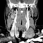
Cervical Lymphadenopathy - Axial

Cervical Lymphadenopathy - Axial

- Left supra-clavicular lymph node, 4.5 x 2.9 cm
- Right posterior submandibular lymph node, 3.5 x 2.7 cm
- Multiple other small cervical lymph nodes, more on the right

Cervical Lymphadenopathy - Coronal

Cervical Lymphadenopathy - Coronal

- Left supra-clavicular lymph node, 4.5 x 2.9 cm
- Right posterior submandibular lymph node, 3.5 x 2.7 cm
- Multiple other small cervical lymph nodes, more on the right

He has undergone several diagnostic procedures during this time including aspirations and core biopsies with inconclusive pathology and he is currently admitted for an excisional biopsy of the supraclavicular lymph node. Infectious and rheumatologic workup has remained negative including: HIV, bartonella, Quantiferon TB Gold, cocci, histo, toxo, CMV, EBV, ANA, RF, ACE.

The patient tolerated the excisional biopsy without complication and was discharged. Preliminary pathology suggests Castleman’s disease.

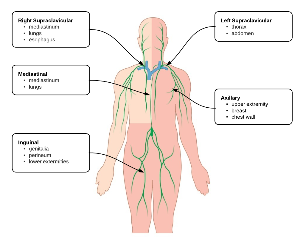
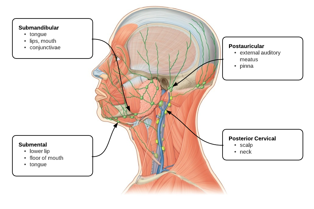
Lymph nodes and their drainage: 1,2

Lymph Nodes of the Body
Lymph Nodes of the Head and Neck

Evaluation of Lymphadenopathy: 2,3,4

Evaluation of Lymphadenopathy

References:

  1. Ferrer R. Lymphadenopathy: differential diagnosis and evaluation. Am Fam Physician. 1998;58(6):1313–1320.
  2. Henry PH, Longo DL. Chapter 59. Enlargement of Lymph Nodes and Spleen. In: Longo DL, Fauci AS, Kasper DL, Hauser SL, Jameson J, Loscalzo J. eds. Harrison’s Principles of Internal Medicine, 18e. New York: McGraw-Hill; 2012.
  3. Armitage JO. Chapter 171. Approach to the Patient With Lymphadenopathy and Splenomegaly In: Cecil, Russell L., Lee Goldman, and Andrew I. Schafer. Goldman’s Cecil medicine. Philadelphia: Elsevier/Saunders, 2011.
  4. Motyckova G, Steensma DP. Why does my patient have lymphadenopathy or splenomegaly? Hematol. Oncol. Clin. North Am. 2012;26(2):395–408– ix. doi:10.1016/j.hoc.2012.02.005.

Hepatic Abscess

HPI:

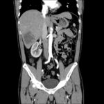
42M with 1.5 weeks of fevers. Initially presented to ER 1wk ago and treated for possible otitis media, however follow-up ENT appointment showed no evidence of OM on exam. Fevers persisted and he developed headaches and went to urgent care where a CT head and LP were negative. A mild elevation of serum transaminases was noted and the following CT abdomen/pelvis was obtained. He denied GI symptoms.

Imaging:

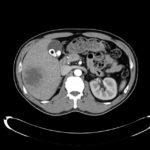
Hepatic Abscess - Axial

Hepatic Abscess - Axial

- 7.4 cm cystic lesion in the inferior right lobe of the liver most consistent in appearance with hepatic abscess.
- Multiple calcified gallstones with a 10 mm gallstone in the neck of the gallbladder or possibly in the cystic duct.

Hepatic Abscess - Coronal

Hepatic Abscess - Coronal

- 7.4 cm cystic lesion in the inferior right lobe of the liver most consistent in appearance with hepatic abscess.
- Multiple calcified gallstones with a 10 mm gallstone in the neck of the gallbladder or possibly in the cystic duct.

Assessment & Plan:

# Liver abscess: likely pyogenic s/p CT-guided drainage with 60cc purulent fluid removed. Gram stain showed GNR and WBC’s, culture grew Klebsiella pneumonia. Treated with ceftriaxone 2g IV q24h, metronidazole 500mg PO TID.

Differential Diagnosis of Hepatic Abscess:1

Differential Diagnosis of Hepatic Abscess

References:

  1. Krige J, Beckingham IJ. Liver abscesses and hydatid disease. BMJ. 2001;322(7285):537–540.

Pediatric Fever

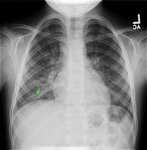
CXR with infiltrates

ID:

5yo girl brought to the pediatric emergency department by her mother due to 3 days of fever.

HPI:

The patient’s fever was first noted 3 days ago, measured at home to 103°F. It is associated with a moist cough, vomiting, and decreased PO intake. Her mother reports that she appears lethargic and has been urinating less frequently. The patient denies headache, changes in vision, burning with urination, or ear pain. No known sick contacts, attends day care.

PMH (Birth History):

No significant medical/surgical history. Ex-term born NSVD with no complications.

PE:

  • VS: 95/65mmHg, 100bpm, 102.6°, 22/min
  • General: Well-appearing, mildly irritated but consolable
  • HEENT: NC/AT, PERRL, oropharynx without erythema, no cervical LAD
  • CV: RRR, no M/G/R
  • Lungs: No evidence of respiratory distress (retractions, flaring), faint crackles over right inferior lung fields
  • Abd: +BS, soft, non-distended, TTP RLQ > LLQ, no rebound/guarding
  • Back: No CVAT

Labs/Imaging:

  • CXR PA/Lateral: RML/RLL infiltrate

Assessment:

5yo with 3 days persistent high fever and cough. These symptoms along with examination findings of crackles warranted further imaging (CXR) which revealed infiltrate in the right inferior lung field. The patient appeared clinically stable and was tolerating PO intake in the ED and was discharged home with azithromycin 5mg/kg/dose (with loading dose), clinic follow-up and strict return precautions.

Evaluation and Management of Pediatric Fever

Algorithm for the Evaluation of Pediatric Fever

A System for Pediatric Fever:

Pathophysiology:

Pathophysiology

Diagnosis:

  • <3mo: 38.0°C, 100.4°F
  • 3-36mo: 39.0°C, 102.2°F
  • Rectal > oral > axillary

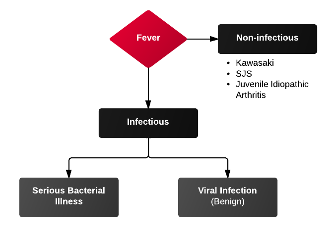
Differential Diagnosis of Pediatric Fever:

Causes Of Fever

Serious Bacterial Illness (SBI):

1) UTI and pyelonephritis

  • Most common cause of SBI
  • Accounts for 3-8% of uncharacterized fevers
  • Female > male, uncircumcised > circumcised
  • Consider BCx, CSF evaluation as 5-10% bacteremic at presentation
  • Urinalysis: LE 75% specificity, Nitrites 97% specificity

2) Pneumonia and sinusitis

  • Sinusitis uncommon <3yo (sinuses unformed)
  • PNA diagnosed with CXR, obtain if findings of respiratory distress (grunting, tachypnea, hypoxemia) or rales on exam

3) Meningitis

  • Diagnose with LP
  • Meningitis suggested if:
    • ANC > 1,000
    • Protein > 80
    • Seizure (particularly complex febrile seizure)

Diagnosis by Age Group:

<3mo

  • Physical exam findings:
    • Tachypnea, hypoxemia → LRT infection
    • Irritability, inconsolability, bulging anterior fontanelle → meningitis
    • Vomiting/diarrhea → non-specific, GE, AOM, UTI, meningitis
  • History
    • Recent immunization: increased risk of SBI (usually UTI) 24-72h after immunization
    • Confirmed bronchiolitis (viral): enterovirus/parainfluenza associated with SBI

3-36mo

  • Physical exam findings:
    • Viral (URTI, GE) → vomiting, diarrhea, rhinorrhea, cough, rash; still playful and responsive
    • UTI → fever, foul-smelling urine, crying when urinating
    • Meningitis → irritability with handling, vomiting, bulging anterior fontanelle, complex febrile seizures

>36mo

  • Physical exam findings: presentation more adult-like
  • Watch for:
    • Group A Streptococcal pharyngitis
    • Infectious mononulceosis
    • Kawasaki: high fever (>5d), strawberry tongue, conjunctivitis, desquamating rash on palms/soles

External Links