Thyroid Emergencies

Hyperthyroidism

Symptoms

Constitutional Weight loss, heat intolerance, perspiration
Cardiopulmonary Palpitations, chest pain, dyspnea
Neuropsychiatric Tremor, anxiety, double vision, muscle weakness
Neck Fullness, dysphagia, dysphonia
Musculoskeletal Extremity swelling
Reproductive Irregular menses, decreased libido, gynecomastia

Signs

Vital signs Tachycardia, widened pulse pressure, fever
Cardiovascular Hyperdynamic precordium, CHF, atrial fibrillation, systolic flow murmur
Ophthalmologic Widened palpebral fissure, periorbital edema, proptosis, diplopia, restricted superior gaze
Neurologic Tremor, hyperreflexia, proximal muscle weakness
Dermatologic Palmar erythema, hyperpigmented plaques or non-pitting edema of tibia
Neck Enlarged or nodular thyroid

Causes

  • Grave disease
    • Mechanism: thyroid-stimulating antibodies
    • Female > Male (10x)
    • Findings: ophthalmopathy (lid lag), infiltrative dermopathy (pretibial)
  • Toxic adenoma, toxic multinodular goiter
    • Mechanism: Excess thyroid hormone production
  • Thyroiditis
    • Mechanism: inflammation results in increased thyroid hormone release, typically followed by depletion and TSH suppression resulting in hypothyroidism
    • Signs/symptoms: tachycardia, weight loss, irritability, sweating, anxiety, heat intolerance
    • Subacute thyroiditis
      • Post-viral
      • Symptoms: hoarseness, dysphagia, painful thyroid
    • Hashimoto
      • Typically hypothyroidism
    • Drug-induced: Lithium, amiodarone
    • Trauma: surgical, direct

Thyroid Storm

  • Essentially an exaggeration of thyrotoxicosis featuring marked hyperthermia (104-106°F), tachycardia (HR > 140bpm), and altered mental status (agitation, delirium, coma).
  • Precipitants
    • Medical: Sepsis, MI, CVA, CHF, PE, visceral ischemia
    • Trauma: Surgery, blunt, penetrating
    • Endocrine: DKA, HHS, hypoglycemia
    • Drugs: Iodine, amiodarone, inhaled anesthetics
    • Pregnancy: post-partum, hyperemesis gravidarum
  • Scoring (Burch, Wartofsky)
  • Management
    • Supportive measures
      • Volume resuscitation (with MVI, Thiamine) and cooling
      • Benzodiazepines for agitation
    • Beta-blockade
      • Propranolol 60-80mg PO q4h
      • Propranolol 0.5-1.0mg IV, repeat q15min then 1-2mg q3h
      • Esmolol continuous infusion
    • MTZ/PTU 1-hour prior to iodine
      • Methimazole 20mg (except pregnancy)
      • Propylthiouracil 600mg (hepatotoxic)
    • Steroids: dexamethasone
    • Iodine
    • Endocrinology consultation

Hypothyroidism

Symptoms

Constitutional Weight gain, cold intolerance, fatigue
Cardiopulmonary Dyspnea, decreased exercise capacity
Neuropsychiatric Impaired concentration and attention
Musculoskeletal Extremity swelling
Gastrointestinal Constipation
Reproductive Irregular menses, erectile dysfunction, decreased libido
Integumentary Coarse hair, dry skin, alopecia, thin nails

Signs

Vital signs Bradycardia, hypothermia
Cardiovascular Prolonged QT, increased ventricular arrhythmia, accelerated CAD, diastolic heart failure, peripheral edema
Neurologic Lethargy, slowed speech, agitation, seizures, ataxia/dysmetria, mononeuropathy, delayed relaxation of reflexes
Musculoskeletal Proximal myopathy, pseudohypertrophy, polyarthralgia
Gastrointestinal Ileus

Causes

  • Hashimoto: auto-antiboids
  • Thyroidectomy
  • Radiation, radioactive iodine ablation

Myxedema Coma

  • Precipitants
    • Critical illness: sepsis (especially PNA), CVA, MI, CHF, trauma, burns
    • Endocrine: DKA, hypoglycemia
    • Drugs: amiodarone, lithium, phenytoin, rifampin, medication non-adherence
    • Environmental: cold exposure
  • Recognition
    • History: hypothyroidism, thyroidectomy scar and acute precipitating illness
    • Hypothermia: temp <95.9°F (or normal in presence of infection)
    • AMS: lethargy, confusion, coma, agitation, psychosis, seizures
    • Hypotension: refractory to volume resuscitation and pressors
    • Bradypnea: with hypercapnia and hypoxia
    • Skin: non-pitting edema of face and hands
    • Hyponatremia
  • Management
    • Airway protection
    • Fluid resuscitation
    • Thyroid hormone replacement
      • Young, otherwise healthy patients: T3 10ug IV q4h
      • Elderly, cardiac compromise: 300ug IV x1
      • Steroids: dexamethasone 1h prior to thyroid hormone
    • Treat precipitating illness

Interpretation of Thyroid Function Tests

Condition TSH T4
None Normal Normal
Hyperthyroidism Low High
Hypothyroidism High Low
Subclinical hyperthyroidism Low Normal
Subclinical hypothyroidism High Normal
Sick euthyroid Low Low

Acid-Base Disturbances

Method

  • Primary disturbance (acidemia/alkalemia)
  • Primary process (metabolic/respiratory)
  • Presence of mixed disorder
    • Increase PCO2 of 10, increases HCO3 by 1 (acute) or 3 (chronic)
    • Decreased PCO2 of 10, decreases HCO3 by 2 (acute) or 5 (chronic)
    • Increase HCO3 of 1, increases PCO2 by 0.7
    • Decreased HCO3, add 15, result should equal PCO2 and number after decimal of pH
  • Anion gap

Causes

  • Anion Gap
    • Methanol
    • Uremia
    • DKA/AKA
    • Paraldehyde, propylene glycol
    • INH
    • Lactate
    • Ethylene glycol
    • Salicylate
  • Non-Anion Gap
    • Fistulae
    • Ureteral fistulae
    • Saline
    • Diarrhea
    • Carbonic anhydrase inhibitors
    • Spironolactone
    • RTA
  • Metabolic Alkalosis
    • Vomiting
    • Volume depletion
    • Diuretics
    • Steroids
  • Respiratory Acidosis
    • CNS lesion
    • Myopathies
    • Chest wall abnormalities
    • Obstructive lung disease
  • Respiratory Alkalosis
    • Anxiety
    • Fever
    • Hyperthyroidism
    • Hypoxia
    • Sympathomimetic

See Also

Dermatologic Emergencies

Urticaria/Anaphylaxis

  • Appearance: diffuse maculopapular, edematous plaques
  • Symptoms: known trigger, transient, pruritic
  • Management: remove trigger, epinephrine, glucagon

EM/SJS/TEN

  • EM
    • Causes: drugs, HSV
    • Appearance: target lesions, symmetric, palm/sole involvement
    • Management: remove offending agent, supportive care
  • SJS (<10% TBSA)
    • Cause: drugs
    • Appearance: >2 mucous membranes
    • Findings: +Nikolsky
    • Symptoms: flu-like
    • Management: burn center, dermatology consult
  • TEN (>30% TBSA)
    • Management: IVIG, steroids, burn center, dermatology consult

SSSS

  • Epidemiology: <6yo, older if immunosuppressed
  • Appearance: painful, diffuse erythema, bullae, no MM involvement
    • Stage 1: tender erythroderma
    • Stage 2: exfoliation
    • Stage 3: desquamation
  • Findings: +Nikolsky
  • Management: antibiotics (cephalosporin), no steroids

Rash Mnemonics

Palmar Rash

  • “sifting rocks scabbed Emma’s palms”
  • Syphilis (2°)
  • RMSF
  • Scabies
  • EM

Nikolsky Sign

  • SJS/TEN
  • SSSS
  • PV

Petechiae/purpura

  • RMSF
  • Meningococcemia
  • DIC
  • Endocarditis
  • TTP/HUS

Meningococcemia

  • Epidemiology: <20yo, dorm, military barracks
  • Appearance: diffuse petechiae, palpable purpura
  • Management: antibiotics, steroids

Necrotizing fasciitis

  • Symptoms: POOP, rapid progression
  • Appearance: bullae, crepitus, systemic toxicity
  • Management: surgery, antibiotics

RMSF

  • Symptoms: flu-like
  • History: tick bite, camping/hiking
  • Appearance: wrist/ankle spreading inward (centrifugal), petechiae
  • Diagnosis: clinical, titers
  • Management: doxycycline (increased mortality if not treated)

PV

  • Epidemiology: 40-60yo
  • Pathophysiology: autoantibodies (desmoglein), causes superficial epidermal separation (pemphigus for superficial)
  • Symptoms: painful oral blisters, small bullae
  • Findings: +Nikolsky
  • Management: steroids (methylprednisolone 1g IV), burn center

BP

  • Epidemiology: >70yo
  • Pathophysiology: autoantibodies, deeper dermal layer (pemphigoid for deep)
  • Symptoms: not painful, no oral lesions
  • Findings: large, tense, unruptured bullae
  • Management: steroids

Toxicology

Drugs of Abuse

Synthetic Cannabinoids (Spice, K2)

  • Symptoms: anxiety, paranoia, tachycardia
  • Unique symptoms compared to traditional cannabinoids: psychosis, seizure, diaphoresis

Hallucinogenic amphetamines (ecstasy, MDMA)

  • Increased serotonergic activity
  • Management: supportive care (IVF, cooling for hyperthermia), benzodiazepines

Gamma-hydroxybutyrate (GHB)

  • Symptoms: euphoria, hypersexuality, rapid onset/clearance
  • Signs: bradycardia, bradypnea, coma with rapid awakening
  • Management: intubation for depressed GCS
  • Withdrawal: symptoms and treatment identical to ethanol withdrawal, consider baclofen

Cathinone (bath salts)

  • Symptoms: hallucinations
  • Signs: tachycardia, hypertension, tremor, mydriasis, diaphoresis, hyperthermia, bruxism
  • Management: benzodiazepines, consider paralysis, avoid beta-blockers

Cocaine

  • MOA: increase catecholamines, Na-channel blockade
  • Toxicity: HTN, tachycardia, hyperthermia, rhabdomyolysis, MI, seizure, VT
  • Management: benzodiazepines, cooling, anti-hypertensives (nitrate, CCB, not B-blocker)

Amphetamine

  • Toxicity: HTN, tachycardia, hyperthermia, rhabdomyolysis, intracranial hemorrhage
  • Management: same as cocaine

Benzodiazepines

  • Toxicity: sedation, respiratory depression
  • Management: consider flumazenil 0.2mg IV q1min x1-5

Toxic Alcohols

  • Overview
    • Toxic metabolites produced by alcohol dehydrogenase which can be inhibited by ethanol or fomepizole
    • Fomepizole: 15mg/kg loading dose, 10mg/kg q12h x4 doses then 15mg/kg q12h (stimulates own metabolism); if dialysis, q4h
  • Diagnosis: osmolar gap (>14), 2Na + Glu/18 + BUN/2.8 + EtOH/4.6
  • Treatment
    • ADH inhibition
    • HCO3
    • Hemodialysis
    • Supportive care
    • Hypoglycemia: dextrose

Methanol

  • Component of antifreeze, windshield washer fluid
  • Metabolite formic acid which causes acidosis and blindness
  • Can give folate

Ethylene glycol

  • Component of antifreeze, automobile coolants, de-icing agents
  • Metabolite oxalic acid which precipitates calcium oxalate crystals and causes acute renal failure
  • Can give thiamine (100mg q6h), pyridoxine (500mg q6h), Mg

Isopropanol

  • Component of rubbing alcohol
  • Metabolite acetone which does not cause acidosis

Analgesics

Acetaminophen

  • Metabolism: glucoronidation, CYP450
    • CYP450 pathway produces toxic metabolite when glucoronidation overwhelmed
    • In pediatrics, sulfation process protective
  • Toxic dose: >150mg/kg, >3g/day
  • Injury: liver (centrilobular necrosis), renal, pancreatic
  • Increased risk: induced CYP450 (chronic EtOH, rifampin, anti-epileptics)
  • Nomogram: applicable to single ingestion at 4-hours
  • Labs: PT/INR, LFT, lipase, chemistry
  • Management: NAC
    • PO: 140mg/kg, 70mg/kg q4h
    • IV: 150mg/kg, 50mg/kg over 4h, 100mg/kg over 16h

NSAID

  • Symptoms
    • Acute: GI upset, low risk UGIB
    • Acute massive: acidosis, coma, seizures
    • Chronic: UGIB, nephropathy, agranulocytosis

Aspirin

  • Signs: tachycardia, hyperthermia, tachypnea/hyperpnea
  • Severe: cerebral and pulmonary edema, CNS hypoglycemia
  • Labs: primary respiratory alkalosis with metabolic acidosis
  • Management
    • Hypoglycemia (CNS) treatment
    • Bicarbonate infusion (urine pH > 8)
    • Hemodialysis for pulmonary edema, cerebral edema, renal failure, acidemia, level >100mg/dL (acute) or > 60mg/dL (chronic)

Opioids

  • Symptoms: respiratory depression, miosis
  • Management: naloxone 0.04mg, 0.4mg, 2mg
  • Withdrawal: nausea/vomiting, diarrhea, abdominal pain, piloerection
    • Neonates: seizure, death
  • Complications with specific agents:
    • Meperidine, tramadol: seizures
    • Methadone: QT prolongation

Anesthetics

Lidocaine

  • Mechanism: Na-channel blockade
  • Types:
    • Ester (one “i”): cocaine, procaine, benzocaine
    • Amide (two “i”): lidocaine, bupivacaine
  • Toxicity
    • Dose: 4mg/kg, 7mg/kg with epinephrine
    • CNS: perioral numbness, slurred speech, seizure
    • CV: VT, VF, AV block
    • Methemoglobinemia: methylene blue
  • Treatment
    • Seizure management
    • Bicarbonate for dysrhythmia
    • Intralipid

Anti-cholinergics

Sympathetic Parasympathetic
Mydriasis Miosis
Bronchodilation Bronchospasm/bronchorrhea
Tachycardia Bradycardia
Urinary retention Urinary incontinence
Hyperglycemia Salivation/lacrimation
Diaphoresis Increased GI motility
  • Examples
    • Atropine
    • Anti-histamine
    • TCA
    • Phenothiazines
    • Jimson weed
  • Symptoms
    • Peripheral: mydriasis, anhidrosis, flushing, hyperthermia, ileus, dry mucous membranes, AUR
    • Central: agitation (passive), delirium, coma, seizure
  • Treatment
    • Supportive
    • Benzodiazepines
    • Theoretically physostigmine
      • Avoid in seizure, QRS-widening, reactive airway disease
      • Possible diagnostic use

Drugs causing miosis (COPS)

  • C: cholinergics
  • O: opioids
  • P: phenothiazines
  • S: sedatives

Drugs causing QT-prolongation

  • Examples:
    • Phenothiazines
    • Anti-arrhythmics
    • Butyrophenones (ex. haloperidol)
    • Macrolides
    • Fluoroquinolones
    • Methadone
    • Ondansetron
    • Atypical antipsychotics
  • Treatment
    • Magnesium sulfate 2g IV over 1min
    • Overdrive pacing (transcutaneous, transvenous if not captured)
    • Consider isoproterenol (pharmacologic overdrive)

Serotonin syndrome

  • Cause: exposure to serotonergic agent(s)
  • Symptoms: agitation, mydriasis, tremor/clonus in lower extremities, tachycardia, hyperthermia
  • Management
    • Supportive care (IVF, vasopressors)
    • Cooling measures and paralysis for hyperthermia
    • Benzodiazepines
    • Cyproheptadine 12mg PO/NG
    • Dexmedetomidine infusion

Anti-emetics

Phenothiazines

  • Examples: compazine (prochlorperazine), phenergan (promethazine)
  • MOA: DA-antagonist
  • AE: sedation, dystonia, parkinsonism
  • Toxicity: seizure, VT, hypotension (TCA-like)

5-HT3 antagonists

  • Examples: zofran (ondansetron), granisetron
  • Toxicity: QT-prolongation

Anti-hypertensives

Calcium channel blockers

  • Toxicity: hypotension, bradycardia, AV blockade, hyperglycemia
  • Management
    • Atropine: 0.5mg IV q2-3min
    • Glucagon: 5mg IV q10min x2 (with anti-emetic)
    • IVF, vasopressors (norepinephrine, epinephrine)
    • Calcium: 3g gluconate, 1-3g chloride
    • High-dose insulin: 1 unit/kg, monitor hypoglycemia/hypokalemia
    • Intralipid: 1.5mL/kg bolus then 0.25mL/kg/minute
    • GI decontamination
    • Pacing, IABP, ECMO

Beta blockers

  • Toxicity: similar to CCB, hypoglycemia
  • Management: similar to CCB, calcium ineffective

Digoxin (foxglove, oleander)

  • MOA: inhibits Na/K ATPase, increases intracellular calcium (inotropic)
  • Toxicity
    • CV: bradycardia, hypotension
    • ECG: bidirectional VT, PVC, scooped ST-segment
    • CNS: agitation, psychosis
    • Visual: yellow-green vision, halo
    • Metabolic: hyperkalemia (acute), hypokalemia, hypomagnesemia
  • Treatment
    • GI decontamination
    • Atropine
    • Transcutaneous pacing (avoid transvenous, irritable myocardium)
    • Digibind
    • Avoid calcium

Clonidine

  • Toxicity: bradycardia, hypotension, opioid mimic (miosis, lethargy, respiratory depression)
  • Management: supportive care, stimulation for respiratory depression, atropine

Sodium-channel blockers

  • Drugs
    • TCA
    • Diphenhydramine
    • Procainamide
    • Carbamazepine
  • ECG
    • QRS prolongation
    • Prominent “R” in aVR
    • RAD
  • Treatment
    • Sodium bicarbonate

Anti-hyperglycemics

Sulfonylurea

  • Symptoms: recurrent severe hypoglycemia
  • Management: octreotide 50-75mcg SQ/IM q6h

Other agents that cause hypoglycemia

  • EtOH
  • B-blocker
  • Quinine
  • Salicylate

Environmental

Carbon monoxide

  • Source: combustion (gas heater, indoor barbeque)
  • Toxicity
    • General: influenza-like, multiple proximate affected individuals
    • GI: abdominal pain, nausea
    • CNS: headache, dizziness, confusion, ataxia, seizure
    • CV: palpitations, arrhythmia, hypotension, MI
  • Treatment
    • T½: RA 6h, NRB 1h, 3atm 0.5h
    • Hyperbaric: neuro deficit, syncope, pregnancy, CV toxicity

Cyanide

  • Mechanism: inhibits oxidative phosphorylation
  • Source: structural fire (wool, silk)
  • Symptoms: syncope, seizure, coma, cardiovascular collapse
  • Detection: severe lactic acidosis, “arterialization” of venous blood, “bitter almond” odor
  • Treatment
    • Hydroxycobalamin (Cyanokit): 5g IV, may repeat x1
    • Sodium thiosulfate 12.5g IV

Methemoglobinemia

  • Mechanism: Fe2+ converted to Fe3+, “functional anemia”
  • Source: nitrite (food), topical/local anesthetics, pyridium, dapsone, reglan
  • Detection: normal PaO2, SpO2 85% unresponsive to supplemental oxygen, ABG with co-oximetry
  • Management: methylene blue 1-2mg/kg IV if symptomatic or MetHb >25%
    • Contraindicated in G6PD deficiency, treat with exchange transfusion or HBO

Hydrogen Sulfide

  • Source: industrial, sulfur spring, sewer
  • Detection: “rotten egg” odor
  • Management: remove from source, supportive care

Hydrocarbon

  • Source: huffing canisters
  • Toxicity: VT/VF from myocardial sensitization
  • Management: beta-blockade
  • Complications: harmless if ingested, aspiration leads to ARDS

Hydrofluoric acid

  • Source: rust remover, wheel cleaner, glass etching
  • Symptoms: pain-out-of-proportion, delayed onset
  • Toxicity: Hypocalcemia (QTc prolongation, VT/VF/TdP), hyperkalemia, hypomagnesemia
  • Management: analgesia, topical calcium gluconate gel, intravenous calcium for large BSA involvement

Alkaline ingestion

  • Symptoms: esophageal perforation, delayed stricture

Acid ingestion

  • Symptoms: gastric perforation (rare), delayed gastric outlet obstruction
  • Findings: metabolic acidosis

Botulism

  • Sources
    • Adult: ingested preformed toxin
    • Infants: ingested spores (achlorhydric), in vivo toxin production
    • Wound: black tar heroin
  • Symptoms: dysphagia, ptosis, diplopia, respiratory failure, descending paralysis
    • Infants: constipation, floppy
  • Management: supportive care, intubation
    • Adults: Anti-toxin from CDC or local Department of Health
    • Infants: 100mg/kg IV x 1 dose (BabyBIG)

Heavy Metals

Iron

  • Dose
    • Ferrous sulfate: 20% elemental iron
    • Toxic: >20mg/kg
    • Lethal: >60mg/kg (1 tablet 325mg ferrous sulfate per kilogram)
  • Toxicity: corrosive, anti-coagulant, hepatotoxic
  • Course
    • Stage I: GI effects, emesis with hematemesis
    • Stage II: Quiescent
    • Stage III: Systemic, multi-organ system dysfunction
    • Stage IV: Resolution, gastric scarring and outlet obstruction
  • Workup
    • CBC/BMP
    • LFT
    • Lactate
    • Fe level
    • KUB (if positive consider WBI)
  • Treatment
    • Decontamination: no activated charcoal, consider WBI
    • Deferoxamine: 15mg/kg/hr

Lead

  • Source: paint, batteries
  • Toxicity
    • Acute: headache, encephalopathy, seizure
    • Chronic: malaise, weight loss, arthralgia, anemia (basophilic stippling)
  • Diagnosis: lead level, wrist drop
  • Management: chelation (BAL, EDTA, DMSA) for level >50ug/dL or asymptomatic >70ug/dL

Lithium

  • Source: iatrogenic, drug-drug interaction
  • Symptoms
    • GI: nausea/vomiting, diarrhea
    • CNS: tremor, coma
    • CV: TWI, QT-prolongation
  • Management
    • IVF, encourage renal elimination
    • Hemodialysis

Other Drugs

Disulfuram

  • MOA: aldehyde dehydrogenase inhibitor
  • Symptoms: increased acetaldehyde leads to flushing, headache, nausea/vomiting, tachycardia, hypotension
  • Management: antihistamine, IVF, vasopressors
  • Other agents causing disulfuram-like reaction: metronidazole, INH, sulfonylurea

Isoniazid

  • Toxicity: seizure
  • Management: pyridoxine 5g IV, repeat x1

Theophyline

  • Toxicity: seizure
  • Management
    • Decontamination: AC
    • Seizures: benzodiazepines
    • Tachyarrhythmia (commonly MAT): beta-blockade
    • Hemodialysis: acute > 100mg/L, chronic >30mg/L

Monoamine oxidase inhibitors

  • Toxicity: food/drug interaction
  • Symptoms: tachycardia, hypertension, hyperthermia, agitation
  • Management: cooling, IVF, management of hyper/hypotension

Phenytoin

  • Oral: cerebellar dysfunction (ataxia), CNS depression
  • IV: hypotension (suspension contains propylene glycol)

Nutritional Supplements

  • Fat-soluble vitamins
    • A: benign intracranial hypertension
    • D: hypercalcemia

Envenomations

Snake

  • Crotalid (rattle), elapidae (coral)
  • Symptoms
    • Local reaction: edema, hemorrhagic bullae
    • Systemic: perioral numbness, fasciculations
    • Severe: thrombocytopenia, decreased fibrinogen
  • Management: Crofab 5 vials

Spider

  • Black widow
    • Identification: hourglass on abdomen
    • Symptoms: painful bite, target-appearance, rarely “acute abdomen”
    • Management: analgesia, anti-venom, tetanus
  • Brown recluse
    • Identification: violin shape on head
    • Geography: Southeast, Midwest
    • Symptoms: painless bite, local reaction, delayed healing with eschar
    • Rare: hemolysis, DIC, shock
    • Management: supportive care, antibiotics if superinfected, consider dapsone, tetanus

Scorpion (Centruroides)

  • Geography: Arizona
  • Symptoms
    • Autonomic: HTN, tachycardia, diaphoresis
    • CNS: opsoclonus, slurred speech, dysphagia
  • Management: anti-venom, supportive care, analgesia, tetanus

Marine

  • Ciguatera
    • Source: toxin bioconcentrated in fish
    • Symptoms: gastroenteritis, hot/cold-reversal, “loose teeth” sensation
    • Management: mannitol
  • Scombroid
    • Source: poorly-refrigerated fish, histamine-like
    • Symptoms: flushing trunk/face (distinguish from allergic reaction), gastroenteritis
    • Management: supportive care, IVF, anti-histamine, bronchodilators if indicated
  • Paralytic shellfish poisoning
    • Source: bivalve
    • Symptoms: gastroenteritis, paralysis
    • Management: supportive, intubation
  • Jellyfish and Cnidaria
    • Source: nematocyst
    • Symptoms: burning pain, pruritus
    • Severe: Irakundji syndrome (HTN, pulmonary edema)
    • Management: supportive, analgesia, box jellyfish antidote, consider vinegar
  • Stingray
    • Source: heat-labile toxin
    • Management: affected area in warm water, tetanus, ciprofloxacin (Vibrio)

Mushrooms

  • Amanita: centrilobular necrosis, similar to acetaminophen
  • Gyronatum: similar to INH (seizure and treatment), may cause methemoglobinemia
  • Symptoms: muscarinic (SLUDGE)
    • Early onset generally benign, delayed onset (>6h) suggests more serious course
  • Management: atropine, glycopyrrolate, IVF

Pesticides

  • Organophosphate: irreversible
  • Carbamate: reversible
  • Symptoms: muscarinic (SLUDGE)
  • Treatment: atropine 2-6mg IV double q5min to control secretions, pralidoxime (for organophosphates)

Strychnine

  • Source: rodenticide
  • Symptoms: myoclonus, opisthotonus, agitation
  • Management: benzodiazepines, airway protection, paralysis

Spontaneous Intracranial Hemorrhage

Brief HPI

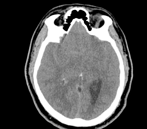
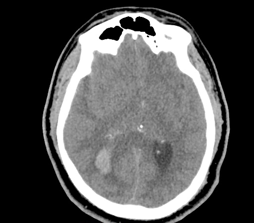
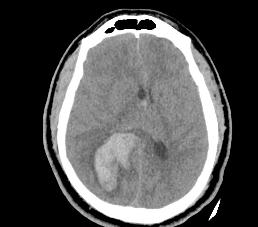
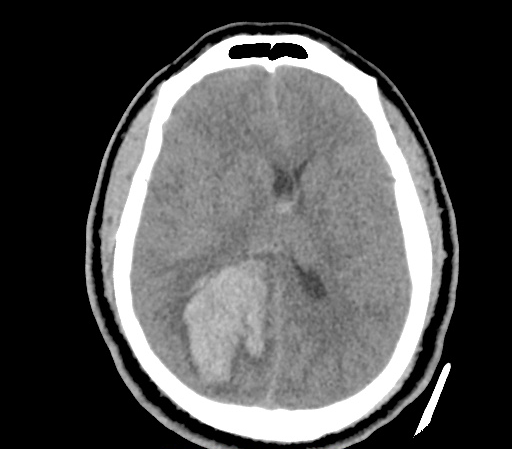
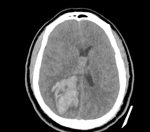
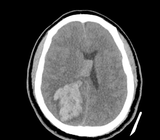
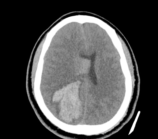
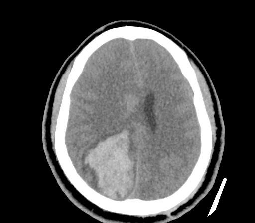
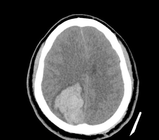
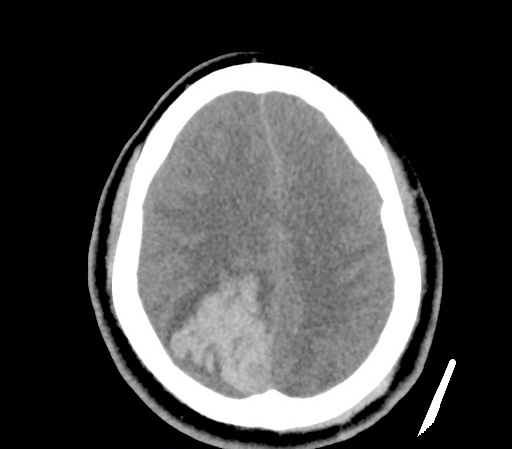
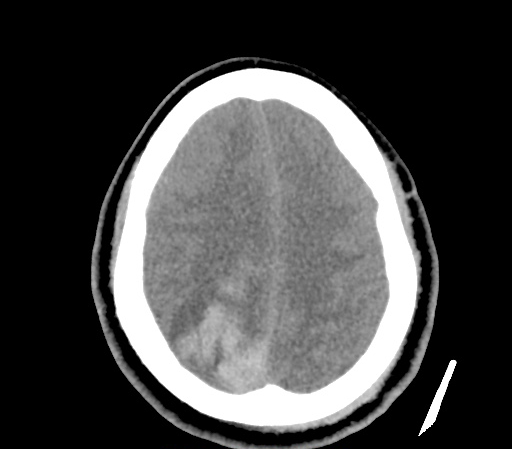
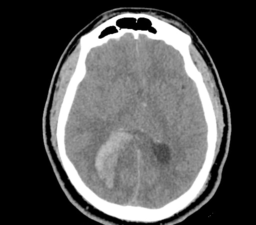
An approximately 40 year-old male with a history of aortic stenosis s/p mechanical aortic valve replacement (on Coumadin) as well as hypertension presented to the emergency department with a chief complaint of severe headache. The patient was in severe distress on arrival and was unable to provide detailed history, he complained of two days of severe left-sided headache while clutching his head and groaning. Examination was notable for sensory localization with directed movements of right hemibody, and no apparent response on the left. He was taken to emergently for CT head non-contrast.

Imaging

ich_0000_Layer-Comp-1
ich_0001_Layer-Comp-2
ich_0002_Layer-Comp-3
ich_0003_Layer-Comp-4
ich_0004_Layer-Comp-5
ich_0005_Layer-Comp-6
ich_0006_Layer-Comp-7
ich_0007_Layer-Comp-8
ich_0008_Layer-Comp-9
ich_0009_Layer-Comp-10
ich_0010_Layer-Comp-11
ich_0011_Layer-Comp-12

CT Head non-contrast

57 mm right posterior parenchymal hemorrhage with intraventricular component. Moderate edema, mass effect and 9 mm of midline shift.

ED Course

Admission INR was 2.9, the patient received 25 units/kg of PCC as well as vitamin K 10mg IV x1. Neurosurgery was consulted and the patient was taken to the operating room for management.

Management of Supratherapeutic INR and Complications of Anti-Coagulation

Management of Supratherapeutic INR

References

  1. Ansell J, Hirsh J, Hylek E, et al. Pharmacology and management of the vitamin K antagonists: American College of Chest Physicians Evidence-Based Clinical Practice Guidelines (8th Edition). Chest 2008; (6 Suppl):160s

Rapid Pediatric Assessment

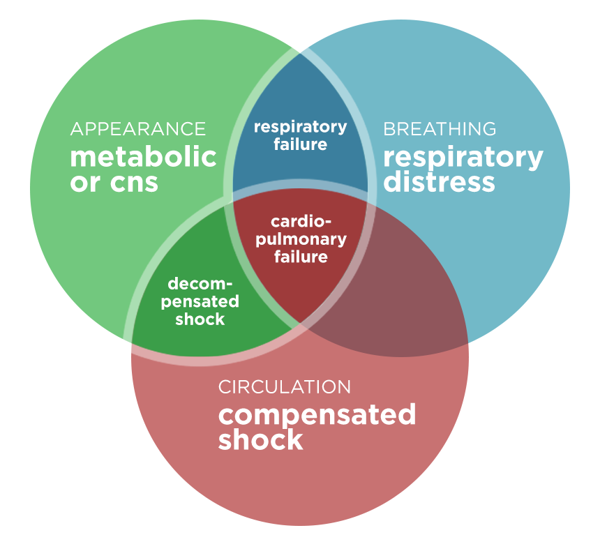
This post presents a tool for the rapid assessment of the cardiopulmonary status and cerebral/metabolic function of critically ill pediatric patients. The purpose is not to establish a diagnosis, rather to identify the particular physiological derangements to prioritize initial interventions. The tool was initially designed as a “triangle” – it has been adapted here (with permission) as a Venn diagram.1

Pediatric Assessment Diagram

Pediatric Assessment Diagram

Assessment of Appearance

  • Tone: Moves spontaneously, resists examination
  • Interactivity: Interacts with environment, reaches for items
  • Consolability: Comforted by caregiver
  • Gaze: Makes eye contact

Assessment of Work of Breathing

  • Airway Sounds: Stridor, grunting, wheezing
  • Position: Tripod
  • Retractions

Assessment of Circulation

  • Pallor
  • Mottling
  • Cyanosis

Management

Impression Interventions
Respiratory distress
  • Position of comfort
  • Oxygen, suction
  • Therapy as appropriate (albuterol, epinephrine, etc)
  • Labs/radiographs as indicated
Respiratory failure
  • Head/airway positioning
  • 100% oxygen
  • Ventilation support (BVM)
  • Advanced airway
Shock (compensated and decompensated)
  • Oxygen
  • Access
  • Fluid resuscitation
  • Specific therapy (antibiotics, surgery)
  • Labs/radiographs as indicated
CNS/Metabolic
  • Pulse oximetry
  • Rapid glucose
  • Labs/radiographs as indicated
Cardiopulmonary Failure
  • Head/airway positioning
  • 100% oxygen
  • Ventilation support (BVM)
  • Chest compressions as needed
  • Specific therapy (defibrillation, epinephrine, amiodarone)
  • Labs/radiographs as indicated

References:

  1. The pediatric assessment triangle: a novel approach for the rapid evaluation of children. Pediatr Emerg Care. 2010;26(4):312-315. doi:10.1097/PEC.0b013e3181d6db37.

Portal Venous Gas

Brief HPI

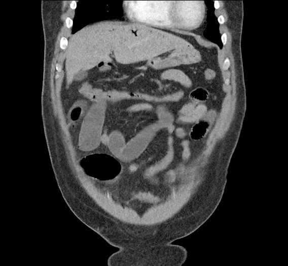
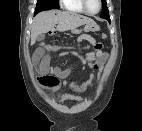
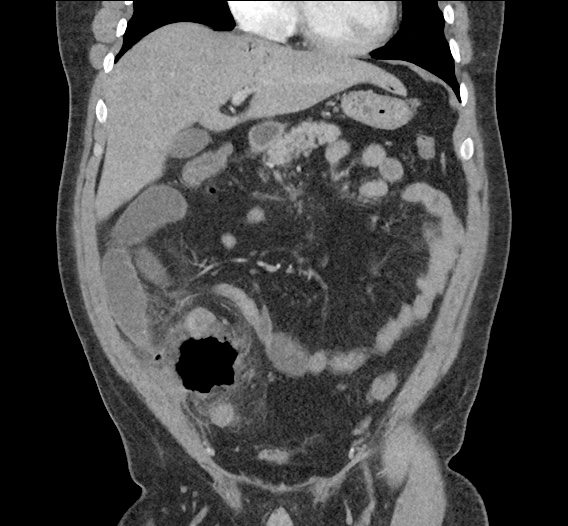
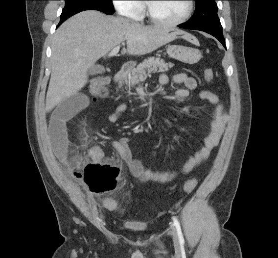
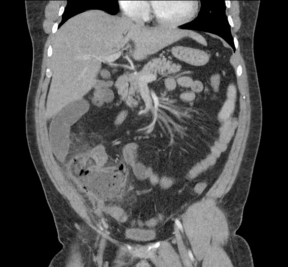
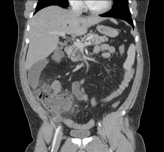
Young male with no significant medical history presenting with progressively worsening right lower quadrant abdominal pain with marked tenderness to palpation and involuntary guarding.

Imaging

portal_venous_gas_0000_Layer-Comp-1
portal_venous_gas_0001_Layer-Comp-2
portal_venous_gas_0002_Layer-Comp-3
portal_venous_gas_0003_Layer-Comp-4
portal_venous_gas_0004_Layer-Comp-5
portal_venous_gas_0005_Layer-Comp-6
portal_venous_gas_0006_Layer-Comp-7
portal_venous_gas_0007_Layer-Comp-8
portal_venous_gas_0008_Layer-Comp-9
portal_venous_gas_0009_Layer-Comp-10
portal_venous_gas_0010_Layer-Comp-11

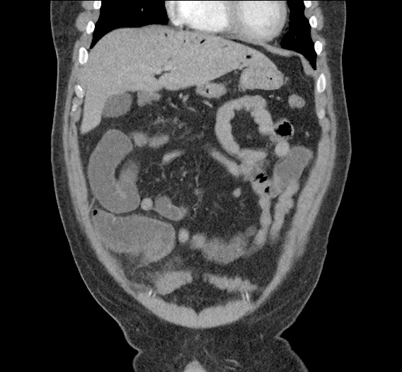
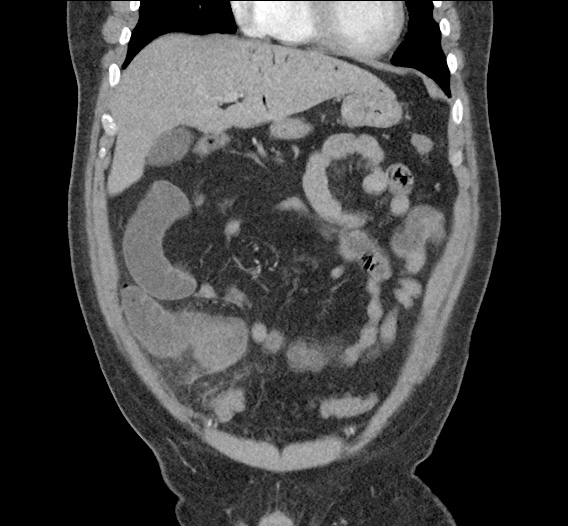
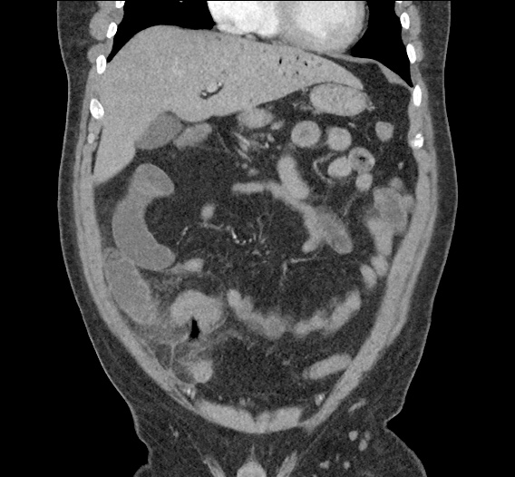
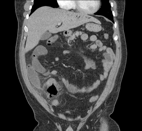
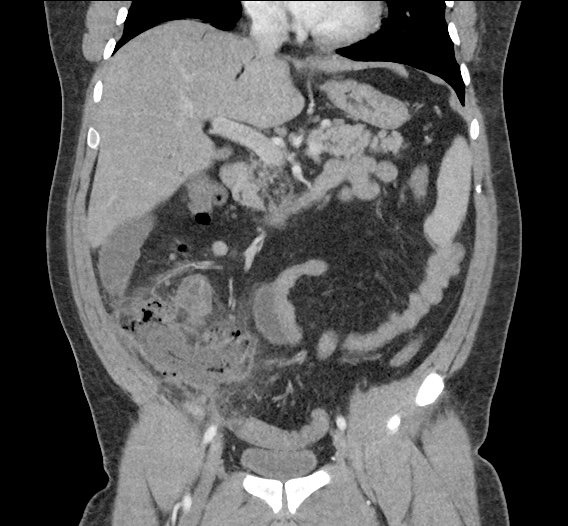
CT Abdomen/Pelvis with Contrast

Inflammatory changes in the right lower quadrant concerning for ruptured appendicitis with approximately 9 cm abscess.
Gas in the liver likely representing portal venous gas which can be seen in the setting of appendicitis vs less likely secondary to bowel ischemia.

Differentiation between Portal Venous Gas and Pneumobilia

Portal venous gas vs. Pneumobilia

References

  1. Rabou Ahmed A and Frank Gaillard. “Pneumobilia.” Radiopaedia. http://radiopaedia.org/articles/pneumobilia.
  2. Morgan Matt A and Donna D’Souza. “Portal venous gas.” Radiopaedia. http://radiopaedia.org/articles/portal-venous-gas
  3. Sebastià C, Quiroga S, Espin E, Boyé R, Alvarez-Castells A, Armengol M. Portomesenteric vein gas: pathologic mechanisms, CT findings, and prognosis. Radiographics. 2000;20(5):1213–24–discussion1224–6. doi:10.1148/radiographics.20.5.g00se011213.
  4. Sherman SC, Tran H. Pneumobilia: benign or life-threatening. J Emerg Med. 2006;30(2):147-153. doi:10.1016/j.jemermed.2005.05.016.

Severe Burns

ED Presentation

34F with no reported medical history BIBA with severe burns after house fire with estimated 70% TBSA involvement. On arrival, the patient was hypoxic, striderous, and unable to provide history. She was intubated for airway protection with some difficulty. Examination revealed deep partial and full-thickness burns to 70% of total body surface area including circumferential burns to bilateral upper extremities and extensive neck and anterior chest involvement. Initial fluid resuscitation and warming measures were instituted. Emergent bedside bronchoscopy revealed copious carbonaceous material throughout with attempts at lavage. Urine output was minimal despite aggressive resuscitation. Critical care transport to local burn facility was arranged where the patient ultimately expired.

Algorithm for the Management of Severe Burns

Algorithm for the Management of Severe Burns

Assessment of Burn Depth

Depth Cause Appearance Sensation
Superficial UV exposure Dry, red
Blanching
Painful
Superficial partial-thickness Scald (splash)
Short flash
Blisters, moist, red
Blanching
Painful to temperature/air
Deep partial-thickness Scald (spill)
Flame, oil, grease
Blisters, waxy dry, white/red
Non-blanching
Pressure
Full-thickness Scald (immersion)
Flame, steam, oil, grease, chemical, electrical
Waxy white, leathery grey, black
Non-blanching
Deep pressure

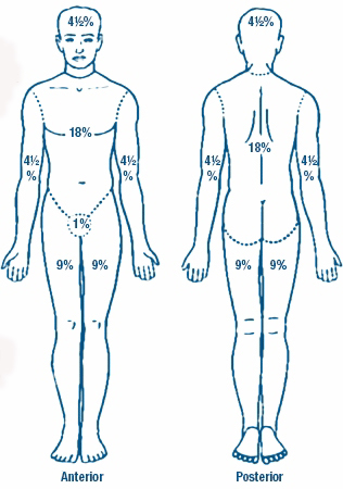
Estimating Burn Surface Area

Burn TBSA

Image from UWHealth.org

  • Trunk: 18% anterior, 18% posterior
  • Lower extremity (each): 9% anterior, 9% posterior
  • Upper extremity (each): 9%
  • Head/neck: 9%
  • Perineum: 1%

Burn Transfer Criteria

  • Partial thickness > 20% TBSA
  • Partial thickness > 10% TBSA for extremes of age (<10 or >50 years-old)
  • Any full-thickness
  • Burns involving face, hands, feet, genitalia, major joints
  • Electrical/chemical
  • Inhalation injury
  • Medical comorbidities impacting management/healing

See Also

References

  1. Monafo WW. Initial management of burns. N Engl J Med. 1996;335(21):1581–1586. doi:10.1056/NEJM199611213352108.
  2. Hettiaratchy S, Papini R. Initial management of a major burn: I–overview. BMJ. 2004;328(7455):1555–1557. doi:10.1136/bmj.328.7455.1555.
  3. Singer AJ, Della-Giustina D. Thermal Burns: Rapid Assessment and Treatment. Emergency Medicine Practice; 2000.
  4. Rice, PL. Emergency care of moderate and severe thermal burns in adults. In: UpToDate, Moreira ME (Ed), UpToDate, Waltham, MA. (Accessed on March 29, 2016)
  5. Gauglitz, GG. Overview of the management of the severely burned patient. In: UpToDate, Jeschke MG (Ed), UpToDate, Waltham, MA. (Accessed on March 29, 2016)

Epiglottitis

Brief H&P:

30 year-old male with no significant medical history presenting with 24 hours of progressively worsening throat pain, difficulty swallowing and voice hoarseness. He reports subjective fevers and chills.
Vital signs notable for Tmax 38.4°C. On physical examination, the patient was sitting upright, unable to swallow secretions with faint inspiratory stridor and dysphonia (though he was able to speak in full sentences and without apparent respiratory distress). Oropharyngeal examination showed minimal right parapharyngeal edema without uvular or palatal deviation and there was exquisite right lateral neck tenderness to palpation.

Labs

  • CBC: 24.2/14.4/43.4/202
  • Wound culture: MSSA
IM-0001-0060
IM-0001-0062
IM-0001-0064
IM-0001-0066
IM-0001-0068
IM-0001-0070
IM-0001-0072
IM-0001-0074
IM-0001-0076
IM-0001-0078
IM-0001-0080

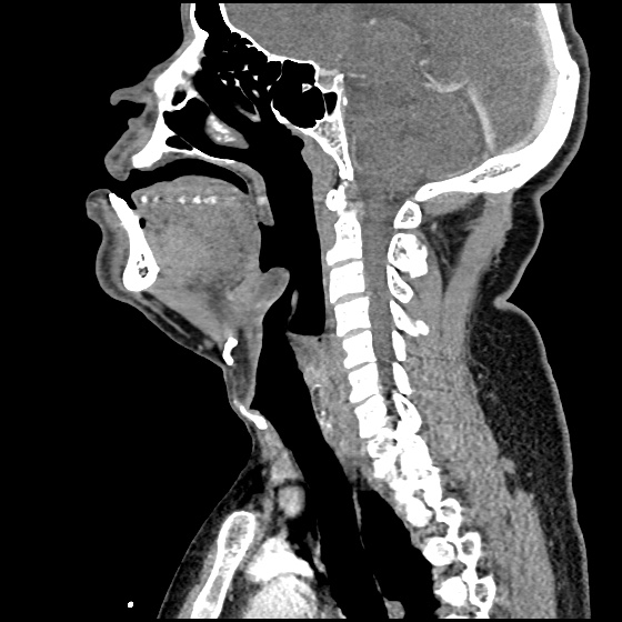
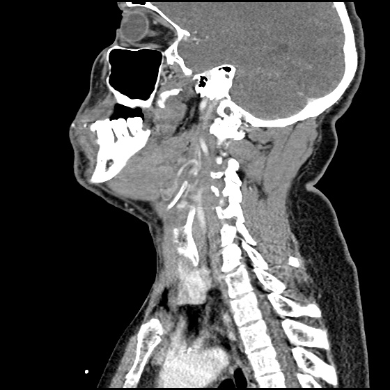
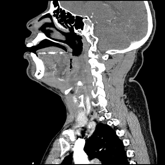
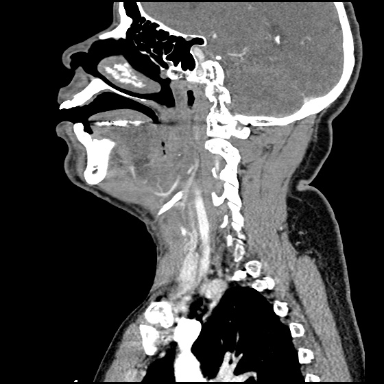
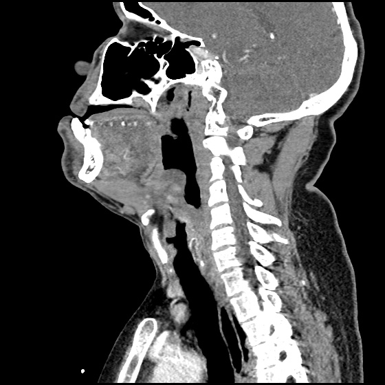
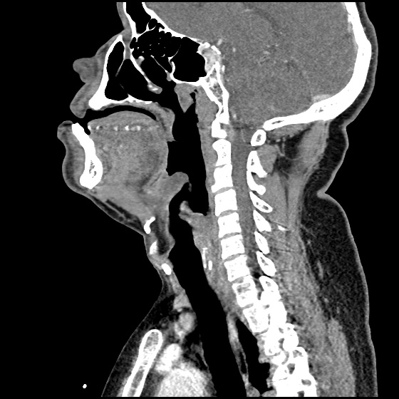
CT Neck/Soft Tissue with Contrast

Edema of the oropharynx/hypopharynx, consistent with epiglottitis and early abscess formation.

ED/Hospital Course

The patient acutely decompensated prior to fiberoptic laryngoscopy and proceeded emergently to the operating room for controlled intubation. The operative report described the following findings: “The patient had diffuse edema of the posterior oropharyngeal wall. The epiglottis was severely thickened, Omega shaped, soft to palpation and with moderate pressure, it appeared to come to a head and pus was expressed from the lingual side of the epiglottis.” The patient was extubated on hospital day three and discharged soon thereafter, he was doing well on follow-up.

Evaluation of Sore Throat – Applied

Evaluation of Sore Throat - Applied

Spinal Epidural Abscess

Case Presentation

HPI:

34M with no PMH presenting with joint pain and rash. The patient was in his usual state of good health until 1 week prior to presentation, noting bilateral shoulder pain. Diagnosed with musculoskeletal process at outside hospital and discharged with analgesics. Presented with partner due to worsening pain involving multiple joints, a non-painful, non-pruritic rash on bilateral lower extremities, and apparent confusion/hallucinations. Social history was non-contributory, no recent procedures or instrumentation.

Objectively, vital signs were notable for tachycardia and elevated core temperature. The patient was ill-appearing, disoriented and unable to provide detailed history. Skin examination was notable for non-blanching petechial rash with areas of confluence most dense in anterior distal lower extremities, rarer proximally, and otherwise without palm/sole involvement. Mucous membranes were dry, neck was supple. There was tenderness to palpation and manipulation of bilateral shoulders. No back tenderness to palpation or percussion was identified. Neurological examination notable for disorientation, intact cranial nerve function, pain-limited weakness in bilateral upper extremities particularly shoulder abduction, and 4/5 hip flexion, knee flexion/extension in bilateral lower extremities.

Labs:

  • CBC: 34.0/11.8/35.7/216
  • Differential: 31 bands
  • INR: 1.94
  • BMP: 131/5.3/102/17/88/2.55/215
  • LFT: AST 93, ALT 57, AP 237, TB 2.9, DB 1.9, Alb 1.4
  • Lactate: 3.3
  • UA: 47WBC, 5RBC
  • Utox: Negative
  • ESR: 83, CRP: 11.9
  • HIV: Nonreactive

Radiology

  • CT head: Negative
  • CXR: Negative
  • XR Shoulder: Negative
  • CT Chest/Abdomen/Pelvis non-contrast: Mild bilateral hydrouereter/hyndronephrosis, L4-L5 grade 2 anterolisthesis.
IM-0001-0005
IM-0001-0006
IM-0001-0007
IM-0001-0008
IM-0001-0009
IM-0001-0010
IM-0001-0011
IM-0001-0012
IM-0001-0013
IM-0001-0014

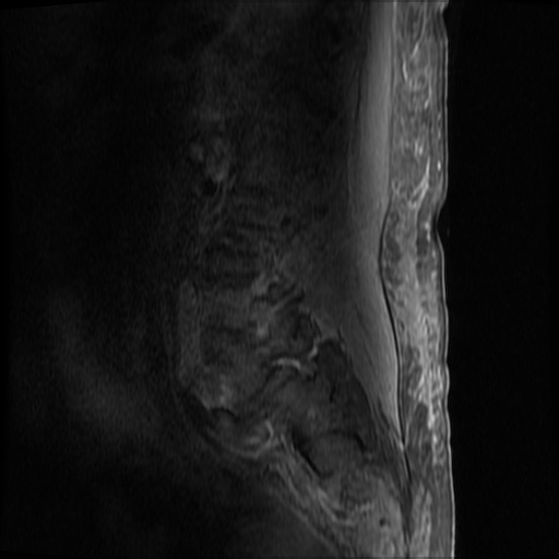
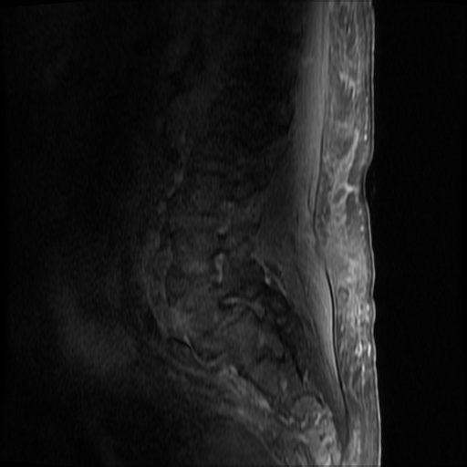
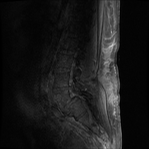
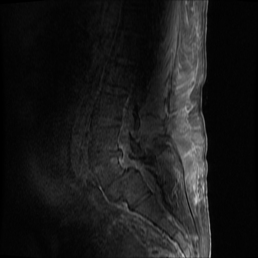
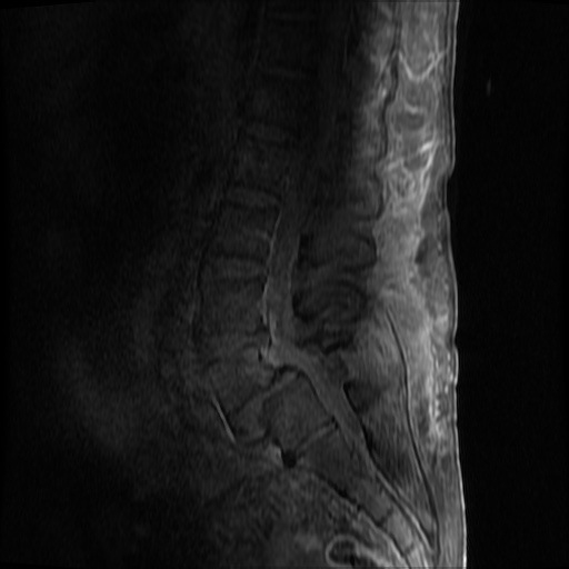
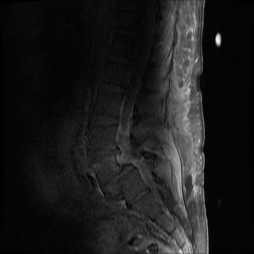
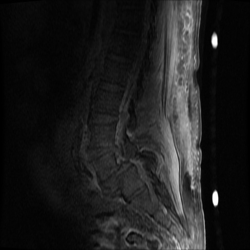
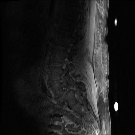
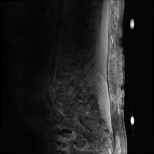
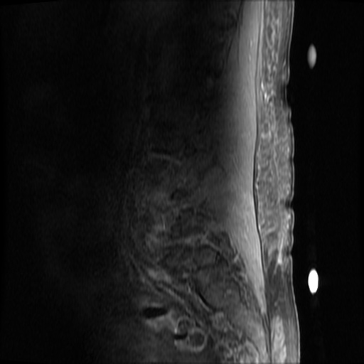
MRI Lumbar Spine w/contrast

Diffuse epidural enhancement posterior to the L4 and L5 vertebral bodies compressing the thecal sac and resulting in moderate severe spinal canal stenosis. Rim enhancement of the 1.5 cm left paraspinal fluid that may be within the L4 tendon sheath or simply paraspinal abscess.

Assessment/Plan:

Severe sepsis with end-organ dysfunction, unclear source (urinary tract involvement unlikely to account for severity of illness). Covered empirically with broad-spectrum anti-microbials including CNS infection given component of encephalitis. Admitted to the intensive care unit.

Hospital Course:

On hospital day 1, the patient underwent non-contrast MRI of the entire neuraxis with findings concerning for L4-L5 and L5-S1 epidural and paraspinal infection resulting in moderate-severe spinal canal stenosis. Blood and urine cultures grew gram-positive cocci in clusters.

On hospital day 2, the patient became increasingly somnolent. Repeat examination by consulting neurology service was concerning for evidence of meningeal irritation. Cultures speciated as methicillin-sensitive staphylococcus aureus and oxacillin was added. MRI was repeated with gadolinium, findings concerning for L4 epidural vs. paraspinal abscess.

On hospital day 3, the patient’s mental status continued to worsen and he was intubated for airway protection. Neurosurgical intervention was deferred due to deteriorating clinical status. Shoulder synovial fluid aspirate culture positive for MSSA, orthopedic surgery consulted for washout/serial arthrocentesis. TTE performed without evidence of valvular vegetation.

On hospital day 4, additional warm joints were aspirated by orthopedic surgery including knee, bilateral ankles, and shoulder each of which ultimately grew MSSA.

On hospital day 6, the patient underwent OR washout of affected joints with intraoperative findings of purulent fluid. TEE performed without evidence of valvular vegetation. The following day, underwent fluoroscopically-guided lumbar puncture, CSF studies inconclusive. Rifampin added for high-grade bacteremia with multiple seeded sites.

The patient was extubated on hospital day 9 and transferred out of the intensive care unit. The following day, he became increasingly tachypneic with evidence of volume overload on examination and was intubated and returned to the intensive care unit. Sustained PEA arrest post-intubation with ROSC, possibly secondary to pneumothorax vs. hypoxia from extensive mucous plugging. Required increasing vasopressor support over the subsequent 12 hours, emergent CVVHD for worsening academia and hypervolemia. The patient sustained another arrest and ultimately expired.

The final impression was that of high-grade bacteremia from unclear source (vague history of proximate hand laceration/infection) with resultant seeding of epidural/paraspinal space, urinary tract, multiple joints, and likely CNS/meninges. Review of abdominal ultrasonography with evidence of cirrhosis, suggesting that some component of initial hepatic synthetic dysfunction may have been chronic and this may have increased the patient’s risk for disseminated infection and SEA. Neurosurgical intervention was not pursued due to unstable clinical status and as the patient’s neurological findings were not consistent with the location of the identified lesion.

Spinal Epidural Abscess (SEA)1

Risk factors:

  • Immunocompromise: diabetes, cirrhosis, CKD, HIV/AIDS
  • Anatomic: DJD, trauma, prior surgery
  • Introduction: IVDA, epidural anesthesia, tattoo

Organism:

  • S. aureus, 2/3
  • S. epidermidis (associated with device, instrumentation)
  • E. coli (urine spread)
  • P. aeruginosa (IVDA)
  • Rare: anaerobes, mycobacteria, fungi

Staging:

  1. Back pain at affected site
  2. Nerve root pain from affected level
  3. Weakness, sensory deficit, bladder/bowel dysfunction
  4. Paralysis

Clinical features:

  • Back pain (75%)
  • Fever (50%)
  • Neuro deficit (33%)

Diagnosis:

  • Labs: Leukocytosis, ESR/CRP, blood cultures
  • Imaging: MRI with gadolinium, 90% sensitivity
  • Clinical findings and laboratory studies are insensitive and non-specific, in one study, approximately ½ of patients had >2 visits.

Prevalence of abnormal physical findings 2

Finding Prevalence
Fever (T>38°C) 19-32%
Focal spinal TTP 52-62%
Diffuse spinal TTP 63-65%
Positive SLR 11-13%
Abnormal sensation 17-27%
Weakness 29-40%
Abnormal reflexes 8-17%
Abnormal rectal tone 5-10%
Saddle anesthesia 2%

Clinical Decision Guideline 3

Spinal Epidural Abscess Clinical Decision Guideline

Management:

  • Neurosurgical evacuation/fusion
  • Antibiotics (vancomycin, oxacillin, cefepime)
  • Neurosurgical intervention may not result in neurological recovery if symptoms present for > 24-36 hours and may be impractical in the setting of panspinal infection.

References:

  1. Darouiche RO. Spinal epidural abscess. N Engl J Med. 2006;355(19):2012–2020. doi:10.1056/NEJMra055111.
  2. Davis DP, Wold RM, Patel RJ, et al. The clinical presentation and impact of diagnostic delays on emergency department patients with spinal epidural abscess. J Emerg Med. 2004;26(3):285–291. doi:10.1016/j.jemermed.2003.11.013.
  3. Davis DP, Salazar A, Chan TC, Vilke GM. Prospective evaluation of a clinical decision guideline to diagnose spinal epidural abscess in patients who present to the emergency department with spine pain. J Neurosurg Spine. 2011;14(6):765–770. doi:10.3171/2011.1.SPINE1091.
  4. WikEM: Epidural abscess (spinal)
Pediatric Sizes and Doses

Below is a rapid reference for essential information related to the care of pediatric patients including sizing estimates for endotracheal tubes and weight-based dosing for critical/common medications (rapid sequence intubation, pediatric advanced life support, seizure management), compiled by Dr. Kelly Young1.

Airway

ETT
4 + Age/4 = uncuffed
Subtract 0.5 for cuffed
Gestational age (weeks) / 10 if premature
Depth = ETTx3
Blade
Newborn: 0
<2yo: 1
2-8yo: 2
>8yo: 3
Other Tubes
NGT = ETT x 2
Chest tube = ETT x 4

Estimating Weight

Age (years) 1 3 5 7 9
Weight (kg) 10 15 20 25 30

Vital Signs

Blood Pressure

Age Measure
Neonate 60mmHg
<1yo 70mmHg
1-10yo 70 + (Age x2)
>10yo 90mmHg

Heart/Respiratory Rate

Age (yrs) HR RR
0-1 140 40
1-4 120 30
4-12 100 20
>12 80 15

Medications

Name Dose
RSI (Paralysis)
Succinylcholine 1mg/kg (x2 infant, x3 neonate)
Rocuronium 1-1.2mg/kg
RSI (Sedation)
Etomidate 0.3mg/kg
Ketamine 2mg/kg
Midazolam 0.1mg/kg
Fentantyl 1mcg/kg
PALS
Defibrillation 2, 4, 10J/kg
Cardioversion 0.5, 1J/kg
Epinephrine 0.01mg/kg (0.1mL/kg of 1:10,000)
Atropine 0.02mg/kg (minimum dose 0.1mg, maximum 0.5mg)
Adenosine 0.1mg/kg (max 6mg), 0.2 mg/kg (max 12mg)
Amiodarone 5mg/kg
Calcium gluconate (10%) 1mL/kg
Calcium chloride (10%) 0.2mL/kg
Magnesium sulfate 25mg/kg
Sodium bicarbonate 1mEq/kg
3% saline 5cc/kg
Mannitol 1g/kg
Fluids
Normal saline (0.9%) 20cc/kg
PRBC 10cc/kg
Maintenance 4cc/kg (first 10kg), 2cc/kg (second 10kg), 1cc/kg thereafter
Dextrose
<1yo D10, 5cc/kg
1-10yo D25, 2cc/kg
>10yo D50, 1cc/kg
Anti-epileptics
Lorazepam, Midazolam 0.1mg/kg x3
Fosphenytoin 20 PE/kg
Keppra 20-40mg/kg
Valproate 20mg/kg
Phenobarbital 20mg/kg
Midazolam infusion 0.1mg/kg/h
Midazolam IN 0.2mg/kg (max 10mg)
Antibiotics
Ceftriaxone 50mg/kg
Amoxicillin 90mg/kg divided BID
Azithromycin 10mg/kg day 1, 5mg/kg days 2-5
Common Medications
Acetaminophen 15mg/kg
Ibuprofen 10mg/kg
Diphenhydramine 1.25mg/kg
Ondansetron 0.15mg/kg
Intranasal Medications
Fentanyl 1.5mcg/kg (max 100mcg)
Midazolam 0.5mg/kg (max 10mg)

Reference:

  1. Young, K. D. (2016, April 18). Pediatric Doses and Sizes. Lecture presented at Harbor-UCLA Medical Center in CA, Torrance.

Nonsustained Ventricular Tachycardia

Case 1

64M with a history of HFrEF (LVEF 20-25%), CAD, AICD (unknown indication), COPD, CKD III presenting with gradual onset shortness of breath, progressive bilateral lower extremity edema.
Examination consistent with severe acute decompensated heart failure presumed secondary to left ventricular dysfunction.
Telemetry monitoring with multiple episodes of nonsustained ventricular tachycardia.

In the ED, the patient developed worsening respiratory failure despite initiation of therapy, requiring endotracheal intubation. Continuous cardiac monitoring revealed persistent salvos of NSVT, progressing to slow ventricular tachycardia without device intervention.
Device interrogation revealed multiple events, 3 shocks, several ATP’s over the recorded period.

Evaluation and Management:

  • NSVT with known (severe) ischemic heart disease
  • For repetitive monomorphic ventricular tachycardia: amiodarone, beta-blockade (if tolerated), procainamide (IIA, C)1

ECG’s

ECG 1
ECG 1

ECG 1

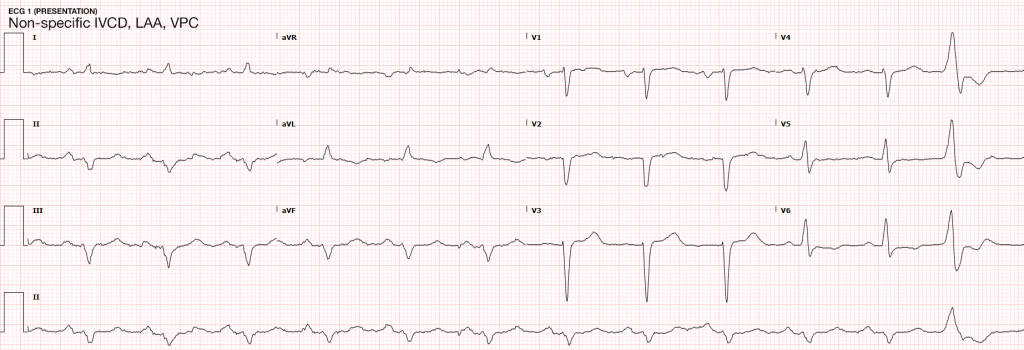
Non-specific IVCD, LAA, VPC

ECG 2
ECG 2

ECG 2

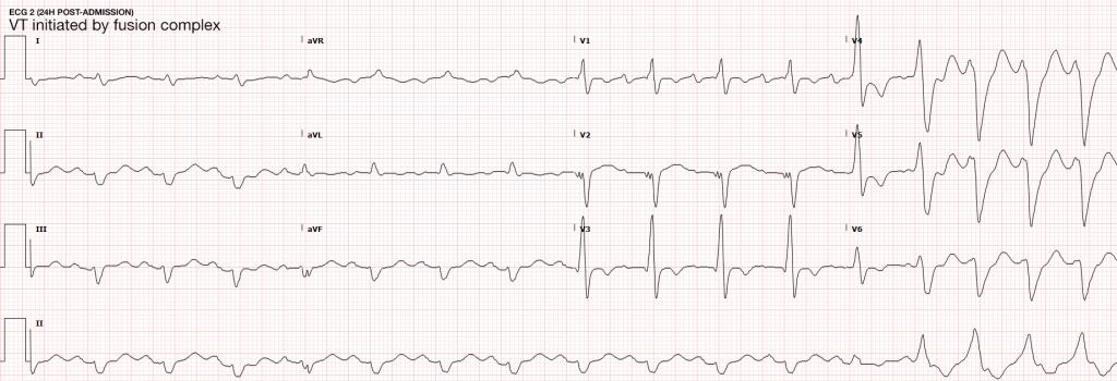
VT initiated by fusion complex

Case 2

31F with autoimmune polyglandular syndrome (adrenal, thyroid and endocrine pancreatic insufficiency), presenting with fever and cough.
Evaluation consistent with sepsis presumed secondary to pulmonary source.
Telemetry monitoring initially with ventricular bigeminy, then nonsustained ventricular tachycardia.

In the ED, the patient developed pulseless ventricular tachycardia – apparently polymorphic. Chest compressions and epinephrine produced return of spontaneous circulation with recovery to baseline neurologic function.
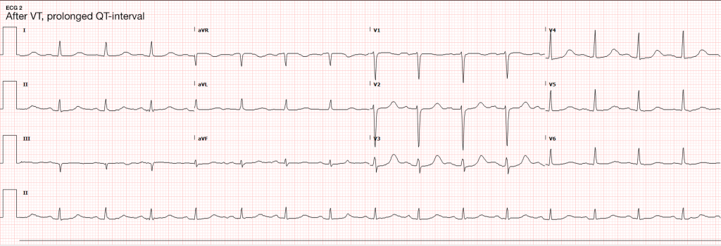
ECG revealed prolonged QTc and chemistry panel notable for critical hypokalemia/hypomagnesemia.

Evaluation and Management:

  • NSVT progressing to VT
  • Initially attributed to electrolyte disturbances. However, serial ECG’s continued to show prolonged QTc (possibly acquired, home medications included metoclopramide and erythromycin). Early echocardiography demonstrated global hypokinesis with EF 30-35% attributed to severe sepsis and recurrent defibrillation. Cardiac CT after resolution of acute illness showed persistently depressed ejection fraction without coronary atherosclerosis. The presence of NICM associated with malignant dysrhythmias warranted ICD placement.
  • Cardioversion for hemodynamic compromise (I, B), B-blockade (I, B), amiodarone if no LQTS (I, C), urgent angiography if ischemia not excluded (I, C)1
  • Correction of electrolyte abnormalities (specifically hypokalemia) may decrease progression to VF.2

ECG’s

ECG 1
ECG 1

ECG 1

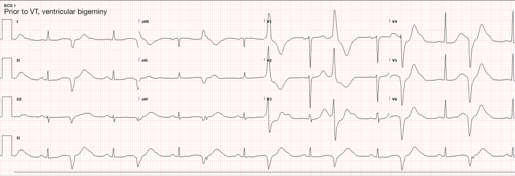
Ventricular bigeminy

ECG 2
ECG 2

ECG 2

Long-QT

VT on Telemetry
VT on Telemetry

VT on Telemetry

Non-sustained ventricular tachycardia noted on telemetry monitoring

Definition3,4

  • > 3-5 consecutive beats originating below the AV node
  • Rate > 100bpm
  • Duration <30s

Epidemiology3,5

  • Occurs in 0-4% of ambulatory patients
  • Increased frequency in males and with increasing age
  • In some patients, NSVT is associated with an increased risk of sustained tachyarrhythmias and sudden cardiac death. In others it is of little prognostic significance.6,7,8

Evaluation

In all patients:
History: including arrhythmogenic medications/substances, pertinent family history
Physical examination
ECG/CXR
TTE
In selected patients:
Exercise testing
Advanced imaging (CT/C-MR)
Electrophysiologic studies
Genetic testing

NSVT in the absence of structural heart disease

NSVT in Idiopathic Ventricular Tachycardia

Ventricular outflow arrhythmias:
RVOT: 70-80%, LBBB pattern
LVOT: 20-30%, RBBB pattern
Mechanism:
Adrenergically mediated
Occur during exercise, resolve as heart-rate increases, recur during recovery
Management:
Exclude arrhythmogenic right ventricular cardiomyopathy (imaging, myocardial biopsy)
If symptomatic, beta-blockade, ± IC anti-arrhythmic, CCB (verapamil) for ILVT
Prognosis:
Good, rare tachycardia-induced cardiomyopathy, rare SCD

NSVT in Polymorphic Ventricular Tachycardia

Mechanism
LQTS (acquired or inherited)
Familial catecholaminergic polymorphic VT
Management
Symptomatic (ex. syncope, cardiac arrest): ICD
Asymptomatic QTc > 550ms: consider ICD
Prognosis
Increased risk SCD

Arrhythmogenic Right Ventricular Cardiomyopathy

Mechanism
Fibrosis, fibro-fatty replacement of myocardium in RVIT/RVOT/RV apex
May occur with only subtle structural abnormalities of the right ventricle
LBBB morphology
Management
Anti-arrhythmics of limited utility
Catheter ablation, ICD backup
Prognosis
Increased risk SCD

NSVT with apparent structural heart disease1

Hypertension and LVH

Mechanism
Stretch-induced abnormal automaticity
Fibrotic tissue
Presence of NSVT correlates with degree of hypertrophy and subendocardial fibrosis
Management
Evaluation for ischemic heart disease
Aggressive medical management of hypertension (including beta-blockade)
Prognosis
Unclear

Valvular Disease

Mechanism
High incidence in AS, severe MR (25%)
Mechanical stress from dysfunctional valvular apparatus
Management
Beta-blockade if symptomatic
Prognosis
No evidence that NSVT is an independent predictor of SCD.

Ischemic Heart Disease9-14

Mechanism
Monomorphic VT associated with re-entry at the borders of ventricular scars
Ischemia induces polymorphic NSVT/VF
Management
Revascularization, beta-blockade, statin, ACE/ARB
MADIT I, MUSTT: ICD for ICM LVEF <40%, NSVT, EPS inducible VT
MADIT II, SCD-HeFT: ICD for moderate-to-severe LV dysfunction irrespective of NSVT or EPS findings
Prognosis
NSTEMI with NSVT >48h after admission 2x risk SCD (MERLIN-TIMI 36)
STEMI with NSVT common, not as predictive of ACM or SCD as LVEF (CARISMA)
NSVT <24h after admission for NSTEMI/STEMI not of prognostic significance.

Hypertrophic Cardiomyopathy

Mechanism
Genetic myocardial disease
Myocyte disarray, fibrosis, ischemia result in arrhythmogenic substrate
Management
Restriction of physical activity
ICD (NSVT, LV thickness, FH SCD, syncope, abnormal BP response to exercise)
Beta-blockade, anti-arrhythmic for symptoms
Prognosis
Increased risk SCD (1% annual)
Other Conditions

  • Non-ischemic dilated cardiomyopathy
  • Giant-cell myocarditis
  • Repaired TOF
  • Amyloidosis
  • Sarcoidosis
  • Chagas cardiomyopathy

Algorithm for the Evaluation of NSVT1

Algorithm for the Evaluation of Nonsustained Ventricular Tachycardia

References

  1. Zipes DP, Camm AJ, Borggrefe M, et al. ACC/AHA/ESC 2006 guidelines for management of patients with ventricular arrhythmias and the prevention of sudden cardiac death–executive summary: A report of the American College of Cardiology/American Heart Association Task Force and the European Society of Cardiology Committee for Practice Guidelines (Writing Committee to Develop Guidelines for Management of Patients with Ventricular Arrhythmias and the Prevention of Sudden Cardiac Death) Developed in collaboration with the European Heart Rhythm Association and the Heart Rhythm Society. Eur Heart J. 2006;27(17):2099–2140. doi:10.1093/eurheartj/ehl199.
  2. Higham PD, Adams PC, Murray A, Campbell RW. Plasma potassium, serum magnesium and ventricular fibrillation: a prospective study. Q J Med. 1993;86(9):609–617.
  3. Katritsis DG, Zareba W, Camm AJ. Nonsustained ventricular tachycardia. J Am Coll Cardiol. 2012;60(20):1993–2004. doi:10.1016/j.jacc.2011.12.063.
  4. Katritsis DG, Camm AJ. Nonsustained ventricular tachycardia: where do we stand? Eur Heart J. 2004;25(13):1093–1099. doi:10.1016/j.ehj.2004.03.022.
  5. Wellens HJ. Electrophysiology: Ventricular tachycardia: diagnosis of broad QRS complex tachycardia. Heart. 2001;86(5):579–585.
  6. Buxton AE, Lee KL, Fisher JD, Josephson ME, Prystowsky EN, Hafley G. A randomized study of the prevention of sudden death in patients with coronary artery disease. Multicenter Unsustained Tachycardia Trial Investigators. N Engl J Med. 1999;341(25):1882–1890. doi:10.1056/NEJM199912163412503.
  7. Jouven X, Zureik M, Desnos M, Courbon D, Ducimetière P. Long-term outcome in asymptomatic men with exercise-induced premature ventricular depolarizations. N Engl J Med. 2000;343(12):826–833. doi:10.1056/NEJM200009213431201.
  8. Udall JA, Ellestad MH. Predictive implications of ventricular premature contractions associated with treadmill stress testing. Circulation. 1977;56(6):985–989.
  9. Preliminary report: effect of encainide and flecainide on mortality in a randomized trial of arrhythmia suppression after myocardial infarction. The Cardiac Arrhythmia Suppression Trial (CAST) Investigators. N Engl J Med. 1989;321(6):406–412. doi:10.1056/NEJM198908103210629.
  10. Goldstein S. Propranolol therapy in patients with acute myocardial infarction: the Beta-Blocker Heart Attack Trial. Circulation. 1983;67(6 Pt 2):I53–7.
  11. Moss AJ. MADIT-I and MADIT-II. J Cardiovasc Electrophysiol. 2003;14(9 Suppl):S96–8.
  12. Moss AJ, Hall WJ, Cannom DS, et al. Improved survival with an implanted defibrillator in patients with coronary disease at high risk for ventricular arrhythmia. Multicenter Automatic Defibrillator Implantation Trial Investigators. N Engl J Med. 1996;335(26):1933–1940. doi:10.1056/NEJM199612263352601.
  13. Buxton AE, Lee KL, Fisher JD, Josephson ME, Prystowsky EN, Hafley G. A randomized study of the prevention of sudden death in patients with coronary artery disease. Multicenter Unsustained Tachycardia Trial Investigators. N Engl J Med. 1999;341(25):1882–1890. doi:10.1056/NEJM199912163412503.
  14. Bardy GH, Lee KL, Mark DB, et al. Amiodarone or an implantable cardioverter-defibrillator for congestive heart failure. N Engl J Med. 2005;352(3):225–237. doi:10.1056/NEJMoa043399.
  15. WikEM: Nonsustained Ventricular Tachycardia

Atypical Antipsychotic Overdose

History & Physical

38M, unknown medical history, brought in after being found unresponsive next to an empty bottle of Seroquel. Presenting vital signs notable for blood pressure of 96/43, heart rate 103. Examination reveals tentatively protected airway (GCS E2 M5 V3, SpO2 100%, RR 14), normal pupil diameter and reactivity, dry mucous membranes with thick vomitus in oral cavity.

Laboratory evaluation was unremarkable, and there was no evidence of aspiration on chest radiography. ECG showed sinus tachycardia without QT prolongation. Blood pressure increased to normal range with fluid resuscitation. The patient’s mental status progressively improved and he was discharged after six hours of uneventful continuous cardiac monitoring.

Toxidrome Summary1

Class Vital Signs Mental Status Pupils Skin Other Examples
Anti-cholinergic T
HR
BP
Delirium
Agitation
Coma
Mydriasis Dry Urinary retention
BS
Anti-histamines
Anti-parkinson
Anti-psychotic
Anti-depressant
Sympathomimetic T
HR
BP
Agitation
Hallucination
Paranoia
Mydriasis Diaphoresis Tremor
Hyperreflexia
Cocaine
Amphetamine
Ephedrine
Opioid/Sedative HR
RR
BP
CNS depression
Coma
Miosis   Hyporeflexia
Needle marks
Opioids
Benzo
Barbiturates

Evaluation1,2

  • POC Glucose
  • ECG (QT interval)
  • Serum acetaminophen, salicylate, EtOH level
  • Serum drug levels if known (anti-epileptics)
  • Urine toxicology screen
  • Chemistry (metabolic acidosis, electrolytes, renal function)
  • LFT (hepatotoxicity)
  • CK (rhabdomyolysis)
  • Serum osmolarity (osmolar gap)
  • UA with microscopy (crystals in ethylene glycol poisoning)
  • ABG (carboxyhemoglobin, methemoglobin)

Pharmacology, Toxicity and Management of Second Generation Antipsychotic (SGA) Overdose3

Pharmacology, Toxicity and Management of Second Generation Antipsychotic (SGA) Overdose

References

  1. Kulig, K. (2013). General Approach to the Poisoned Patient. In Rosen’s Emergency Medicine – Concepts and Clinical Practice (8th ed., Vol. 1, pp. 1954-1959). Elsevier Health Sciences.
  2. Wittler, M., & Lavonas, E. (2013). Antipsychotics. In Rosen’s Emergency Medicine – Concepts and Clinical Practice (8th ed., Vol. 1, pp. 2047-2051). Elsevier Health Sciences.
  3. Levine M, Ruha A-M. Overdose of atypical antipsychotics: clinical presentation, mechanisms of toxicity and management. CNS Drugs. 2012;26(7):601–611.
  4. WikEM: Antipsychotic toxicity

Back Pain

Causes of Back Pain

Causes of Back Pain

Key Historical Findings

Onset
Acute onset with associated activity suggests mechanical process
Acute onset without trigger, particularly if severe pain may suggest vascular process
Progressive onset without trigger suggests non-mechanical process (i.e. malignancy)
Aggravating/Alleviating Factors
Worsening with cough/valsalva suggests herniated disk
Relief with flexion associated with spinal stenosis
Location/Radiation
Radicular pain typically extends below knee, associated with nerve root involvement
Radiation to/from chest or abdomen suggests visceral source
Flank location suggests retroperitoneal source
History/Associated Symptoms
Fever
Medications (particularly anti-coagulants)
Hematuria
Malignancy
IVDA
Vascular disease

Key Physical Findings

  • Abnormal vital signs

    • Fever: abscess, osteomyelitis, discitis
    • Hypertension: dissection
    • Shock: AAA
  • Localize point of greatest tenderness
  • Examine abdomen for pulsatile mass
  • Perform thorough neurological examination including rectal tone and perianal sensation
  • Positive straight leg raise associated with sciatic nerve root irritation and is sensitive (but not specific) for disk disease.

References

  1. Mahoney, B. (2013). Back Pain. In Rosen’s Emergency Medicine – Concepts and Clinical Practice (8th ed., Vol. 1, pp. 278-284). Elsevier Health Sciences.
  2. WikEM: Lower back pain

Acute Pelvic Pain

Evaluation of Acute Pelvic Pain

Acute Pelvic Pain

Key Historical Findings

Location
Lateralized: suggests process related to tube or ovary, consider unilateral urinary tract process. On right, add appendicitis to differential; on left, add diverticulitis (particularly if age >40.
Central: suggests process involving uterus, bladder or bilateral adnexa
Diffuse: suggests PID
Radiation
Radiation to rectum suggests pooling of fluid or blood in cul-de-sac
Onset
Abrupt: suggests acute intrapelvic hemorrhage (from ruptured ectopic or ovarian cyst), ovarian torsion, urolithiasis
Gradual: inflammatory process such as PID
Chronic/recurrent: suggests endometriosis, recurrent ovarian cyst, ovarian mass
Associated Symptoms
Fevers/chills: suggests infectious process
Nausea/vomiting: suggests process involving gastrointestinal tract, though may accompany pregnancy or severe pain associated with ovarian torsion, urolithiasis.
Dysuria: suggests process involving urinary tract, though may be associated with local vulvar/vaginal process
Urinary urgency: more specific for bladder or urethral irritation
Obstetric History
History of recurrent spontaneous abortions or prior ectopic pregnancy increases likelihood of recurrence.
Ongoing fertility treatments increase likelihood for ectopic/heterotopic (occurs in 1:100 with assisted reproduction compared to 1:8000 in general population)
Vaginal Bleeding
In non-pregnant: suggests PID, DUB, cervical or uterine cancer
In early pregnancy: may be associated with ectopic pregnancy, non-viable IUP, or subchorionic hemorrhage
In late pregnancy: may be associated with placental pathology (previa, abruption)

Key Physical Findings

  • Pelvic examination: assists with localization of lateralized process. Should be preceded by ultrasound if >20 weeks.
  • Abnormal vaginal discharge: suggests vaginitis, cervicitis, PID, or retained foreign body.
  • Cervical motion tenderness: suggests reproductive tract inflammation or irritation of adjacent structures (appendicitis, cystitis)
  • Unilateral adnexal mass/tenderness: associated with ovarian cyst/mass, TOA, ectopic, or ovarian torsion.

References:

  1. Hart, D., & Lipsky, A. (2013). Acute Pelvic Pain in Women. In Rosen's Emergency Medicine – Concepts and Clinical Practice (8th ed., Vol. 1, pp. 266-272). Elsevier Health Sciences.
  2. WikEM: Pelvic pain

Gastrointestinal Bleeding

Evaluation and Management of Gastrointestinal Bleeding

Evaluation and Management of Gastrointestinal Bleeding

Key Historical Features

Quantity
Patient’s estimate
Symptoms suggestive of anemia/volume depletion: (pre)syncope, dyspnea
Appearance/Location
Distinguish upper from lower GI bleding
PMH
Prior episodes and source
History of aortic aneurysm graft
Comorbidities: presence of CAD, CHF, liver disease or diabetes increases mortality
Medications/substance use
Gastrotoxic, anti-coagulants, anti-platelet agents
Alcohol abuse

Key Physical Findings

Vital signs
Tachycardia or hypotension
Eyes
Conjuntival pallor suggests anemia
Scleral icterus suggests liver disease
Abdomen
Hyperactive bowel sounds may be present in UGIB (blood is cathartic)
Epigastric tenderness to palpation suggests PUD
Diffuse tenderness suggests bowel ischemia, obstruction/ileus, or perforation
Rectal (digital, anoscopy)
May reveal fissures, hemorrhoids or polyps

Labs/Diagnostic Tests

  • CBC: consider transfusion for Hb <8-10g/dL particularly in elderly or those with CAD
  • BMP: BUN:creatinine > 36 in the absence of renal failure suggests UGIB
  • PT/PTT/INR: coagulopathy
  • Lactate: elevated in bowel ischemia or systemic hypoperfusion
  • T&S or T&C
  • ECG: screen for myocardial ischemia

Blatchford Scoring System

Item Value Points
BUN 18-22 2
22-28 3
28-70 4
>70 6
Hb (male) 12-13 1
10-12 3
<10 6
Hb (female) 10-12 1
<10 6
SBP 100-109 1
90-99 2
<90 3
Other HR > 100 1
Melena 1
Syncope 2
Liver disease 2
Heart failure 2

References:

  1. Goralnick, E., & Meguerdichian, D. (2013). Gastrointestinal Bleeding. In Rosen's Emergency Medicine – Concepts and Clinical Practice (8th ed., Vol. 1, pp. 248-253). Elsevier Health Sciences.

Aortic Dissection

Imaging

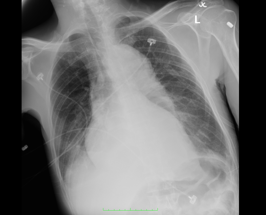
Prominent cardiomediastinal silhouette, which may be due to patient position.

Prominent cardiomediastinal silhouette, which may be due to patient position.

IM-0001-0040
IM-0001-0048
IM-0001-0056
IM-0001-0064
IM-0001-0072
IM-0001-0080
IM-0001-0088
IM-0001-0096
IM-0001-0104
IM-0001-0112
IM-0001-0120
IM-0001-0128

CT Angiography Aorta

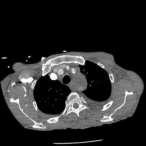
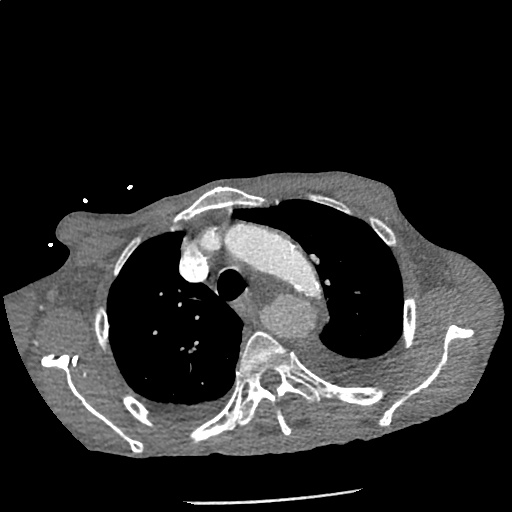
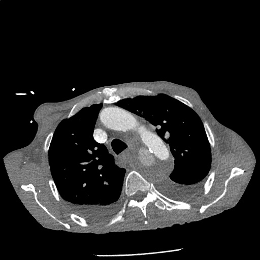
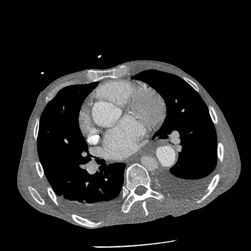
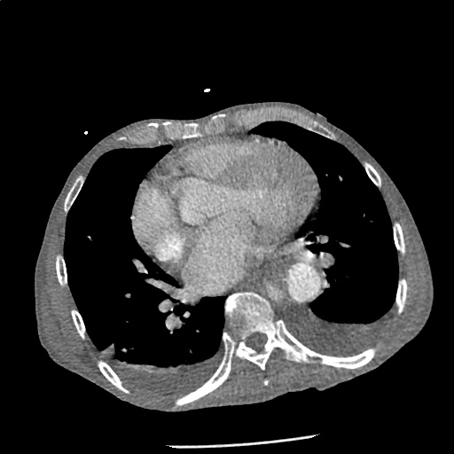
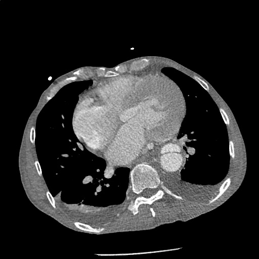
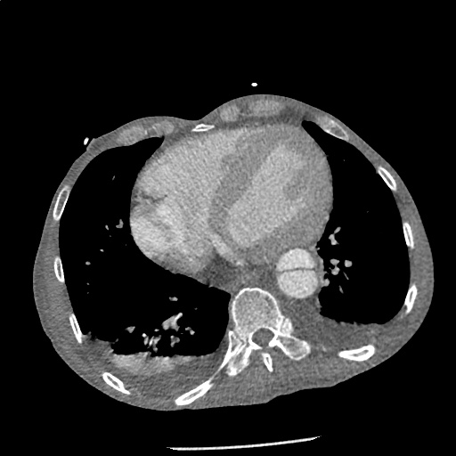
Highly complex type B aortic dissection originating at the distal arch (just distal to the left subclavian artery) and terminating at the level of diaphragm. The dissection contains multiple false lumens containing blood products of differing ages (thrombus and contrast-opacified blood). No apparent involvement of the left common carotid or left subclavian artery.

Mediastinum Anatomy

Mediastinal Masses

Anterior
Retrosternal goiter
Thymoma
Germ-cell tumor
Lymphadenopathy (lymphoma)
Middle
Aortic arch aneurysm
Dilated pulmonary artery
Tracheal lesion
Posterior
Esophageal lesions
Hiatal hernia
Descending aortic aneurysm
Paraspinal abscess

References:

  1. Faiz, O., & Moffat, D. (2002). Anatomy at a glance. Malden, MA: Blackwell Science.
  2. Whitten CR, Khan S, Munneke GJ, Grubnic S. A diagnostic approach to mediastinal abnormalities. Radiographics. 2007;27(3):657–671. doi:10.1148/rg.273065136.
  3. WikEM: Widened mediastinum

Nausea and Vomiting

Pathophysiology of Nausea and Vomiting

Pathophysiology of Nausea and Vomiting

Complications of Nausea and Vomiting

  • Hypovolemia: activates RAAS
  • Metabolic alkalosis: loss of hydrogen ions in vomitus
  • Hypokalemia: RAAS promotes sodium retention and potassium excretion
  • Esophageal injury: Mallory-Weiss tear, Boerhaave syndrome
  • Aspiration

Key Historical Findings

Duration of vomiting
Acute: Episodic and occurring for <1 week. Suggestive of obstructive, toxic/metabolic, infectious, ischemic or neurologic process.
Chronic: Episodic and occurring for >1 month. Suggestive of partial obstruction, motility disorder or neurologic process.
Onset
Acute onset: suggests pancreatitis, gastroenteritis, or cholecystitis.
Timing
Post prandial: delayed >1 hour suggests gastric outlet obstruction or gastroparesis.
Contents
Bile: presence of bile suggests patent connection between duodenum and stomach (no GOO)
Undigested food: suggests upper GI tract process (achalasia, esophageal stricture, Zenker)
Feculent: suggests distal bowel obstruction
Associated symptoms
Headache: suggests elevated ICP

Causes of Nausea and Vomiting

Causes of Nausea and Vomiting

References

  1. Zun, L. (2013). Nausea and Vomiting. In Rosen’s Emergency Medicine – Concepts and Clinical Practice (8th ed., Vol. 1, pp. 238-247). Elsevier Health Sciences.