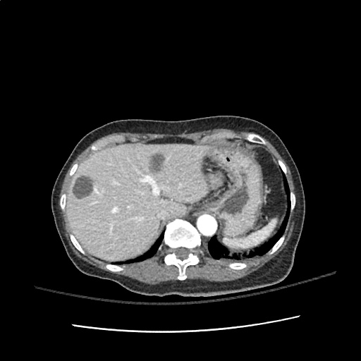
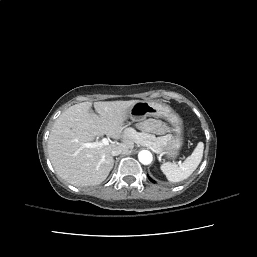
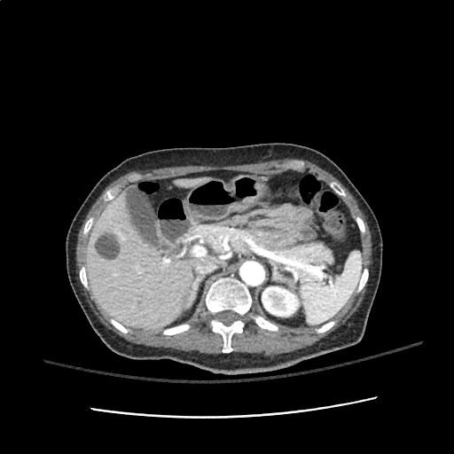
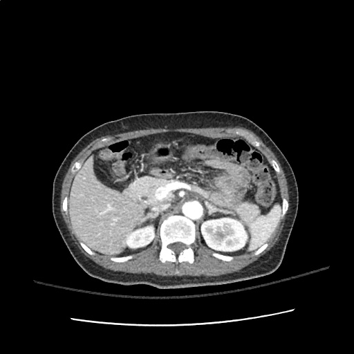
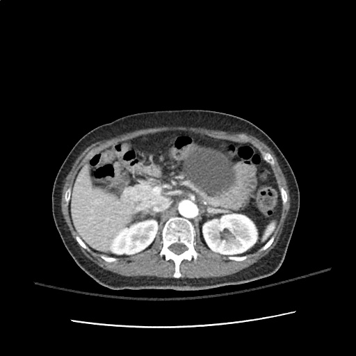
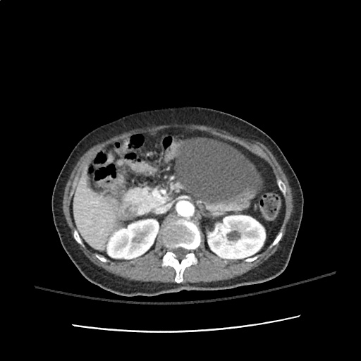
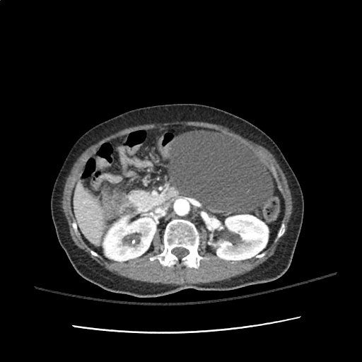
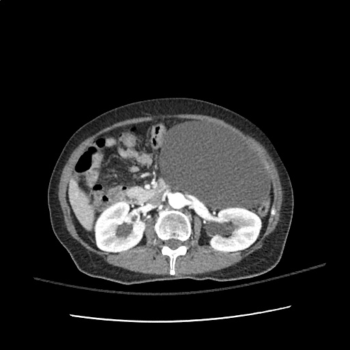
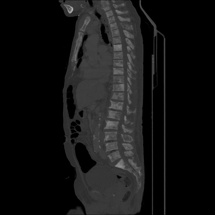
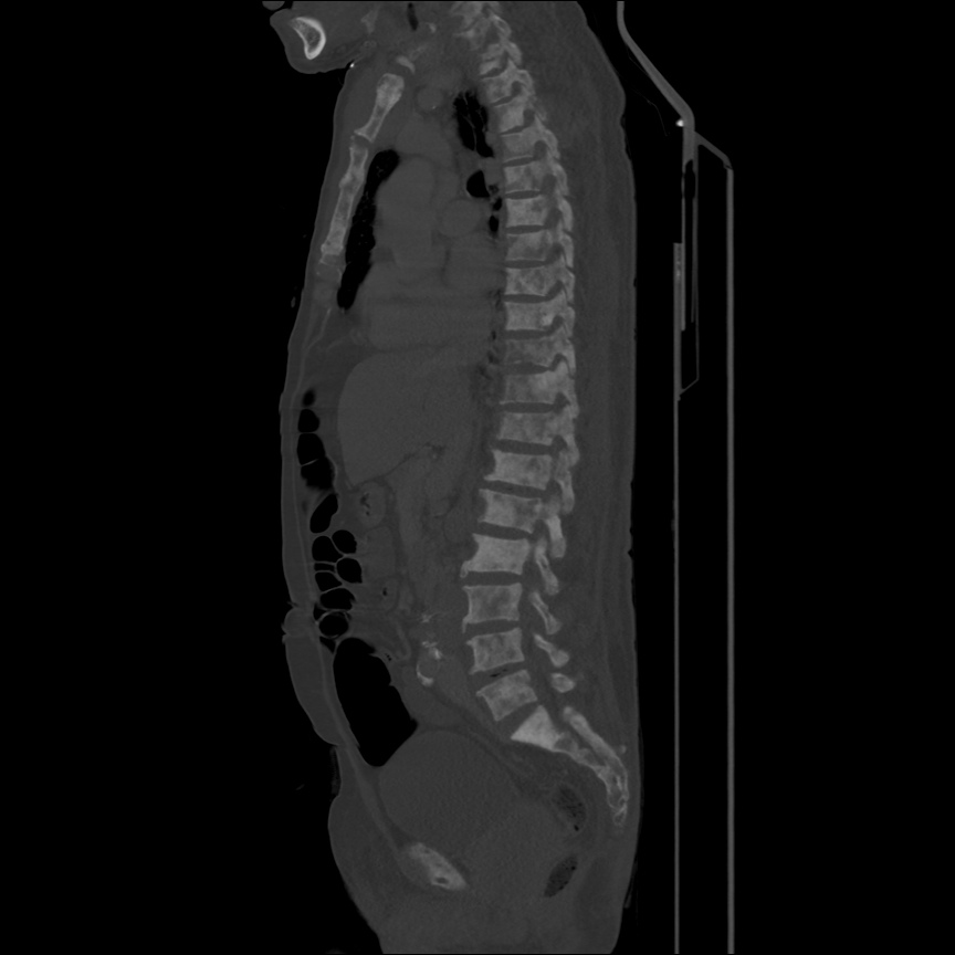
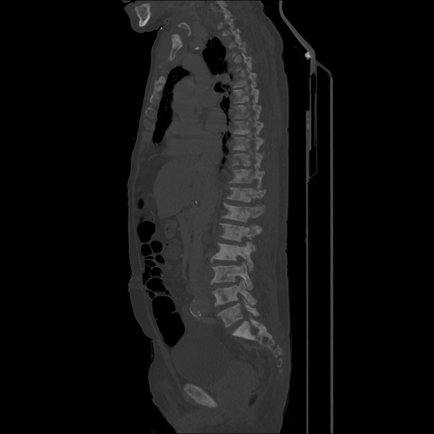
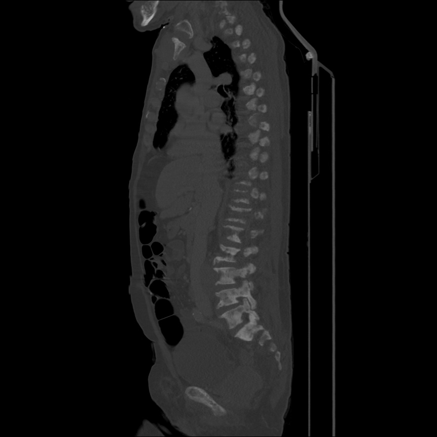
Abdominal Pain

Pathophysiology of Abdominal Pain

  1. Visceral: distension of hollow organs or capsular stretch of solid organs.
  2. Somatic: parietal peritoneal irritation
  3. Referred

    • Extra-abdominopelvic

      • Epigastric: inferior MI
      • Pelvic: hip
      • Abdominal: lower lobe pneumonia/infarction
    • Abdominopelvic

      • Shoulder: diaphragmatic irritation (ex. perforated duodenal ulcer, splenic pathology)
      • Mid-back: aortopathy, pancreatitis
      • Flank: renal pathology
      • Low back: uterus, rectum

Concerning Historical Features

  • Elderly: increased probability for severe disease with poor clinical diagnostic accuracy
  • Immunocompromised: HIV/AIDS, uncontrolled diabetes, chronic liver disease, chemotherapy, other immunosuppression
  • Pain preceding nausea/vomiting: increased likelihood of surgical process
  • Abrupt onset, duration <48h, constant timing
  • Prior abdominal surgical history: consider bowel obstruction
  • No prior episodes of similar pain
  • Recent antibiotic or steroid use: may mask signs of infection
  • Cardiac risk factors (HTN, vascular disease, atrial fibrillation: increased risk for mesenteric ischemia or aortic aneurysm
  • Heavy NSAID use or anticoagulation: increase concern for gastrointestinal bleeding

Imaging

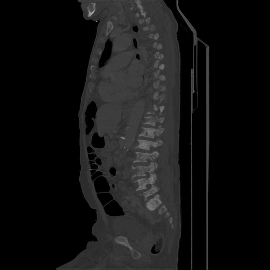
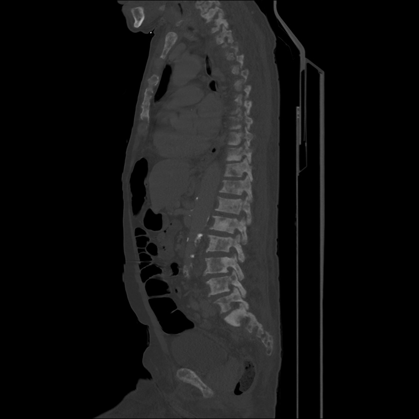
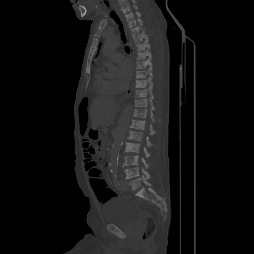
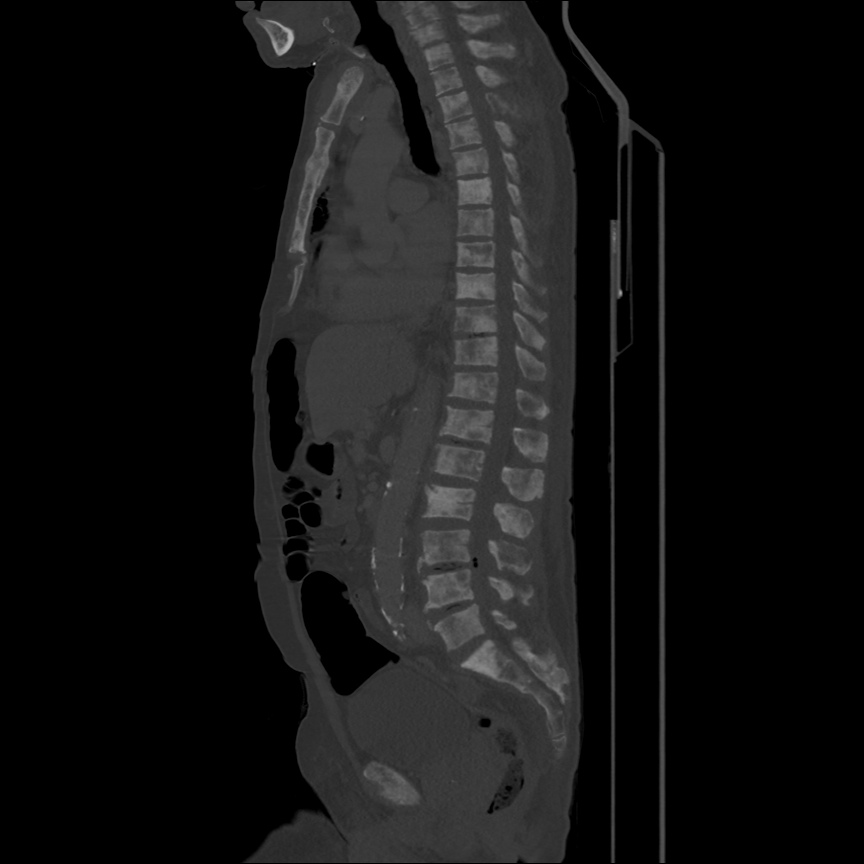
  • Plain film reserved for those who would otherwise not undergo CT. XR abdomen for bowel obstruction or radiopaque foreign body.
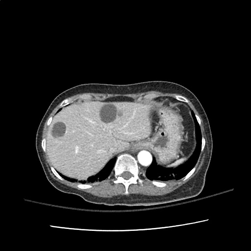
  • CT abdomen/pelvis with IV contrast, particularly if elderly or immunocompromised.
  • Ultrasound preferred for hepatobiliary pathology
  • Bedside ultrasound for identification of IUP, free intraperitoneal fluid, cholecystitis, CBD dilation, ascites, hydronephrosis, aortopathy, volume status.

Causes of Abdominal Pain

Causes of Abdominal Pain

References

  1. Budhram, G., & Bengiamin, R. (2013). Abdominal Pain. In Rosen’s Emergency Medicine – Concepts and Clinical Practice (8th ed., Vol. 1, pp. 223-231). Elsevier Health Sciences.

Chest Pain

An Algorithm for the Evaluation of Chest Pain

Algorithm for the Evaluation of Chest Pain

NOTE: Algorithm revised in November, 2017. The prior version is no longer supported but remains available here.

Guided Lecture

EM Ed
Watch “Chest Pain: It’s Giving Me Angina” from EM Ed. In this lecture Dr. Celedon reviews the critical differential diagnosis for chest pain and how to safely and effectively work up patient’s with this challenging chief complaint.

References

  1. Brown, J. (2013). Chest Pain. In Rosen’s Emergency Medicine – Concepts and Clinical Practice (8th ed., Vol. 1, pp. 214-222). Elsevier Health Sciences.

Endocrine Emergencies

HPI

30 year-old female with a history of autoimmune polyglandular syndrome (adrenal, thyroid and endocrine pancreatic insufficiency), polysubstance use, brought to the emergency department by ambulance with reported chief complaint of fever. On presentation, the patient reported fever for one day, associated with cough. She was lethargic and confused, answering yes/no questions but unable to provide detailed history. She states that she has been taking her home medications as prescribed, which include hydrocortisone, fludrocortisone, synthroid and insulin. No collateral information was immediately available.

Additional history was obtained from chart review upon discharge. The patient was hospitalized two weeks prior with pneumonia and discharged after two days. For 2-3 days prior to presentation, she reported the following symptoms to family members: nausea/vomiting, cough, decreased oral intake, fevers, and palpitations – she did not take her home medications during this time.

Physical Exam

VS: T 38.6 HR 112 RR 18 BP 149/82 O2 90% RA
Gen: Alert, fatigued, slow responses.
HEENT: No meningeal irritation, dry mucous membranes.
Pulmonary: Tachypnea, inspiratory wheezing and faint crackles at left and right inferior lung fields, appreciated anteriorly as well.
Neuro: Alert, oriented to self, situation, not month/year. PERRL, EOMI, facial muscles symmetric, tongue protrudes midline without fasciculation. Peripheral sensation grossly intact to light touch and moves all extremities on command.

Labs

  • VBG: alkalemia, primary respiratory
  • CBC: no leukocytosis, normal differential, normocytic anemia
  • BMP: 131, 2.5 , 94, 28, 11, 1.6, 115
  • Mg: 1.3
  • Lactate: 1.0
  • TSH: 17 , T4: 1.03
  • Troponin: 0.129

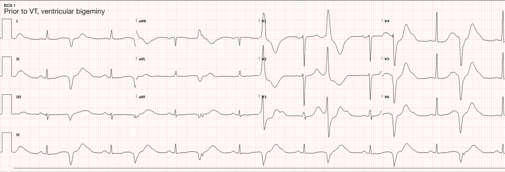
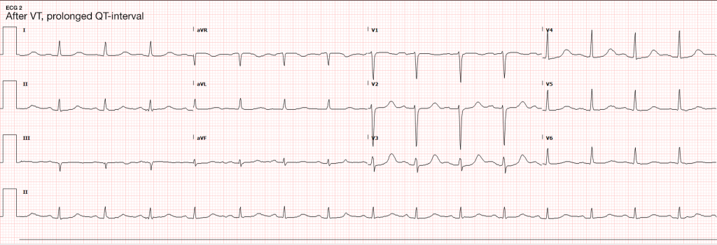
ECG

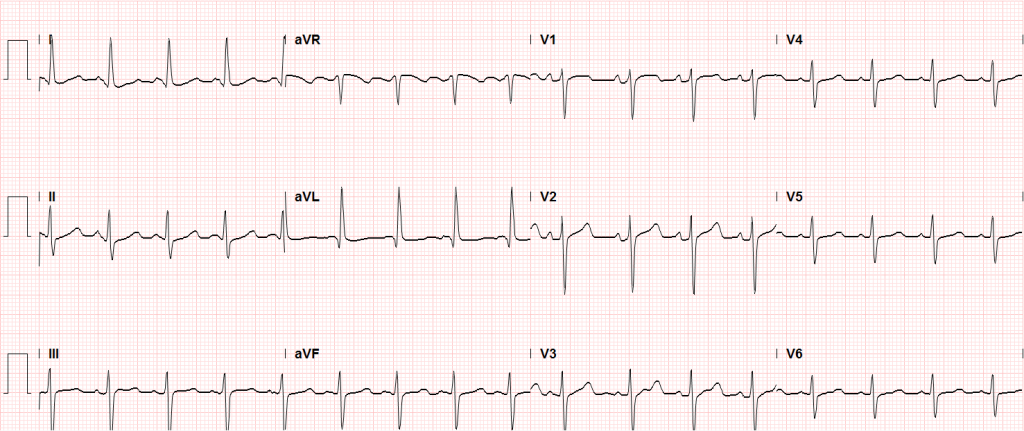
ECG 1
ECG 2

Imaging

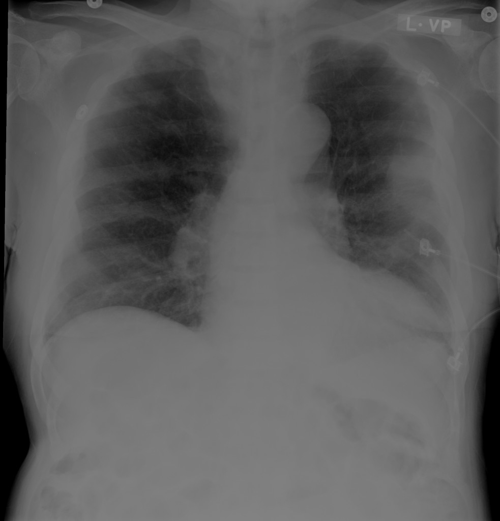
  • CXR: Negative acute.
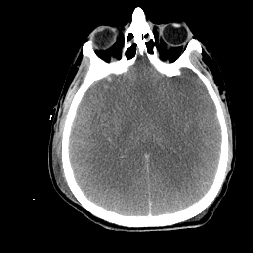
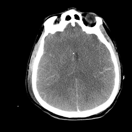
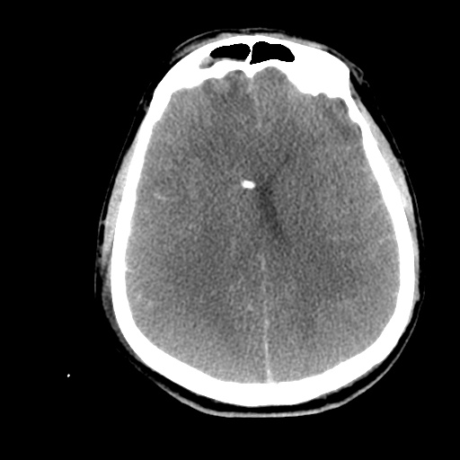
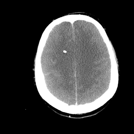
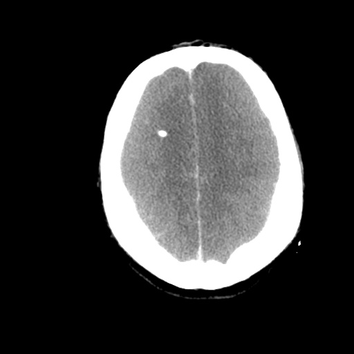
  • CT Head: Negative acute.
  • CT Cardiac: NICM, EF 35%.
IM-0001-0026
IM-0001-0030
IM-0001-0034
IM-0001-0038
IM-0001-0042
IM-0001-0046
IM-0001-0050
IM-0001-0054
IM-0001-0058
IM-0001-0062
IM-0001-0066
IM-0001-0070

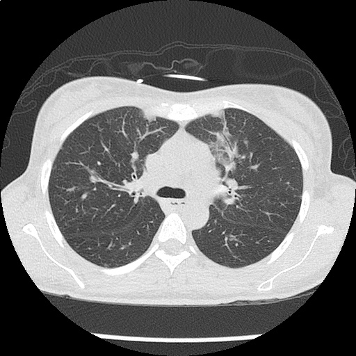
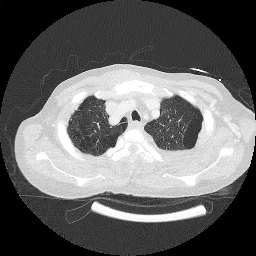
CT Chest non-contrast

  • Diffuse patchy GGO (pulmonary edema, atypical pneumonia, alveolar hemorrhage, others).
  • Multiple bilateral pulmonary nodules.
  • Possible pulmonary arterial hypertension.

Hospital Course

The patient’s evaluation in the emergency department was concerning for severe sepsis secondary to suspected pulmonary source (given association of fever with cough, hypoxia and abnormal chest imaging findings). The patient had persistent alteration in mental status concerning for CNS infection. While preparing for lumbar puncture, cardiac monitoring revealed sustained polymorphic ventricular tachycardia without appreciable pulse. CPR was initiated, amiodarone 150mg IV push administered and at first pulse check a perfusing sinus rhythm was noted with immediate recovery of prior baseline mental status. Amiodarone load was continued and additional potassium sulfate (PO and IV) was administered. Review of telemetry monitoring revealed preceding 30-45 minutes of non-sustained ventricular tachycardia. The patient had two more episodes of sustained ventricular tachycardia requiring defibrillation. The patient was admitted to the medical intensive care unit for continued management.

#Sustained Ventricular Tachycardia
Initially attributed to critical hypokalemia and hypomagnesemia. However, after appropriate repletion serial ECG’s continued to demonstrate prolonged QT interval (possibly acquired secondary to medications, later review revealed multiple promotility agents for treatment of gastroparesis which could contribute to QT-prolongation including erythromycin and metoclopramide, also associated with endocrinopathies). Early echocardiography demonstrated global hypokinesis with estimated EF 30-35%. This was initially attributed to severe sepsis, as well as recurrent defibrillation. However, cardiac CT after resolution of acute illness showed persistent depressed ejection fraction, no evidence of coronary atherosclerosis. The presence of non-ischemic cardiomyopathy (may be attributable to chronic endocrine dysfunction or prior history of methamphetamine abuse) associated with malignant dysrhythmias warranted ICD placement for secondary prevention which the patient was scheduled to receive.

#Severe Sepsis
Attributed to pulmonary source given CT findings, healthcare associated and covered broadly. Mental status gradually improved and returned to baseline. CT head was negative, lumbar puncture deferred.

#Hypokalemia
Unclear etiology. Adrenal insufficiency commonly associated with hyperkalemia and no history of surreptitious fludrocortisone use. Possibly secondary to GI losses. Improved with repletion.

#Autoimmune Polyglandular Syndrome
Started on stress-dose steroids in emergency department. Transiently developed DKA which was reversed appropriately and hydrocortisone was tapered to home regimen. Home levothyroxine was resumed.

Endocrine Emergencies: Hyperthyroidism

Symptoms

Constitutional Weight loss, heat intolerance, perspiration
Cardiopulmonary Palpitations, chest pain, dyspnea
Neuropsychiatric Tremor, anxiety, double vision, muscle weakness
Neck Fullness, dysphagia, dysphonia
Musculoskeletal Extremity swelling
Reproductive Irregular menses, decreased libido, gynecomastia

Signs

Vital signs Tachycardia, widened pulse pressure, fever
Cardiovascular Hyperdynamic precordium, CHF, atrial fibrillation, systolic flow murmur
Ophthalmologic Widened palpebral fissure, periorbital edema, proptosis, diplopia, restricted superior gaze
Neurologic Tremor, hyperreflexia, proximal muscle weakness
Dermatologic Palmar erythema, hyperpigmented plaques or non-pitting edema of tibia
Neck Enlarged or nodular thyroid

Thyroid Storm

Essentially an exaggeration of thyrotoxicosis featuring marked hyperthermia (104-106°F), tachycardia (HR > 140bpm), and altered mental status (agitation, delirium, coma).

Precipitants
Medical: Sepsis, MI, CVA, CHF, PE, visceral ischemia
Trauma: Surgery, blunt, penetrating
Endocrine: DKA, HHS, hypoglycemia
Drugs: Iodine, amiodarone, inhaled anesthetics
Pregnancy: post-partum, hyperemesis gravidarum

Scoring (Burch, Wartofsky)

Fever
99-100 5
100-101 10
101-102 15
102-103 20
103-104 25
>104 30
Tachycardia
90-110 5
110-120 10
120-130 15
130-140 20
>140 25
Mental Status
Normal 0
Mild agitation 10
Extreme lethargy 20
Coma, seizure 30
CHF
Absent 0
Mild (edema) 5
Moderate (rales, atrial fibrillation) 10
Pulmonary edema 15
GI
None 0
Nausea/vomiting, abdominal pain 10
Jaundice 20
Precipitating Event
None 0
Present 10
  • >45: thyroid storm
  • 25-44: impending thyroid storm
  • <25: unlikely thyroid storm

Management

Supportive measures
Volume resuscitation and cooling
Benzodiazepines for agitation
Beta-blockade
Propranolol 60-80mg PO q4h
Propranolol 0.5-1.0mg IV, repeat q15min then 1-2mg q3h
Esmolol continuous infusion
Endocrinology consultation
PTU, SSKI

Endocrine Emergencies: Hypothyroidism

Symptoms

Constitutional Weight gain, cold intolerance, fatigue
Cardiopulmonary Dyspnea, decreased exercise capacity
Neuropsychiatric Impaired concentration and attention
Musculoskeletal Extremity swelling
Gastrointestinal Constipation
Reproductive Irregular menses, erectile dysfunction, decreased libido
Integumentary Coarse hair, dry skin, alopecia, thin nails

Signs

Vital signs Bradycardia, hypothermia
Cardiovascular Prolonged QT, increased ventricular arrhythmia, accelerated CAD, diastolic heart failure, peripheral edema
Neurologic Lethargy, slowed speech, agitation, seizures, ataxia/dysmetria, mononeuropathy, delayed relaxation of reflexes
Musculoskeletal Proximal myopathy, pseudohypertrophy, polyarthralgia
Gastrointestinal Ileus

Myxedema Coma

Precipitants
Critical illness: sepsis (especially PNA), CVA, MI, CHF, trauma, burns
Endocrine: DKA, hypoglycemia
Drugs: amiodarone, lithium, phenytoin, rifampin, medication non-adherence
Environmental: cold exposure
Recognition
History: hypothyroidism, thyroidectomy scar and acute precipitating illness
Hypothermia: temp <95.9°F (or normal in presence of infection)
AMS: lethargy, confusion, coma, agitation, psychosis, seizures
Hypotension: refractory to volume resuscitation and pressors
Bradypnea: with hypercapnia and hypoxia
Hyponatremia

Management

  • Airway protection
  • Fluid resuscitation
  • Thyroid hormone replacement
    • Young, otherwise healthy patients: T3 10ug IV q4h
    • Elderly, cardiac compromise: 300ug IV x1
  • Hydrocortisone: 50-100mg IV q6-8h
  • Treat precipitating illness

Interpretation of Thyroid Function Tests

Condition TSH T4
None Normal Normal
Hyperthyroidism Low High
Hypothyroidism High Low
Subclinical hyperthyroidism Low Normal
Subclinical hypothyroidism High Normal
Sick euthyroid Low Low

Endocrine Emergencies: Adrenal Insufficiency

Either primary due to adrenal gland failure (often secondary to autoimmune destruction), or secondary most often due to exogenous glucocorticoid administration (usually requiring more than 30mg/day for > 3wks).

Symptoms

Constitutional Weakness, fatigue
Gastrointestinal Anorexia, nausea, cramping
Neuropsychiatric Depression, apathy
Reproductive Amenorrhea, decreased libido
Musculoskeletal Myalgia, arthralgia

Signs

General Hyponatremia, orthostatic hypotension, low-grade fever
Primary Hyperpigmentation, hyperkalemia, hyperchloremia, acidosis
Secondary Hypoglycemia

Management

Maintenance
Hydrocortisone 20mg qAM, 10mg qPM
Fludrocortisone 50-100ug daily
Minor illness (x2)
Hydrocortisone 40mg qAM, 20mg qPM
Fludrocortisone 50-200ug daily
Adrenal Crisis
Dexamethasone 4mg IV or hydrocortisone 100mg IV
2-3L 0.9% NaCl
Treat precipitating illness

Life-Threatening Electrolyte Abnormalities3

Critical Hypokalemia

Causes
GI losses (diarrhea, laxative use)
Renal losses (hyperaldosteronism, diuretics)
Cellular shifts (alkalosis)
ECG changes
U-waves 4
T-wave flattening
Ventricular arrhythmias (exacerbated with digoxin use)
Treatment
Maximum rate 10-20mEq/h with ECG monitoring
If malignant ventricular arrhythmias or arrest imminent, consider more rapid administration (10mEq over 5 minutes)


Critical Hypomagnesemia

Causes
GI, renal losses
Thyroid dysfunction
Treatment
1-2g IV over 5-60 minutes or IVP for Torsades

Conclusion

Unfortunately, this patient’s comprehensive clinical picture does not fit neatly into a particular category of endocrinologic pathology. Her underlying autoimmune disorder manifests both primary adrenal and thyroid dysfunction. Components of the patient’s presentation are suggestive of critical hypothyroidism (myxedema coma) including alteration in mental status, QT-prolongation and hyponatremia as well as possible precipitant of pneumonia. However, despite elevated TSH, the patient’s free T4 level was within normal range. Also absent was hypoventilation (the patient was appropriately tachypneic for degree of hypoxia and with resultant respiratory alkalosis) or bradycardia/hypothermia.
Similarly, adrenal insufficiency is typically associated with hyperkalemia, whereas our patient had critical hypokalemia that was determined to be at least a contributory factor to her ventricular dysrhythmia. The etiology of the patient’s hypokalemia remained unexplained.

References:

  1. Sharma, A., & Levy, D. (2009). Thyroid and Adrenal Disorders. In Rosen’s Emergency Medicine (8th ed., Vol. 2, pp. 1676-1692). Elsevier Health Sciences.
  2. Savage MW, Mah PM, Weetman AP, Newell-Price J. Endocrine emergencies. Postgrad Med J. 2004;80(947):506–515. doi:10.1136/pgmj.2003.013474.
  3. ECC Committee, Subcommittees and Task Forces of the American Heart Association. 2005 American Heart Association Guidelines for Cardiopulmonary Resuscitation and Emergency Cardiovascular Care. Circulation. 2005;112(24 Suppl):IV1–203. doi:10.1161/CIRCULATIONAHA.105.166550.
  4. Levis JT. ECG diagnosis: hypokalemia. Perm J. 2012;16(2):57.

Dyspnea

Causes of Dyspnea

Causes of Dyspnea

Findings in Selected Causes of Dyspnea

Condition History Symptoms Findings Evaluation
Anaphylaxis Exposure to allergen Abrupt onset, facial swelling Stridor, wheezing, hives  
PE Immobilization, malignancy, prior DVT/PE, surgery, OCP Abrupt onset, pleuritic chest pain Tachycardia, hypoxia ECG (RV strain)
CT PA, D-dimer
LE US (DVT)
Pneumonia Exposure, tobacco use Fever, productive cough Focal rales CXR
CBC
Blood/respiratory cultures
Pneumothorax Trauma, thin male Abrupt onset, chest pain Decreased BS, subQ emphysema, JVD and tracheal deviation if tension CXR
US
Fluid overload Dietary indiscretion, medication non-adherence Orthopnea, PND JVD, S3/S4, peripheral edema CXR
US
ECG
BNP
COPD/Asthma Tobacco use, personal/family history Progressive Retractions, accessory muscle use, wheezing CXR
US (distinguish from fluid overload)
Malignancy Tobacco use, weight loss Hemoptysis   CXR
CT Chest

References

  1. Braithwaite, S., & Perina, D. (2013). Dyspnea. In Rosen’s Emergency Medicine – Concepts and Clinical Practice (8th ed., Vol. 1, pp. 206-213). Elsevier Health Sciences.

Altered Mental Status Applied

H&P

58 year-old female with no known past medical history, brought to emergency department by husband due to fatigue and weakness. The patient does not speak and cannot provide history. Her husband describes a progressive decline from normal baseline two weeks ago, noting lethargy/fatigue. Noted decreased speech and attention one week ago, and absent speech and requiring assistance with ambulation for the past two days. Thorough review of systems unremarkable excepting vomiting with decreased oral intake (tolerating fluids) and prior headache which resolved.

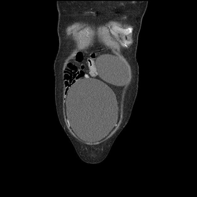
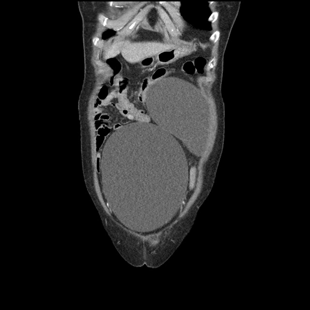
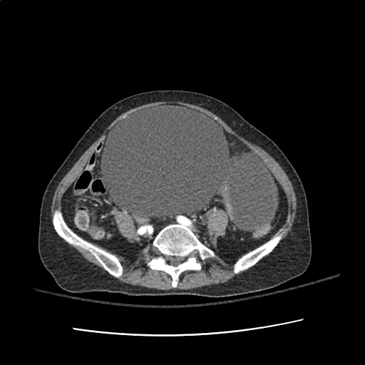
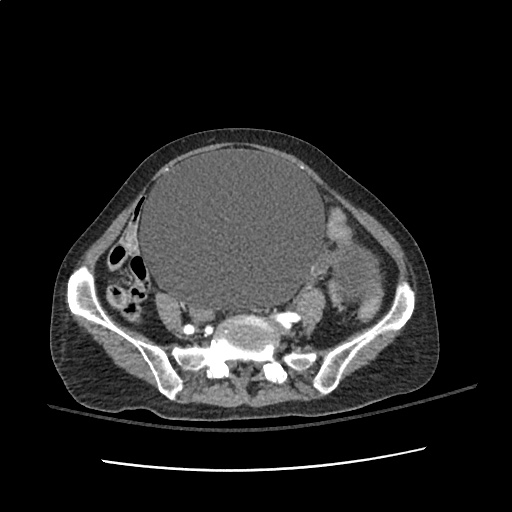
On examination, vital signs were normal, the patient was lying in bed and in no acute distress. The patient was non-verbal and did not follow commands (GCS E4-M5-V2). She was unable to comply with a thorough neurological examination, however pupils were equal and reactive, eyes tracked without nystagmus, no facial asymmetry noted, reflexes 1+ and symmetric in UE/LE, cannot participate in strength/sensory testing. Abdominal examination notable for infraumbilical and left-sided mass which elicits groans with palpation, though no rigidity or guarding. Mucous membranes moist, no skin tenting.

Labs

  • CBC: 13.5 (97% neutrophils) , 12.9, 38.2, 240
  • BMP: 107, 2.4, 70, 28, 9, 10, 0.44, 102
  • Serum osmolarity: 224
  • Urine osmolarity: 239
  • UNa: 20

Imaging

IM-0001-0022
IM-0001-0024
IM-0001-0026
IM-0001-0028
IM-0001-0032
IM-0001-0036
IM-0001-0040
IM-0001-0044
IM-0001-0048
IM-0001-0052
IM-0001-0056
IM-0001-0060
IM-0001-0064
IM-0001-0068
IM-0001-0072
IM-0001-0076
IM-0001-0080
IM-0001-0084
IM-0001-0088
IM-0001-0092
IM-0001-0010
IM-0001-0014
IM-0001-0018
IM-0001-0022
IM-0001-0026
IM-0001-0030
IM-0001-0034
IM-0001-0038
IM-0001-0042
IM-0001-0046
IM-0001-0050

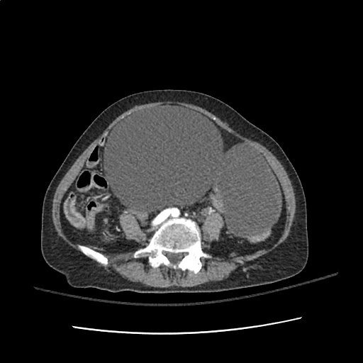
CT abdomen/pelvis with intravenous contrast

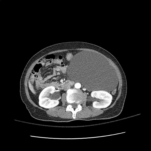
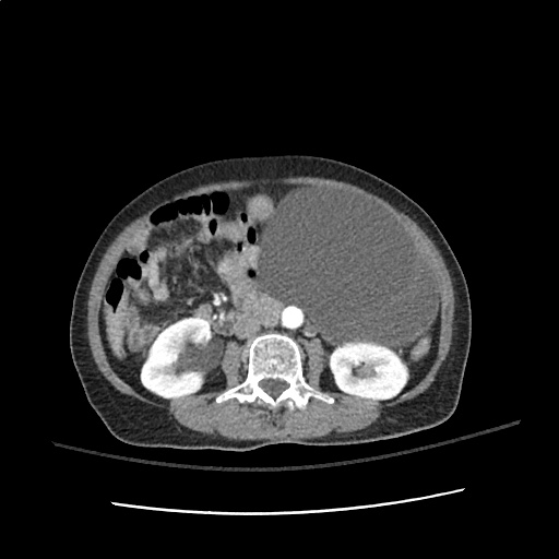
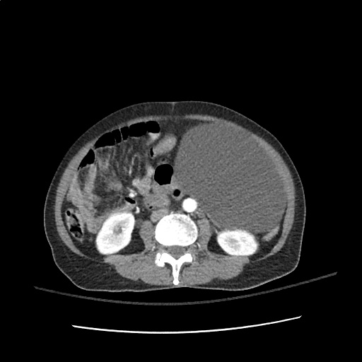
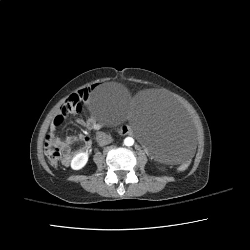
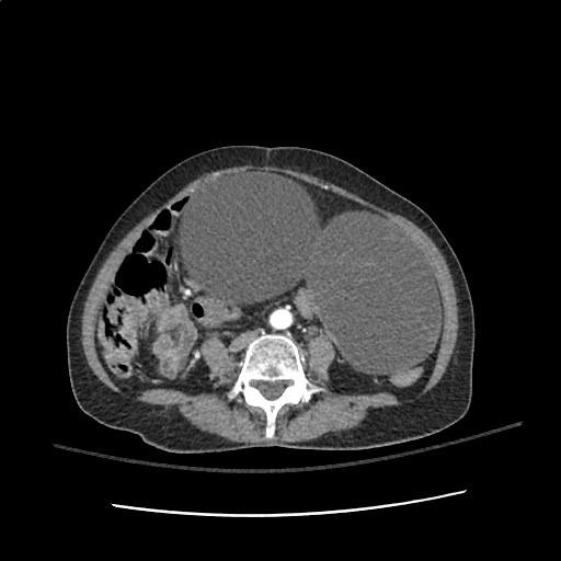
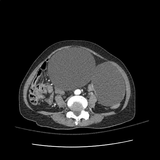
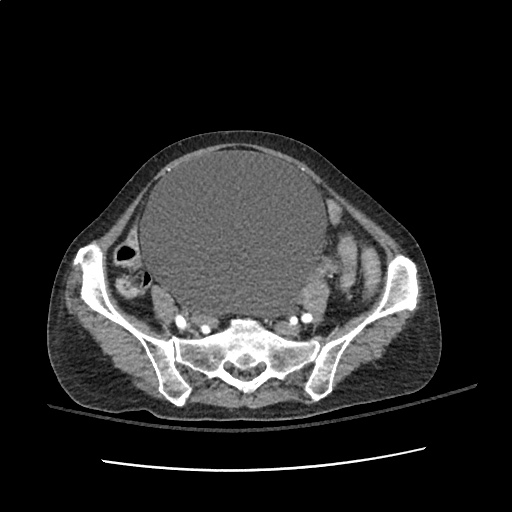
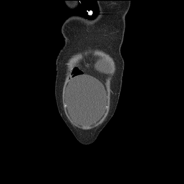
  • Large, 15 cm cystic mass in the left abdomen, which likely arises from the mesentery. This mass is suspicious for neoplasm.
  • Multiple low-density cystic lesions in the liver, which measure up to 4.5 cm in diameter and are concerning for metastatic disease. Alternatively, these may represent benign hepatic cysts which are unrelated to the mesenteric mass.
  • Massively distended bladder, with moderate bilateral hydronephrosis and mild hydroureter.

Hospital Course

The patient was admitted to the medical intensive care unit. The following problem list details findings from the extensive inpatient evaluation.

#Altered Mental Status: The patient’s dramatically depressed level of consciousness improved gradually with correction of hyponatremia and the patient was alert, oriented and at baseline at the time of discharge. Evaluation included MRI brain which showed only chronic microvascular changes. A lumbar puncture was notable for isolated elevation of CSF protein. The patient was treated empirically for HSV encephalitis until CSF HSV PCR resulted negative. Neurology was consulted and identified increased CSF oligoclonal bands of unclear significance.

#Hyponatremia: Nephrology consulted, presumed SIADH based on urine studies (secondary to infection or malignancy). Corrected upon discharge.

#Pelvic Mass: Initially thought to arise from small bowel on CT abdomen/pelvis, after bladder decompression and transvaginal ultrasound, thought to arise from adnexa. Gynecology consulted, cyst characteristics (homogenous, fluid-filled) suggest benign process and tumor markers within normal limits. No acute intervention, drainage or biopsy warranted.

#Bladder distension: Unclear etiology, associated with mild/moderate hydronephrosis. Thought to be secondary to bladder outlet obstruction secondary to pelvic mass. Indwelling catheter placed, discontinued prior to discharge with successful spontaneous voiding trial and normal post-void residual.

Hyponatremia Applied

Hyponatremia Applied

Altered Mental Status Applied

Altered Mental Status Applied

Sore Throat

Evaluation of Sore Throat

Evaluation of Sore Throat

Physical Examination:

Neck
Stiffness, limitation of extension suggestive of retropharyngeal abscess.
Jaw
Trismus associated with peritonsillar cellulitis or abscess.
Oral Cavity
Dry mucous membranes suggest dehydration (from odynophagia) and indicates severity of symptoms.
Tongue elevation, sublingual/submental induration, poor dentition (particularly of mandibular molars) associated with Ludwig Angina.
Unilateral tonsillar enlargement with contralateral uvular deviation suggests peritonisllar abscess. Fluctuance may be palpated.
Tonsilar exudates suggest infectious pharyngitis (non-specific).
Palatal petechiae suggest bacterial pharyngitis.
Ulcerations of the anterior oral cavity are associated with herpes infection, lesions on the soft palate are suggestive of coxsackievirus infection.
Rarely, a grey membrane in the posterior pharynx will suggest diphtheria.
Lymphadenopathy
Tender anterior cervical lymphadenopathy may suggest bacterial pharyngitis.
Posterior cervical lymphadenopathy is associated with infectious mononucleosis.
Large, firm, non-mobile lymph nodes may suggest malignancy.
Eyes
Presence of conjunctivitis (also rhinorrhea, exanthema) associated with viral pharyngitis.
Skin
Ulcers involving the hands, feet, in addition to pharyngeal lesions suggest coxsackievirus infection.
Scarlatiniform rash associated with pharyngitis (particularly in school-age children) suggests streptococcal pharyngitis.
Abdomen
Splenomegaly is associated with infectious mononucleosis.

Centor Criteria (Modified)

  • +1: Fever
  • +1: Tonsillar Exudate
  • +1: Tender anterior cervical lymphadenopathy
  • +1: Absence of cough
  • -1: Age >45yo

Incidence of GABHS by Centor Criteria

  • 0, -1: 1%
  • 1: 10%
  • 2: 17%
  • 3: 35%
  • 4: 51%

References:

  1. Newman, D., & Shreves, A. (2013). Sore Throat. In Rosen’s Emergency Medicine – Concepts and Clinical Practice (8th ed., Vol. 1, pp. 198-202). Elsevier Health Sciences.
  2. King, B. R., & Charles, R. A. (2004). Pharyngitis In The ED Diagnostic Challenges And Management Dilemmas. Emergency medicine practice, 6(5), 1–24.

Dizziness and Vertigo

Types of Dizziness

Types of Dizziness

Distinguishing Central vs. Peripheral Vertigo

Characteristic Peripheral Central
Onset Sudden Gradual
Intensity Severe Mild
Duration Minutes Weeks
Timing Intermittent Continuous
Nystagmus Horizontal Vertical, bidirectional
Exacerbation with head movement +
Auditory symptoms +
Neurological findings +

Causes of Vertigo

Causes of Vertigo

Characteristics of common causes of vertigo

Cause Mechanism Onset Symptoms Findings
Peripheral
BPPV Otolith Brief, positional episodes Nausea, vomiting, absent auditory symptoms. Dix-Hallpike positive
Vestibular neuronitis Viral, post-viral inflammation of vestibular portion of CNVIII Acute and severe, subsiding over days. Nausea, vomiting, absent auditory symptoms. Head thrust abnormal
Meniere Endolymphatic hydrops Recurrent, lasting hours Tinnitus, hearing loss. SNHL
Central
Vertebrobasilar insufficiency Atherosclerosis (vascular risk factors) Acute onset, recurrent episodes if TIA Headache, gait impairment, diplopia, absent auditory symptoms. Neurologic deficits
Cerebellar stroke Atherosclerosis (vascular risk factors) Acute and severe Headache, dysphagia, gait impairment Dysmetria, dysdiadochokinesia, ataxia, CN palsy
Brainstem stroke Atherosclerosis (vascular risk factors), dissection Acute and severe Dysphagia, dysphonia, gait impairment, sensory disturbances Loss of pain/temperature on ipsilateral face, contralateral body, palatal/pharyngeal paralysis
MS Demyelination Subacute onset History of other, variable symptoms INO

History

  • Onset, duration, timing, severity, exacerbating factors
  • Vascular risk factors: age, male, HTN, CAD, DM, atrial fibrillation
  • Vestibulotoxic medications: aminoglycosides, AED

Key Physical Examination Findings

  • VS: Presence of hypotension suggests presyncope
  • Head: Examine for evidence of trauma
  • Neck: Auscultate for carotid bruit
  • Ear: Effusion or perforation suggests peripheral process (possible perilymphatic fistula)
  • Eye: Examine for pupillary defects (CNIII), papilledema, extraoccular muscles
  • Neuro: Cerebellar testing

Positional Testing

Dix-Hallpike
Turn head 45°
Upright sitting → supine (head overhanging bed)
Positive: nystagmus + symptoms on one side
Roll
Supine
Turn head 90°
Positive: nystagmus + symptoms on both sides, more severe on affected

HINTS1

Normal head impulse, direction-changing nystagmus, or skew deviation suggests stroke.

Head impulse
Focus on examiner’s nose
Rapidly turn head 10° in horizontal plan
Presence of corrective saccade suggests defect of peripheral vestibular nerve
Nystagmus
Peripheral: Horizontal, unidirectional. Increases on gaze in direction of fast phase (decreases or resolves opposite)
Central: Direction changing
Skew deviation
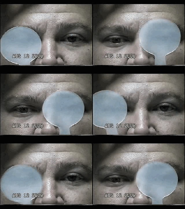
Cross cover
Presence of vertical disconjugate gaze suggests brainstem dysfunction

HINTS Gallery

Positive Head Impulse Test
Central Changing Nystagmus
Skew Deviation

Labs

  • Glucose
  • CBC/Chemistry
  • ECG

Imaging

  • Warranted if findings concerning for central process
  • MRI preferred

Management

Specific etiologies
Vestibular neuronitis: steroids
Meniere: dietary changes
BPPV: canalith repositioning
Symptomatic relief
Promethazine (Phenergan) 12.5-25mg PO
Ondansetron (Zofran) 4mg IV
Lorazepam (Ativan) 1-2mg PO/IV
Meclizine (Antivert) 25mg PO q6-8h PRN

References

  1. Kattah, J. C., Talkad, A. V., Wang, D. Z., Hsieh, Y.-H., & Newman-Toker, D. E. (2009). HINTS to diagnose stroke in the acute vestibular syndrome: three-step bedside oculomotor examination more sensitive than early MRI diffusion-weighted imaging. Stroke; a journal of cerebral circulation, 40(11), 3504–3510. doi:10.1161/STROKEAHA.109.551234
  2. Chang, A., & Olshaker, J. (2013). Dizziness and Vertigo. In Rosen’s Emergency Medicine – Concepts and Clinical Practice (8th ed., Vol. 1, pp. 162-169). Elsevier Health Sciences.

Seizure

Definition

Seizure
Pathologic neuronal activation leading to abnormal function
Epilepsy
Recurrent unprovoked seizures

Classification

  • Cause
    • Primary: Unprovoked
    • Secondary: Provoked, caused by trauma, illness, intoxication, metabolic disturbances, etc.
  • Effect on mentation
    • Generalized: involvement of both hemispheres with associated loss of consciousness (tonic-clonic, absence, atonic, myoclonic)
    • Focal: Involving single hemisphere with preserved level of consciousness
  • Status epilepticus
    • Witnessed convulsions lasting >5min
    • Recurrent seizure without recovery from postictal period

Causes of Seizures

Causes of Seizures

Management of Seizures

Management of Seizures

Medications for Treatment of Seizures

Medication Dose (adult) Dose (peds) Comment
1st Line
Lorazepam 4mg IV <13kg: 0.1mg/kg (max 2mg)
13-39kg: 2mg

>39kg: 4mg
Repeat in 10min
Midazolam 10mg IM 0.2mg/kg IM (max 5mg) Repeat in 10min
Midazolam 10mg buccal 0.5mg/kg buccal (max 5mg) Repeat in 10min
2nd Line
Fosphenytoin 20mg PE/kg IV    
Phenytoin 20mg/kg IV   May cause hypotension
3rd Line
Midazolam 0.05-2mg/kg/hr    
Propofol 1-2mg/kg bolus then 20-200mcg/kg/min    
Pentobarbital 5-15mg/kg bolus then 0.5-5mg/kg/hr    
Special Conditions
Glucose 50mL D50/W   Hypoglycemia
MgSO4 6g IV over 15min   Eclampsia (20wks gestation to 6wks post-partum)
Pyridoxine 0.5g/min until seizures stop, max 5g   INH ingestion
3% saline 100-200mL over 1-2h   Confirmed hyponatremia

History

Points suggestive of seizure over alternative process
Abrupt onset
Duration < 120s
LOC
Purposeless activity: automatisms, tonic-clonic
Provocation: fever in children, substance withdrawal
Postictal state
Retrograde amnesia
Incontinence, oral trauma (buccal maceration, tongue laceration)
Rapidly resolving lactic acidosis
Important historical points for patients with seizure history
Recent illness
Medications (adherence, changes, interactions)
Substance use
Ictogenic factors
Recent/remote head trauma
Developmental abnormalities
Substance use
Sleep deprivation
Pregnancy

Key Physical Examination Findings

  • Vital sign abnormalities persisting beyond immediate postictal state (may suggest drug/toxin exposure, CNS lesion)
  • Nuchal rigidity
  • Signs of IVDA
  • Sequela

    • Head trauma
    • Tongue laceration
    • Shoulder dislocation (posterior)
  • Neurological exam

    • Stroke
    • Elevated ICP
    • Failure to note improvement in postictal confusion (encephalopathy, subclinical seizures)

Labs

  • Glucose
  • BMP (Na, Ca, Mg)
  • AED levels
  • CBC (leukocytosis and bandemia common post-seizure)
  • CSF
  • B-hCG
  • LFT (hepatic dysfunction, alcoholic hepatitis)
  • Lactate (rapidly resolves on repeat)

Indications for Imaging

  • New seizures
  • History of trauma
  • History of malignancy
  • Immunocompromised
  • Headache
  • Anti-coagulation
  • Focal neurological exam
  • Persistent AMS

References

  1. McMullan, J., Davitt, A., & Pollack, C. (2013). Seizures. In Rosen’s Emergency Medicine – Concepts and Clinical Practice (8th ed., Vol. 1, pp. 156-161). Elsevier Health Sciences
  2. WikEM: Seizure

Altered Mental Status

Components of Consciousness

Components of Consciousness

Causes of Altered Mental Status

Causes of Altered Mental Status

History

Rate of onset
Abrupt: CNS
Gradual: Systemic

Physical Examination

  • Vital Signs

    • Blood Pressure: low (shock), high (SAH, stroke, ICP)
    • Heart Rate: low (medication overdose, conduction block), high (hypovolemia, infection, anemia, thyrotoxicosis, drug/toxin)
    • Temperature: low/high (infection, drug/toxin, environmental)
    • Respiratory Rate: low/high (CNS, drug/toxin, metabolic derangement)
  • Eyes

    • Unilateral dilation: CNS/structural cause
    • Papilledema: ICP
    • EOM: cranial nerve dysfunction
    • Oculocephalic: brainstem function
  • Head: trauma
  • Mucous membranes: hydration, laceration
  • Neck: meningeal irritation
  • Pulmonary: respiratory effort
  • CV: murmur, arrhythmia, CO
  • Abdomen: pulsatile mass, sequelae of liver failure
  • Skin: rash, needle tracks

Labs

  • Glucose
  • ECG: arrhythmia, ischemia, electrolyte abnormalities
  • BMP: electrolytes, renal failure, anion gap
  • ABG: hypoxemia, hypercarbia
  • Urinalysis: infection, SG
  • Utox
  • CBC: leukocytosis, leukopenia, severe anemia, thrombocytopenia
  • Ammonia: hepatic encephalopathy
  • TFT: thyrotoxicosis, myxedema coma
  • CSF: meningitis, encephalitis

Imaging

  • CT head: Non-contrast sufficient to identify ICH. Use contrast if mass/infection suspected
  • CTA head/neck: If aneurysm, AVM, venous sinus thrombosis or vertebrobasilar insufficiency suspected
  • CXR: PNA

References

  1. Bassin, B., & Cooke, J. (2013). Depressed Consciousness and Coma. In Rosen’s Emergency Medicine – Concepts and Clinical Practice (8th ed., Vol. 1, pp. 142-150). Elsevier Health Sciences.

Syncope

Causes of Syncope

Causes of Syncope

History

  • Rate of onset
  • Position at onset
  • Duration, rate of recovery
  • Preceding features

    • Obstruction: associated with exertion
    • Neurocardiogenic: associated with emotion, micturition, bowel movement, emesis, neck movement
  • Following features

    • Seizure: Postictal confusion
    • Hypotension: Initial VS
    • Associated trauma

Physical Examination

  • VS: rhythm, BP, temperature
  • HEENT: mucous membranes (laceration, dry), trauma, papilledema
  • CV: murmur (AS), rub (pericarditis), bruit (cerebrovascular disease), JVD (obstruction)
  • Lungs: crackles (CHF)
  • Abdomen: pulsatile mass (AAA)
  • Extremities: pulse discrepancy (dissection)
  • Neuro: focal findings (stroke, mass, seizure)

Evaluation

  • ECG: arrhythmia (PR, QT, Brugada, unanticipated hypertrophy, RV strain, pericarditis)
  • Orthostatic VS
  • CBC: anemia
  • BMP: electrolyte abnormalities (hyponatremia, hyper/hypokalemia)
  • Glucose: hypoglycemia
  • Troponin: ischemia
  • B-hCG: ectopic
  • Utox: drugs
  • CXR: dissection
  • CT head: focal neurological findings
  • CT PA: concern for PE
  • US abdomen: AAA

San Francisco Syncope Rules (CHESS)

  • CHF
  • Hematocrit <30%
  • ECG abnormality
  • SBP <90mmHg
  • SOB

References

  1. De Lorenzo, R. (2013). Syncope. In Rosen’s Emergency Medicine – Concepts and Clinical Practice (8th ed., Vol. 1, pp. 135-141). Elsevier Health Sciences.

Weakness

View Algorithm
There is a ddxof algorithm for the evaluation of weakness. View it here.

Motor Neuron Signs

Upper Motor Neuron:
Spasticity
Hyperreflexia
Pronator drift
Babinski
Lower Motor Neuron:
Flaccidity
Hyporeflexia
Fasciculation
Atrophy

Causes of Weakness

Lesion Critical Emergent
Non-neurological Shock (VS, clinical assessment)
Hypoglycemia (POC glucose)
Electrolyte derangement (BMP)
Anemia (POC Hb, CBC)
MI (ECG, troponin)
CNS depression (Utox, EtOH)
 
Cortex Stroke Tumor
Abscess
Demyelination
Brainstem Stroke Demyelination
Spinal Cord Ischemia
Compression (disk, abscess, hematoma)
Demyelination (transverse myelitis)
Peripheral Acute demyelination (GBS) Compressive plexopathy
Muscle Rhabdomyolysis Inflammatory myositis

Weakness Syndromes

Unilateral weakness, ipsilateral face
Lesion: Contralateral cortex, internal capsule
Causes: Stroke (sudden onset), demyelination/mass (gradual onset)
Symptoms: Neglect, visual field cut, aphasia
Findings: UMN signs
Key features: Association with headache suggests hemorrhage or mass
Unilateral weakness, contralateral face
Lesion: Brainstem
Causes: Vertebrobasilar insufficiency, demyelination
Symptoms: Dysphagia, dysarthria, diplopia, vertigo, nausea/vomiting
Findings: CN involvement, cerebellar abnormalities
Unilateral weakness, no facial involvement
Lesion: Contralateral medial cerebral cortex, discrete internal capsule
Causes: Stroke
Rare Cause: Brown-Sequard if contralateral hemibody pain and temperature sensory disturbance
Unilateral weakness single limb (monoparesis/plegia)
Lesion: Spinal cord, peripheral nerve, NMJ
UMN signs: Brown-Sequard if contralateral pain and temperature sensory disturbance
LMN signs: Radiculopathy if associated sensory disturbance
Normal reflexes, normal sensation: Consider NMJ disorder
Bilateral weakness of lower extremities (paraparesis/plegia)
Lesion: Spinal cord, peripheral nerve
UMN signs: Anterior cord syndrome (compression, ischemia, demyelination) if contralateral pain and temperature sensory disturbance
Cauda equina: Loss of perianal sensation, loss of rectal tone, or urinary retention
GBS: If no signs of cauda equina and sensory disturbances paralleling ascending weakness (with hyporeflexia)
Bilateral weakness of upper extremities
Lesion: Central cord syndrome
Causes: Syringomyelia, hyperextension injury
Findings: Pain and temperature sensory disturbances in upper extremities (intact proprioception)
Bilateral weakness of all four extremities (quadriparesis/plegia)
Lesion: Cervical spinal cord
Findings: UMN signs below level of injury, strength/sensory testing identifies level
Bilateral weakness, proximal groups
Lesion: Muscle
Causes: Rhabdomyolysis, polymyositis, dermatomyositis, myopathies
Findings: Muscle tenderness to palpation, no UMN signs, no sensory disturbances
Facial weakness, upper and lower face
Lesion: CNVII
Causes: Bell’s palsy, mastoiditis, parotitis
Other CN involvement suggests brainstem lesion, multiple cranial neuropathies, or NMJ

Review of Spinal Cord Anatomy

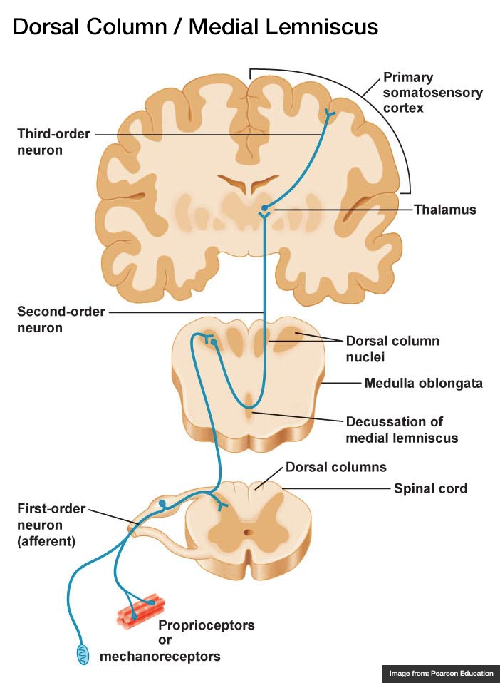
  • Dorsal Column – Medial Lemniscus (fine touch, proprioception)
    1. Afferent sensory fibers with cell body in DRG
    2. Ascend in ipsilateral posterior column
    3. Synapse in medulla, decussate, ascend in contralateral medial lemniscus
    4. Synapse in thalamus (VPL)
    5. Synapse in sensory strip of post-central gyrus
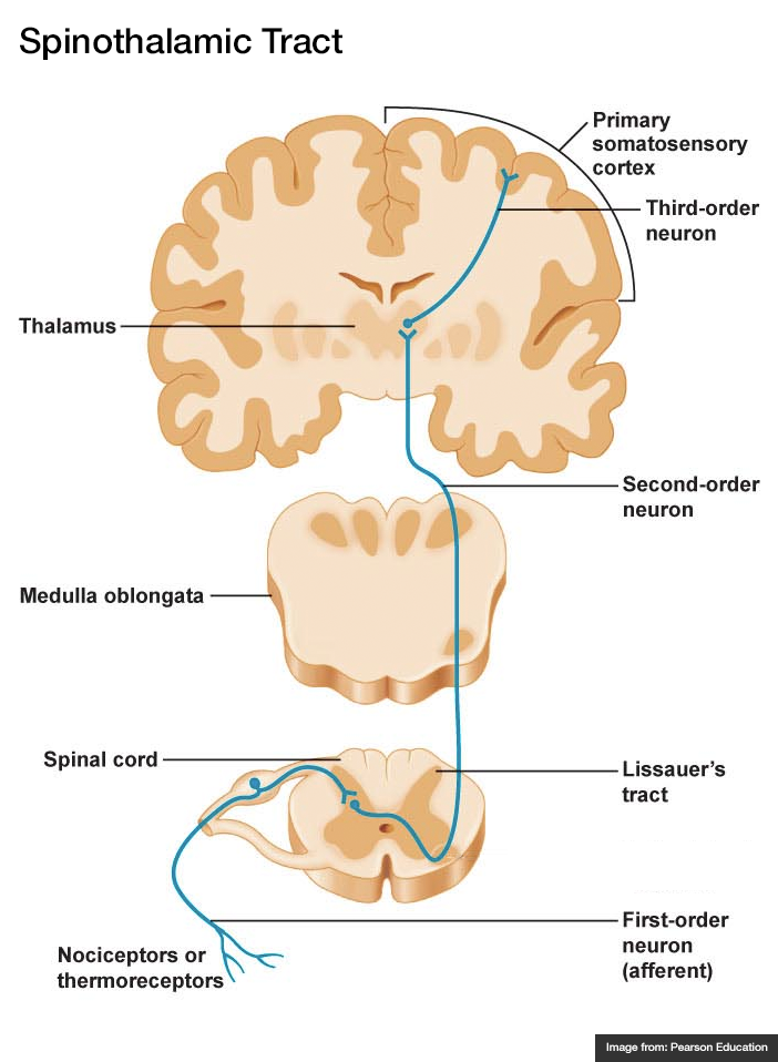
  • Spinothalamic Tract (pain, temperature)

    1. Afferent sensory fibers with cell body in DRG
    2. Ascends 1-2 levels
    3. Synapse in ipsilateral spinal cord, decussate, ascend in contralateral lateral spinothalamic tract
    4. Synapse in thalamus (VPL)
    5. Synapse in sensory strip of post-central gyrus
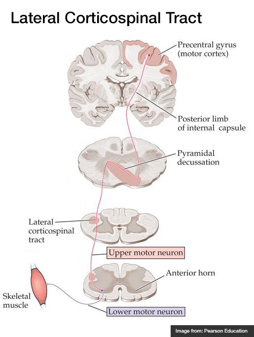
  • Lateral Corticospinal Tract (motor)

    1. Efferent cell body in motor strip of pre-central gyrus
    2. Descends through internal capsule
    3. Decussates in pyramid of medulla, descends in contralateral lateral corticospinal tract
    4. Synapse in anterior horn, lower motor neuron to muscle fiber
Spinal Cord Syndromes
Spinothalamic Tract
Dorsal Column / Medial Lemniscus
Lateral Corticospinal Tract

References

  1. Morchi, R. (2013). Weakness. In Rosen’s Emergency Medicine – Concepts and Clinical Practice (8th ed., Vol. 1, pp. 124-128). Elsevier Health Sciences.

Fever

Causes of Fever

Causes of Fever

Key Features

  • Morbidity and mortality increase with age and comborbidities
  • Most common sources in elderly: respiratory, genitourinary, skin/soft-tissue
  • Atypical presentations: functional decline, altered mental status

Immediate Evaluation and Management

  • Critical Findings

    • Altered mental status
    • Respiratory distress
    • Hemodynamic instability
  • Critical Interventions

    • Airway management, supplemental O2
    • Cardiac monitoring
    • Fluid resuscitation
    • Empiric antibiotics
    • Cooling measures (T>41.0°C)

Pathophysiology of Fever

Production of endogenous or exogenous pyrogens
Increase temperature set point in hypothalamus
Patient experiences chills when core temperature < set point
Vasoconstriction, shivering causes fever
Patient experiences euthermia, though may feel malaise, fatigue
Resolution
Patient experiences sweats until core temperature returns to normal set point

References

  1. Blum, F., & Biros, M. (2013). Fever in the Adult Patient. In Rosen’s Emergency Medicine – Concepts and Clinical Practice (8th ed., Vol. 1, pp. 119-123). Elsevier Health Sciences.

ECG Guide

The format of this article is atypical for the structure and concept of the website – but it’s always been about learning. Here is a simplified guide to ECG interpretation with a focus on the aspects I find more challenging to understand or recall.

Grid and Leads

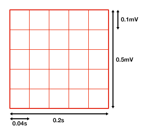
The ECG grid
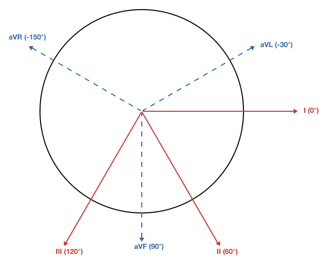
Limb leads
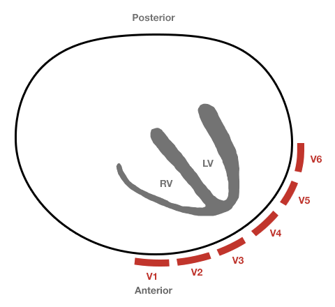
Precordial Leads

Axis

ECG axes

Atrial Enlargement

Atrial enlargement

Normal:
First portion of deflection is RA, second is LA
Right Atrial Enlargement:
P-wave amplitude > 2.5mm in inferior leads
Normal duration P-wave
Left Atrial Enlargement:
P-wave duration increased (terminal negative portion >0.04s)
Amplitude of terminal negative component >1mm below isoelectric line in V1

Ventricular Hypertrophy

Right Ventricular Hypertrophy:
Right axis deviation
Abnormal R-wave progression

  • Increased R-wave amplitude in leads overlying the right ventricle (V1)
  • Increased S-wave amplitude in leads overlying the left ventricle (V6)
Criteria

  • V1: R>S
  • V6: S>R
Left Ventricular Hypertrophy:
Left axis deviation
Increased R-wave amplitude in leads overlying the LV (I, aVL, V5, V6)
Increased S-wave amplitude in leads overlying the RV (V1)
Criteria:

  • Precordial Leads
    • R-wave in V5/V6 + S-wave in V1/V2 > 35mm
    • R-wave in V5 > 26mm
    • R-wave in V6 > 20mm
  • Limb Leads
    • R-wave in aVL > 11mm
    • R-wave in aVF > 20mm
  • Combined
    • R-wave in aVL + S-wave in V3 > 20mm (F), 28mm (M)

Secondary Repolarization Abnormalities

Secondary repolarization abnormality

  • Downsloping ST-segment depression
  • Asymmetric T-wave inversion

Bundle Branch Blocks

Left Bundle Branch Block

Left bundle branch block

  • QRS duration > 0.12s (3 boxes)
  • Broad or notched R-wave with prolonged upstroke in I, aVL, V5, V6
  • Associated ST-segment depression and T-wave inversion
  • Reciprocal changes in V1, V2 (deep S-wave)
  • Possible LAD

Right Bundle Branch Block

Right bundle branch block

  • QRS duration > 0.12s (3 boxes)
  • RSR’ in V1, V2
  • Reciprocal changes in I, aVL, V5, V6 (deep S-wave)

Hemiblocks

His-Purkinje system and hemiblocks (anterior fascicular block, posterior fascicular block)

Other Blocks

  • Non-specific intraventricular conduction delay: QRS >0.10s without BBB
  • Incomplete BBB: LBBB/RBBB pattern with non-prolonged QRS
  • Bifascicular block: RBBB + LAFB/LPFB (by axis deviation)

Ischemia and Infarction

ECG changes associated with ischemia and infarction

  1. Hyperacute T-waves
  2. T-wave inversion: Symmetric, compared to TWI associated with repolarization abnormalities
  3. ST-elevation: Unlike J-point elevation, ST-segment merges with T-wave
  4. Q-waves
    1. Duration > 0.04s
    2. Amplitude > 1/3 R-wave
    3. Normal in aVR

Coronary Artery Territories

Coronary artery territories

Distribution Coronary Artery Leads Reciprocal Changes
1. Inferior RCA, PDA II, III, aVF Anterior, Lateral
2. Lateral LCx I, aVL, V5, V6 Inferior
3. Anterior LAD V1-V6 Inferior
4. Posterior RCA Posterior Anterior (esp. V1)

External Links

Diplopia

History and Physical

38F with no medical history, presenting with double vision. The patient reported six weeks of intermittent diplopia for which she had presented to this hospital previously. She was briefly admitted for evaluation of possible cranial nerve IV palsy. Extensive imaging was unremarkable, without mass lesion, infarction, vascular malformation, or meningeal enhancement. She was discharged with outpatient follow-up including ophthalmology clinic and further imaging.

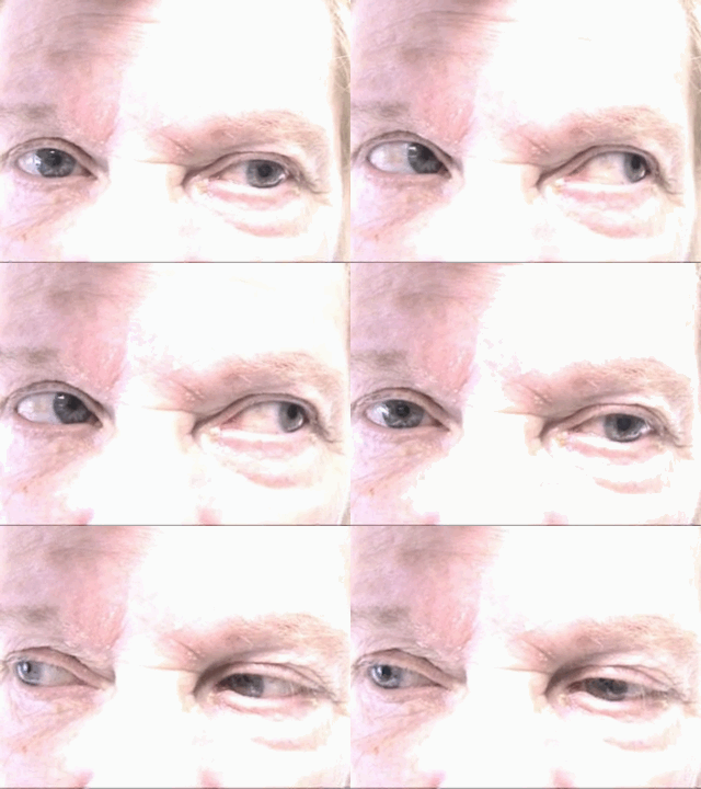
The patient represented due to persistent diplopia that is worse with right gaze. The diplopia is predominantly vertical, alleviated by head tilt. Now associated with three days of right ptosis as well as two weeks of progressive weakness and fatigue – most notable when climbing stairs.

Examination notable for right hypertropia increased on right or downward gaze suggestive of isolated inferior rectus weakness. Pupils were equal and reactive. There was marked fatigable ptosis with 2mm right palpebral fissure compared to 10mm on contralateral side. Symmetrical muscle weakness was noted, 4/5 neck flexion, elbow extension, wrist flexion/extension, shoulder abduction, hip flexion. Gait was wide-based. Application of ice for 5 minutes improved right palpebral fissure opening to >7mm.

Further evaluation included CXR and CT chest with intravenous contrast which did not identify a mediastinal mass. The patient’s respiratory status remained stable throughout hospitalization as assessed by measurements of forced vital capacity. On hospital day one, an edrophonium test was performed which was positive. The patient was started on pyridostigmine, completed a course of IVIG and was discharged with outpatient neurology follow-up.

Evaluation of Diplopia 1

History

  1. Onset/cadence
  2. Direction of gaze with worst diplopia
  3. Orientation (vertical/horizontal)
  4. Associated symptoms (headache, vertigo, dysarthria, eye pain)

Terms Describing Eye Position

Terms describing eye position

Tropias are always present, phorias are identified by cross-cover testing (break fusion)

Algorithm for the Evaluation of Diplopia 2

Algorithm for the Evaluation of Diplopia

Causes of Diplopia 3,4,5,6

Finding EOM Causes Features
Mechanical orbitopathy Variable. Abrupt restriction of movement Orbital cellulitis Pain, erythema
Orbital pseudotumor Autoimmune
Trauma History
Thyroid eye disease Bilateral
Isolated CN III Limited adduction/upgaze/downgaze Microvascular ischemia Pain, risk factors, pupil-sparing
Aneurysm Pupil involvement
Demyelination MRI
Isolated CN IV Limited downgaze (hypertropia) Trauma May be mild
Microvascular ischemia Less common than CN III
ICP Fundoscopy, imaging
Demyelination MRI
Isolated CN VI Limited abduction

(esotropia)

ICP Fundoscopy, imaging
Demyelination MRI
Microvascular ischemia Less common than CN III
INO Limited adduction

(exotropia)

Demyelination MRI
Stroke Dysarthria, ataxia, facial weakness
Multiple CN involvement (III, IV, VI) Variable Cavernous sinus process Retroorbital pain, conjunctival injection or chemosis
Brainstem deficits Variable Brainstem stroke Weakness, dysmetria, tremor
Basilar artery occlusion Vertigo, slurred speech
Wernicke AMS, ataxia, nystagmus
Basilar meningitis Fever, photophobia, meningismus
Miller-Fisher Ataxia, areflexia
Neuromuscular process Variable Myasthenia gravis Fatigability, ice test

References

  1. Alves, M., Miranda, A., Narciso, M. R., Mieiro, L., & Fonseca, T. (2015). Diplopia: a diagnostic challenge with common and rare etiologies. The American journal of case reports, 16, 220–223. doi:10.12659/AJCR.893134
  2. Borooah, S., Wright, M., & Dhillon, B. (2011). Pocket Tutor Ophthalmology. JP Medical Limited. Retrieved from https://books.google.com/books?id=z\_CfWj8-ftoC
  3. Dinkin, M. (2014). Diagnostic approach to diplopia. Continuum (Minneapolis, Minn.), 20(4 Neuro-ophthalmology), 942–965. doi:10.1212/01.CON.0000453310.52390.58
  4. Rucker, J. C., & Tomsak, R. L. (2005). Binocular diplopia. A practical approach. The neurologist, 11(2), 98–110. doi:10.1097/01.nrl.0000156318.80903.b1
  5. Friedman, D. I. (2010). Pearls: diplopia. Seminars in neurology, 30(1), 54–65. doi:10.1055/s-0029-1244995
  6. Guluma, K. (2013). Diplopia. In Rosen’s Emergency Medicine – Concepts and Clinical Practice (8th ed., Vol. 1, pp. 176-183). Elsevier Health Sciences.
  7. WikEM: Diplopia

Anemia

HPI

70M with a history of dementia presenting with 3 days of fatigue. The patient was unable to provide detailed history, however family members reported worsening fatigue with the patient requiring assistance with ambulation for several days. The patient was referred from an outside clinic after point-of-care hemoglobin of 6.7. No reported history of anemia, and no history suggestive of obvious external bleeding.

Vital signs stable, tachycardia and tachypnea noted with minimal exertion but saturating well on ambient air and in no acute distress. Examination notable for conjunctival pallor without scleral icterus, systolic flow murmur, brown stool guaiac negative.

CBC with hemoglobin of 7.5 , MCV 80.3 , RDW 22.4 , no leukocytosis and normal platelets. Also noted was an alkaline phosphatase of 828 , normal total and direct bilirubin, and undetectable serum troponin. Chest x-ray showed a possible pleural-based mass.

The patient was transfused two units of PRBC’s and admitted for further evaluation. CT chest/abdomen/pelvis revealed sternal and rib-based pleural soft-tissue mass, prostate mass, pelvic and retroperitoneal lymphadenopathy as well as extensive bony metastatic disease consistent with primary prostate cancer with diffused metastasis. Serum PSA was 2,087 . Iron studies suggested anemia of chronic disease. Reticulocytes were not obtained but may have suggested inadequate production index given extensive bony metastases and possible associated myelosuppression. The patient was symptomatically improved after transfusion and discharged with outpatient follow-up for discussions regarding possible biopsy and treatment.

Images

Chest x-ray with pleural-based mass

Areas of pleural thickening. Possible pleural based mass in left mid lung.

ct_chest_anemia_01
ct_chest_anemia_02
ct_chest_anemia_03
ct_chest_anemia_04
ct_chest_anemia_05
ct_chest_anemia_06
ct_chest_anemia_07
ct_chest_anemia_08
ct_chest_anemia_09
ct_chest_anemia_10
ct_chest_anemia_11
ct_chest_anemia_12
ct_chest_anemia_13

CT Chest: Lung Window

  • Rib-based pleural soft tissue masses.
  • Large 5.6 x 4.4cm anterior sternal soft-tissue mass.
ct_bone_anemia_01
ct_bone_anemia_02
ct_bone_anemia_03
ct_bone_anemia_04
ct_bone_anemia_05
ct_bone_anemia_06
ct_bone_anemia_07
ct_bone_anemia_08

CT Body: Bone Window

  • Extensive bony metastatic disease.
  • Prostate mass, large pelvic and retroperitoneal lymphadenopathy.
  • Consistent with primary prostate cancer with diffuse metastasis.

Algorithm for the Evaluation of Anemia 1,2

Algorithm for the Evaluation of Anemia

References:

  1. Zaiden R, Rana F. Evaluation of Anemia. BMJ Best Practice. Oct 2014. http://us.bestpractice.bmj.com/best-practice/monograph/93/overview.html. Last accessed 15 May 2015.
  2. Janz, T. G., Johnson, R. L., & Rubenstein, S. D. (2013). Anemia in the emergency department: evaluation and treatment. Emergency medicine practice, 15(11), 1–15– quiz 15–6.
  3. WiKEM: Anemia

Conjunctivitis and the Red Eye

Differential diagnosis of Conjunctivitis 1,2,3

Condition Pain Visual Acuity Photophobia Discharge Conjunctiva Lymphadenopathy Laterality Associated Features
Viral conjunctivitis None Unaffected None + watery ++ follicular pattern Pre-auricular Unilateral, spreads bilateral Viral URI
Bacterial conjunctivitis None Unaffected None ++ purulent +++ papillary pattern Occasional Unilateral, spreads bilateral Otitis media
Allergic conjunctivitis None Unaffected None + mucoid + None Bilateral Atopy

Differential Diagnosis of Red Eye 4,5

Condition Comment Hyperemia Pupil Pain Visual Acuity Cornea
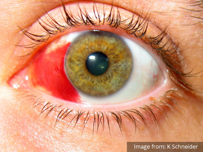
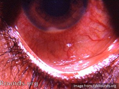
Subconjuntival hemorrhage Subconjunctival Hemorrhage Associated with trauma, coagulopathy, hypertension. Unilateral, sharply circumscribed Unaffected None Unaffected Clear
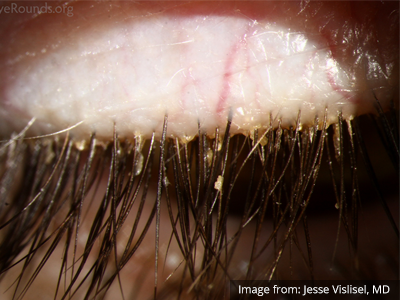
Blepharitis Blepharitis Acute/chronic inflammation of eyelid. Diffuse Unaffected Foreign body sensation Unaffected Clear
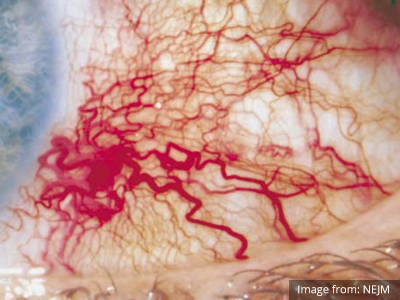
Epislceritis Episcleritis Recurrent, self-limited episodes, possible autoimmune association. Engorged, radially-oriented vessels Unaffected Mild Unaffected Clear
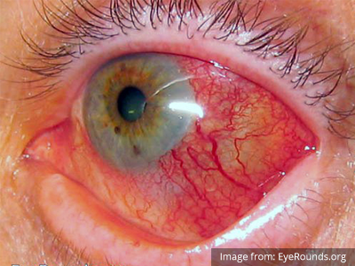
Scleritis Scleritis Vascular or connective tissue disease. Focal or diffuse, pink sclera Unaffected Moderate Reduced Clear
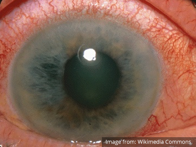
Acute angle-closure glaucoma Acute Angle-Closure Glaucoma Mydriasis leading to decreased outflow of aqueous humor. Circumcorneal injection Semi-dilated Severe Reduced Hazy
Acute anterior uveitis Uveitis Inflammation of iris or ciliary body. Circumcorneal injection Constricted Moderate Reduced Hazy
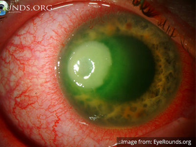
Keratitis Keratitis Inflammation of corneal epithelium. Caused by infection, contact lenses, UV exposure. Multiple punctate erosions, stain with fluorescein Unaffected Moderate Reduced Hazy

Algorithm for the Evaluation of the Red Eye 6

Algorithm for the Evaluation of the Red Eye

References

  1. Teoh, D. L., & Reynolds, S. (2003). Diagnosis and management of pediatric conjunctivitis. Pediatric emergency care, 19(1), 48–55.
  2. Azari, A. A., & Barney, N. P. (2013). Conjunctivitis. JAMA: the journal of the American Medical Association, 310(16), 1721. doi:10.1001/jama.2013.280318
  3. Cronau, H., Kankanala, R. R., & Mauger, T. (2010). Diagnosis and management of red eye in primary care. American family physician, 81(2), 137–144.
  4. Leibowitz, H. M. (2000). The red eye. New England Journal of Medicine, 343(5), 345–351. doi:10.1056/NEJM200008033430507
  5. Richards, A., & Guzman-Cottrill, J. A. (2010). Conjunctivitis. Pediatrics in review / American Academy of Pediatrics, 31(5), 196–208. doi:10.1542/pir.31-5-196
  6. Borooah, S., Wright, M., & Dhillon, B. (2011). Ophthalmology. JP Medical Limited.

Severe Traumatic Brain Injury

HPI:

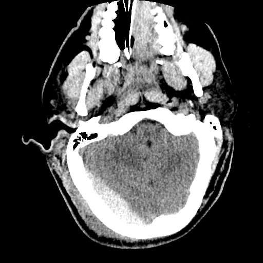
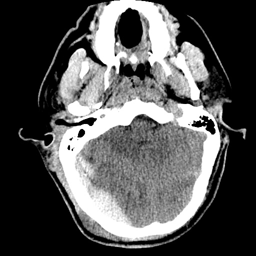
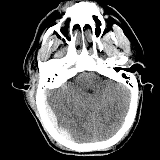
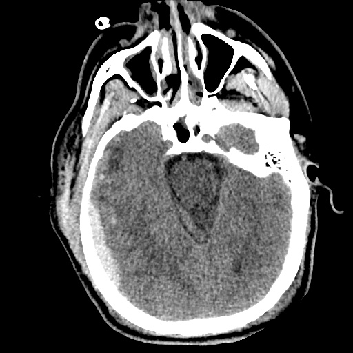
34 year-old male brought in by ambulance s/p assault. Field GCS reportedly 7, in trauma bay assessed as E2-V4-M6. Witnessed seizure in CT scanner, resolved with lorazepam. Intubated for airway protection, underwent external ventricular drain placement and transferred to surgical ICU.

Initial imaging revealed bifrontal subdural hematomas and right temporal hemorrhagic contusion with generalized edema. Repeat imaging one hour later showed interval development of large extra-axial hemorrhage overlying the right occipital and parietal lobes (2.2cm), representing subdural or epidural hematoma.

The patient’s ICU course was complicated by continued seizures and refractory elevation in intracranial pressure. A pentobarbital infusion was started and titrated to adequate burst suppression and hyperosmolar therapy with both mannitol and hypertonic saline continued. Additional imaging revealed stable hemorrhage but continued diffuse cerebral edema evidenced by sulcal effacement.

On hospital day 5, examination revealed bilateral fixed and dilated pupils. Imaging revealed effacement of basilar cisterns, pre-pontine cistern, and cisterna magna suggestive of impending/ongoing transtentorial and tonsillar herniation. Pentobarbital was weaned and conventional cerebral angiography as well as cerebral perfusion studies were consistent with brain death.

Images

tbi1_01
tbi1_02
tbi1_03
tbi1_04
tbi1_05
tbi1_06
tbi1_07
tbi1_08
tbi1_09
tbi1_10
tbi1_11
tbi1_12

CT head without contrast one hour after presentation

  • Large extra-axial posterior hemorrhages. Hemorrhagic contusions in the right frontal and temporal lobes.
  • The cerebral sulci appear effaced – findings suggest diffuse cerebral edema.
  • S/p EVD using a right frontal approach.
tbi2_01
tbi2_02
tbi2_03
tbi2_04
tbi2_05
tbi2_06
tbi2_07
tbi2_08
tbi2_09
tbi2_11
tbi2_12

CT head without contrast on hospital day 5

  • Interval evidence of global hypoxic/ischemic injury to the brain.
  • Interval apparent effacement of the basilar cisterns, pre-pontine cistern, and cisterna magna suggesting impending/ongoing downward transtentorial herniation and tonsillar herniation.
  • Stable supra/infratentorial subdural/epidural hematoma.

Algorithm for the Management of Severe Traumatic Brain Injury1,2

Algorithm for the Management of Severe Traumatic Brain Injury

References

  1. Brain Trauma Foundation, American Association of Neurological Surgeons, Congress of Neurological Surgeons, Joint Section on Neurotrauma and Critical Care, AANS/CNS, Carney, N. A., & Ghajar, J. (2007). Guidelines for the management of severe traumatic brain injury. Introduction. Journal of neurotrauma, 24 Suppl 1, S1–2. doi:10.1089/neu.2007.9997
  2. Stocchetti, N., & Maas, A. I. R. (2014). Traumatic intracranial hypertension. The New England journal of medicine, 370(22), 2121–2130. doi:10.1056/NEJMra1208708
  3. WikEM: Severe traumatic brain injury

Lactic Acidosis

HPI:

59F with a reported history of congestive heart failure, presenting with intermittent chest discomfort for three days.

She characterized this discomfort as “heartburn”, describing a mid-epigastric burning sensation radiating up her neck, not associated with exertion, lasting 1-2 hours and resolving with antacids. The patient has poor exercise tolerance at baseline and for the past several years has been able to ambulate only short distances around her home, and states that these symptoms have been worsening in the past week. She denies chest pain on exertion, orthopnea or paroxysmal nocturnal dyspnea. She states that she was diagnosed with congestive heart failure five years ago, but was never prescribed medications.

On further questioning, the patient reports several weeks of mouth and lip pain which has limited oral intake, though no dysphagia to solids or liquids. She otherwise denies fevers/chills, abdominal pain, nausea/vomiting, cough, changes in urinary or bowel habits.

In the emergency department, the patient was noted to have an elevated serum troponin, though ECG showed no changes of acute ischemia/infarction.

PMH:

  • Congestive heart failure

PSH:

  • None

FH:

  • Mother with diabetes
  • Father with MI at age 65

SHx:

  • 4-5 drinks of alcohol/day
  • No tobacco or drug use

Meds:

  • None

Allergies:

NKDA

Physical Exam:

VS: T 37.4 HR 106 RR 18 BP 145/82 O2 100% RA
Gen: Morbidly obese female, lying in bed, in no acute respiratory distress, speaking in complete sentences.
HEENT: Dry, cracked lips, slightly erythematous, otherwise moist mucous membranes, poor dentition. Mild scleral icterus. No cervical lymphadenopathy.
CV: Rapid rate, regular rhythm, normal S1/S2, II/VI systolic ejection murmur at LUSB, no radiation appreciated. No jugular venous distension.
Lungs: Clear to auscultation in posterior lung fields bilaterally, no crackles appreciated.
Chest: Well-circumscribed erythematous patch in folds beneath left breast, no underlying fluctuance, no significant tenderness to palpation. On contralateral breast, some hyperpigmentation but no erythema.
Abdomen: Obese, non-tender, non-distended. Patch of erythema below pannus, mildly tender to palpation.
Ext: Bilateral lower extremities with marked edema and overlying scaly plaques, some slightly ulcerated weeping serous fluid. Peripheral pulses are difficult to palpate, capillary refill difficult to assess.

Labs/Studies:

  • CBC: 11.1/11.1/34.5/212 (MCV 114.2)
  • BMP: 140/4.5/97/20/10/1.14/64
  • Anion Gap: 23
  • LFT: AST: 73, ALT: 26, AP: 300, TB: 4.6, DB: 2.1, Alb: 3.0, INR 1.3
  • BNP: 158
  • Troponin: 1.284
Sinus tachycardia, LVH, secondary repolarization abnormalities

Sinus tachycardia, LVH, secondary repolarization abnormalities

Imaging:

CT Pulmonary Angiography:
No evidence of central pulmonary embolism, thoracic aortic dissection, or thoracic aortic aneurysm. Evaluation of the peripheral vessels is limited due to motion artifact. No focal consolidation or pneumothorax.

CT Abdomen/Pelvis non-contrast:
No evidence of intra-abdominal abscess or definite source of infection. Marked hepatic steatosis.

CT Lower Extremity non-contrast:
Diffuse circumferential subcutaneous edema involving both lower extremities from the level of the mid thighs distally through the feet. There are bilateral subcutaneous calcifications which are likely venous calcifications in the setting of chronic venous stasis disease. There is some overlying skin thickening.

TTE:
There is moderate concentric left ventricular hypertrophy with hyperdynamic LV wall motion. The Ejection Fraction estimate is >70%. Grade I/IV (mild) LV diastolic dysfunction. No hemodynamically significant valve abnormalities.

US Abdomen:
Hepatomegaly, echogenic liver suggesting fatty infiltration. Moderately blunted hepatic vein waveforms suggesting decreased hepatic parenchymal compliance.

Assessment/Plan:

The patient was admitted to the cardiology service for management of NSTEMI and evaluation of undiagnosed CHF. She was started on a heparin continuous infusion. In addition, a CT pulmonary angiogram was obtained to evaluate for pulmonary embolism as an explanation of her progressive dyspnea on exertion. No PE, consolidation or effusion was identified.

Despite the patient’s reported history of congestive heart failure, there was no evidence that her symptoms were a result of an acute exacerbation with only a mildly elevated BNP but no jugular venous distension or evidence of pulmonary edema. The patient’s significant lower extremity edema was more suggestive of chronic venous stasis.

One notable laboratory abnormality that was explored was her elevated anion gap metabolic acidosis. Studies submitted included serum lactate, salicylates, osmolarity, CK, and urinalysis for ketonuria. This evaluation was notable for an elevated serum lactate of 13.2mmol/L and an arterial blood gas that showed adequate respiratory compensation (and no A-a gradient). Given the patient’s modest leukocytosis (with neutrophil predominance), and tachycardia, the concern for sepsis was increased though the source remained unclear. Prominent possibilities included a skin and soft-tissue infection vs. less likely intra-abdominal source though the patient’s physical examination was not suggestive of a process that would produce such a substantial lactic acidosis. Blood cultures were drawn and the patient was started on empiric antibiotics for the suspected sources. In addition, the patient was cautiously volume resuscitated given her reported history of CHF while pending a transthoracic echocardiogram to evaluate cardiac function. Additional imaging including CT abdomen/pelvis and lower extremities was obtained (though without contrast due to the patient’s recent exposure), and no obvious source was identified.

Over the next two days, the patient’s serum lactate downtrended to normal range, as did the serum troponin. A transthoracic echocardiogram showed an LVEF >70% with mild concentric hypertrophy and diastolic dysfunction. Blood and urine cultures were without growth.

Additional issues managed during the hospitalization included elevated serum transaminases (AST > ALT), conjugated hyperbilirubinemia and evidence of decreased hepatic synthetic function with hypoalbuminemia and elevated INR. Given the patient’s history of EtOH use, as well as other corroborating findings including macrocytic anemia, hypomagnesemia, folate and B12 deficiency, this was attributed to alcoholic hepatitis (discriminant function <32). Infectious hepatitis serologies were negative. The patient was started on nutritional supplements. Finally, the patient persistently complained of lip and oral mucosal pain. Examination was without discrete lesions but some mucosal redness was identified. Despite poor dentition, there was no evidence of abscess and HSV/HIV testing was negative. This was thought to be stomatitis caused by her identified nutritional deficiencies.

Differential Diagnosis of Elevated Serum Lactate 1,2

Differential Diagnosis of Elevated Serum Lactate

Algorithm for Evaluation of Acidemia 3,4

Algorithm for Evaluation of Acidemia

Algorithm for Evaluation of Alkalemia 3,4

Algorithm for Evaluation of Alkalemia

References:

  1. Fall, P. J., & Szerlip, H. M. (2005). Lactic acidosis: from sour milk to septic shock. Journal of intensive care medicine, 20(5), 255–271. doi:10.1177/0885066605278644
  2. Luft, F. C. (2001). Lactic acidosis update for critical care clinicians. Journal of the American Society of Nephrology : JASN, 12 Suppl 17, S15–9.
  3. Ingelfinger, J. R., Berend, K., de Vries, A. P. J., & Gans, R. O. B. (2014). Physiological Approach to Assessment of Acid–Base Disturbances. The New England journal of medicine, 371(15), 1434–1445. doi:10.1056/NEJMra1003327
  4. Ingelfinger, J. R., & Seifter, J. L. (2014). Integration of Acid–Base and Electrolyte Disorders. The New England journal of medicine, 371(19), 1821–1831. doi:10.1056/NEJMra1215672

Pleural Effusion

HPI:

64F with a history of CAD, MI, CHF, CLL and rheumatoid arthritis who presented to the emergency department after transfer from a rehabilitation facility for respiratory distress. The patient reported several days of progressive shortness of breath with dyspnea on exertion. She also noted some associated orthopnea and lower extremity edema. The patient was recently hospitalized for similar symptoms and was diagnosed with CHF at the time. At the rehab facility, the patient became hypoxemic and hypertensive, reporting shortness of breath and chest pain prior to presentation.

Hospital Course:

The patient was initially managed with BiPAP and nitroglycerin continuous infusion, but was then stable on supplemental O2 via nasal cannula and was transitioned to long-acting nitrates and anti-hypertensives. The patient’s hypoxemic respiratory failure was initially attributed to acute exacerbation of left-ventricular heart failure, and the patient was managed with spot diuresis. However, there was no symptomatic improvement and the patient became hypernatremic so diuresis was held as alternative diagnoses were explored.

A transthoracic echocardiogram showed preserved LVEF (50-55%), but some diastolic dysfunction and elevated PAP/RAP. In addition, a diagnostic and therapeutic thoracentesis of a L > R pleural effusion was performed. Pleural fluid studies were suggestive of a transudative process, though with some abnormal characteristics (including lymphocyte predominance, as well as presence of signet cells).

Rheumatology and pulmonology services were consulted for input and recommendations for further evaluation were appreciated. Per rheumatology, the patient’s diagnosis of rheumatoid arthritis may not be consistent with her presentation or prior serologic studies. Her pleural fluid analysis was also not consistent with rheumatoid disease. According to pulmonary consult, the patient’s hypoxemia remains most consistent with left ventricular dysfunction though primary pulmonary processes cannot be excluded (and would warrant further evaluation with imaging and pulmonary function testing).

PMH:

  • CHF
  • CAD
  • CVA
  • Myocardial Infarction
  • HTN
  • Hypothyroidism
  • CLL
  • Anemia

PSH:

  • TAH-BSO

FH:

  • No family history of autoimmune disease.
  • Mother: DM

SHx:

  • Denies tobacco/EtOH/drug use
  • Lives at home, at SNF since discharge

Meds:

  • Furosemide 20mg p.o. daily
  • Gabapentin 300mg p.o. t.i.d.
  • Hydralazine 50mg p.o. t.i.d.
  • Hydrochlorothiazide 25mg p.o. b.i.d.
  • Hydroxychloroquine 200mg p.o. daily
  • Levothyroxine 25mcg p.o. daily
  • Minoxidil 2.5mg p.o. b.i.d.
  • Pantoprazole 40mg p.o. daily
  • Prednisone 15mg p.o. daily

Allergies:

  • Shellfish
  • Physical Exam:

    VS: T 37.2 HR 84 RR 15 BP 147/75 O2 97% 4LNC
    Gen: Elderly female, alert and oriented to self and place, responding appropriately to questions.
    HEENT: Mucous membranes moist, sclera anicteric, no cervical lymphadenopathy.
    CV: Regular rate and rhythm, normal S1/S2, no additional heart sounds. III/VI mid-systolic murmur heard best at LLSB with diastolic component, no radiation appreciated. Non-displaced PMI. JVP measured to 14cm.
    Lungs: Decreased breath sounds in left lung field to inferior 2/3 with crackles above, on right crackles to inferior 1/2 of lung fields posteriorly. Dullness to percussion of inferior left lung field posteriorly.
    Abdomen: Soft, non-tender, non-distended, no hepatosplenomegaly, no appreciable fluid wave.
    Ext: Bilateral lower extremities with 2+ pitting edema to knees, some hyperpigmentation to right lower extremity.

    Labs/Studies:

    • CBC: 11.2/10.2/32.7/179
    • BMP: 141/3.9/103/30/15/0.85/107
    • INR: 0.9
    • BNP: 1857
    • UA: WBC 4, RBC 29 , +Bacteria, UCr 90, UPr 14
    • Rheum: CCP <16, ANCA neg, RF 936
    • Pleural Fluid: LDH 98 (serum 237), Protein 2.8 (serum 6.0), Glucose 107
    • Cytology: Reactive mesothelial cells, histiocytes, lymphocytes, signet cells

    Imaging

    CXR
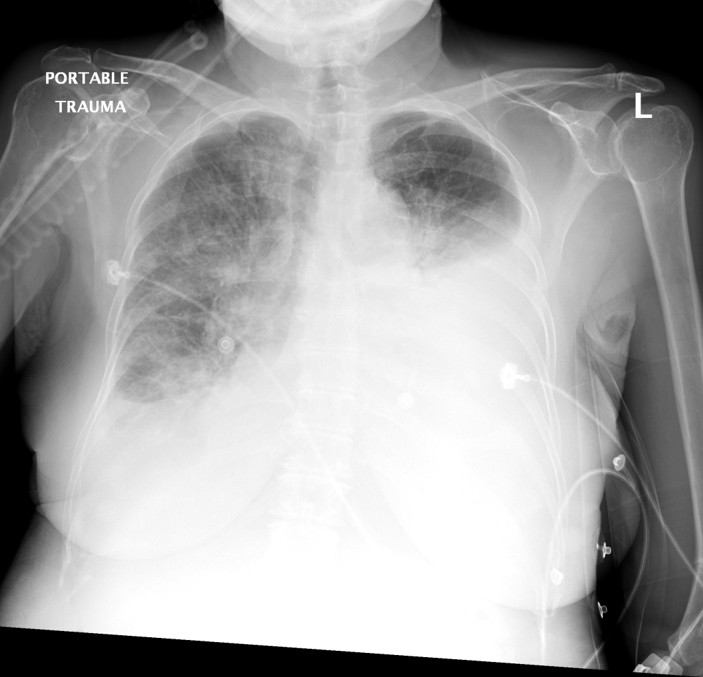
    CXR: Pleural Effusion

    There is a large left pleural effusion obscuring the lower half of the left hemi thorax. The cardiac silhouette is also obscured. There is pulmonary venous vascular congestion. There is also a right pleural effusion with fluid tracking into the minor fissure. Pulmonary interstitial edema is also noted.

    CT Chest (High-Resolution):

    • Bilateral, left greater than right, pleural effusions with adjacent atelectasis and collapse versus consolidation of the left lower lobe.
    • Prominent main pulmonary artery measuring 3.3 cm in diameter, which can be seen with pulmonary arterial hypertension.

    TTE:

    • LVEF is 50-55%.
    • Impaired left ventricular relaxation, which is associated with grade I/IV or mild diastolic dysfunction.
    • Moderate aortic stenosis with mild regurgitation (AVA 1.4 cm3, mean gradient 14mmHg, peak velocity 2.4 m/s).
    • Severe pulmonary hypertension (est PASP 52-62mmHg).
    • The inferior vena cava appeared dilated and decreased <50% with respiration (RAP 10-20 mmHg).
    • Minimal pericardial effusion without echocardiographic evidence of tamponade.

    Assessment/Plan:

    64F with history of CAD (prior MI), CHF, hypertension, CLL, hypothyroidism presented from a SNF with progressive shortness of breath, orthopnea and LE swelling, found to have bilateral (L>R) pleural effusion now s/p thoracentesis with transudative fluid.

    #Acute hypoxic respiratory failure: Large pleural effusions, s/p thoracentesis with pleural fluid suggestive of transudative process. Most likely secondary to left ventricular diastolic dysfunction. Improved after thoracentesis and diuresis. High-resolution CT chest performed without evidence of autoimmune-related pulmonary fibrosis or ILD (though persistent pleural effusions, pulmonary vascular congestion).

    #Pleural fluid signet cells: Identified on cytology, potentially related to history of untreated CLL or alternative primary malignancy.

    #Left ventricular diastolic dysfunction, decompensated: Associated with pleural effusions and hypoxemic respiratory failure. Management with diuresis.

    #Pulmonary Hypertension: Severe, noted on transthoracic echocardiography, may be secondary to hypoxemia associated with pleural effusions, consider repeat imaging once euvolemic or right-heart catheterization.

    #Microscopic Hematuria: No evidence of infection, no symptoms suggestive of nephrolithiasis. No casts identified or significant proteinuria. Plan for renal ultrasound.

    #Rheumatoid Arthritis: History of rheumatoid arthritis, on prednisone and hydroxychloroquine. Imaging without evidence of inflammatory arthropathy, RF elevated but CCP negative. Per rheumatology, the patient’s symptoms are not consistent with RA, continuing home medications while evaluation is ongoing. Pleural effusions unlikely associated with RA as transudative, and without monocyte predominance or low glucose.

    Case 2: Malignant Pleural Effusion

    malig_01
    malig_02
    malig_03
    malig_04
    malig_05
    malig_06
    malig_07
    malig_08
    malig_09
    malig_10
    malig_11
    malig_12
    malig_13
    malig_14
    Within the lungs there are ground-glass opacities bilaterally, and a left pleural effusion with adjacent consolidation vs compressive atelectasis.
    • Protein: 2.6 (serum: 4.9)
    • LDH: 1275 (serum: 219)
    • Cytology: Malignant cells

    Case 3: Traumatic Thoracentesis

    trauma_01
    trauma_02
    trauma_03
    trauma_04
    trauma_05
    trauma_06
    trauma_07
    trauma_08
    trauma_09
    trauma_10
    trauma_11
    trauma_12
    trauma_13
    trauma_14
    trauma_15
    trauma_16
    trauma_17
    trauma_18
    trauma_19
    Moderate right pleural effusion, some fluid in non-dependent portions suggestive of loculation. Diffuse nodules and opacification in right lung with compressive atelectasis. Left pleural effusion with high density material at the posterior costophrenic angle. Left chest tube.
    • Protein: 2.7 (serum 6.4)
    • LDH: 344 (serum 236)
    • Cell count: 100,000 RBC

    Case 4: Pneumonia

    pna_01
    pna_02
    pna_03
    pna_04
    pna_05
    pna_06
    pna_07
    pna_08
    pna_09
    pna_10
    pna_11
    pna_12
    pna_13
    pna_14
    pna_15
    Loculated right pleural effusion with foci of atelectasis and consolidative changes concerning for pneumonia. Minimal left-sided pleural effusion with consolidative changes. Enlarged mediastinal lymph nodes, possibly reactive.
    • Protein: 4.3 (serum 6.7)
    • LDH: 377 (serum 108)
    • pH: 7.46
    • Glucose: 153
    • Neutrophils: 84%

    Etiology of Pleural Effusions: 1

    Etiology Frequency (%)
    CHF 35
    Pneumonia 22
    Malignancy 15
    Pulmonary Embolism 11

    Clinical Features in the Diagnosis of Pleural Effusions and Identifying Etiology: 1,2

    Pleural effusions can be easily identified on chest radiography, physical examination findings include dullness to percussion, decreased tactile fremitus and decreased (or absent) breath sounds.

    • Hemoptysis: Malignancy, PE, TB
    • Weight Loss: Malignancy, TB
    • Ascites: Cirrhosis, ovarian cancer
    • Unilateral Leg Swelling: PE
    • Bilateral Leg Swelling: CHF, cirrhosis, nephrotic syndrome
    • Jugular Venous Distension: CHF

    Differential Diagnosis of Pleural Effusions: 1,2,3,4

    Differential Diagnosis of Pleural Effusions

    References:

    1. Light, R. W. (2002). Clinical practice. Pleural effusion. The New England journal of medicine, 346(25), 1971–1977. doi:10.1056/NEJMcp010731
    2. McGrath, E. E., & Anderson, P. B. (2011). Diagnosis of pleural effusion: a systematic approach. American journal of critical care : an official publication, American Association of Critical-Care Nurses, 20(2), 119–27– quiz 128. doi:10.4037/ajcc2011685
    3. Thomsen, T. W., DeLaPena, J., & Setnik, G. S. (2006). Videos in clinical medicine. Thoracentesis. The New England journal of medicine (Vol. 355, p. e16). doi:10.1056/NEJMvcm053812
    4. Wilcox, M. E., Chong, C. A. K. Y., Stanbrook, M. B., Tricco, A. C., Wong, C., & Straus, S. E. (2014). Does this patient have an exudative pleural effusion? The Rational Clinical Examination systematic review. JAMA : the journal of the American Medical Association, 311(23), 2422–2431. doi:10.1001/jama.2014.5552
    5. WikEM: Pleural effusion