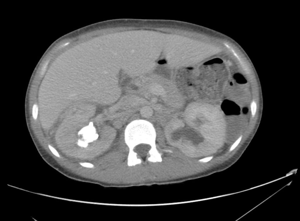
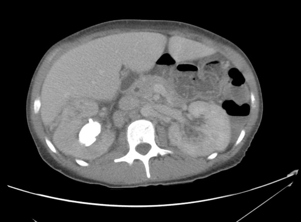
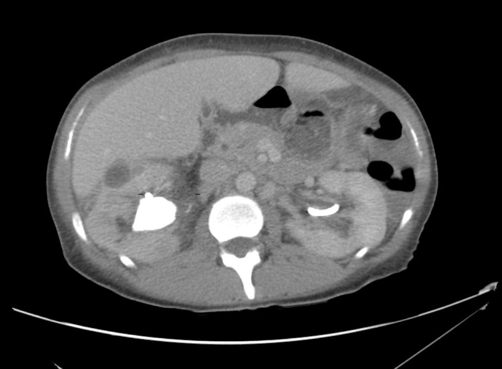
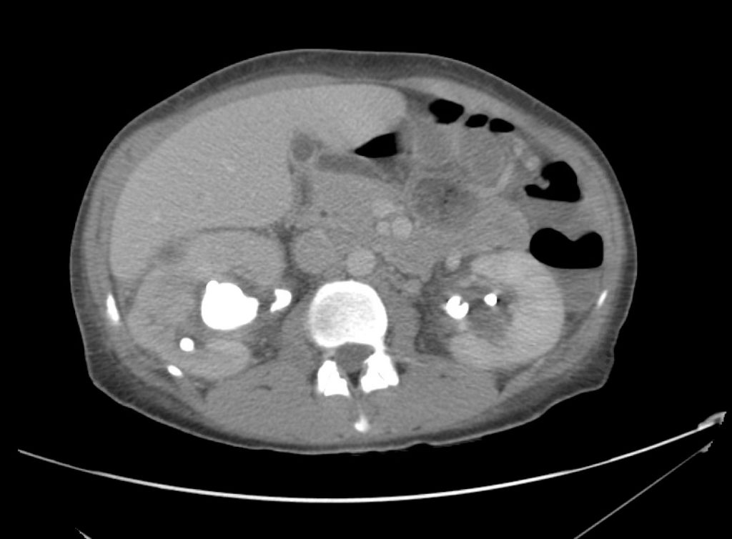
Hypoxia

Brief H&P:

A 67 year-old male with a history of hypertension and diabetes presents to the emergency department after a syncopal episode. He had been completing his normal morning routine when he developed a sensation of lightheadedness and awoke on the ground of his kitchen. He denies associated chest pain, palpitations, diaphoresis, or recent illness. He has no known sick contacts nor exposures to individuals undergoing evaluation for COVID-19.

On arrival in the emergency department, the patient was noted to be hypoxic with pulse oximetry measuring 74%. He was placed on supplemental oxygen via non-rebreather with improvement of oxygen saturation to 94%. Examination demonstrated diminished alertness (requiring constant stimulation for responses) and generalized motor weakness. Cardiac and pulmonary examinations were unremarkable with the exception of tachypnea and no extremity edema was appreciated.

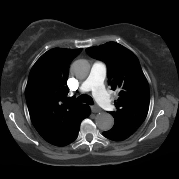
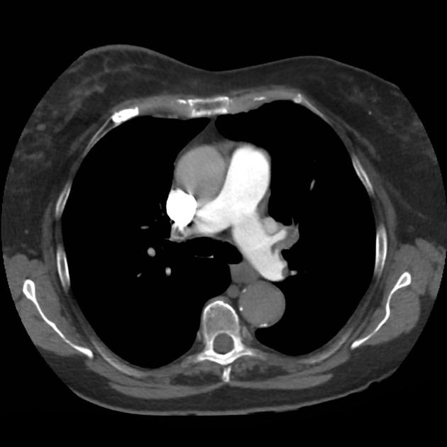
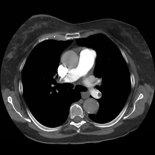
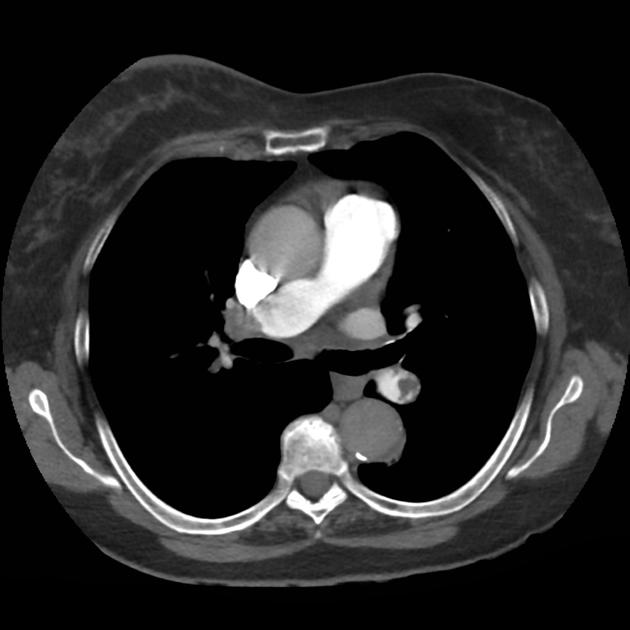
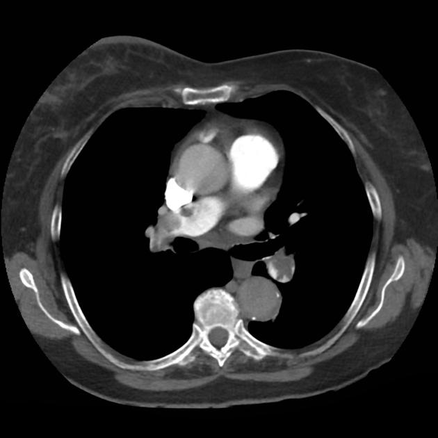
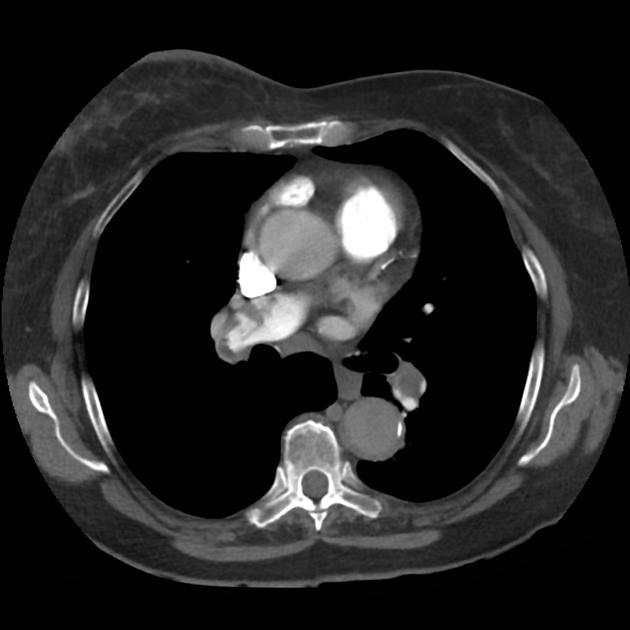
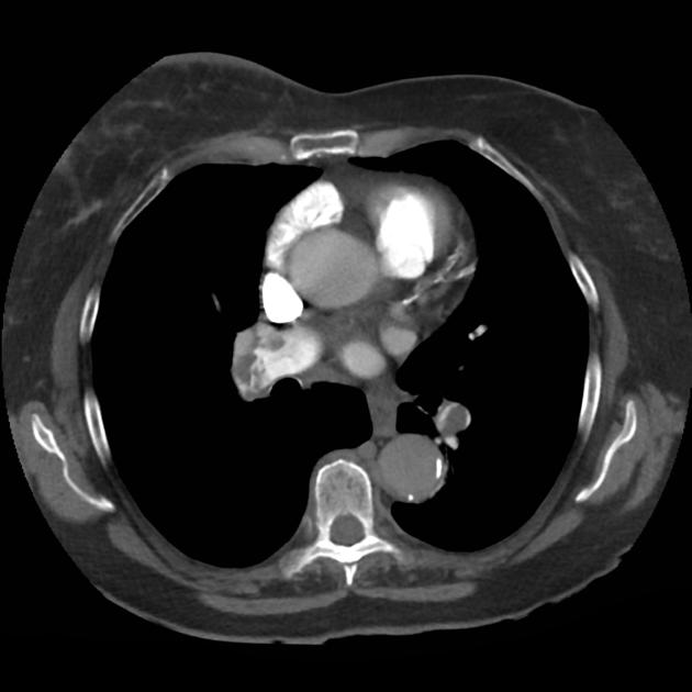
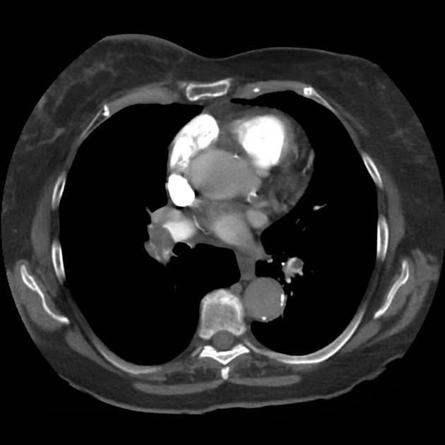
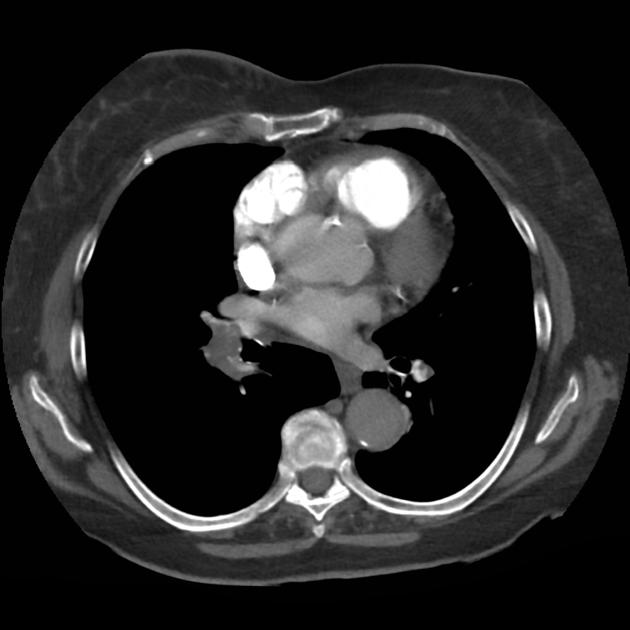
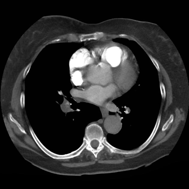
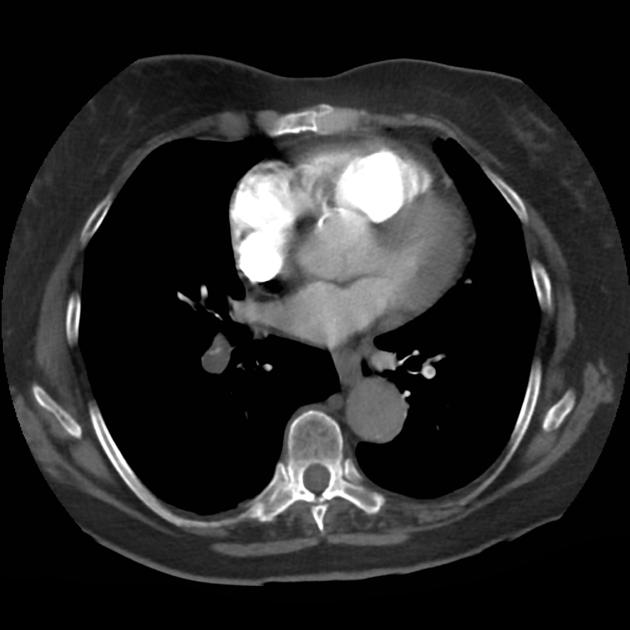
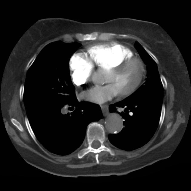
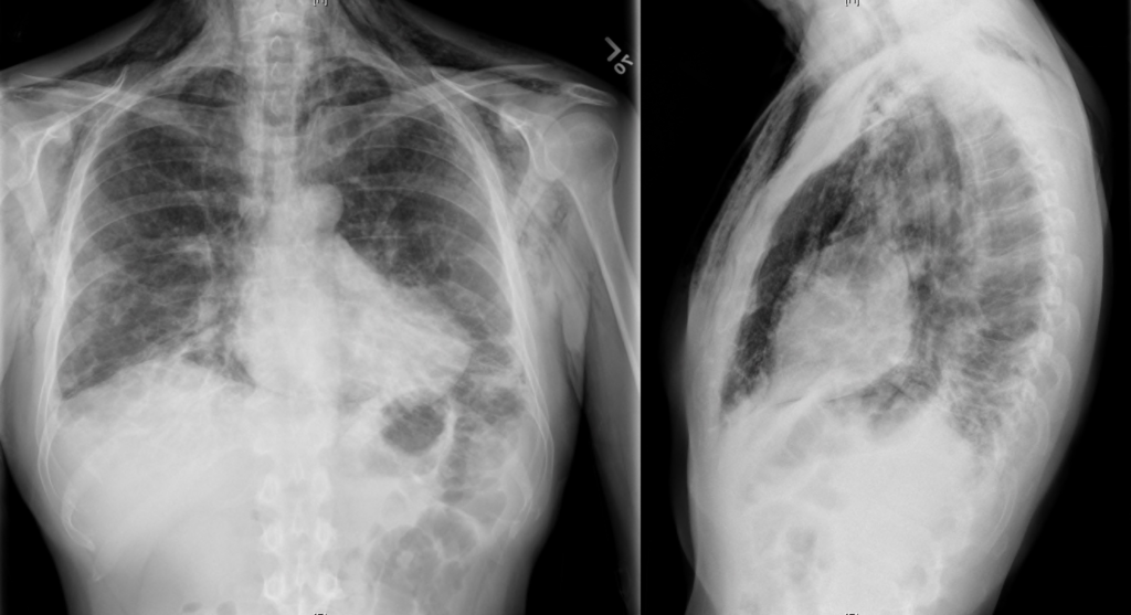
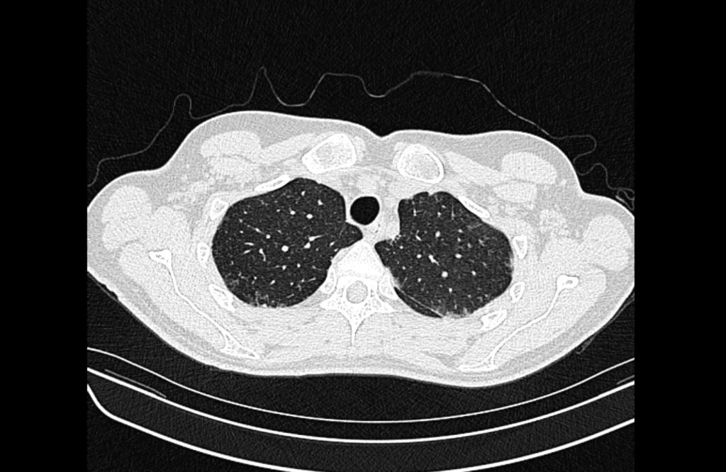
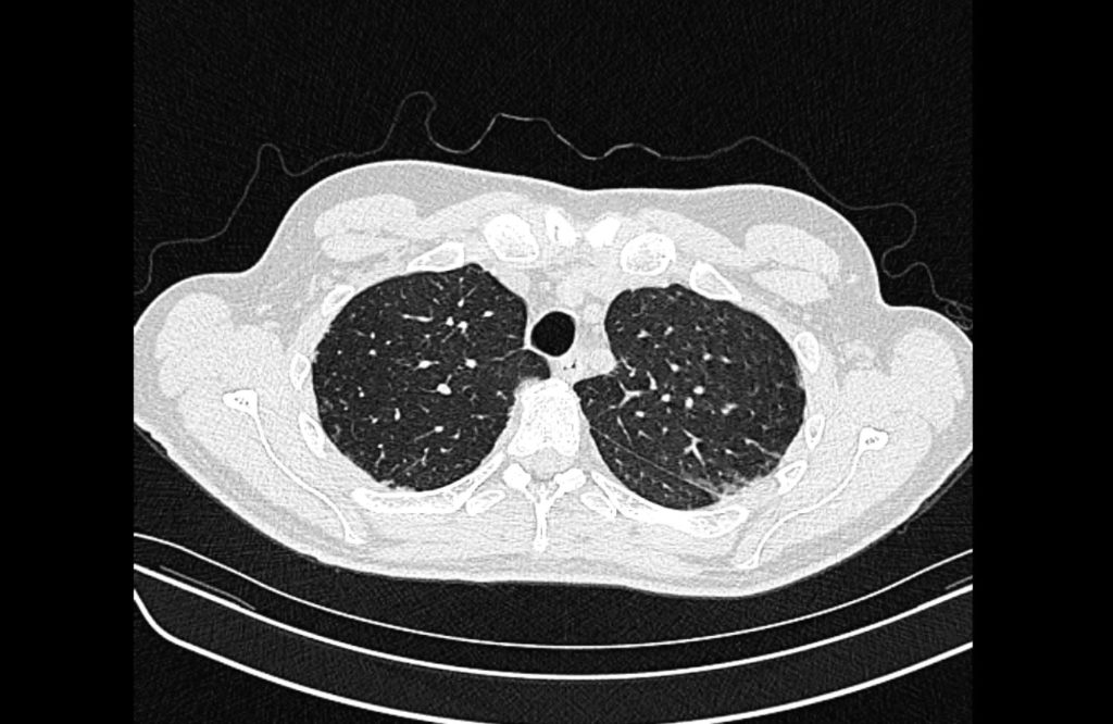
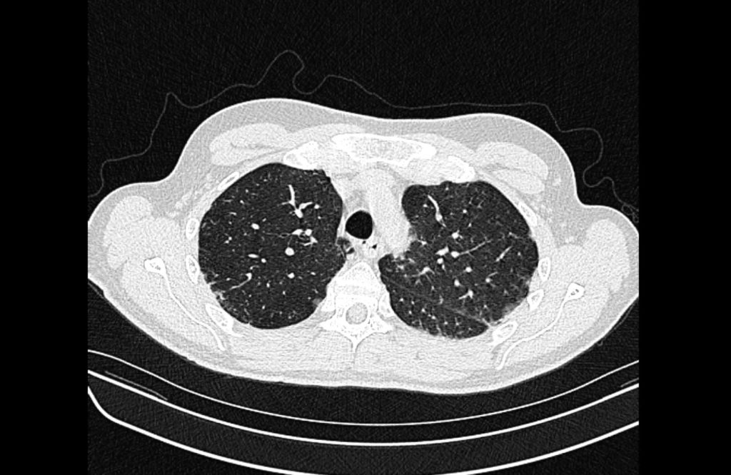
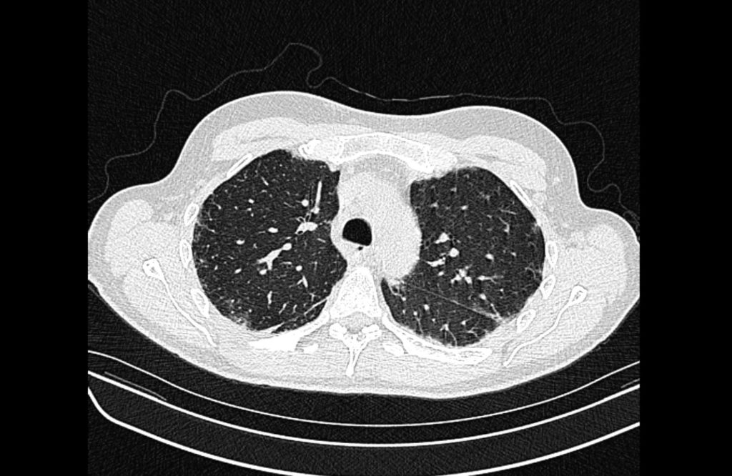
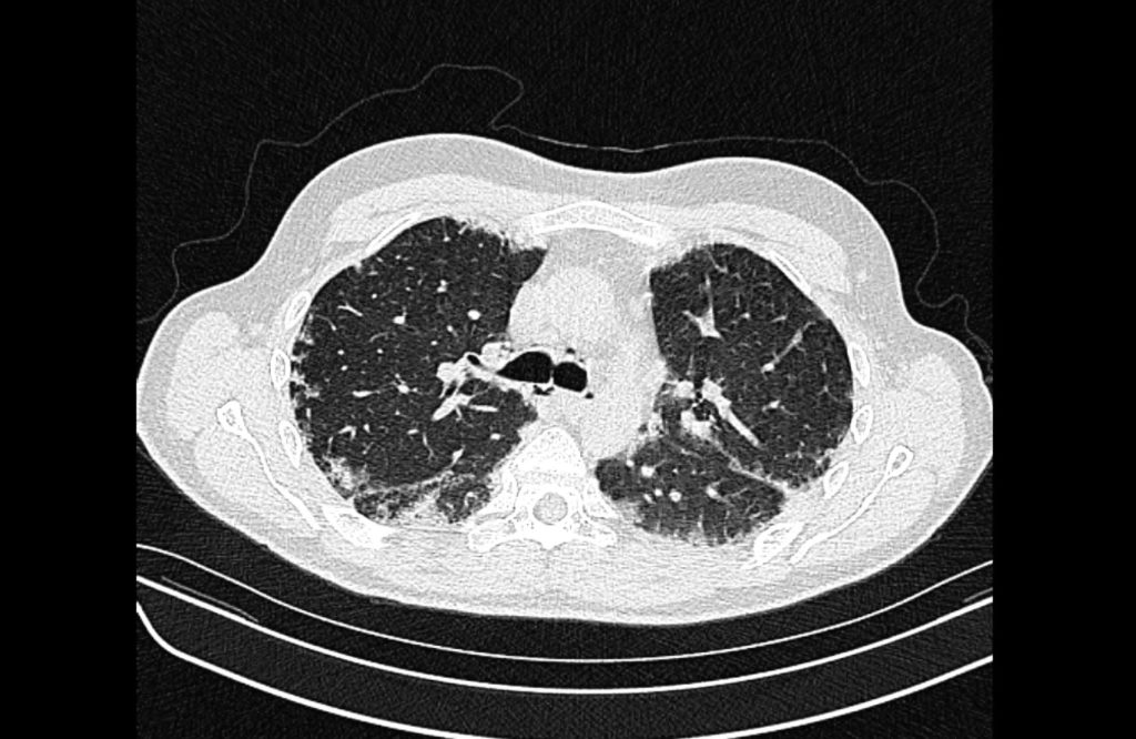
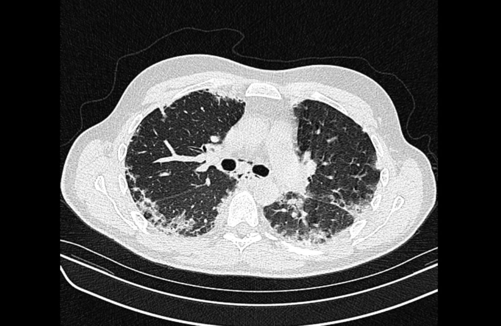
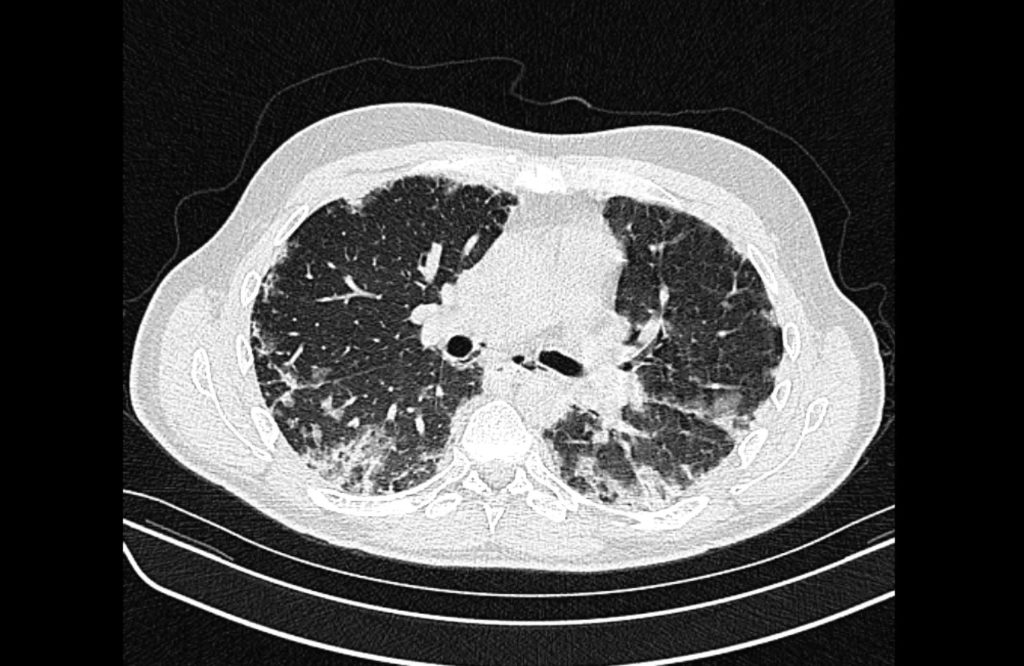
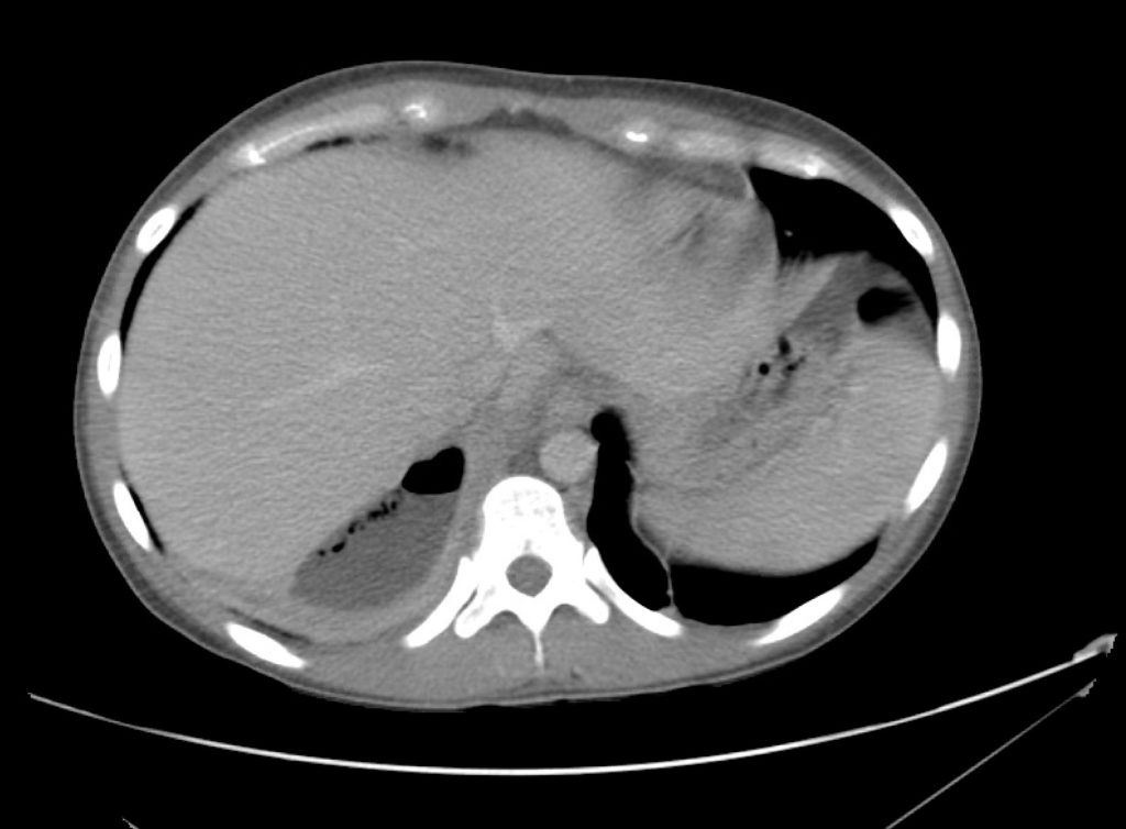
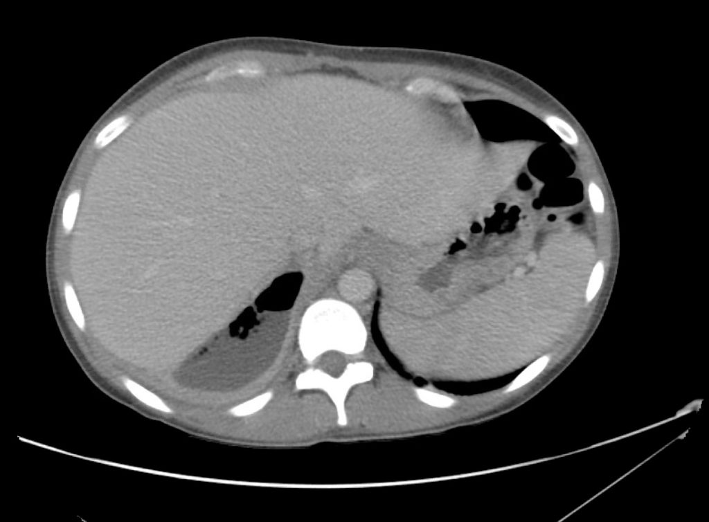
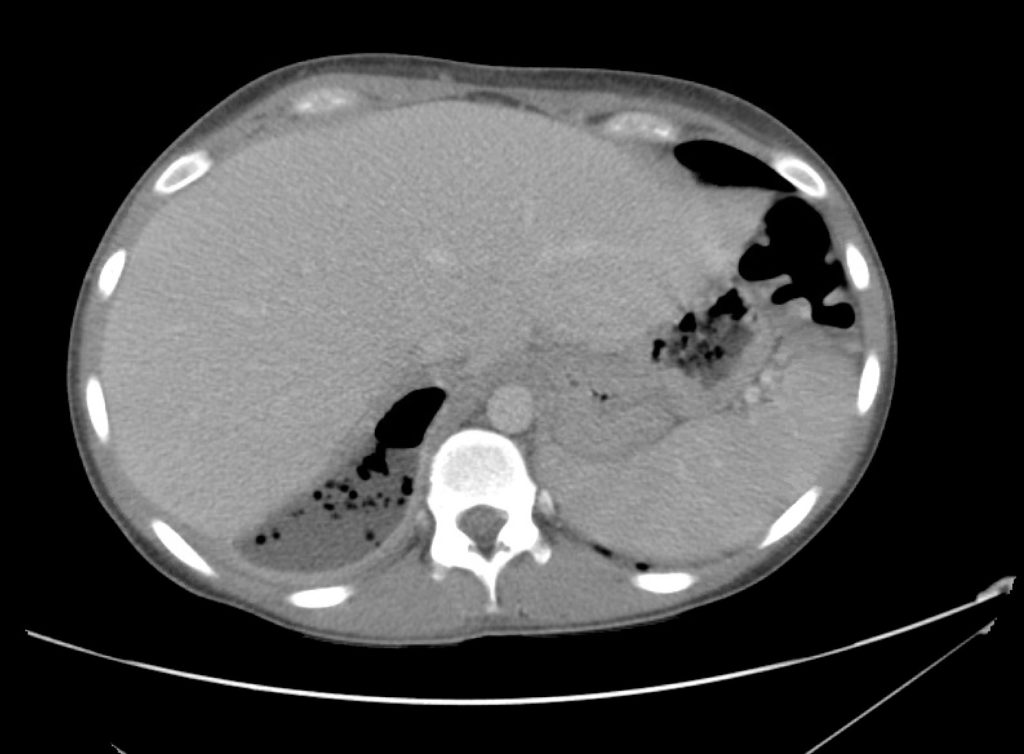
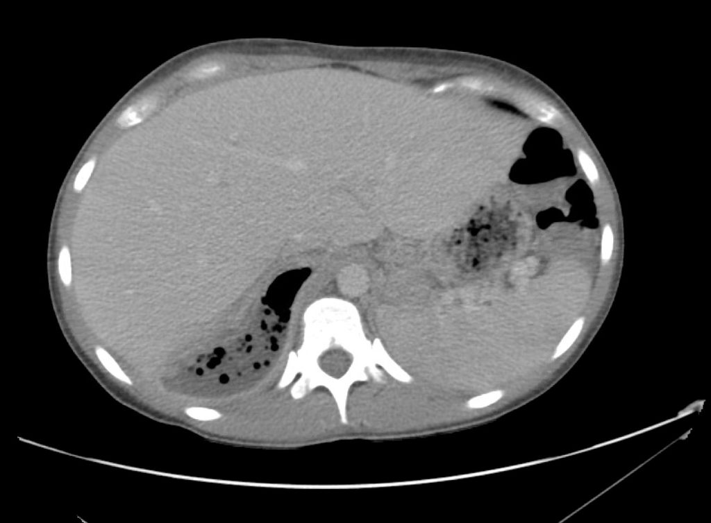
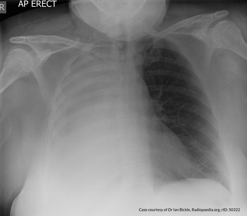
A chest radiograph was obtained which demonstrated platelike atelectasis. An arterial blood gas was obtained with PaO2 of 72mmHg suggesting a prominent A-a gradient. CT pulmonary angiography was obtained:

01_pe
02_pe
03_pe
04_pe
05_pe
06_pe
07_pe
08_pe
09_pe
10_pe
11_pe
12_pe

CT Pulmonary Angiography:

Bilateral pulmonary emboli. Case courtesy of Associate Prof Frank Gaillard, Radiopaedia.org, rID: 19636

Upon return, the patient’s mental status worsened associated with hypotension and he was intubated for airway protection and received systemic thrombolysis. He was subsequently taken for emergent endovascular treatment of massive pulmonary embolus.

An Algorithm for the Differential Diagnosis of Hypoxemia & Hypoxia1-7

Hypoxemia is defined as low PaO2 while hypoxia is insufficient global or local tissue oxygen content.

An Algorithm for the Differential Diagnosis of Hypoxemia and Hypoxia

References

  1. Stapczynski J. Respiratory Distress. In: Tintinalli JE, Ma O, Yealy DM, Meckler GD, Stapczynski J, Cline DM, Thomas SH. eds. Tintinalli’s Emergency Medicine: A Comprehensive Study Guide, 9e New York, NY: McGraw-Hill; . http://accessmedicine.mhmedical.com/content.aspx?bookid=2353&sectionid=219642010. Accessed April 12, 2020.
  2. Gas Transport & pH. In: Barrett KE, Barman SM, Brooks HL, Yuan JJ. eds. Ganong’s Review of Medical Physiology, 26e New York, NY: McGraw-Hill; . http://accessmedicine.mhmedical.com/content.aspx?bookid=2525&sectionid=204297654. Accessed April 12, 2020.
  3. Loscalzo J. Hypoxia and Cyanosis. In: Jameson J, Fauci AS, Kasper DL, Hauser SL, Longo DL, Loscalzo J. eds. Harrison’s Principles of Internal Medicine, 20e New York, NY: McGraw-Hill; . http://accessmedicine.mhmedical.com/content.aspx?bookid=2129&sectionid=192012521. Accessed April 12, 2020.
  4. West NE, Lechtzin N. Chapter 93. Hypoxia. In: McKean SC, Ross JJ, Dressler DD, Brotman DJ, Ginsberg JS. eds. Principles and Practice of Hospital Medicine New York, NY: McGraw-Hill; 2012. http://accessmedicine.mhmedical.com/content.aspx?bookid=496&sectionid=41304065. Accessed April 12, 2020.
  5. Pulmonary Physiology. In: Kibble JD, Halsey CR. eds. Medical Physiology: The Big Picture New York, NY: McGraw-Hill; 2014. http://accessmedicine.mhmedical.com/content.aspx?bookid=1291&sectionid=75576764. Accessed April 12, 2020.
  6. Petersson, J., Glenny, R. (2014). Gas exchange and ventilation–perfusion relationships in the lung European Respiratory Journal 44(4), 1023-1041. https://dx.doi.org/10.1183/09031936.00037014
  7. Morchi, R. (2011). Diagnosis Deconstructed: The Case of the Patient with No Chief Complaint Emergency Medicine News XXXIII(3)
    Rodríguez-Roisin, R., Roca, J. (2005). Mechanisms of hypoxemia Intensive Care Medicine 31(8), 1017-1019. https://dx.doi.org/10.1007/s00134-005-2678-1

COVID-19

Brief HPI:

A 38 year-old male with a history of hypertension presents to the emergency department with fever, cough and shortness of breath. He notes 4 days of symptoms which have been gradually worsening despite over-the-counter treatments. He denies recent travel or sick contacts. While he attempted to remain isolated – his symptoms grew intolerable.

On arrival in the emergency department, vital signs were notable for tachycardia and hypoxia (SpO2 85%, improving to 92% on 4L by nasal cannula). Physical examination demonstrated tachypnea and accessory muscle use but clear lung fields, and no extremity edema nor jugular venous distension. A chest radiograph revealed patchy airspace opacities. A presumptive diagnosis of COVID-19 pneumonia was made.

While awaiting hospitalization, the patient’s hypoxia worsened though he remained otherwise alert and oriented. He was placed on 15L via non-rebreather and instructed regarding self-prone positioning. He was admitted to the intensive care unit.

An Algorithm for the Management of COVID-19 Hypoxic Respiratory Failure1-6

An algorithm for the management of COVID-19 respiratory failure

References

  1. Whittle, J., Pavlov, I., Sacchetti, A., Atwood, C., Rosenberg, M. (2020). Respiratory Support for Adult Patients with COVID‐19 Journal of the American College of Emergency Physicians Open https://dx.doi.org/10.1002/emp2.12071
  2. Hui, D., Chow, B., Chu, L., Ng, S., Lee, N., Gin, T., Chan, M. (2012). Exhaled Air Dispersion during Coughing with and without Wearing a Surgical or N95 Mask PLoS ONE  7(12), e50845. https://dx.doi.org/10.1371/journal.pone.0050845
  3. Hui, D., Chow, B., Lo, T., Ng, S., Ko, F., Gin, T., Chan, M. (2015). Exhaled Air Dispersion During Noninvasive Ventilation via Helmets and a Total Facemask Chest  147(5), 1336-1343. https://dx.doi.org/10.1378/chest.14-1934
  4. Hui, D., Chow, B., Lo, T., Tsang, O., Ko, F., Ng, S., Gin, T., Chan, M. (2019). Exhaled air dispersion during high-flow nasal cannula therapy versus CPAP via different masks European Respiratory Journal  53(4), 1802339. https://dx.doi.org/10.1183/13993003.02339-2018
  5. Sun, Q., Qiu, H., Huang, M., Yang, Y. (2020). Lower mortality of COVID-19 by early recognition and intervention: experience from Jiangsu Province Annals of Intensive Care  10(1), 33. https://dx.doi.org/10.1186/s13613-020-00650-2
  6. Roca, O., Caralt, B., Messika, J., Samper, M., Sztrymf, B., Hernández, G., García-de-Acilu, M., Frat, J., Masclans, J., Ricard, J. (2018). An Index Combining Respiratory Rate and Oxygenation to Predict Outcome of Nasal High-Flow Therapy American Journal of Respiratory and Critical Care Medicine  199(11), 1368-1376. https://dx.doi.org/10.1164/rccm.201803-0589oc

Pneumomediastinum

Brief HPI:

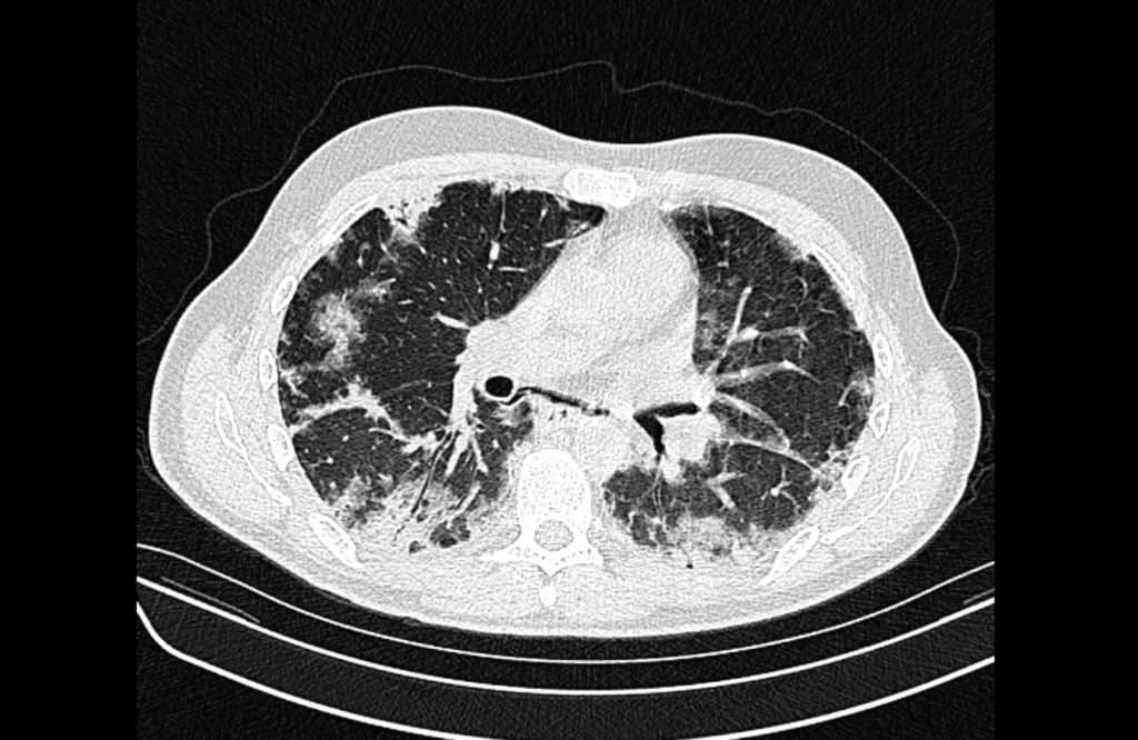
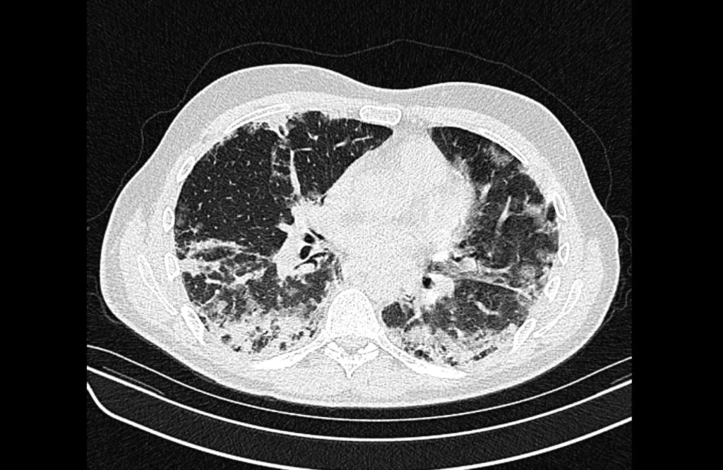
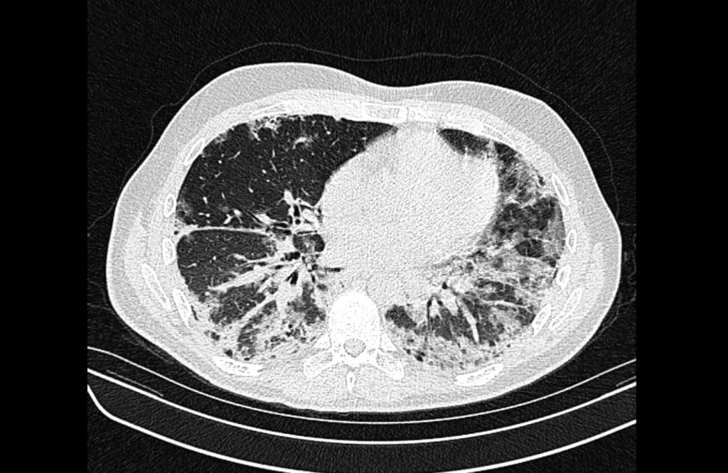
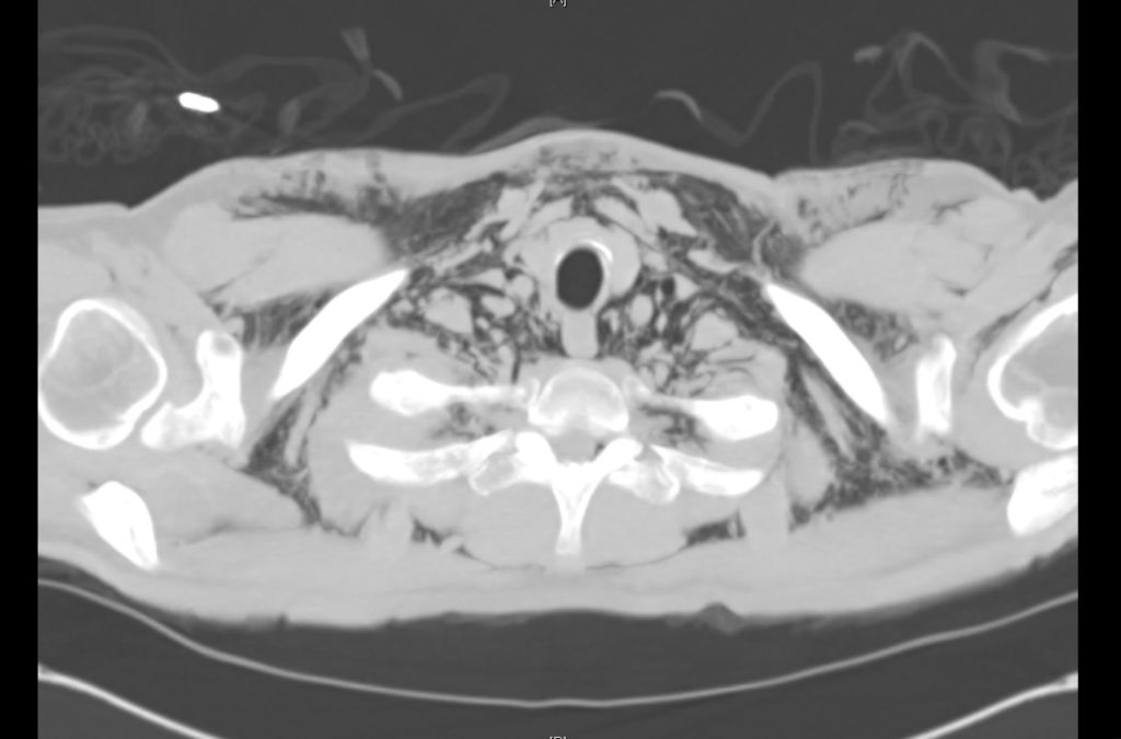
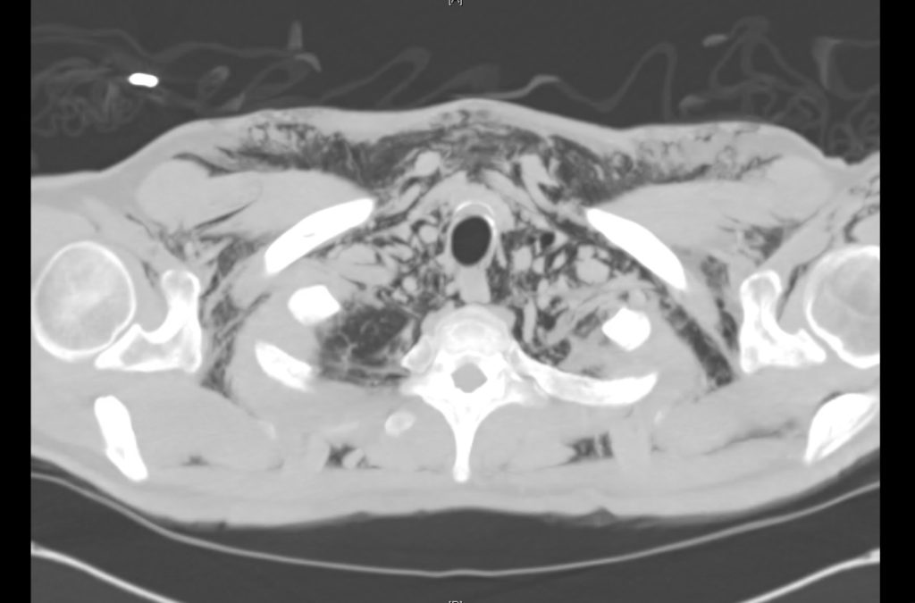
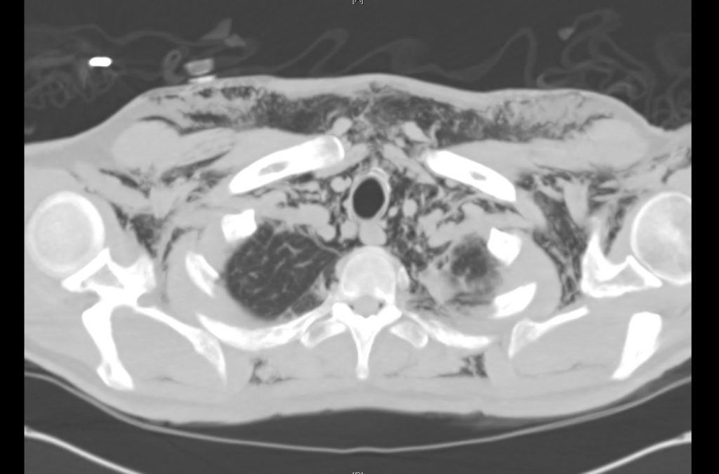
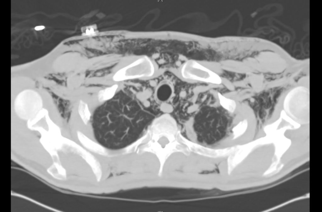
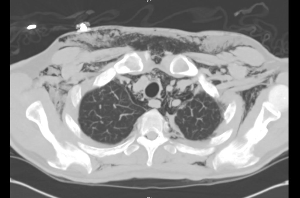
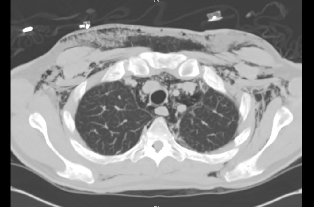
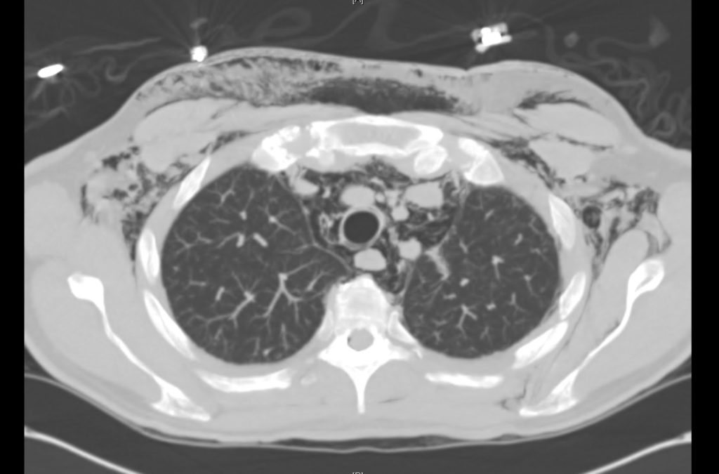
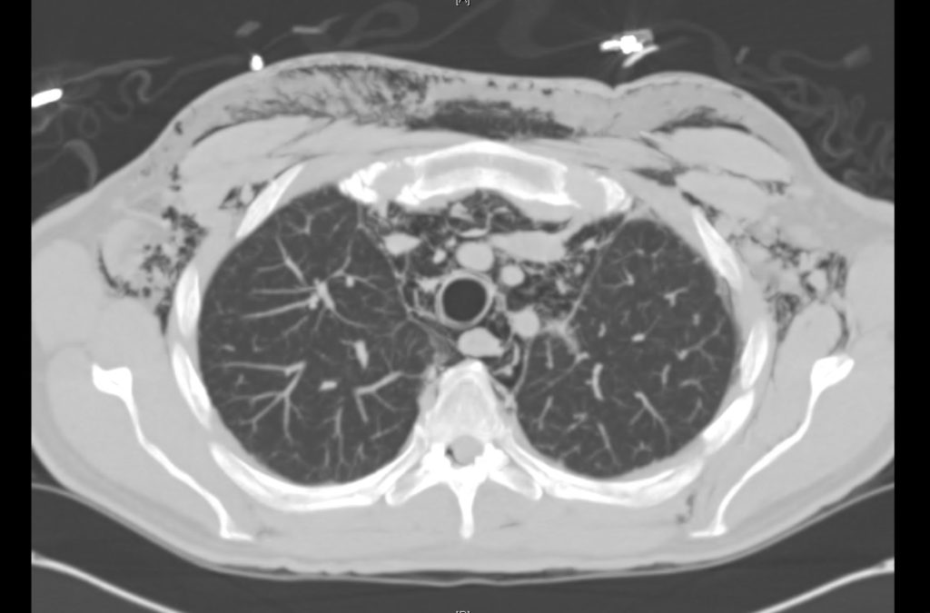
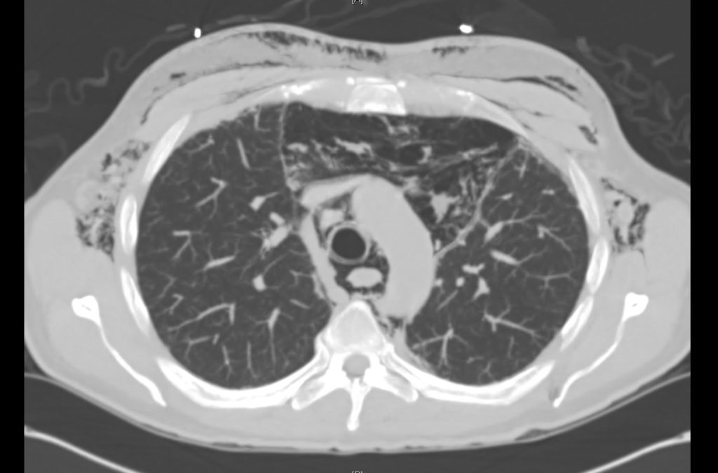
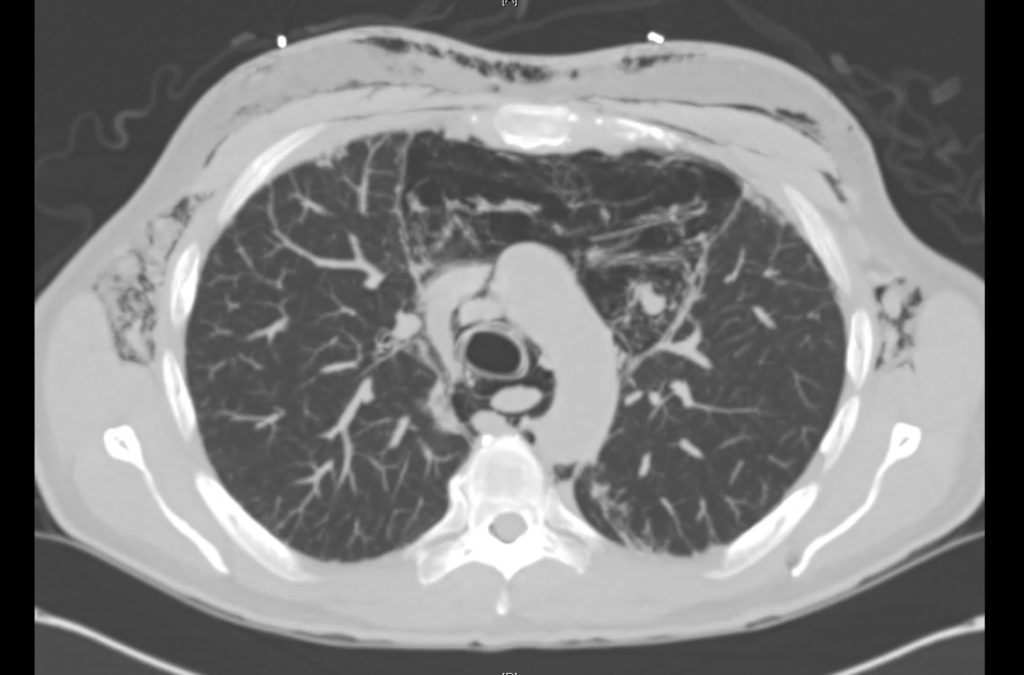
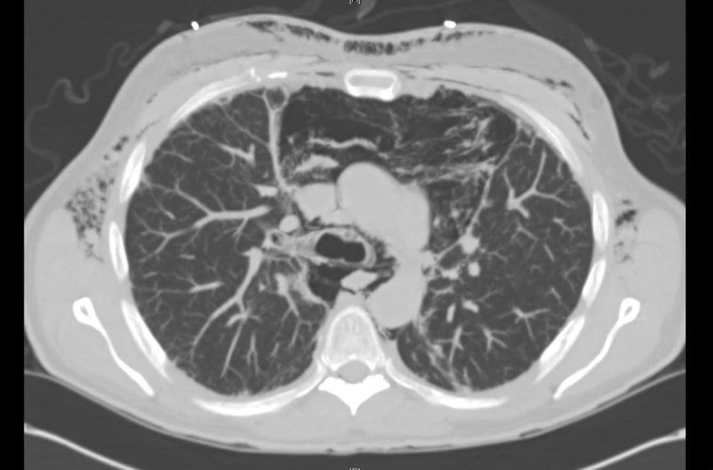
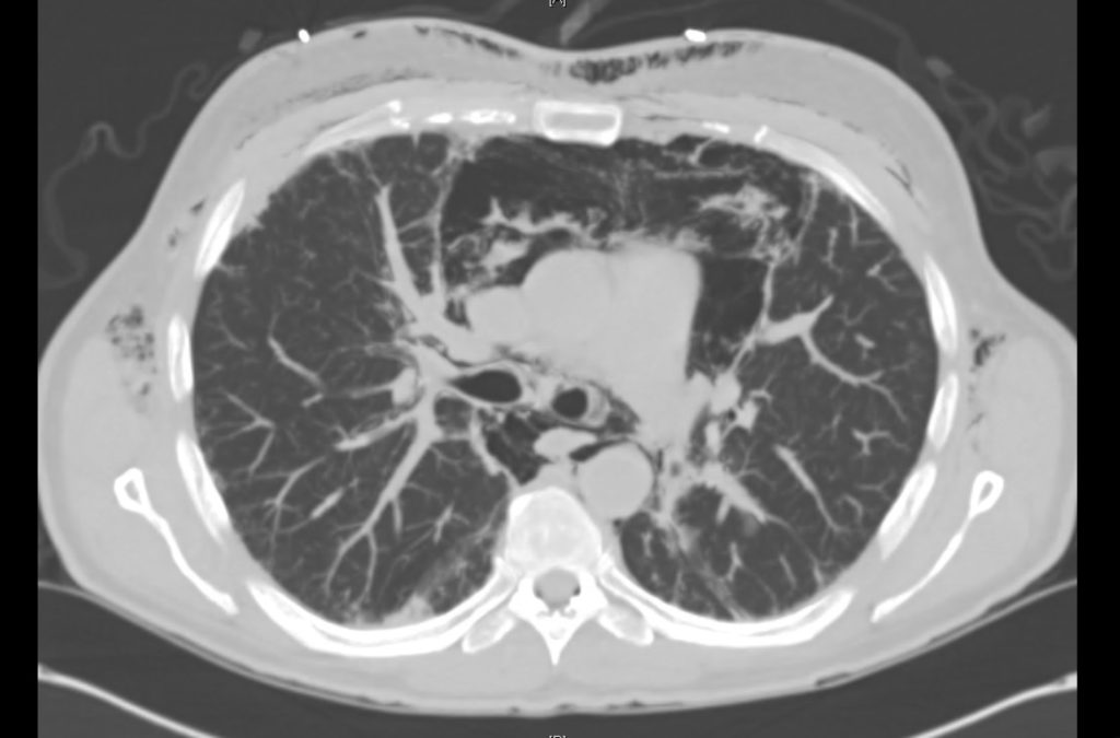
A 34-year-old male with a history of rheumatoid arthritis and interstitial lung disease presents to the emergency department with joint pain unimproved with home medications. He suspects the precipitant is a recent illness, describing cough and nasal congestion. He also noted a “crunching” sensation when turning his neck not otherwise associated with fevers, recurrent vomiting, chest pain, abdominal pain or difficulty breathing.

A chest radiograph was obtained which demonstrated pneumomediastinum.

Chest x-ray showing pneumomediastinum

Imaging from several months prior to presentation is shown below:

ct1_1
ct1_2
ct1_3
ct1_4
ct1_5
ct1_6
ct1_7
ct1_8
ct1_9
ct1_10

Prior CT Chest:

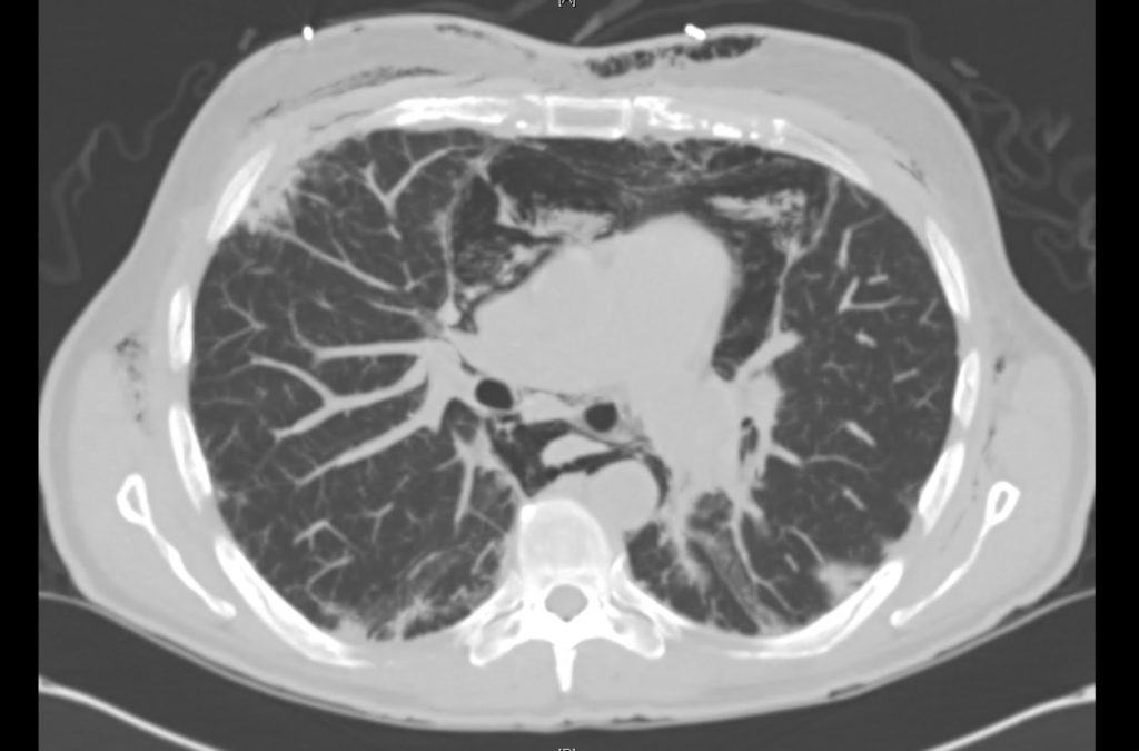
Extensive peripheral reticular and ground glass opacities and traction bronchiectasis predominates in the lower lobes. Imaging findings are most suggestive of usual interstitial pneumonia. Small focus of pneumomediastinum at carina.

The patient was placed on supplemental oxygen, a repeat chest CT was obtained.

ct2_1
ct2_2
ct2_3
ct2_4
ct2_5
ct2_6
ct2_7
ct2_8
ct2_9
ct2_10
ct2_11
ct2_12
ct2_13
ct2_14
ct2_15
ct2_16

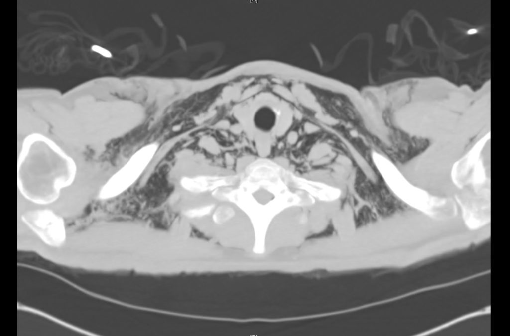
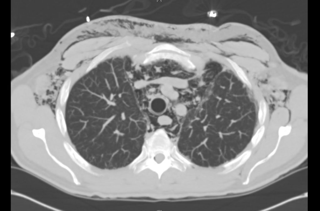
Current CT Chest:

Large pneumomediastinum extends superiorly into the bilateral lower neck and bilateral anterior and posterior chest walls. It extends inferiorly to the anterior diaphragmatic space. This most likely represents spontaneous pneumomediastinum in the clinical setting of interstitial lung disease. Pneumorrhachis is seen, related to pneumomediastinum.

The etiology of the patient’s spontaneous pneumomediastinum was deemed to be related to his underlying interstitial lung disease provoked by viral respiratory tract infection related coughing. He was observed for two days without decompensation and was discharged with outpatient follow-up.

Pathophysiology of Pneumomediastinum

Spontaneous pneumomediastinum results from the rupture of terminal alveoli with subsequent tracking of gas along the bronchovascular tree through interstitial lung tissue to the mediastinum and adjacent structures (pleural, pericardial, retropharyngeal, retroperitoneal, intraperitoneal and subcutaneous spaces)1.

Secondary pneumomediastinum arises from non-alveolar sources including the gastrointestinal tract (most gravely, esophageal rupture though also from other intraperitoneal sources2), and upper respiratory tract (including facial fractures3).

Management of Pneumomediastinum5-7

The management of spontaneous pneumomediastinum focuses on treatment of the underlying precipitant, supportive care, administration of supplemental oxygen (to promote gas reabsorption) and observation for complications including rare progression to tension pneumomediastinum4.

Secondary pneumomediastinum is of significantly more concern and should be suspected in patients with any of the following features:

Symptoms

  • History of forceful vomiting
  • Dysphagia

Signs

  • Fever
  • Hemodynamic instability
  • Left-sided pleural effusion
  • Abdominal tenderness
  • Leukocytosis

Management is aggressive including resuscitation, maintenance of NPO status, broad-spectrum antibiotics, and emergent surgical consultation.

Differential Diagnosis of Pneumomediastinum5-11

An Algorithm for the Evaluation of Pneumomediastinum

References:

  1. Macklin, M., Macklin, C. (1944). Malignant Interstitial Emphysema of the Lungs and Mediastinum as an Important Occult Complication in Many Respiratory Diseases and Other Conditions: an Interpretation of the Clinical Literature in the Light of Laboratory Experiment Medicine 23(4)
  2. Fosi, S., Giuricin, V., Girardi, V., Caprera, E., Costanzo, E., Trapano, R., Simonetti, G. (2014). Subcutaneous Emphysema, Pneumomediastinum, Pneumoretroperitoneum, and Pneumoscrotum: Unusual Complications of Acute Perforated Diverticulitis Case Reports in Radiology 2014(), 1-5. https://dx.doi.org/10.1155/2014/431563
  3. Luca, G., Petteruti, F., Tanga, M., Luciano, A., Lerro, A. (2011). Pneumomediastinum and Subcutaneous Emphysema Unusual Complications of Blunt Facial Trauma Indian Journal of Surgery 73(5), 380-381. https://dx.doi.org/10.1007/s12262-011-0310-x
  4. Shennib, H., Barkun, A., Matouk, E., Blundell, P. (1988). Surgical Decompression of a Tension Pneumomediastinum Chest 93(6), 1301-1302. https://dx.doi.org/10.1378/chest.93.6.1301
  5. Bakhos, C., Pupovac, S., Ata, A., Fantauzzi, J., Fabian, T. (2014). Spontaneous pneumomediastinum: an extensive workup is not required. Journal of the American College of Surgeons 219(4), 713-7. https://dx.doi.org/10.1016/j.jamcollsurg.2014.06.001
  6. Iyer, V., Joshi, A., Ryu, J. (2009). Spontaneous Pneumomediastinum: Analysis of 62 Consecutive Adult Patients Mayo Clinic Proceedings 84(5), 417-421. https://dx.doi.org/10.4065/84.5.417
  7. Takada, K., Matsumoto, S., Hiramatsu, T., Kojima, E., Shizu, M., Okachi, S., Ninomiya, K., Morioka, H. (2009). Spontaneous pneumomediastinum: an algorithm for diagnosis and management. Therapeutic advances in respiratory disease 3(6), 301-7. https://dx.doi.org/10.1177/1753465809350888
  8. Al-Mufarrej, F., Badar, J., Gharagozloo, F., Tempesta, B., Strother, E., Margolis, M. (2008). Spontaneous pneumomediastinum: diagnostic and therapeutic interventions. Journal of cardiothoracic surgery 3(1), 59. https://dx.doi.org/10.1186/1749-8090-3-59
  9. Takada, K., Matsumoto, S., Hiramatsu, T., Kojima, E., Watanabe, H., Sizu, M., Okachi, S., Ninomiya, K. (2008). Management of spontaneous pneumomediastinum based on clinical experience of 25 cases Respiratory Medicine 102(9), 1329-1334. https://dx.doi.org/10.1016/j.rmed.2008.03.023
  10. Bejvan, S., Godwin, J. (1996). Pneumomediastinum: old signs and new signs. American Journal of Roentgenology 166(5), 1041-1048. https://dx.doi.org/10.2214/ajr.166.5.8615238
  11. Langwieler, T., Steffani, K., Bogoevski, D., Mann, O., Izbicki, J. (2004). Spontaneous pneumomediastinum The Annals of Thoracic Surgery 78(2), 711-713. https://dx.doi.org/10.1016/j.athoracsur.2003.09.021

Numbness

Case 1

Brief HPI:

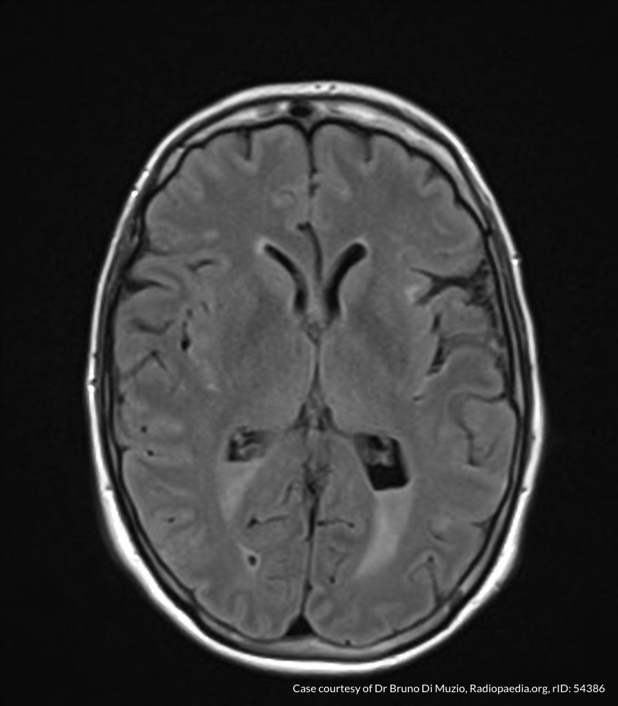
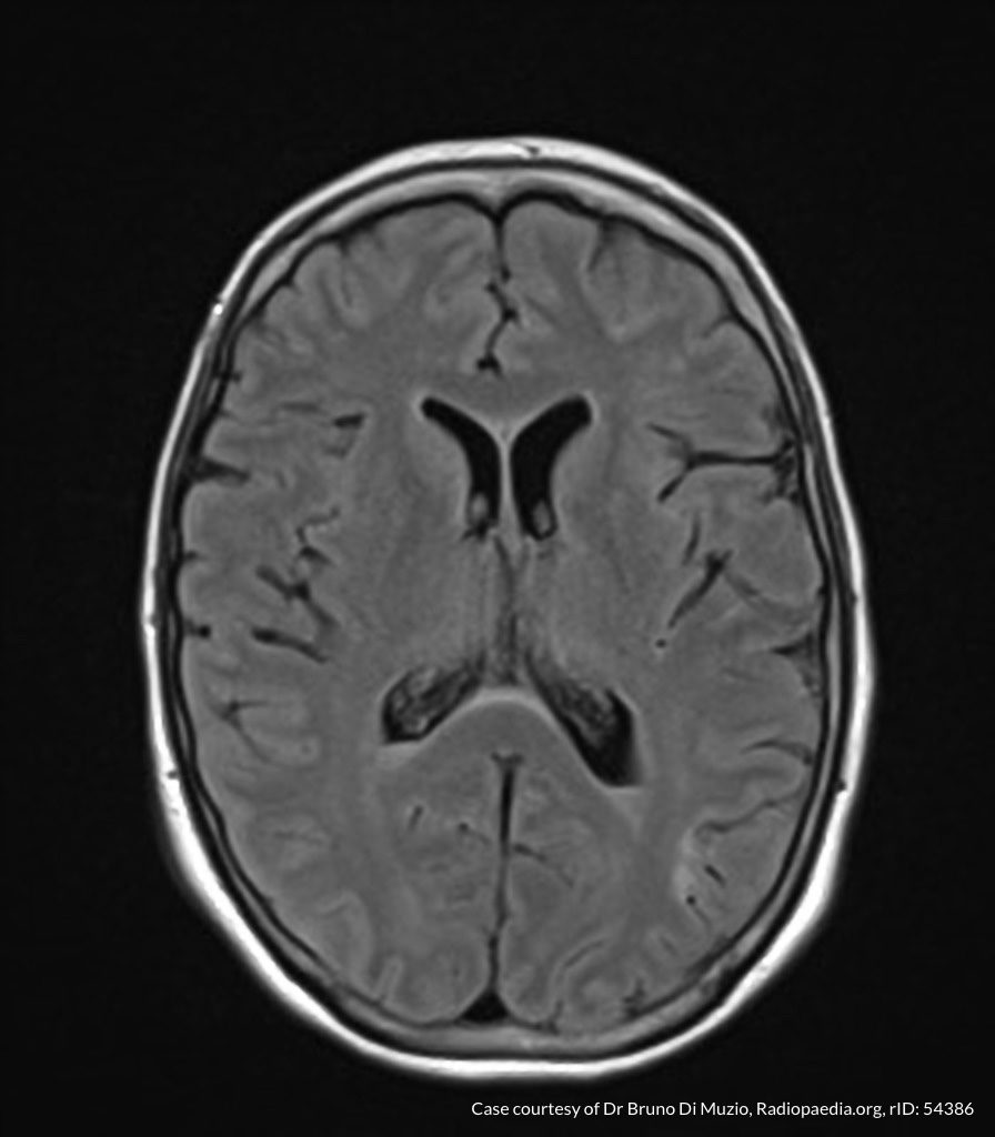
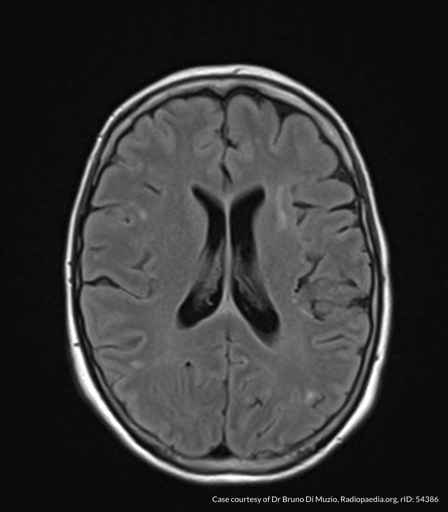
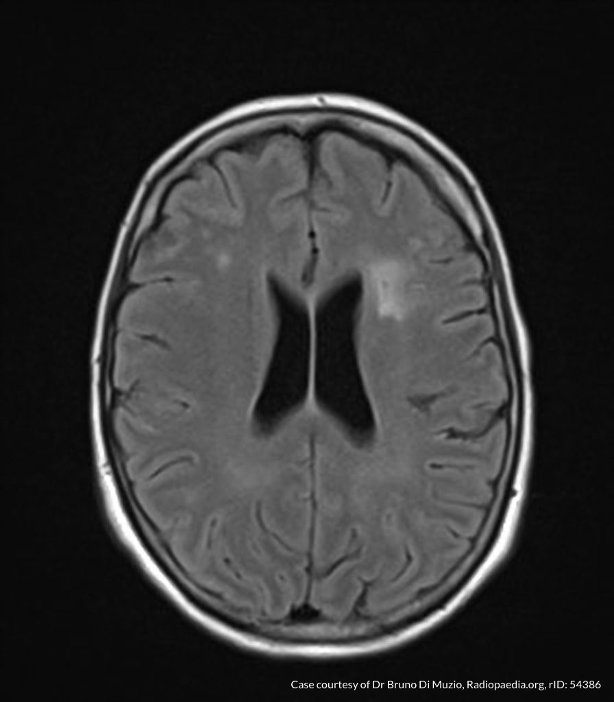
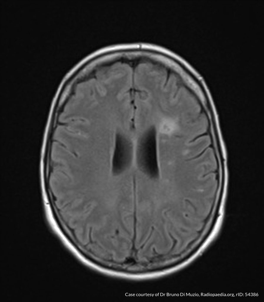
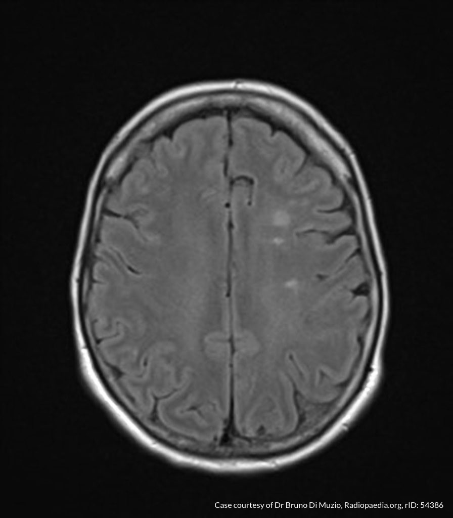
A 66-year-old male with a history of hypertension, diabetes, hyperlipidemia and prior stroke presents with acute-onset right-sided numbness. Examination demonstrates decreased sensation to light touch and pinprick in right upper- and-lower extremities as well as right face. Strength, cranial nerve and cerebellar testing is normal.

acute-lacunar-infarct (0)
acute-lacunar-infarct (1)
acute-lacunar-infarct (2)
acute-lacunar-infarct (3)
acute-lacunar-infarct (4)
acute-lacunar-infarct (5)
acute-lacunar-infarct (6)
acute-lacunar-infarct (7)
acute-lacunar-infarct (8)
acute-lacunar-infarct (9)
acute-lacunar-infarct (10)

MRI Brain:

Small focus of restricted diffusion within the left insular subcortical white matter consistent with acute lacunar infarct.1

Discussion:

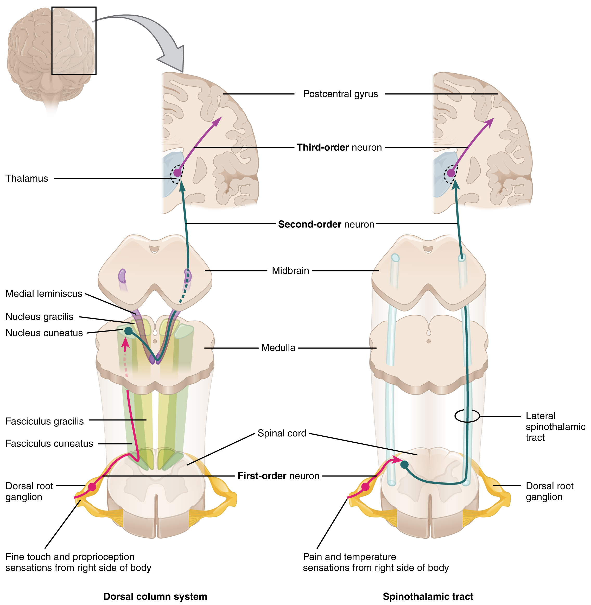
Infarction of the ventral posterolateral nucleus of the thalamus disrupts relays from the medial lemniscus and spinatholamic tracts extending to the cortex.

Case 2

Brief HPI:

A 34-year-old female with no significant medical history presents with headache and neck pain. Examination is notable for decreased strength with decreased sensation to pinprick (and preserved light touch) in bilateral upper extremities.

chiari-1-malformation-with-secondary-cervicothoracic-syringomyelia (0)
chiari-1-malformation-with-secondary-cervicothoracic-syringomyelia (1)
chiari-1-malformation-with-secondary-cervicothoracic-syringomyelia (2)
chiari-1-malformation-with-secondary-cervicothoracic-syringomyelia (3)
chiari-1-malformation-with-secondary-cervicothoracic-syringomyelia (4)
chiari-1-malformation-with-secondary-cervicothoracic-syringomyelia (6)
chiari-1-malformation-with-secondary-cervicothoracic-syringomyelia (8)

MRI C-Spine:

Chiari I malformation with associated cervical syringomyelia (79mm length, 11mm anteroposterior diameter) extending from C3 to T1.2

Discussion:

The central cord lesion affects the adjacent, medial fibers of the corticospinal tract resulting in upper extremity weakness. Disruption of crossing spinothalamic tract fibers results in diminished pain and temperature sensation at the level of the lesion.

Anatomy3,4

Dorsal column and spinothalamic tracts

Sensory information is gathered from specialized receptors in skin and soft-tissues which detect a variety of stimuli including temperature, pressure, vibration, and pain. This is subsequently transmitted through peripheral nerves which in certain regions coalesce into larger bundles before entering the spinal cord through the dorsal nerve root. Upon entering the spinal cord, sensory information is divided into two tracts:

  1. Spinothalamic: Small, poorly-myelinated fibers carrying pain, temperature and touch stimuli synapse with second-order neurons over several levels in the ipsilateral dorsal horn crossing to the contralateral side in the anterior commissure anterior to the central canal. Touch information ascends in the anterior spinothalamic tract while pain and temperature information ascends in the lateral spinothalamic tract.
  2. Dorsal column: Large, myelinated fibers carrying vibration and proprioception information ascend in the ipsilateral posteromedial spinal cord. Fibers from the thoracic and lumbar region occupy the more medial (gracile) column, while fibers from the cervical region occupy the more lateral (cuneate) column. These fibers synapse in their respective nuclei in the medulla before crossing to the contralateral medial lemniscus.

Both tracts proceed to the ventral posterolateral (VPL) nucleus of the thalamus, through the internal capsule before terminating in the somatotopically-arranged primary somatosensory cortex in the parietal lobe.

Understanding the neuroanatomy supports a systematic approach to the evaluation of sensory disturbances. It is important to note that the transmission of light touch sensation involves both pathways and offers less localizing value when compared to specific assessment of proprioception or pain detection. Sensory disturbances are often accompanied by motor abnormalities  which can further aid with localization. Other key distinguishing features include acuity of onset wherein abrupt presentations may suggest ischemia or infarction, compared to more indolent processes with broader differentials (including compressive mass lesions, demyelination, or autoimmune disease).

An Algorithm for the Evaluation of Sensory Disturbances5

References

  1. Case courtesy of Dr Bruno Di Muzio, Radiopaedia.org, rID: 45066
  2. Case courtesy of Dr Bahman Rasuli, Radiopaedia.org, rID: 65655
  3. Aminoff MJ. Numbness, Tingling, and Sensory Loss. In: Kasper D, Fauci A, Hauser S, Longo D, Jameson J, Loscalzo J. eds. Harrison’s Principles of Internal Medicine, 19e New York, NY: McGraw-Hill; 2014. http://accessmedicine.mhmedical.com/content.aspx?bookid=1130&sectionid=79724797. Accessed February 07, 2020.
  4. Sensory Disorders. In: Simon RP, Aminoff MJ, Greenberg DA. eds. Clinical Neurology, 10e New York, NY: McGraw-Hill; . http://accessmedicine.mhmedical.com/content.aspx?bookid=2274&sectionid=176234164. Accessed February 07, 2020.
  5. Berkowitz AL. Numbness: A Localization-Based Approach. In: McKean SC, Ross JJ, Dressler DD, Scheurer DB. eds. Principles and Practice of Hospital Medicine, 2e New York, NY: McGraw-Hill; . http://accessmedicine.mhmedical.com/content.aspx?bookid=1872&sectionid=146977205. Accessed February 07, 2020.

Pediatric Status Asthmaticus

Brief HPI:

A 6 year-old boy with a history of asthma presents to the emergency department via EMS for dyspnea. The patient is agitated on exam with nasal flaring and intercostal retractions. The parents report that his difficulty breathing started two days ago. The first day his MDI inhaler provided transient relief; however, over the next 24 hours he required nebulized albuterol 3 times with no significant relief. They deny any recent infections or steroid use and state that his immunizations are up-to-date.

On evaluation, vital signs are notable for BP 93/61, HR 140, RR 47, and SpO2 90%. He is afebrile; capillary glucose 113mg/dL. On examination, the patient is agitated with nasal flaring, intercostal retractions, shallow breathing with diminished breath sounds throughout.

Algorithm for the Management of Pediatric Asthma1-11

Algorithm for the Management of Pediatric Asthma

PASS12

Wheezing Work of Breathing Prolonged Expiration
Mild (0) None or end-expiration Normal or minimal retractions Normal or minimally prolonged
Moderate (1) Throughout expiration Intercostal retractions Moderately prolonged
Severe (2) Severe wheezing or absent Suprasternal retractions, abdominal wall movement Severely prolonged
This algorithm was developed by Dr. Joshua Niforatos. Joshua is an emergency medicine resident at The Johns Hopkins School of Medicine and an alumnus of the Cleveland Clinic Lerner College of Medicine.

Special thanks to Dr. Kelly Young, Director of the Pediatric Emergency Medicine Fellowship at Harbor-UCLA Medical Center and Dr. Adeola Kosoko, Assistant Professor, Assistant Residency Program Director, Director Of Diversity, Inclusion, And Mission at McGovern Medical School for their review of the algorithm.

References

  1. Rowe, B., Bretzlaff, J., Bourdon, C., Bota, G., Camargo, C. (2000). Magnesium sulfate for treating exacerbations of acute asthma in the emergency department. The Cochrane database of systematic reviews https://dx.doi.org/10.1002/14651858.cd001490
  2. Camargo, C., Spooner, C., Rowe, B. (2003). Continuous versus intermittent beta-agonists in the treatment of acute asthma. The Cochrane database of systematic reviews https://dx.doi.org/10.1002/14651858.cd001115
  3. Camargo, C., Rachelefsky, G., Schatz, M. (2009). Managing asthma exacerbations in the emergency department: summary of the National Asthma Education And Prevention Program Expert Panel Report 3 guidelines for the management of asthma exacerbations. Proceedings of the American Thoracic Society 6(4), 357 – 366. https://dx.doi.org/10.1513/pats.p09st2
  4. Gouin, S., Robidas, I., Gravel, J., Guimont, C., Chalut, D., Amre, D. (2010). Prospective evaluation of two clinical scores for acute asthma in children 18 months to 7 years of age. Academic emergency medicine : official journal of the Society for Academic Emergency Medicine 17(6), 598 – 603. https://dx.doi.org/10.1111/j.1553-2712.2010.00775.x
  5. Travers, A., Milan, S., Jones, A., Camargo, C., Rowe, B. (2012). Addition of intravenous beta(2)-agonists to inhaled beta(2)-agonists for acute asthma. The Cochrane database of systematic reviews 12(), CD010179. https://dx.doi.org/10.1002/14651858.cd010179
  6. Jat, K., Chawla, D. (2012). Ketamine for management of acute exacerbations of asthma in children. The Cochrane database of systematic reviews 11(), CD009293. https://dx.doi.org/10.1002/14651858.cd009293.pub2
  7. Ortiz-Alvarez, O., Mikrogianakis, A., Committee, C. (2012). Managing the paediatric patient with an acute asthma exacerbation. Paediatrics & child health 17(5), 251 – 262. https://dx.doi.org/10.1093/pch/17.5.251
  8. Jones, B., Paul, A. (2013). Management of acute asthma in the pediatric patient: an evidence-based review. Pediatric emergency medicine practice 10(5), 1 – 23- quiz 23-4.
  9. Nievas, I., Anand, K. (2013). Severe acute asthma exacerbation in children: a stepwise approach for escalating therapy in a pediatric intensive care unit. The journal of pediatric pharmacology and therapeutics : JPPT : the official journal of PPAG 18(2), 88 – 104. https://dx.doi.org/10.5863/1551-6776-18.2.88
  10. Rehder, K. (2017). Adjunct Therapies for Refractory Status Asthmaticus in Children. Respiratory care 62(6), 849 – 865. https://dx.doi.org/10.4187/respcare.05174
  11. Carroll, C., Sala, K. (2013). Pediatric status asthmaticus. Critical care clinics 29(2), 153 – 166. https://dx.doi.org/10.1016/j.ccc.2012.12.001
  12. Gorelick, M., Stevens, M., Schultz, T., Scribano, P. (2004). Performance of a novel clinical score, the Pediatric Asthma Severity Score (PASS), in the evaluation of acute asthma. Academic Emergency Medicine 11(1), 10 – 18. https://dx.doi.org/10.1197/s1069-6563(03)00579-7

Emphysematous Urinary Tract Infections

Brief HPI:

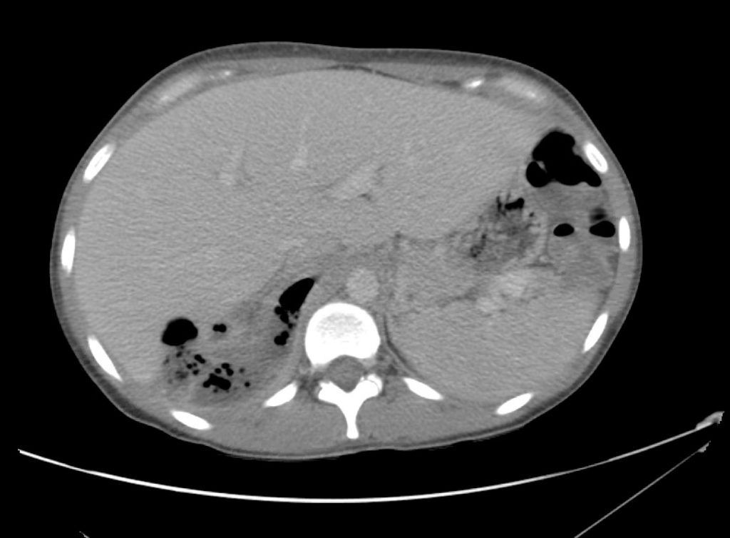
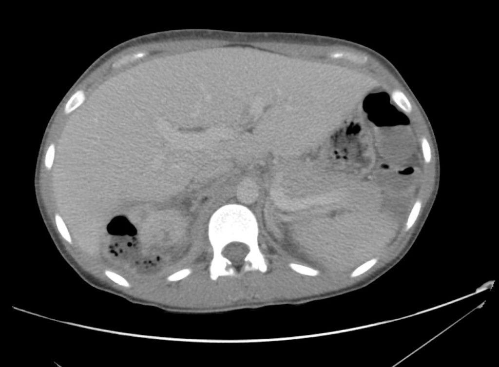
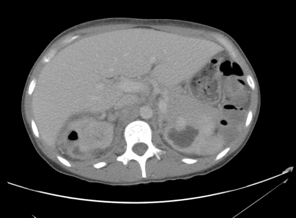
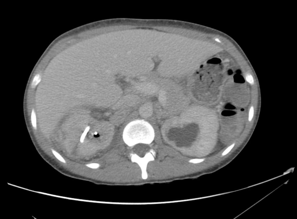
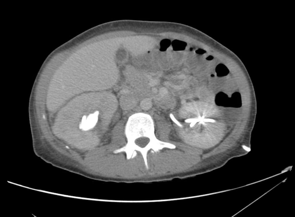
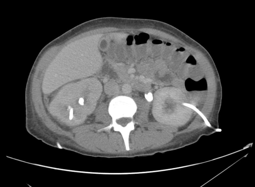
A 45 year-old female with a history of ureterolithiasis s/p bilateral percutaneous nephrostomies, hypertension and diabetes presents to the emergency department with flank pain and dysuria for two days. She noted that output from her right nephrostomy had diminished. On evaluation, her vital signs are notable for fever and tachycardia but are otherwise normal. Examination demonstrates right costovertebral angle tenderness to percussion. Drain sites appeared normal, without overlying erythema. Urinalyses from both nephrostomy collection bags were submitted. Computed tomography of the abdomen and pelvis was obtained to evaluate for nephrostomy malposition.

01_pyelo
03_pyelo
04_pyelo
05_pyelo
06_pyelo
07_pyelo
08_pyelo
09_pyelo
10_pyelo
11_pyelo
12_pyelo
13_pyelo
14_pyelo
15_pyelo
16_pyelo
17_pyelo

CT Abdomen/Pelvis Interpretation

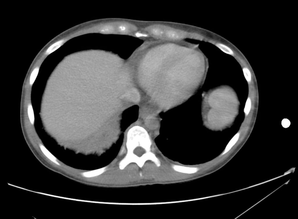
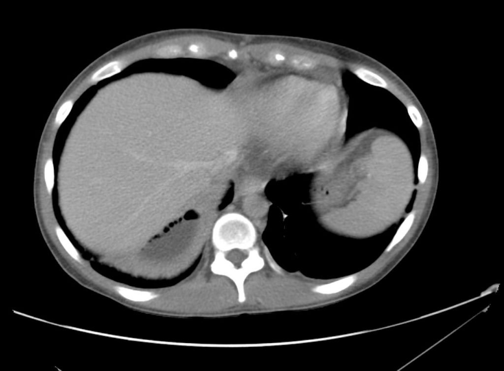
Complex perirenal fluid collection with gas suggestive of emphysematous pyelonephritis with abscess.

Hospital Course

The patient was treated with parenteral antibiotics based on prior culture data and was admitted to the intensive care unit with urology consultation and plan for interventional radiology percutaneous drainage. The patient underwent uncomplicated perinephric drain placement and nephrostomy exchange and was discharged on hospital day five to complete a course of oral antibiotics.

An Algorithm for the Evaluation and Management of Emphysematous Urinary Tract Infections

An Algorithm for the Evaluation and Management of Emphysematous Urinary Tract Infections

References

  1. Evanoff GV, Thompson CS, Foley R, Weinman EJ. Spectrum of gas within the kidney. Emphysematous pyelonephritis and emphysematous pyelitis. Am J Med. 1987;83(1):149-154.
  2. Wan YL, Lee TY, Bullard MJ, Tsai CC. Acute gas-producing bacterial renal infection: correlation between imaging findings and clinical outcome. Radiology. 1996;198(2):433-438. doi:10.1148/radiology.198.2.8596845.
  3. Shokeir AA, El-Azab M, Mohsen T, El-Diasty T. Emphysematous pyelonephritis: a 15-year experience with 20 cases. Urology. 1997;49(3):343-346. doi:10.1016/S0090-4295(96)00501-8.
  4. Chen MT, Huang CN, Chou YH, Huang CH, Chiang CP, Liu GC. Percutaneous drainage in the treatment of emphysematous pyelonephritis: 10-year experience. JURO. 1997;157(5):1569-1573.
  5. Huang JJ, Tseng CC. Emphysematous pyelonephritis: clinicoradiological classification, management, prognosis, and pathogenesis. Arch Intern Med. 2000;160(6):797-805.
  6. Roy C, Pfleger DD, Tuchmann CM, Lang HH, Saussine CC, Jacqmin D. Emphysematous pyelitis: findings in five patients. Radiology. 2001;218(3):647-650. doi:10.1148/radiology.218.3.r01fe14647.
  7. Park BS, Lee S-J, Kim YW, Huh JS, Kim JI, Chang S-G. Outcome of nephrectomy and kidney-preserving procedures for the treatment of emphysematous pyelonephritis. Scand J Urol Nephrol. 2006;40(4):332-338. doi:10.1080/00365590600794902.
  8. Grupper M, Kravtsov A, Potasman I. Emphysematous cystitis: illustrative case report and review of the literature. Medicine (Baltimore). 2007;86(1):47-53. doi:10.1097/MD.0b013e3180307c3a.
  9. Mokabberi R, Ravakhah K. Emphysematous urinary tract infections: diagnosis, treatment and survival (case review series). Am J Med Sci. 2007;333(2):111-116.
  10. Yao J, Gutierrez OM, Reiser J. Emphysematous pyelonephritis. Kidney Int. 2007;71(5):462-465. doi:10.1038/sj.ki.5002001.
  11. Thomas AA, Lane BR, Thomas AZ, Remer EM, Campbell SC, Shoskes DA. Emphysematous cystitis: a review of 135 cases. BJU Int. 2007;100(1):17-20. doi:10.1111/j.1464-410X.2007.06930.x.
  12. Falagas ME, Alexiou VG, Giannopoulou KP, Siempos II. Risk factors for mortality in patients with emphysematous pyelonephritis: a meta-analysis. JURO. 2007;178(3 Pt 1):880–5–quiz1129. doi:10.1016/j.juro.2007.05.017.
  13. Somani BK, Nabi G, Thorpe P, et al. Is percutaneous drainage the new gold standard in the management of emphysematous pyelonephritis? Evidence from a systematic review. J Urol. 2008;179(5):1844-1849. doi:10.1016/j.juro.2008.01.019.
  14. Aswathaman K, Gopalakrishnan G, Gnanaraj L, Chacko NK, Kekre NS, Devasia A. Emphysematous pyelonephritis: outcome of conservative management. Urology. 2008;71(6):1007-1009. doi:10.1016/j.urology.2007.12.095.
  15. Kapoor R, Muruganandham K, Gulia AK, et al. Predictive factors for mortality and need for nephrectomy in patients with emphysematous pyelonephritis. BJU Int. 2010;105(7):986-989. doi:10.1111/j.1464-410X.2009.08930.x.
  16. Ubee SS, McGlynn L, Fordham M. Emphysematous pyelonephritis. BJU Int. 2011;107(9):1474-1478. doi:10.1111/j.1464-410X.2010.09660.x.
  17. Lu Y-C, Chiang B-J, Pong Y-H, et al. Predictors of failure of conservative treatment among patients with emphysematous pyelonephritis. BMC Infect Dis. 2014;14(1):418. doi:10.1186/1471-2334-14-418.

Hyponatremia

HPI:

A 62 year-old male with a history of hepatitis C cirrhosis complicated by hepatocellular carcinoma s/p radiofrequency ablation presenting after referral from hepatology clinic for hyponatremia. One week ago, the patient developed abdominal distension and shortness of breath that resolved after large-volume paracentesis and was started on furosemide 40mg p.o. daily and aldactone 100mg p.o. daily. After initiating diuretics, the patient noted worsening lower extremity edema, and increased thirst/fluid intake.

He reports two days of fatigue and intermittent confusion supported by family members who reported slowed speech. He otherwise denies abdominal pain, distension, nausea/vomiting, diarrhea/constipation, chest pain or shortness of breath. In the ED, the patient received 1L NS bolus.

PMH:

  • Hepatitis C cirrhosis c/b HCC s/p RFA
  • Rheumatoid arthritis

PSH:

  • None

Family History:

  • Non-contributory.

Social History:

  • Lives with partner, denies current or prior t/e/d abuse
  • HepC contracted from blood transfusions

Medications:

  • Furosemide 40mg p.o. daily
  • Spironolactone 100mg p.o. daily
  • Rifaximin 550mg p.o. b.i.d.

Allergies:

  • NKDA

Physical Exam:

VS: T 98.2 HR 80 RR 14 BP 95/70 O2 98% RA
Gen: Elderly male in no acute distress, alert and answering questions appropriately.
HEENT: NC/AT, PERRL, EOMI, faint scleral icterus, MMM.
CV: RRR, normal S1/S2, no murmurs. JVP 8cm.
Lungs: Faint basilar crackles on bilateral lung bases.
Abd: Normoactive bowel sounds, mildly distended but non-tender, without rebound/guarding.
Ext: 2+ pitting edema in lower extremities to knees bilaterally. 2+ peripheral pulses, warm and well perfused.
Neuro: AAOx3. CN II-XII intact. No asterixis. Normal gait. Normal FTN/RAM.

Labs/Studies:

  • BMP (admission): 112/5.6/88/22/28/1.1/97
  • BMP (+10h): 118/5.4/93/23/26/1.0/133
  • sOsm: 264
  • Urine: Na <20, K 26, Osm 453
  • BNP: 40
  • AST/ALT/AP/TB/Alb: 74/57/91/2.4/2.2

Assessment/Plan:

62M with HepC cirrhosis, with e/o decompensation (new-onset ascites) and hyponatremia.

  1. #Hyponatremia: Sodium 114, likely chronic, patient currently asymptomatic without concerning findings on neurological exam. Clinical findings suggestive of hypervolemic hyponatremia 2/2 decompensated cirrhosis resulting in decreased effective arterial blood volume and volume retention. However, the recent initiation of diuretics, mild AKI and early response to isotonic fluids in the ED suggests possible hypovolemic component.
    • 1L fluid restriction
    • q.4.h. sodium check, goal increase of 8mEq per 24h
    • hold diuretics
  2. #Hyperkalemia: Potassium 5.6, asymptomatic, AKI vs. medication-induced (aldactone). Continue monitoring.
  3. #AKI: Elevated creatinine 1.1 from baseline 0.7. Likely pre-renal given recent initiation of diuretics. Consider hepatorenal syndrome given decompensated cirrhosis. Follow-up repeat creatinine after 1L NS bolus in ED.
  4. #Hepatitis C: decompensated with new-onset ascites. No e/o encephalopathy, continue home rifaximin.

Physiology of Hyponatremia: 1,2,3,4

Physiology of Hyponatremia

Differential Diagnosis of Hyponatremia: 5

Differential Diagnosis of Hyponatremia

Evaluation of Hyponatremia: 2

  1. Identification of onset (acute vs. chronic)
  2. Presence of symptoms (HA, nausea, confusion, seizures)
  3. Assessment of volume status (edema, JVD, skin turgor, postural BP)
  4. Medical history (cardiac, liver, renal disease), drug history

References:

  1. Freda BJ, Davidson MB, Hall PM. Evaluation of hyponatremia: a little physiology goes a long way. Cleve Clin J Med. 2004;71(8):639–650.
  2. Biswas M, Davies JS. Hyponatraemia in clinical practice. Postgrad Med J. 2007;83(980):373–378. doi:10.1136/pgmj.2006.056515.
  3. Adrogué HJ, Madias NE. Hyponatremia. N. Engl. J. Med. 2000;342(21):1581–1589. doi:10.1056/NEJM200005253422107.
  4. Marx JA, Hockberger RS, Walls RM, Adams JG. Rosen’s emergency medicine: concepts and clinical practice. 2010;1.
  5. Milionis HJ, Liamis GL, Elisaf MS. The hyponatremic patient: a systematic approach to laboratory diagnosis. CMAJ. 2002;166(8):1056–1062.

Transfusion Reactions

Brief HPI:

A 28 year-old female with a history of systemic lupus erythematosus and end-stage renal disease without access to scheduled hemodialysis presents to the emergency department with 1 week of worsening dyspnea, fatigue and leg swelling. Her symptoms are reminiscent of prior episodes resolving with hemodialysis. On evaluation, vital signs are normal and laboratory tests demonstrate microcytic anemia (Hb 5.9g/dL) but no hyperkalemia. A plain chest radiograph is normal and the patient ambulates without hypoxia.

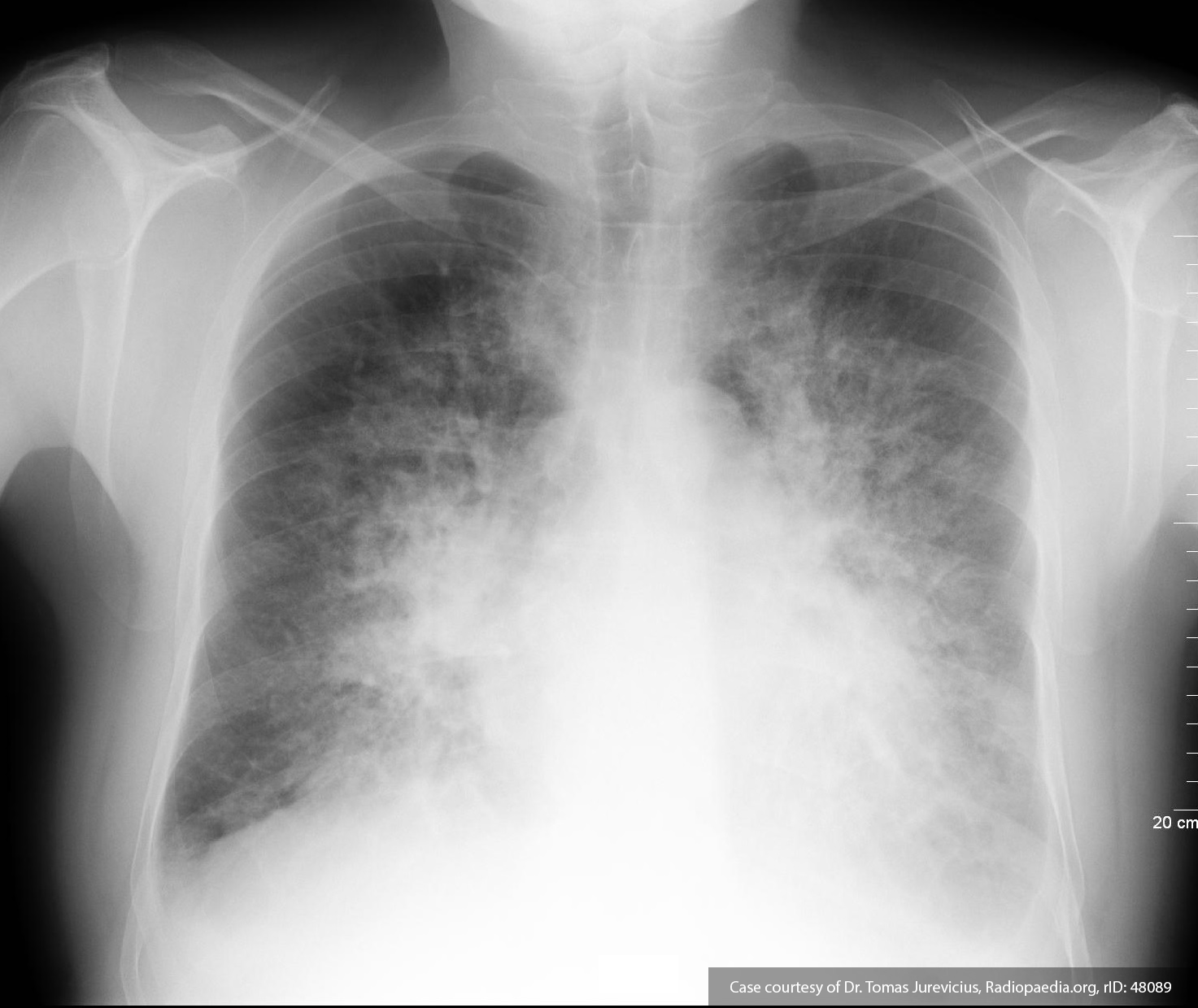
The patient was deemed to not meet any requirements for emergent hemodialysis. One unit of packed red blood cells was ordered for transfusion for symptomatic anemia. During transfusion, the patient developed worsening dyspnea and was found to be hypertensive and hypoxic. A chest radiograph was obtained and is shown below.

Chest x-ray with pulmonary edema

Pulmonary vascular congestion and bilateral pleural effusions.


The transfusion was discontinued, the patient was placed on non-invasive positive pressure ventilation, and emergent hemodialysis was initiated with subsequent resolution of presumed transfusion associated circulatory overload.

Algorithm for the Evaluation and Management of Transfusion Reactions

Algorithm for the Evaluation and Management of Transfusion Reactions

This algorithm was developed by Dr. Eric Madden, chief resident in emergency medicine at McGovern Med EM.

References

  1. Carson JL, Triulzi DJ, Ness PM. Indications for and Adverse Effects of Red-Cell Transfusion. N Engl J Med. 2017;377(13):1261-1272. doi:10.1056/NEJMra1612789.
  2. Delaney M, Wendel S, Bercovitz RS, et al. Transfusion reactions: prevention, diagnosis, and treatment. Lancet. 2016;388(10061):2825-2836. doi:10.1016/S0140-6736(15)01313-6.
  3. Goel R, Tobian AAR, Shaz BH. Noninfectious transfusion-associated adverse events and their mitigation strategies. Blood. 2019;133(17):1831-1839. doi:10.1182/blood-2018-10-833988.
  4. Osterman JL, Arora S. Blood product transfusions and reactions. Emerg Med Clin North Am. 2014;32(3):727-738. doi:10.1016/j.emc.2014.04.012.
  5. Silvergleid AJ. Approach to the patient with a suspected acute transfusion reaction. Post TW, ed. UpToDate. Waltham, MA: UpToDate Inc. https://www.uptodate.com (Accessed on September 01, 2019.)
  6. Suddock JT, Crookston KP. Transfusion Reactions. January 2019.

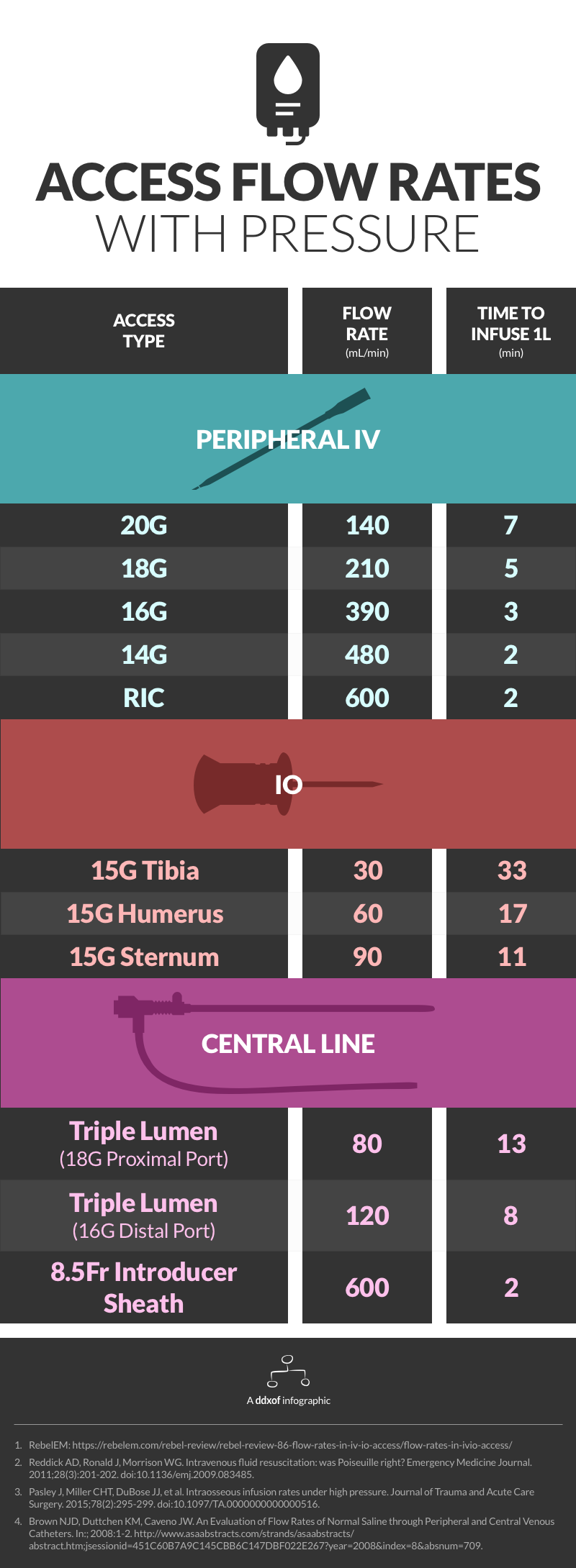
Infographic: Access Flow Rates

Infographic for IV flow rates

Notes

Data on access flow rates are highly variable. This infographic uses flow rates achieved through dedicated rapid infusers (ex. Level 1 ®) or at a pressure of 300mmHg. It is possible that rapid infusers and specialized pressure tubing achieve higher flow rates. The main references and additional sources are listed below. See this post from REBEL EM for gravity flow rates.

References

  1. Reddick AD, Ronald J, Morrison WG. Intravenous fluid resuscitation: was Poiseuille right? Emergency Medicine Journal. 2011;28(3):201-202. doi:10.1136/emj.2009.083485.
  2. Pasley J, Miller CHT, DuBose JJ, et al. Intraosseous infusion rates under high pressure. Journal of Trauma and Acute Care Surgery. 2015;78(2):295-299. doi:10.1097/TA.0000000000000516.
  3. Brown NJD, Duttchen KM, Caveno JW. An Evaluation of Flow Rates of Normal Saline through Peripheral and Central Venous Catheters. In:; 2008:1-2. http://www.asaabstracts.com/strands/asaabstracts/abstract.htm;jsessionid=451C60B7A9C145CBB6C147DBF022E267?year=2008&index=8&absnum=709.

Additional Sources

  1. Ngo AS-Y, Oh JJ, Chen Y, Yong D, Ong MEH. Intraosseous vascular access in adults using the EZ-IO in an emergency department. Int J Emerg Med. 2009;2(3):155-160. doi:10.1007/s12245-009-0116-9.
  2. Traylor S, Bastani A, Emergency NB-DAO, 2016. 311 Are Three Ports Better Than One? an Evaluation of Flow Rates Using All Ports of a Triple Lumen Central Venous Catheter in Volume Resuscitation. doi:10.1016/j.annemergmed.2016.08.327.
  3. Hammer N, Möbius R, Gries A, Hossfeld B, Bechmann I, Bernhard M. Comparison of the Fluid Resuscitation Rate with and without External Pressure Using Two Intraosseous Infusion Systems for Adult Emergencies, the CITRIN (Comparison of InTRaosseous infusion systems in emergency medicINe)-Study. Raju R, ed. PLoS ONE. 2015;10(12):e0143726–15. doi:10.1371/journal.pone.0143726.
  4. Ong MEH, Chan YH, Oh JJ, Ngo AS-Y. An observational, prospective study comparing tibial and humeral intraosseous access using the EZ-IO. Am J Emerg Med. 2009;27(1):8-15. doi:10.1016/j.ajem.2008.01.025.
  5. Philbeck TE, Miller LJ, Montez D, Puga T. Hurts so good. Easing IO pain and pressure. JEMS. 2010;35(9):58–62–65–6–68–quiz69. doi:10.1016/S0197-2510(10)70232-1.
  6. FRCA SIK, MRCA PRG, FRCA KP, MBChB SW, FRCA TS, MRCP PRG. Flow rates through intravenous access devices: an in vitro study. J Clin Anesth. 2016;31:101-105. doi:10.1016/j.jclinane.2016.01.048.
  7. Puga T, Montez D, Care TPC, 2016. 263: ADEQUACY OF INTRAOSSEOUS VASCULAR ACCESS INSERTION SITES FOR HIGH-VOLUME FLUID INFUSION. journalslwwcom
  8. Tan BKK, Chong S, Koh ZX, Ong MEH. EZ-IO in the ED: an observational, prospective study comparing flow rates with proximal and distal tibia intraosseous access in adults. Am J Emerg Med. 2012;30(8):1602-1606. doi:10.1016/j.ajem.2011.10.025.

Hypertensive Emergency

Brief HPI:

A 62 year-old female with a history of hypertension, diabetes and coronary artery disease is brought to the emergency department with altered mental status. The patient is confused and unable to provide history. Her family note that symptoms have been gradually worsening for the past one day and she had previously been in her usual state of good health. There was no history of recent illness, medication changes, recreational substance use, sick contacts, or travel.

On evaluation, vital signs were notable for hypertension (224/120mmHg, comparable on all extremities) though otherwise normal including afebrile core temperature – capillary glucose was 114mg/dL. On examination, the patient was awake and alert, making coordinated movements symmetrically in all four extremities without hyperreflexia or increased tone. Speech was unintelligible and the patient was unable to follow simple commands.

Labs/Imaging

Laboratory tests were notable for a serum creatinine of 1.2mg/dL (baseline unknown) but otherwise normal including CBC, troponin, TSH, and UA. ECG demonstrated left ventricular hypertrophy without acute ischemic changes. Imaging including chest radiograph and CT head non-contrast and CTA brain/neck were normal. Lumbar puncture was performed and CSF was normal.

Hospital Course

The patient was initiated on a continuous infusion of nicardipine for presumed hypertensive encephalopathy and admitted to the medical intensive care unit. An MRI was performed on hospital day 1 and demonstrated chronic microvascular ischemic changes. The patient’s mental status gradually improved over the course of her hospitalization and she was discharged home on hospital day 4.

An Algorithm for the Evaluation and Management of Hypertensive Emergencies

An Algorithm for the Evaluation and Management of Hypertensive Emergencies

References

General

  1. Lloyd-Jones DM, Morris PB, Ballantyne CM, et al. 2017 Focused Update of the 2016 ACC Expert Consensus Decision Pathway on the Role of Non-Statin Therapies for LDL-Cholesterol Lowering in the Management of Atherosclerotic Cardiovascular Disease Risk: A Report of the American College of Cardiology Task Force on Expert Consensus Decision Pathways. In: Vol 70. 2017:1785-1822. doi:10.1016/j.jacc.2017.07.745.
  2. Janke AT, McNaughton CD, Brody AM, Welch RD, Levy PD. Trends in the Incidence of Hypertensive Emergencies in US Emergency Departments From 2006 to 2013. J Am Heart Assoc. 2016;5(12). doi:10.1161/JAHA.116.004511.
  3. Rodriguez MA, Kumar SK, De Caro M. Hypertensive crisis. Cardiology in Review. 2010;18(2):102-107. doi:10.1097/CRD.0b013e3181c307b7.
  4. Katz JN, Gore JM, Amin A, et al. Practice patterns, outcomes, and end-organ dysfunction for patients with acute severe hypertension: the Studying the Treatment of Acute hyperTension (STAT) registry. Am Heart J. 2009;158(4):599–606.e1. doi:10.1016/j.ahj.2009.07.020.
  5. Elliott WJ. Clinical features in the management of selected hypertensive emergencies. Prog Cardiovasc Dis. 2006;48(5):316-325. doi:10.1016/j.pcad.2006.02.004.
  6. Aggarwal M, Khan IA. Hypertensive crisis: hypertensive emergencies and urgencies. Cardiol Clin. 2006;24(1):135-146. doi:10.1016/j.ccl.2005.09.002.
  7. Varon J, Marik PE. Clinical review: the management of hypertensive crises. Crit Care. 2003;7(5):374-384. doi:10.1186/cc2351.
  8. Shayne PH, Pitts SR. Severely increased blood pressure in the emergency department. YMEM. 2003;41(4):513-529. doi:10.1067/mem.2003.114.
  9. Vaughan CJ, Delanty N. Hypertensive emergencies. The Lancet. 2000;356(9227):411-417. doi:10.1016/S0140-6736(00)02539-3.

Ischemic Stroke

  1. Powers WJ, Rabinstein AA, Ackerson T, et al. 2018 Guidelines for the Early Management of Patients With Acute Ischemic Stroke: A Guideline for Healthcare Professionals From the American Heart Association/American Stroke Association. Stroke. 2018;49(3):e46-e110. doi:10.1161/STR.0000000000000158.

Hemorrhagic Stroke

  1. Hemphill JC, Greenberg SM, Anderson CS, et al. Guidelines for the Management of Spontaneous Intracerebral Hemorrhage: A Guideline for Healthcare Professionals From the American Heart Association/American Stroke Association. Stroke. 2015;46(7):2032-2060. doi:10.1161/STR.0000000000000069.

Subarachnoid Hemorrhage

  1. Connolly ES, Rabinstein AA, Carhuapoma JR, et al. Guidelines for the management of aneurysmal subarachnoid hemorrhage: a guideline for healthcare professionals from the American Heart Association/american Stroke Association. Stroke. 2012;43(6):1711-1737. doi:10.1161/STR.0b013e3182587839.

Renal

  1. Gillies MA, Kakar V, Parker RJ, Honoré PM, Ostermann M. Fenoldopam to prevent acute kidney injury after major surgery-a systematic review and meta-analysis. Crit Care. 2015;19(1):449. doi:10.1186/s13054-015-1166-4.
  2. Tumlin JA, Dunbar LM, Oparil S, et al. Fenoldopam, a dopamine agonist, for hypertensive emergency: a multicenter randomized trial. Fenoldopam Study Group. Academic Emergency Medicine. 2000;7(6):653-662.
  3. Shusterman NH, Elliott WJ, White WB. Fenoldopam, but not nitroprusside, improves renal function in severely hypertensive patients with impaired renal function. Am J Med. 1993;95(2):161-168.

Aortic Disease

  1. Hiratzka LF, Bakris GL, Beckman JA, et al. 2010 ACCF/AHA/AATS/ACR/ASA/SCA/SCAI/SIR/STS/SVM guidelines for the diagnosis and management of patients with Thoracic Aortic Disease: a report of the American College of Cardiology Foundation/American Heart Association Task Force on Practice Guidelines, American Association for Thoracic Surgery, American College of Radiology, American Stroke Association, Society of Cardiovascular Anesthesiologists, Society for Cardiovascular Angiography and Interventions, Society of Interventional Radiology, Society of Thoracic Surgeons, and Society for Vascular Medicine. Circulation. 2010;121(13):e266-e369. doi:10.1161/CIR.0b013e3181d4739e.

Pregnancy

  1. Townsend R, O’Brien P, Khalil A. Current best practice in the management of hypertensive disorders in pregnancy. Integr Blood Press Control. 2016;9:79-94. doi:10.2147/IBPC.S77344.
  2. Al-Safi Z, Imudia AN, Filetti LC, Hobson DT, Bahado-Singh RO, Awonuga AO. Delayed Postpartum Preeclampsia and Eclampsia. Obstet Gynecol. 2011;118(5):1102-1107. doi:10.1097/AOG.0b013e318231934c.
  3. Hypertension in pregnancy: diagnosis and management. National Institute for Health and Care Excellence. https://www.nice.org.uk/guidance/cg107. Published August 1, 2010. Accessed May 20, 2019.

Cerebrospinal Fluid

Brief HPI:

An approximately 70 year-old male with unknown medical history is brought to the emergency department with altered mental status. A community member contacted police after not seeing the patient for the past three days which was unusual. Upon entering the patient’s home, EMS found the patient on the ground, unresponsive. Capillary glucose was normal and naloxone was administered without appreciable effect.

On arrival in the emergency department, the patient remained unresponsive to verbal and noxious stimulation and was intubated for airway protection. Vital signs were notable for hypotension (BP 88/45mmHg) and a core temperature of 96.5°F. Physical examination demonstrated cool extremities and ecchymosis and edema involving the right upper and lower extremities. The patient’s blood pressure improved with fluid resuscitation and empiric broad-spectrum antibiotics were administered due to concern for infection in the setting of hypothermia.

Laboratory/Imaging Results

Laboratory tests were notable for leukocytosis and creatine kinase above the threshold for detection. Radiology preliminary interpretation of non-contrast head imaging was normal. A lumbar puncture was performed with grossly purulent cerebrospinal fluid.

00_ventriculitis
01_ventriculitis
02_ventriculitis
03_ventriculitis
04_ventriculitis
05_ventriculitis
06_ventriculitis
07_ventriculitis
08_ventriculitis
09_ventriculitis

MRI Brain

Dependent material within the occipital horns of the lateral ventricles consistent with ventriculitis.

Hospital Course

The patient was admitted for the treatment of presumed meningitis. Radiology final interpretation of non-contrast head computed tomography commented on ventricular debris suggestive of ventriculitis which was later confirmed on magnetic resonance imaging1,2. Due to poor response to systemic antibiotics, neurosurgery was consulted, a ventricular drain was placed with administration of intrathecal antibiotics. The patient’s condition continued to deteriorate and family members elected to allow his natural death.

An Algorithm for the Analysis of Cerebrospinal Fluid (CSF)3-14

An Algorithm for the Analysis of Cerebrospinal Fluid (CSF)

References

  1. Lesourd A, Magne N, Soares A, et al. Primary bacterial ventriculitis in adults, an emergent diagnosis challenge: report of a meningoccal case and review of the literature. BMC Infect Dis. 2018;18(1):226. doi:10.1186/s12879-018-3119-4.
  2. Gofman N, To K, Whitman M, Garcia-Morales E. Successful treatment of ventriculitis caused by Pseudomonas aeruginosa and carbapenem-resistant Klebsiella pneumoniae with i.v. ceftazidime-avibactam and intrathecal amikacin. Am J Health Syst Pharm. 2018;75(13):953-957. doi:10.2146/ajhp170632.
  3. Dorsett M, Liang SY. Diagnosis and Treatment of Central Nervous System Infections in the Emergency Department. Emerg Med Clin North Am. 2016;34(4):917-942. doi:10.1016/j.emc.2016.06.013.
  4. Perry JJ, Alyahya B, Sivilotti MLA, et al. Differentiation between traumatic tap and aneurysmal subarachnoid hemorrhage: prospective cohort study. BMJ. 2015;350:h568. doi:10.1136/bmj.h568.
  5. Lee SCM, Lueck CJ. Cerebrospinal fluid pressure in adults. J Neuroophthalmol. 2014;34(3):278-283. doi:10.1097/WNO.0000000000000155.
  6. Brouwer MC, Thwaites GE, Tunkel AR, van de Beek D. Dilemmas in the diagnosis of acute community-acquired bacterial meningitis. Lancet. 2012;380(9854):1684-1692. doi:10.1016/S0140-6736(12)61185-4.
  7. Wright BLC, Lai JTF, Sinclair AJ. Cerebrospinal fluid and lumbar puncture: a practical review. J Neurol. 2012;259(8):1530-1545. doi:10.1007/s00415-012-6413-x.
  8. Gorchynski J, Oman J, Newton T. Interpretation of traumatic lumbar punctures in the setting of possible subarachnoid hemorrhage: who can be safely discharged? Cal J Emerg Med. 2007;8(1):3-7.
  9. Deisenhammer F, Bartos A, Egg R, et al. Guidelines on routine cerebrospinal fluid analysis. Report from an EFNS task force. Eur J Neurol. 2006;13(9):913-922. doi:10.1111/j.1468-1331.2006.01493.x.
  10. Seehusen DA, Reeves MM, Fomin DA. Cerebrospinal fluid analysis. Am Fam Physician. 2003;68(6):1103-1108.
  11. Shah KH, Edlow JA. Distinguishing traumatic lumbar puncture from true subarachnoid hemorrhage. J Emerg Med. 2002;23(1):67-74.
  12. Walker HK, Hall WD, Hurst JW. Clinical Methods: The History, Physical, and Laboratory Examinations. 1990.
  13. Mayefsky JH, Roghmann KJ. Determination of leukocytosis in traumatic spinal tap specimens. Am J Med. 1987;82(6):1175-1181.
  14. Geiseler PJ, Nelson KE, Levin S, Reddi KT, Moses VK. Community-acquired purulent meningitis: a review of 1,316 cases during the antibiotic era, 1954-1976. Rev Infect Dis. 1980;2(5):725-745.

Pleural Fluid

Brief HPI:

A 43 year-old female with no reported medical history presents with shortness of breath. She notes 2 months of gradually worsening symptoms associated with unproductive cough and intermittent subjective fevers. Symptoms are worsened with activity and when laying flat. She has no history of similar symptoms in the past.

Vital signs are notable for tachycardia, tachypnea and hypoxia. Examination demonstrates absent breath sounds in the entire right lung field. A plain chest radiograph is obtained and shown below. The patient was placed on non-invasive positive pressure with minimal improvement and an emergent therapeutic thoracentesis was performed. Pleural fluid was exudative and a large volume was submitted for cytology.

Whiteout right lung field Whiteout right lung field

An Algorithm for the Analysis of Pleural Fluid

An Algorithm for the Analysis of Pleural Fluid

References

  1. Light RW, Girard WM, Jenkinson SG, George RB. Parapneumonic effusions. Am J Med. 1980;69(4):507-512.
  2. Heffner JE, Brown LK, Barbieri CA. Diagnostic value of tests that discriminate between exudative and transudative pleural effusions. Primary Study Investigators. Chest. 1997;111(4):970-980. doi:10.1378/chest.111.4.970.
  3. Romero S, Martinez A, Hernandez L, et al. Light’s criteria revisited: consistency and comparison with new proposed alternative criteria for separating pleural transudates from exudates. Respiration. 2000;67(1):18-23. doi:10.1159/000029457.
  4. Light RW. Clinical practice. Pleural effusion. N Engl J Med. 2002;346(25):1971-1977. doi:10.1056/NEJMcp010731.
  5. Sahn SA, Huggins JT, San Jose E, Alvarez-Dobano JM, Valdes L. The Art of Pleural Fluid Analysis. Clinical Pulmonary Medicine. 2013;20(2):77-96. doi:10.1097/CPM.0b013e318285ba37.
  6. Light RW. The Light criteria: the beginning and why they are useful 40 years later. Clinics in Chest Medicine. 2013;34(1):21-26. doi:10.1016/j.ccm.2012.11.006.
  7. Aggarwal AN, Agarwal R, Sehgal IS, Dhooria S, Behera D. Meta-analysis of Indian studies evaluating adenosine deaminase for diagnosing tuberculous pleural effusion. Int J Tuberc Lung Dis. 2016;20(10):1386-1391. doi:10.5588/ijtld.16.0298.

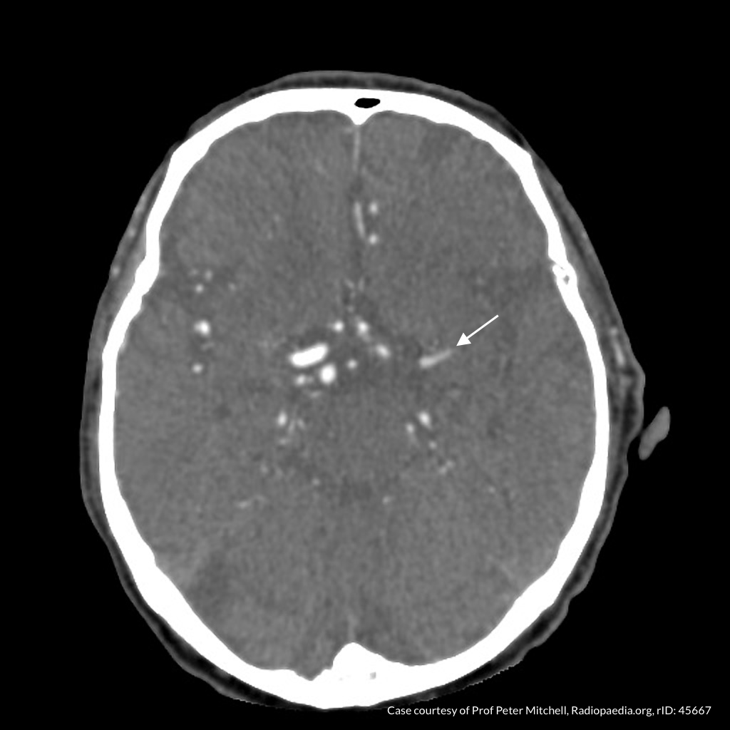
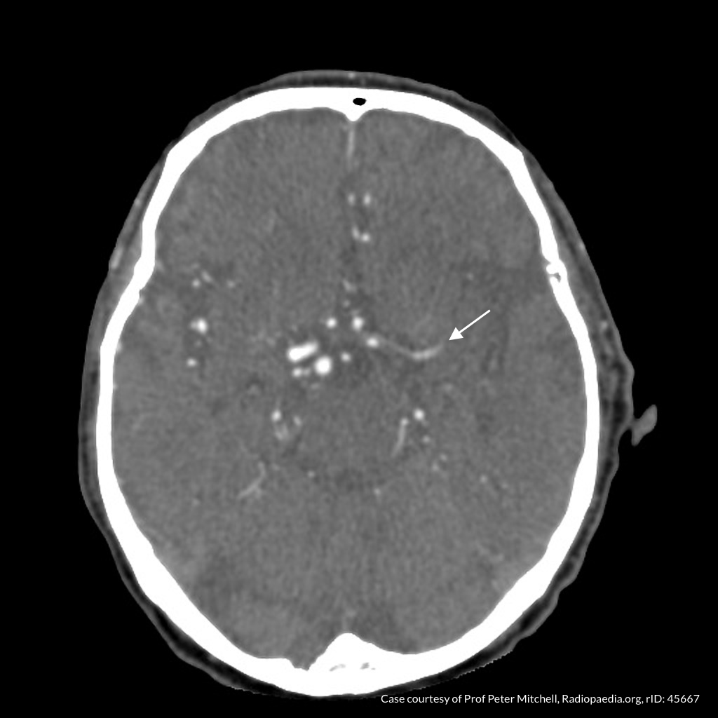
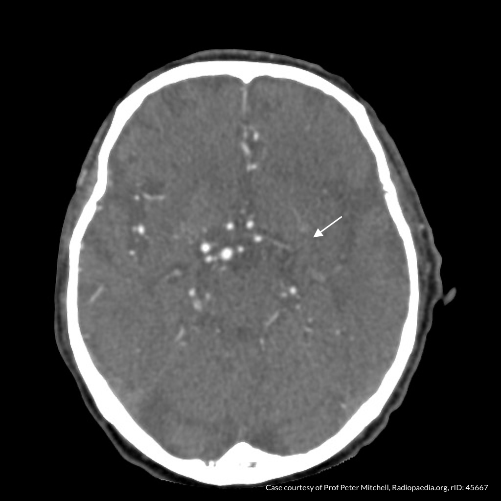
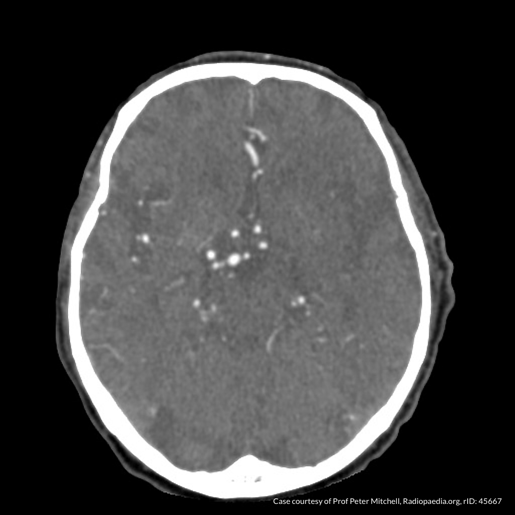
Stroke

Brief HPI:

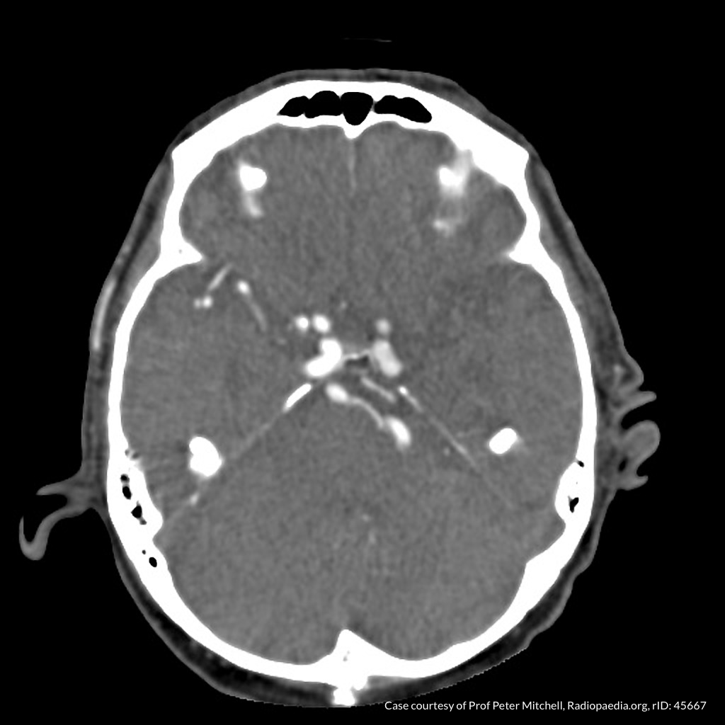
An approximately 60 year-old male with a history of hypertension and diabetes is brought to the emergency department after noting difficulty speaking and right-sided weakness upon awakening. Prehospital capillary glucose measured 268mg/dL. He went to sleep at 10pm on the evening prior to presentation.

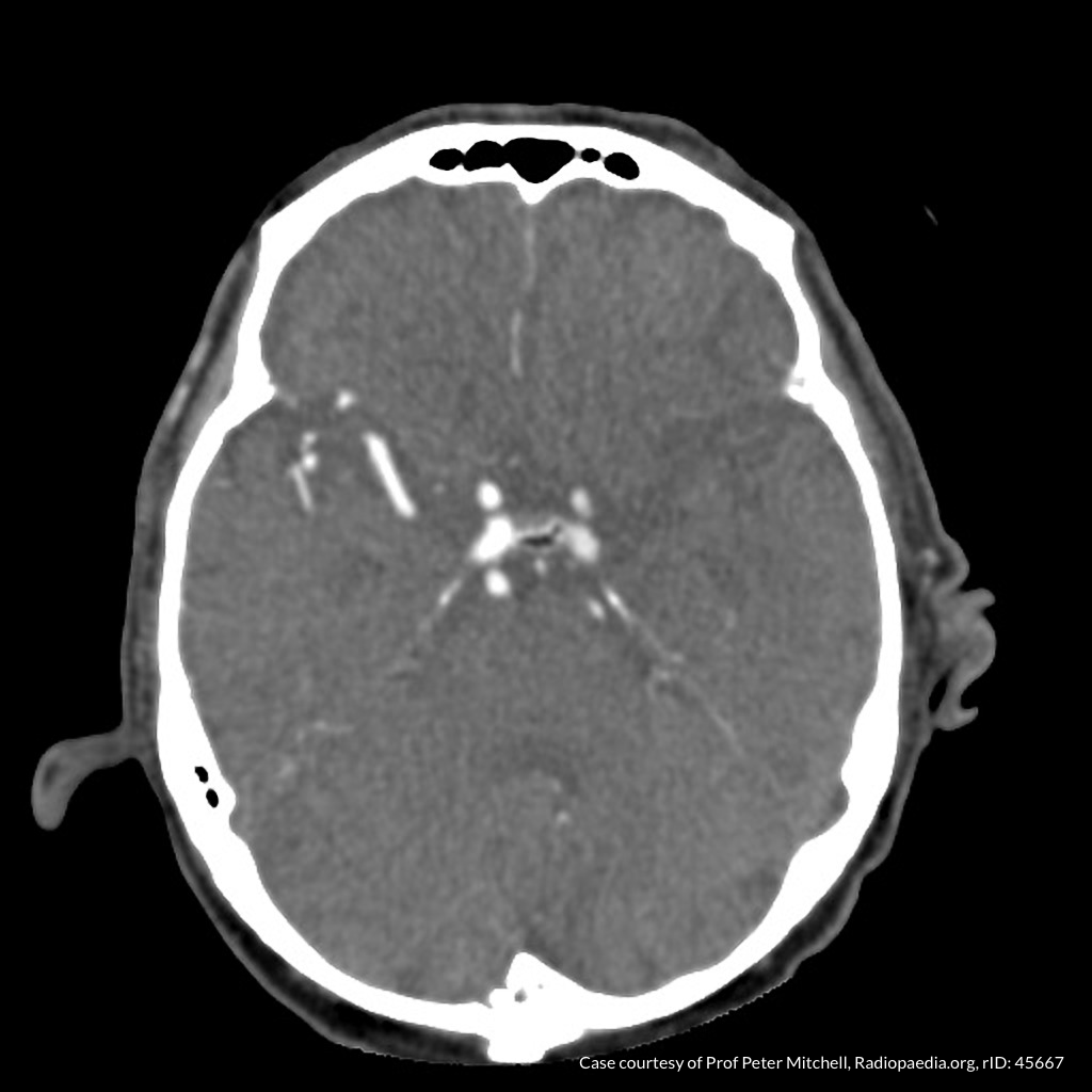
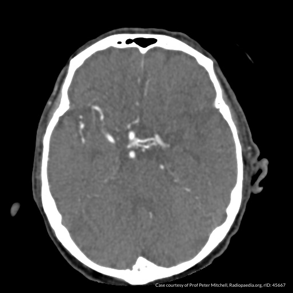
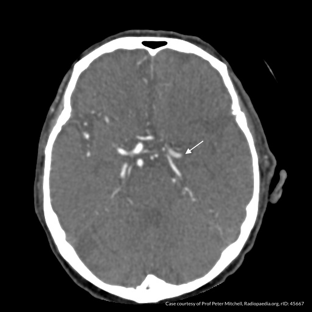
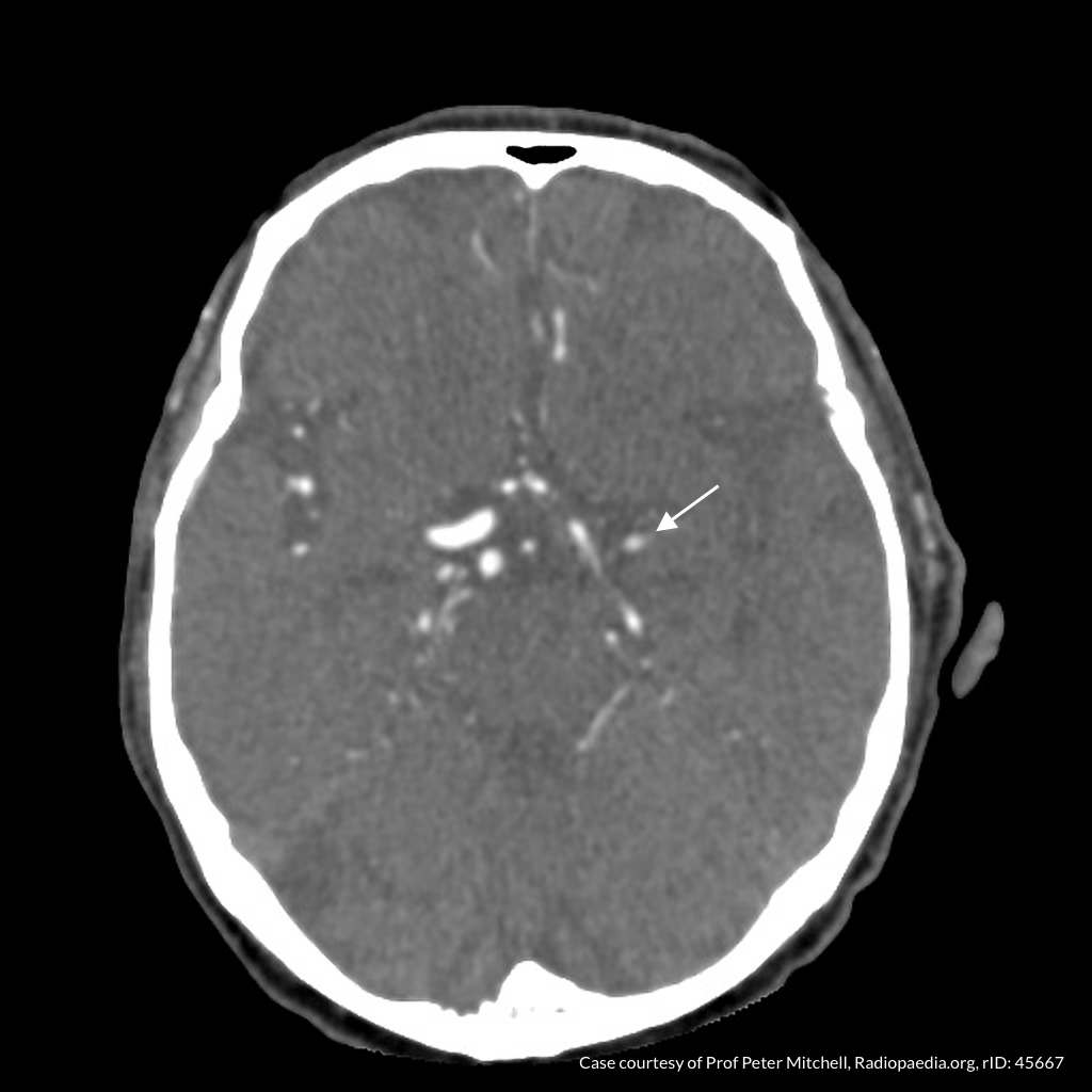
The patient arrives in the emergency department awake and alert at 9am. He was unable to provide history due to speech difficulty but is able to follow commands. Examination demonstrates right upper and lower extremity weakness. Computed tomography of the head and neck is obtained, non-contrast imaging shows no hemorrhage and angiography demonstrates left MCA occlusion. He proceeds emergently to the angiography suite where mechanical thrombectomy restores normal perfusion. The patient is discharged to an inpatient rehabilitation facility for intensive physical therapy three days later.

CT Angiography

01_mca
02_mca
03_mca
04_mca
05_mca
06_mca
07_mca
08_mca
09_mca

CT Angiography

Left MCA M1 occlusion

Code Stroke Algorithm

Code Stroke Algorithm

References

  1. Goldstein LB, Simel DL. Is this patient having a stroke? JAMA. 2005;293(19):2391-2402. doi:10.1001/jama.293.19.2391.
  2. Hemmen TM, Meyer BC, McClean TL, Lyden PD. Identification of nonischemic stroke mimics among 411 code strokes at the University of California, San Diego, Stroke Center. J Stroke Cerebrovasc Dis. 2008;17(1):23-25. doi:10.1016/j.jstrokecerebrovasdis.2007.09.008.
  3. Prabhakaran S, Ruff I, Bernstein RA. Acute stroke intervention: a systematic review. JAMA. 2015;313(14):1451-1462. doi:10.1001/jama.2015.3058.
  4. Yew KS, Cheng EM. Diagnosis of acute stroke. Am Fam Physician. 2015;91(8):528-536.
  5. Hemphill JC, Greenberg SM, Anderson CS, et al. Guidelines for the Management of Spontaneous Intracerebral Hemorrhage: A Guideline for Healthcare Professionals From the American Heart Association/American Stroke Association. Stroke. 2015;46(7):2032-2060. doi:10.1161/STR.0000000000000069.
  6. Hankey GJ. Stroke. Lancet. 2017;389(10069):641-654. doi:10.1016/S0140-6736(16)30962-X.
  7. Powers WJ, Rabinstein AA, Ackerson T, et al. 2018 Guidelines for the Early Management of Patients With Acute Ischemic Stroke: A Guideline for Healthcare Professionals From the American Heart Association/American Stroke Association. Stroke. 2018;49(3):e46-e110. doi:10.1161/STR.0000000000000158.
  8. Hasan TF, Rabinstein AA, Middlebrooks EH, et al. Diagnosis and Management of Acute Ischemic Stroke. Mayo Clin Proc. 2018;93(4):523-538. doi:10.1016/j.mayocp.2018.02.013.
  9. Thomalla G, Simonsen CZ, Boutitie F, et al. MRI-Guided Thrombolysis for Stroke with Unknown Time of Onset. N Engl J Med. 2018;379(7):611-622. doi:10.1056/NEJMoa1804355.

Synovial Fluid

Brief HPI:

A 38 year-old female with a history of obesity and obstructive sleep apnea presents with right knee pain. She cannot identify a clear precipitant for her symptoms which she first noted 2 weeks ago. Her pain is worsened with ambulation and while previously tolerable, has grown more severe despite over-the-counter analgesics over the past two days. She denies fevers, intravenous drug use, recent travel or instrumentation.

On evaluation, vital signs are normal. Physical examination demonstrates a moderate-sized right knee effusion with overlying warmth though no edema. There is minimal pain with range of motion, no pain with heel percussion, and she is ambulatory independently with a mildly antalgic gait. Clinical suspicion for septic arthritis was low. A diagnostic arthrocentesis was performed without complication. Synovial fluid was less-viscous than normal with slight debris. Laboratory analysis revealed 14,230 white blood cells with 85% neutrophils and no crystals visualized. The patient was discharged with supportive care and outpatient follow-up – cultures were ultimately negative.

An Algorithm for the Analysis of Synovial Fluid

An Algorithm for the Analysis of Synovial Fluid

References

  1. Margaretten ME, Kohlwes J, Moore D, Bent S. Does this adult patient have septic arthritis? JAMA. 2007;297(13):1478-1488. doi:10.1001/jama.297.13.1478.
  2. Brannan SR, Jerrard DA. Synovial fluid analysis. J Emerg Med. 2006;30(3):331-339. doi:10.1016/j.jemermed.2005.05.029.
  3. Couderc M, Pereira B, Mathieu S, et al. Predictive value of the usual clinical signs and laboratory tests in the diagnosis of septic arthritis. CJEM. 2015;17(4):403-410. doi:10.1017/cem.2014.56.
  4. MD HJC, MD LAB, MD ML. Septic Arthritis. Hospital Medicine Clinics. 2014;3(4):494-503. doi:10.1016/j.ehmc.2014.06.009.

Ascitic Fluid

Brief HPI:

A 56 year-old male with a history of alcoholic cirrhosis complicated by esophageal varices presents to the emergency department with abdominal distension. He notes gradually worsening symptoms over the past 2 weeks – roughly correlating with the timing of his last paracentesis. He has limited access to medical care and typically presents to emergency departments for palliative paracenteses. He is otherwise in his usual state of health and denies fevers, chills, abdominal pain, vomiting blood, or dark/bloody stools.

Vital signs are notable for a heart rate of 97bpm and blood pressure of 110/65mmHg – otherwise normal. Examination demonstrates a distended abdomen which is non-tender, dull to percussion and with a palpable fluid wave. Bedside ultrasonography shows large, homogenous-appearing ascites with readily-accessible pockets for drainage in the bilateral lower quadrants. A palliative paracentesis is performed with uncomplicated extraction of 4 liters of translucent, straw-colored fluid. Ascitic fluid analysis shows 90 white blood cells of which 10% are polymorphonuclear. The patient is observed briefly in the emergency department, noted symptomatic improvement and was discharged with a plan for telephone follow-up of fluid culture results.

An Algorithm for the Analysis of Ascitic Fluid

Algorithm for the Analysis of Ascitic Fluid

References

  1. Runyon BA. Care of patients with ascites. N Engl J Med. 1994;330(5):337-342. doi:10.1056/NEJM199402033300508.
  2. Wong CL, Holroyd-Leduc J, Thorpe KE, Straus SE. Does this patient have bacterial peritonitis or portal hypertension? How do I perform a paracentesis and analyze the results? JAMA. 2008;299(10):1166-1178. doi:10.1001/jama.299.10.1166.
  3. Tarn AC, Lapworth R. Biochemical analysis of ascitic (peritoneal) fluid: what should we measure? Ann Clin Biochem. 2010;47(Pt 5):397-407. doi:10.1258/acb.2010.010048.
  4. Li PK-T, Szeto CC, Piraino B, et al. ISPD Peritonitis Recommendations: 2016 Update on Prevention and Treatment. Perit Dial Int. 2016;36(5):481-508. doi:10.3747/pdi.2016.00078.
  5. MacIntosh T. Emergency Management of Spontaneous Bacterial Peritonitis – A Clinical Review. Cureus. 2018;10(3):e2253. doi:10.7759/cureus.2253.

Resuscitative Thoracotomy

Brief HPI:

A call is received from pre-hospital providers regarding an inbound trauma patient. An estimated 30 year-old male with unknown history sustained a penetrating wound to the right flank. On EMS arrival the patient was unresponsive but had a weakly-palpable radial pulse which was lost en-route. Their estimated time of arrival is 5 minutes.

Algorithm for the Selection of Patients for Resuscitative Thoracotomy

Algorithm for the Selection of Patients for Resuscitative Thoracotomy

References:

  1. Seamon MJ, Haut ER, Van Arendonk K, Barbosa RR, Chiu WC, Dente CJ, et al. An evidence-based approach to patient selection for emergency department thoracotomy: A practice management guideline from the Eastern Association for the Surgery of Trauma. J Trauma Acute Care Surg. 2015 Jul;79(1):159–73.
  2. Burlew CC, Moore EE, Moore FA, Coimbra R, McIntyre RC, Davis JW, et al. Western Trauma Association critical decisions in trauma: resuscitative thoracotomy. J Trauma Acute Care Surg. 2012 Dec;73(6):1359–63.
  3. Sherren PB, Reid C, Habig K, Burns BJ. Algorithm for the resuscitation of traumatic cardiac arrest patients in a physician-staffed helicopter emergency medical service. Crit Care. 2013 Mar 12;17(2):308.
  4. Cothren CC, Moore EE. Emergency department thoracotomy for the critically injured patient: Objectives, indications, and outcomes. World J Emerg Surg. 2006 Mar 24;1:4.

Red and Painful Eye

Brief HPI

A 60-year-old female with rheumatoid arthritis presents with unilateral eye pain and redness without reported vision changes. Physical examination demonstrates radially-oriented engorged episcleral vessels and normal visual acuity – she was diagnosed with episcleritis and discharged with follow-up.

Algorithm for the Evaluation of the Red and Painful Eye

Algorithm for the Evaluation of the Red and Painful Eye

Overview

The red or painful eye is a common presentation in the emergency department and the rapid identification and management of potentially sight-threatening causes is critical.

The diagnostic approach to the red or painful eye begins with identifying a history of caustic exposure where immediate and copious irrigation (even before detailed examination) may limit further injury. Alkaline agents induce more severe liquefactive necrosis leading to keratoconjunctivitis, while acidic agents are generally less destructive. Management is identical for both: irrigation with lactated Ringer solution through a Morgan lens applied to a topically anesthetized eye for 5-10 minutes repeated until the pH of the eye is neutral1.

Ocular or facial trauma presents a spectrum of differential diagnoses. Suspicion for globe rupture is increased by a suggestive mechanism such as a high-velocity projectile or high-impact blunt facial trauma. Characteristic examination findings include obvious globe deformity, an irregularly-shaped pupil, extrusion of vitreous, markedly decreased visual acuity, or parting of fluorescein (Seidel sign). If globe rupture is suspected, further manipulation is unadvisable and the affected eye should be shielded. Measures should be taken to avoid increases in intra-ocular pressure including elevation of the patient’s head-of-bed, anti-emetics (to prevent intra-ocular hypertension with vomiting), and the avoidance of medications potentially implicated in intra-ocular hypertension (ketamine, succinylcholine). Tetanus and antimicrobial prophylaxis should be provided while awaiting emergent ophthalmology consultation2,3.

Another traumatic diagnosis warranting rapid identification and possible intervention is retrobulbar hematoma – identified by proptosis, eye pain, decreased visual acuity, and elevated intra-ocular pressure. Pressures exceeding 40mmHg warrant lateral cantholysis in conjunction with medical management to prevent optic nerve ischemia and preserve vision4,5.

Diagnostic Approach

The evaluation of the non-traumatic red or painful eye follows a systematic and anatomically-based approach, starting with external components and moving inward:

1. External

The examination begins externally with an assessment of function (visual acuity) and inspection and palpation of the periorbital region. Periorbital edema, erythema, and tenderness to palpation in the setting of systemic illness (fever) is concerning for orbital cellulitis. When associated with elevated intra-ocular pressure or proptosis, a retrobulbar abscess may be present. Both warrant admission and parenteral antibiotics and the latter may require operative management such as aspiration or cantholysis. Less severe features without impact on visual acuity is suggestive of a periorbital cellulitis which may be treated as an outpatient with close follow-up2,6.

2. Lids and Lashes

Several non-emergent processes may affect the lids and lashes including blepharitis (inflammation of the eyelid margin), chalazion (inflammation of the Meibomian glands), hordeolum (eyelash follicle abscess), or dacrocystitis (infection of the lacrimal sac)2,7,8.

3. Conjunctiva and Sclera

Again, proceeding from superficial to deeper structures we encounter the epithelial layer (including palpebral and bulbar components) covering the sclera which is subject to allergic or infectious inflammation. Conjunctivitis is characterized by engorgement of superficial conjunctival blood vessels, potentially associated with conjunctival edema (chemosis), or discharge. Most conjunctivitis is self-limited and not sight-threatening, treatment is aimed at symptomatic relief though topical antibiotics have few adverse effects and may be prescribed if the diagnosis of bacterial conjunctivitis is unclear9.

When associated with pain, a deeper inflammatory process is implicated. Scleritis is a frequently immune-mediated inflammatory process (though infection, malignancy and medications have been implicated) associated with pain, photophobia, and examination findings of globe tenderness and engorged scleral blood vessels. Management in the emergency department is trivial (systemic NSAID’s), however ophthalmology consultation should be secured due to the risk of vision-compromising complications, as well as the intimation of an underlying systemic disorder9,10. Episcleritis is similarly immune-mediated, though generally self-limiting. The diagnosis is made by identification of characteristic, radially-oriented engorged episcleral vessels. When the diagnosis of scleritis versus episcleritis or conjunctivitis is in question the application of a topical vasoconstrictor (phenylephrine 2.5%) will blanch vessels in the conjunctival or superficial episcleral plexuses – sparing scleral vessels10,11.

4. Cornea

Keratitis can be caused by infection, ultraviolet light exposure, or contact lens use. Patients may have photophobia and a foreign-body sensation. Gross inspection or slit-lamp examination will show epithelial erosions that stain with fluorescein or the characteristic dendritic pattern accompanying herpes simplex virus infection. Management includes ophthalmology consultation, topical antibiotics if a bacterial process is suggested, and close follow-up7-9,12.

5. Anterior Chamber

A critical process occurring in the anterior chamber is angle-closure glaucoma. The patient commonly presents with severe pain, circumcorneal injection, and a pupil fixed at mid-dilation. Diagnosis is confirmed by the measurement of elevated intra-ocular pressure (greater than 20mmHg). Reduction of intra-ocular pressure with topical and systemic agents should begin immediately while awaiting emergent ophthalmology consultation13.

The slit-lamp microscope facilitates examination of the anterior chamber. The presence of cells (floating white and red blood cells, or layering hypopyon or hyphema) and flare (protein) suggest inflammation in the anterior segment caused by a systemic inflammatory process, infection, or trauma and warrants close ophthalmologic follow-up2,7,9.

6. Vitreous

An ocular examination mimicking orbital cellulitis with evidence of anterior chamber involvement, particularly in a patient with a history of recent ocular surgery or trauma suggests endophthalmitis. Management requires admission for parenteral antibiotics with ophthalmology consultation2.

Additional Diagnostic Modalities

Advanced imaging may be useful in the diagnosis of traumatic and non-traumatic orbital pathology. Multi-detector computed tomography (MD-CT) is readily available and rapidly performed in the emergency department and can aid in the diagnosis of critical infectious processes, including extension beyond the orbital septum in orbital cellulitis, scleral thickening in endophthalmitis, and characterization of hematoma or abscess in the retrobulbar space. The addition of intravenous contrast media can identify critical vascular processes such as cavernous sinus thrombosis14,15. For traumatic pathology, CT can assist with the evaluation of globe integrity, lens position, vitreous/retinal detachment, and foreign bodies16. Imaging cannot be relied upon exclusively to exclude pathology, and the patient’s presentation and clinician’s examination should determine the need for consultation and evaluation. For globe rupture, for example, in one study of 59 patients with severe ocular trauma and diagnostic uncertainty regarding the presence of globe rupture, CT failed to diagnosed open globe injury in 1/3 of patients (with surgical scleral inspection as a reference standard)17. Another retrospective analysis of 48 eyes sustaining trauma revealed sensitivity ranging from 56-68% for CT identification of open globe injury18.

In addition to potential diagnostic inaccuracy, computed tomography exposes patients to risks including ionizing radiation, and the possibility of contrast-induced nephropathy19. Ultrasound is becoming increasingly accessible and comfortable for the emergency physician, and has the benefit of being relatively non-invasive – including facilitating ocular examination in patients with significant periorbital swelling limiting eye-opening. Ocular ultrasound may aid with the diagnosis of a wide variety of ocular pathology including vitreous hemorrhage, retinal detachment, central retinal arterial/venous occlusions, foreign body identification, lens dislocation and retrobulbar hematoma. In one study of 61 patients presenting with trauma or acute vision changes, ultrasound interpretation agreed with criterion standard (orbital computed tomography or ophthalmology evaluation) for 98% of cases20.

View Ocular Ultrasound Algorithm

References

  1. Messman AM. Ocular Injuries: New Strategies In Emergency Department Management. Emergency Medicine Practice. 2015;17(11):1–21–quiz21–2.
  2. Wright JL, Wightman JM. Red and painful eye. … Concepts and Clinical Practice 8th ed …. 2014.
  3. Romaniuk VM. Ocular trauma and other catastrophes. Emerg Med Clin North Am. 2013;31(2):399-411. doi:10.1016/j.emc.2013.02.003.
  4. Babineau MR, Sanchez LD. Ophthalmologic Procedures in the Emergency Department. Emerg Med Clin North Am. 2008;26(1):17-34. doi:10.1016/j.emc.2007.11.003.
  5. Rowh AD, Ufberg JW, Chan TC, Vilke GM, Harrigan RA. Lateral canthotomy and cantholysis: emergency management of orbital compartment syndrome. J Emerg Med. 2015;48(3):325-330. doi:10.1016/j.jemermed.2014.11.002.
  6. Henderson M, Tierney L, Smetana G. The Patient History: Evidence-Based Approach. McGraw Hill Professional; 2012.
  7. Alteveer JG, Mccans KM, Hemphill RR. The Red Eye, The Swollen Eye, And Acute Vision Loss. … Practice+ Em Practice …. 2002.
  8. Leibowitz HM. The red eye. N Engl J Med. 2000;343(5):345-351. doi:10.1056/NEJM200008033430507.
  9. Mahmood AR, Narang AT. Diagnosis and management of the acute red eye. Emerg Med Clin North Am. 2008. doi:10.1016/j.emc.2007.10.002.
  10. Albini TA, Rao NA, Smith RE. The Diagnosis and Management of Anterior Scleritis. International Ophthalmology Clinics. 2005;45(2):191.
  11. Roscoe M, Landis T. How to diagnose the acute red eye with confidence. JAAPA. 2006;19(3):24–30–quiz45–6.
  12. Deborah S Jacobs MD. Evaluation of the red eye. UpToDate. https://www.uptodate.com/contents/evaluation-of-the-red-eye. Published February 24, 2016. Accessed April 18, 2017.
  13. Prum BE, Herndon LW, Moroi SE, et al. Primary Angle Closure Preferred Practice Pattern(®) Guidelines. Ophthalmology. 2016;123(1):P1-P40. doi:10.1016/j.ophtha.2015.10.049.
  14. LeBedis CA, Sakai O. Nontraumatic orbital conditions: diagnosis with CT and MR imaging in the emergent setting. Radiographics. 2008;28(6):1741-1753. doi:10.1148/rg.286085515.
  15. Platnick J, Crum AV, Soohoo S, Cedeño PA, Johnson MH. The globe: infection, inflammation, and systemic disease. YSULT. 2011;32(1):38-50. doi:10.1053/j.sult.2010.12.003.
  16. Dunkin JM, Crum AV, Swanger RS, Bokhari SAJ. Globe trauma. YSULT. 2011;32(1):51-56. doi:10.1053/j.sult.2010.09.003.
  17. Hoffstetter P, Schreyer AG, Schreyer CI, et al. Multidetector CT (MD-CT) in the diagnosis of uncertain open globe injuries. Rofo. 2010;182(2):151-154. doi:10.1055/s-0028-1109659.
  18. Arey ML, Mootha VV, Whittemore AR, Chason DP, Blomquist PH. Computed tomography in the diagnosis of occult open-globe injuries. Ophthalmology. 2007;114(8):1448-1452. doi:10.1016/j.ophtha.2006.10.051.
  19. Custer PL, Kent TL. Pitfalls of ophthalmic radiographic imaging. Curr Opin Ophthalmol. 2014;25(5):432-435. doi:10.1097/ICU.0000000000000064.
  20. Blaivas M, Theodoro D, Sierzenski PR. A study of bedside ocular ultrasonography in the emergency department. Academic Emergency Medicine. 2002;9(8):791-799.
Pericardial Effusion

HPI:

43F with a history of HTN and diastolic heart failure presenting with two days of shortness of breath. Reports that symptoms are worse at night when lying down to sleep and associated with a cough productive of white sputum. She also reports intermittent left-sided chest pain, described as sharp and exacerbated by cough or deep inspiration. She denies fevers/chills, nausea/vomiting, sick contacts or recent travel.
m

PMH:

  • Hypertension
  • Diabetes Mellitus (Type II)
  • Hyperlipidemia
  • Diastolic heart failure

PSH:

  • Cesarean section

FH:

  • Father with MI at 76 years-old

SHx:

  • Lives at home.
  • Denies tobacco, alcohol or drug abuse.

Meds:

  • Lasix 40mg p.o. daily
  • Lisinopril 20mg p.o. daily
  • Atenolol 50mg p.o. daily
  • Omeprazole 20mg p.o. daily
  • Lantus 14 units daily
  • Novolin 6 units t.i.d

Allergies:

NKDA

Physical Exam:

VS: T 98.2 HR 81 RR 19 BP 219/91 O2 95% RA
Gen: Adult female in no acute distress, alert and responding appropriately to questions.
HEENT: PERRL, EOMI, mucous membranes moist.
CV: RRR, no murmurs appreciated, no JVD.
Lungs: Crackles at right lung base.
Abd: Soft, non-tender, non-distended, without rebound/guarding.
Ext: 1+ pitting edema in bilateral lower extremities to knee.
Neuro: AAOx4, grossly normal peripheral sensation and motor strength.

Labs/Studies:

  • Troponin: 0.15
  • Procalcitonin: 0.15
  • CBC: 10.9/9.1/26.4/296
  • BMP: 134/4.6/104/22/56/2.87/214

Imaging:

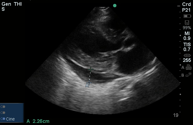
Pericardial Effusion

Pericardial Effusion

Measured in the largest dimension, suggestive of a moderate to large pericardial effusion.

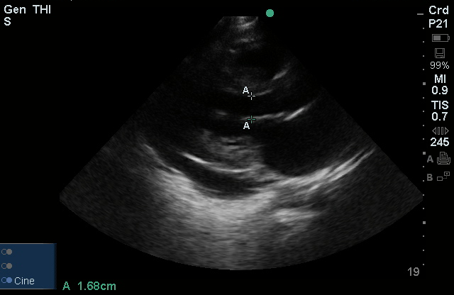
E-Point Septal Separation

E-Point Septal Separation

E-Point Septal Separation (EPSS), estimated here is the smallest distance between the anterior leaflet of the mitral valve and intraventricular septum. Values > 12mm are suggestive of depressed ejection fraction.

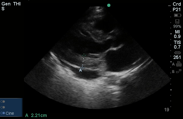
Left Ventricular Hypertrophy

Left Ventricular Hypertrophy

Thickened left ventricular wall.

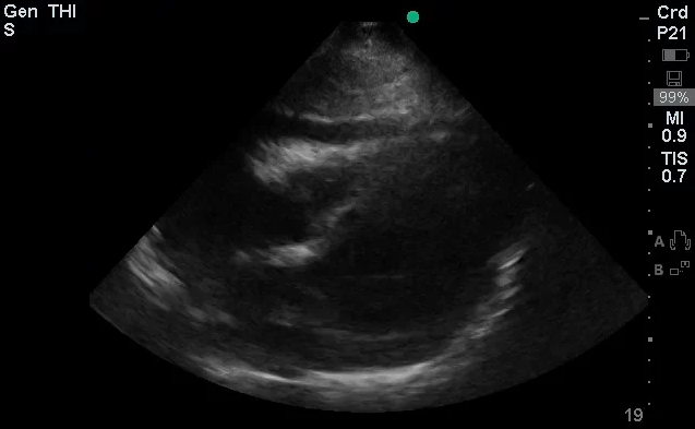
Pericardial Effusion - Subxiphoid

Pericardial Effusion - Subxiphoid

Pericardial Effusion - Parasternal Long

Pericardial Effusion - Parasternal Long

Pericardial Effusion - Parasternal Short

Pericardial Effusion - Parasternal Short

  • CXR: Consolidation involving the majority of the right lung, cardiomegaly.
  • Bedside Echo: LVEF 55%, concentric LVH, no wall motion abnormality, moderate pericardial effusion noted, RV not collapsed.

Assessment/Plan:

43F with a history of HTN, diastolic heart failure presenting with SOB.

#SOB: CXR finding of right-sided consolidation with history of productive cough, evidence of leukocytosis with neutrophil predominance, and relative hypoxemia suggestive of community-acquired pneumonia. No evidence of systemic inflammatory response. PE unlikely, patient is not bed-bound and alternative diagnosis more likely.
– Start empiric antimicrobial therapy ceftriaxone 1g IV q24h, azithromycin 500mg IV q24h.

#Pericardial Effusion: Noted on bedside echo, no evidence of RV collapse to suggest cardiac tamponade. Also, no JVD and pulsus paradoxus measured at 8mmHg.
– Obtain formal transthoracic echocardiogram to evaluate effusion.
– Consult cardiology if worsening hemodynamics

#Elevated Troponin: No ECG changes suggestive of acute ST-elevation MI. May represent NSTEMI though historical features not consistent with ACS.
– Trend troponin/EKG q.8.h. x3
– Give aspirin 325mg, consider anti-coagulation.
– Consider stress echo prior to discharge

#Elevated Creatinine: Baseline unknown, likely acute component with or without chronic kidney disease.
– Volume resuscitation as tolerated, follow repeat chemistry.

#Hypertension: Asymptomatic, resume home medications.

Physiology of Cardiac Tamponade 1

  • Intrapericardial pressure (IPP) normally reflects intrathoracic pressure (ITP).
  • Inspiration: low ITP → low RAP → increased RA filling.
  • Expiration: high ITP → low LAP → increased LA filling.
  • Increased pericardial fluid → increased IPP → increased LA/RA filling pressures (diastolic dysfunction) → increased variation with respiration.
  • Earliest hemodynamic change in cardiac tamponade is JVD or IVC dilation.

IVC variation as marker for RAP 1

IVC Diameter (cm) Change with Respiration (%) RAP (mmHg)
<2.1 >50% 0-5
<2.1 <50% 5-10
>2.1 >50% 5-10
>2.1 <50% >15

Grading Pericardial Effusions 1

Grade Echo-free space (mm) Size (mL)
Small <10 100
Moderate 10-20 100-500
Large >20 >500

History and Physical Exam in Patients with Acute Pericarditis 2,3

Symptom/Sign ACS Pericarditis PE
Quality Pressure Sharp Sharp
Pleuritic No Yes Yes
Positional No Yes (worse when supine) No
Duration Minutes to hours Hours to days Hours to days
Improves with NG Yes No No
Friction Rub No Yes No
S3 Maybe No No

Differential Diagnosis of Pericardial Effusion 2-8

Differential Diagnosis of Pericardial Effusion

References:

  1. Schairer, J. R., Biswas, S., Keteyian, S. J., & Ananthasubramaniam, K. (2011). A Systematic Approach to Evaluation of Pericardial Effusion and Cardiac Tamponade. Cardiology in Review, 19(5), 233–238. doi:10.1097/CRD.0b013e31821e202c
  2. Khandaker MH, Espinosa RE, Nishimura RA, et al. Pericardial Disease: Diagnosis and Management. Mayo Clinic Proceedings. 2010;85(6):572-593. doi:10.4065/mcp.2010.0046.
  3. Lange, RA, Hillis, LD. Clinical practice. Acute pericarditis. The New England journal of medicine. 2004;351(21), 2195–2202. doi:10.1056/NEJMcp041997
  4. Imazio M, Adler Y. Management of pericardial effusion. Eur Heart J. 2013;34(16):1186-1197. doi:10.1093/eurheartj/ehs372.
  5. LeWinter MM. Clinical practice. Acute pericarditis. N Engl J Med. 2014;371(25):2410-2416. doi:10.1056/NEJMcp1404070.
  6. Vakamudi S, Ho N, Cremer PC. Pericardial Effusions: Causes, Diagnosis, and Management. Prog Cardiovasc Dis. 2017;59(4):380-388. doi:10.1016/j.pcad.2016.12.009.
  7. Imazio M, Mayosi BM, Brucato A, et al. Triage and management of pericardial effusion. J Cardiovasc Med (Hagerstown). 2010;11(12):928-935. doi:10.2459/JCM.0b013e32833e5788.
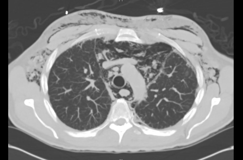
  8. Maisch B, Seferović PM, Ristić AD, et al. Guidelines on the diagnosis and management of pericardial diseases executive summary; The Task force on the diagnosis and management of pericardial diseases of the European society of cardiology. Eur Heart J. 2004;25(7):587-610. doi:10.1016/j.ehj.2004.02.002.

Low Voltage ECG

Definition

  • QRS in limb leads <5mm
  • QRS in precordial leads <10mm

General Causes

  • Fluid, fat or air attenuating signal
  • Myocardial infiltration
  • Loss of viable myocardium

Example

Low Voltage ECG
Low Voltage ECG

Low Voltage ECG

ECG of patient with pericardial effusion

Baseline ECG
Baseline ECG

Baseline ECG

Old ECG from same patient

Differential Diagnosis of Low Voltage ECG

Differential Diagnosis of Low-Voltage ECG

References

  1. Madias JE. Low QRS voltage and its causes. J Electrocardiol. 2008;41(6):498–500. doi:10.1016/j.jelectrocard.2008.06.021.
  2. WikEM: Low ECG voltage

Palpitations

Brief H&P

48F with a history of Grave disease (off medications for 4 months), presenting with palpitations. Noted gradual onset of palpitations while at rest, describing a pounding sensation lasting 3-4 hours and persistent (though improved) on presentation. Symptoms not associated with chest pain, shortness of breath, loss of consciousness, nor triggered by exertion. She reported a history of 8-10 episodes in the past for which she did not seek medical attention. Review of systems notable only for heat intolerance.

On physical examination, vital signs were notable for tachycardia (HR 138bpm). No alteration in mental status, murmur, tremor or hyperreflexia appreciated.

Labs

  • Hb: 14.7
  • Urine hCG: negative
  • TSH: <0.01
  • Total T3: 311ng/dL
  • Free T4: 2.64ng/dL

ECG

Palpitations - Sinus Tachycardia

Sinus Tachycardia

Impression/Plan

Palpitations due to sinus tachycardia from symptomatic hyperthyroidism secondary to medication non-adherence. Improved with propranolol, discharged with methimazole and PMD follow-up.

Algorithm for the Evaluation and Management of Palpitations1, 2

Algorithm for the Evaluation and Management of Palpitations

Evaluation of Palpitations

History and Physical

Subjective description of symptom quality
Rapid and regular beating suggests paroxysmal SVT or VT. Rapid and irregular beating suggests atrial fibrillation, atrial flutter, or variable conduction block.
Stop/start sensation: PAC or PVC
Rapid fluttering: Sustained supraventricular or ventricular tachycardia
Pounding in neck: Produced by canon A waves from AV dissociation (VT, complete heart block, SVT)
Onset and offset
Random, episodic, lasting instants: Suggests PAC or PVC
Gradual onset and offset: Sinus tachycardia
Abrupt onset and offset: SVT or VT
Syncope
Suggests hemodynamically significant arrhythmia, often VT
Examination
Identify evidence of structural, valvular heart disease

ECG1

ECG Finding Presumed etiology
Short PR, Delta waves WPW, AVRT
LAA, LVH Atrial fibrillation
PVC, BBB Idiopathic VT
Q-waves Prior MI, VT
QT-prolongation VT (polymorphic)
LVH, septal Q-waves HCM
Blocks  

References

  1. Zimetbaum P, Josephson ME. Evaluation of patients with palpitations. N Engl J Med. 1998;338(19):1369-1373. doi:10.1056/NEJM199805073381907.
  2. Probst MA, Mower WR, Kanzaria HK, Hoffman JR, Buch EF, Sun BC. Analysis of emergency department visits for palpitations (from the National Hospital Ambulatory Medical Care Survey). The American Journal of Cardiology. 2014;113(10):1685-1690. doi:10.1016/j.amjcard.2014.02.020.
  3. Abbott AV. Diagnostic approach to palpitations. Am Fam Physician. 2005;71(4):743-750.