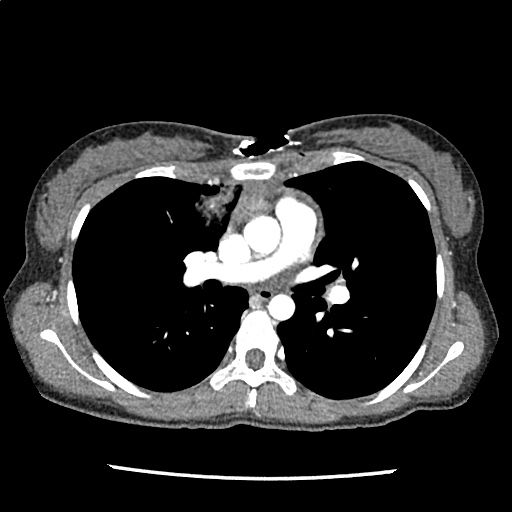
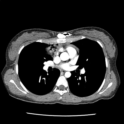
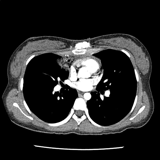
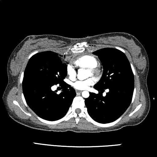
Palpitations

Brief H&P

48F with a history of Grave disease (off medications for 4 months), presenting with palpitations. Noted gradual onset of palpitations while at rest, describing a pounding sensation lasting 3-4 hours and persistent (though improved) on presentation. Symptoms not associated with chest pain, shortness of breath, loss of consciousness, nor triggered by exertion. She reported a history of 8-10 episodes in the past for which she did not seek medical attention. Review of systems notable only for heat intolerance.

On physical examination, vital signs were notable for tachycardia (HR 138bpm). No alteration in mental status, murmur, tremor or hyperreflexia appreciated.

Labs

  • Hb: 14.7
  • Urine hCG: negative
  • TSH: <0.01
  • Total T3: 311ng/dL
  • Free T4: 2.64ng/dL

ECG

Palpitations - Sinus Tachycardia

Sinus Tachycardia

Impression/Plan

Palpitations due to sinus tachycardia from symptomatic hyperthyroidism secondary to medication non-adherence. Improved with propranolol, discharged with methimazole and PMD follow-up.

Algorithm for the Evaluation and Management of Palpitations1, 2

Algorithm for the Evaluation and Management of Palpitations

Evaluation of Palpitations

History and Physical

Subjective description of symptom quality
Rapid and regular beating suggests paroxysmal SVT or VT. Rapid and irregular beating suggests atrial fibrillation, atrial flutter, or variable conduction block.
Stop/start sensation: PAC or PVC
Rapid fluttering: Sustained supraventricular or ventricular tachycardia
Pounding in neck: Produced by canon A waves from AV dissociation (VT, complete heart block, SVT)
Onset and offset
Random, episodic, lasting instants: Suggests PAC or PVC
Gradual onset and offset: Sinus tachycardia
Abrupt onset and offset: SVT or VT
Syncope
Suggests hemodynamically significant arrhythmia, often VT
Examination
Identify evidence of structural, valvular heart disease

ECG1

ECG Finding Presumed etiology
Short PR, Delta waves WPW, AVRT
LAA, LVH Atrial fibrillation
PVC, BBB Idiopathic VT
Q-waves Prior MI, VT
QT-prolongation VT (polymorphic)
LVH, septal Q-waves HCM
Blocks  

References

  1. Zimetbaum P, Josephson ME. Evaluation of patients with palpitations. N Engl J Med. 1998;338(19):1369-1373. doi:10.1056/NEJM199805073381907.
  2. Probst MA, Mower WR, Kanzaria HK, Hoffman JR, Buch EF, Sun BC. Analysis of emergency department visits for palpitations (from the National Hospital Ambulatory Medical Care Survey). The American Journal of Cardiology. 2014;113(10):1685-1690. doi:10.1016/j.amjcard.2014.02.020.
  3. Abbott AV. Diagnostic approach to palpitations. Am Fam Physician. 2005;71(4):743-750.

Seizure

Brief HPI

A 72 year-old male with a history of hypertension and hiatal hernia presents to the emergency department with one week of generalized weakness. His family report decreased oral intake with frequent emesis over the past four days. He denies chest pain, shortness of breath, abdominal pain, or other complaints. During the interview he has a generalized tonic-clonic seizure which persists for five minutes despite the administration of 4mg of lorazepam.

An Algorithm for the Management of Seizures

The management of active seizures is algorithmic, starting with a rapid assessment of airway patency, supporting ventilation (with appropriate positioning, nasopharyngeal airway adjuncts and bag-valve mask if needed) and ensuring adequate perfusion. Patients should have continuous vital sign monitoring, supplemental oxygen to maintain oxygen saturation >92% and intravenous access1.

Pharmacologic treatment follows a stepwise approach, detailed in the algorithm below. The focus is on immediate stabilization and progressively escalating anti-epileptic drugs eventually requiring endotracheal intubation and continuous infusions of sedatives2-4.

An Algorithm for the Management of Seizures

Pathophysiology

Seizures are caused by excessive and disorganized neuronal activation, typically induced by global alterations in the production and transmission of impulses (electrolyte derangements, drugs/toxins, infection), or foci of increased irritability (hemorrhage, stroke, mass) – a pathophysiologic motif that mimics cardiac tachyarrhythmias (sympathomimetic toxicity or scarred myocardium for example)1. Status epilepticus, defined as a seizure lasting greater than five minutes or recurrent seizures without a return to normal baseline, shares an equally high short-term mortality – greater than 20%5.

Syncope vs. Seizure

The algorithm below details historical and examination features that may assist with distinguishing epileptic seizure from non-epileptic activity6,7.

Clinical Features Distinguishing Seizure from Syncope

Case Conclusion

The patient continued to seize and a point-of-care chemistry panel revealed a serum sodium of 108mEq/L. Seizures abate after the infusion of hypertonic saline (100mL of 3% saline over 10 minutes, repeated until cessation of seizures). While hyponatremia is generally corrected slowly – owing to the risk of osmotic demyelination – immediate correction in this setting is critical8.


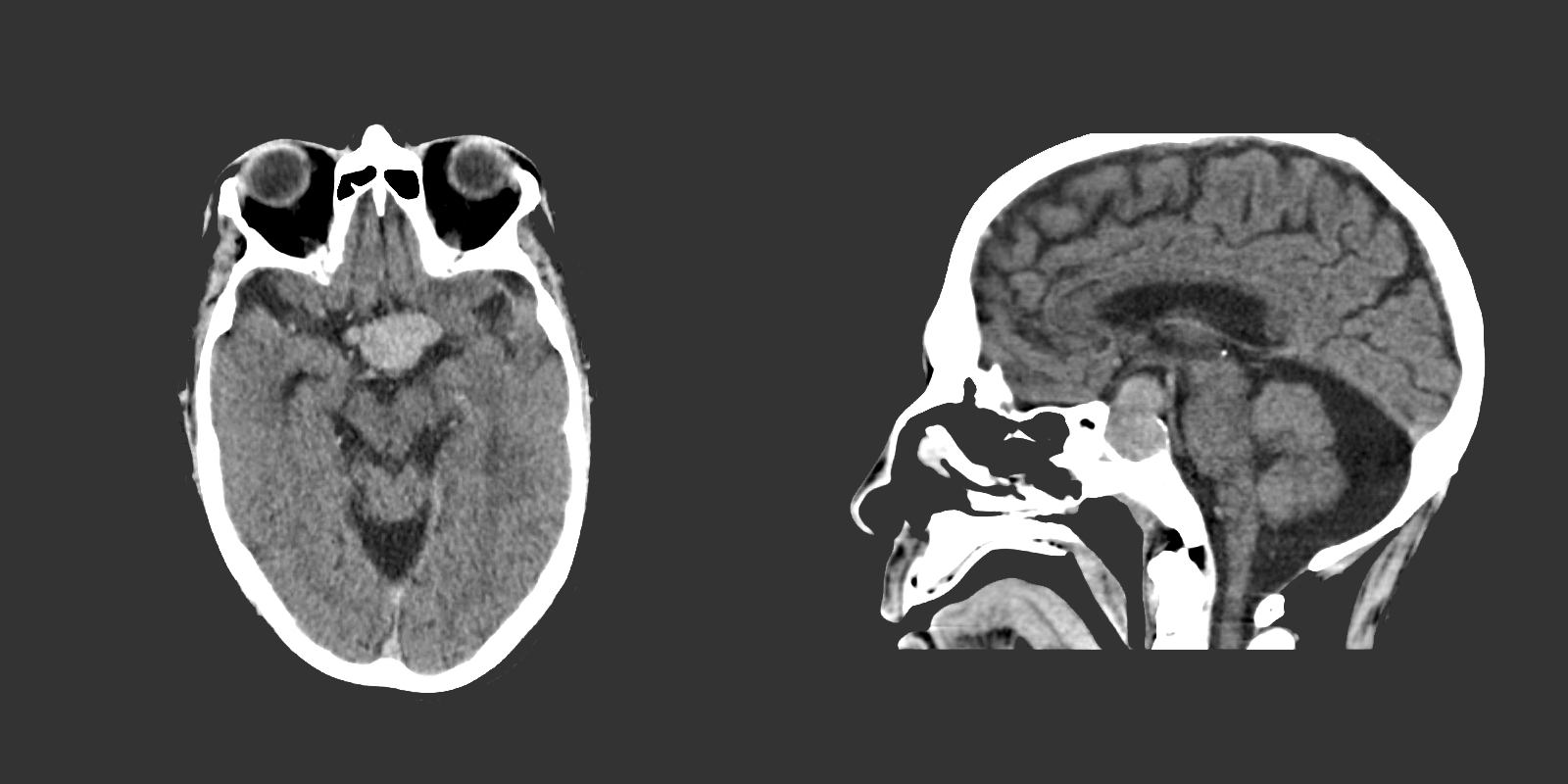
The remainder of the patient’s evaluation demonstrated urine osmolarity is 389mOsm/kg and urine sodium is 53mmol/L, in the setting of relative euvolemia on examination these findings were consistent with SIADH. Head computed tomography is obtained and reveals a sellar mass.

View Hyponatremia Algorithm

References

  1. McMullan JT, Davitt AM, Pollack CV Jr. Seizures. In: Rosen’s Emergency Medicine. Mosby Incorporated; 2002:2808. doi:10.1016/S1474-4422(06)70350-7.
  2. Billington M, Kandalaft OR, Aisiku IP. Adult Status Epilepticus: A Review of the Prehospital and Emergency Department Management. J Clin Med. 2016;5(9):74. doi:10.3390/jcm5090074.
  3. Huff JS, Morris DL, Kothari RU, Gibbs MA, Emergency Medicine Seizure Study Group. Emergency department management of patients with seizures: a multicenter study. Academic Emergency Medicine. 2001;8(6):622-628.
  4. Prasad M, Krishnan PR, Sequeira R, Al-Roomi K. Anticonvulsant therapy for status epilepticus. Prasad M, ed. Cochrane Database Syst Rev. 2014;16(9):CD003723. doi:10.1002/14651858.CD003723.pub3.
  5. Logroscino G, Hesdorffer DC, Cascino G, Annegers JF, Hauser WA. Short-term mortality after a first episode of status epilepticus. Epilepsia. 1997;38(12):1344-1349.
  6. Sheldon R, Rose S, Ritchie D, et al. Historical criteria that distinguish syncope from seizures. J Am Coll Cardiol. 2002;40(1):142-148.
  7. McKeon A, Vaughan C, Delanty N. Seizure versus syncope. Lancet Neurol. 2006;5(2):171-180. doi:10.1016/S1474-4422(06)70350-7.
  8. Goh KP. Management of hyponatremia. Am Fam Physician. 2004;69(10):2387-2394.

Cardiac Arrest

Brief HPI:

An overhead page alerts you to an arriving patient with cardiac arrest. An approximately 35-year-old male was running away from police officers and collapsed after being shot with a stun gun. The patient was found to be pulseless, CPR was started by police officers and the patient is en route.

An Algorithm for the Evaluation and Management of Cardiac Arrest with Ultrasonography

An Algorithm for the Evaluation and Management of Cardiac Arrest with Ultrasonography

Causes of Cardiac (and non-cardiac) Arrest

Sudden cardiac arrest (SCA) leading to sudden cardiac death (SCD) if not successfully resuscitated, refers to the unexpected collapse of circulatory function. Available epidemiologic data for in-hospital and out-of-hospital cardiac arrest (OHCA) point appropriately to cardiac processes as the most common cause, though extra-cardiac processes (most frequently respiratory), comprise up to 40% of cases1-3.

Identifying the underlying cause is critical as several reversible precipitants require rapid identification. However, the usual diagnostic techniques may be challenging, limited or absent – including patient history, detailed examination, and diagnostic studies.

The initial rhythm detected upon evaluation is most suggestive of the etiologic precipitant. Pulseless ventricular tachycardia (pVT) or ventricular fibrillation (VF) is suggestive of a cardiac process – most commonly an acute coronary syndrome although heart failure or other structural and non-structural heart defects associated with dysrhythmias may be at fault4.

Pulseless electrical activity (PEA) presents a broader differential diagnosis as it essentially represents severe shock. The most common extra-cardiac cause is hypoxia – commonly secondary to pulmonary processes including small and large airway obstruction (bronchospasm, aspiration, foreign body, edema). Other causes include substance intoxication, medication adverse effect5,6, or electrolyte disturbances7. Finally, any precipitant of shock may ultimately lead to PEA, including hypovolemia/hemorrhage, obstruction (massive pulmonary embolus8, tamponade, tension pneumothorax), and distribution (sepsis).

Asystole is the absence of even disorganized electrical discharge and is the terminal degeneration of any of the previously-mentioned rhythms if left untreated.

Management of Cardiac Arrest

Optimizing survival outcomes in patients with cardiac arrest is dependent on early resuscitation with the prioritization of interventions demonstrated to have survival benefit. When advanced notice is available, prepare the resuscitation area including airway equipment (with adjuncts to assist ventilation and waveform capnography devices). Adopt the leadership position and assign roles for chest compressions, airway support, application of monitor/defibrillator, and establishment of peripheral access.

High-quality chest compressions with minimal interruptions are the foundation of successful resuscitation – and guideline changes prioritizing compressions have demonstrated detectable improvements in rates of successful resuscitation9,10. Measurement of quantitative end-tidal capnography can guide adequacy of chest compressions11,12 and an abrupt increase may signal restoration of circulation without necessitating interruptions of chest compressions13,14. Sustained, low measures of end-tidal CO2 despite appropriate resuscitation may signal futility and (alongside other factors) guides termination of resuscitation11,12.

The next critical step in restoring circulation is prompt defibrillation of eligible rhythms (pVT/VF) when detected. The immediate delivery of 200J (uptitrated to the device maximum for subsequent shocks) of biphasic energy and restoration of a perfusing rhythm is one of few interventions with clear benefits. For pVT/VF that persists despite multiple countershocks (more than three), the addition of an intravenous antiarrhythmic appears to improve survival to hospital admission. The ARREST trial was a randomized controlled study comparing amiodarone to its diluent as placebo for OHCA with refractory pVT/VF showing significant improvement in survival to hospital admission for the amiodarone group15. This was followed by the ALIVE trial comparing amiodarone and lidocaine which showed significantly higher rates of survival to hospital admission in the amiodarone group16. However, a more recent randomized trial comparing amiodarone (in a novel diluent less likely to cause hypotension), lidocaine, and placebo in a similar patient population showed less convincing results, with no detected difference in survival or the secondary outcome of favorable neurological outcome for either amiodarone or lidocaine compared with placebo17. The heterogeneity of available data contributed to current guidelines which recommend that either amiodarone or lidocaine may be used for shock-refractory pVT/VF18.

Current guidelines also recommend the administration of vasopressors (epinephrine 1mg every 3-5 minutes). In one randomized controlled trial exploring the long-standing guideline recommendations, epinephrine was associated with increased rates of restoration of spontaneous circulation, though no significant impact on the primary outcome of survival to hospital discharge was identified19. Physiologically, increased systemic vascular resistance combined with positive beta-adrenergic impact on cardiac output would be expected to complement resuscitative efforts. However, more recent studies have suggested that arrest physiology and unanticipated pharmacologic effects may complicate this simplistic interpretation – particularly when patient-centered outcomes are emphasized. Research exploring the timing and amount of epinephrine suggest that earlier administration and higher cumulative doses are associated with negative impacts on survival to hospital discharge and favorable neurological outcomes20-22.

Ultimately, treatment should focus on optimal execution of measures with clear benefits (namely chest compressions and early defibrillation of eligible rhythms). Other management considerations with which the emergency physician is familiar with including establishing peripheral access and definitive airway management can be delayed.

Rapid Diagnostic Measures for the Identification of Reversible Processes

Traditional diagnostic measures are generally unavailable during an ongoing cardiac arrest resuscitation. The emergency medicine physician must rely on the physical examination and point-of-care tests with the objective of identifying potentially reversible processes. Measurement of capillary blood glucose can exclude hypoglycemia as a contributor. Point-of-care chemistry and blood gas analyzers can identify important electrolyte derangements, as well as clarifying the primary impulse in acid-base disturbances.

End-tidal capnography was discussed previously for the guidance of ongoing resuscitation, but it may have diagnostic utility in patients with SCD. In one study the initial EtCO2 was noted to be significantly higher for primary pulmonary processes (with PEA/asystole as presenting rhythm) compared to primary cardiac processes (with pVT/VF as presenting rhythm)23.

The use of point-of-care ultrasonography, particularly in PEA arrest where non-cardiac etiologies dominate, may help identify the etiology of arrest and direct therapy. Bedside ultrasonography should be directed first at assessment of cardiac function – examining the pericardial sac and gross abnormalities in chamber size. A pericardial effusion may suggest cardiac tamponade, ventricular collapse can be seen with hypovolemia, and asymmetric right-ventricular dilation points to pulmonary embolus where thrombolysis should be considered8. If cardiac ultrasound is unrevealing, thoracic ultrasound can identify pneumothorax24-27.

In the absence of ultrasonographic abnormalities, attention turns to other rapidly reversible precipitants first. If opioid toxicity is a consideration, an attempt at reversal with naloxone has few adverse effects. If any detected rhythm is a polymorphic ventricular tachycardia characteristic of torsades de pointes – rapid infusion of magnesium sulfate should follow defibrillation. Other potentially reversible medications or toxins should be managed as appropriate.

Post-Resuscitation Steps

After successful restoration of circulation, the next management steps are critical to the patient’s long-term outcomes. A definitive airway should be established if not already secured (and if restoration of circulation was not associated with neurological recovery sufficient for independent airway protection). Circulatory support should continue with fluid resuscitation and vasopressors to maintain end-organ perfusion.

An immediate ECG should be performed to identify infarction, ischemia or precipitants of dysrhythmia. ST-segment elevation after return of spontaneous circulation (ROSC) warrants emergent angiography and possible intervention. However, given the prevalence of cardiac causes (of which coronary disease is most common) for patients with pVT/VF arrest, the presence of ST elevations is likely of insufficient sensitivity to identify all patients who would benefit from angiography. Several studies and meta-analyses have explored a more inclusive selection strategy for angiography (patients without obvious non-cardiac causes for arrest), all of which identified survival benefits with angiography and successful angioplasty when possible28-30.

Finally, the induction of hypothermia (or targeted temperature management) has significant benefits in survivors of cardiac arrest and can be instituted in the emergency department. Studies first targeted a core temperature of 32-24°C, with a randomized controlled trial demonstrating higher rates of favorable neurological outcome and reduced mortality31. More recent studies suggest that a more liberal temperature target does not diffuse the positive effects of induced hypothermia. A randomized trial of 939 patients with OHCA comparing a targeted temperature of 33°C vs 36°C suggested that a lower temperature target did not confer higher benefit to mortality or recovery of neurological function32. The more liberal temperature target may alleviate adverse effects associated with hypothermia which include cardiovascular effects (bradycardia), electrolyte derangements (during induction and rewarming), and possible increased risk of infections33. Targeted temperature management is achieved with external cooling measures or infusion of cooled fluids, rarely requiring more invasive measures34. Aggregate review of available data in a recent meta-analysis further supports the use of targeted temperature management after cardiac arrest as standard-of-care35.

References

  1. Bergum D, Nordseth T, Mjølstad OC, Skogvoll E, Haugen BO. Causes of in-hospital cardiac arrest – Incidences and rate of recognition. Resuscitation. 2015;87:63-68. doi:10.1016/j.resuscitation.2014.11.007.
  2. Wallmuller C, Meron G, Kurkciyan I, Schober A, Stratil P, Sterz F. Causes of in-hospital cardiac arrest and influence on outcome. Resuscitation. 2012;83(10):1206-1211. doi:10.1016/j.resuscitation.2012.05.001.
  3. Vaartjes I, Hendrix A, Hertogh EM, et al. Sudden death in persons younger than 40 years of age: incidence and causes. European Journal of Cardiovascular Prevention & Rehabilitation. 2009;16(5):592-596. doi:10.1097/HJR.0b013e32832d555b.
  4. Zheng ZJ, Croft JB, Giles WH, Mensah GA. Sudden cardiac death in the United States, 1989 to 1998. Circulation. 2001;104(18):2158-2163.
  5. Hoes AW, Grobbee DE, Lubsen J, Man in ‘t Veld AJ, van der Does E, Hofman A. Diuretics, beta-blockers, and the risk for sudden cardiac death in hypertensive patients. Ann Intern Med. 1995;123(7):481-487.
  6. Siscovick DS, Raghunathan TE, Psaty BM, et al. Diuretic therapy for hypertension and the risk of primary cardiac arrest. N Engl J Med. 1994;330(26):1852-1857. doi:10.1056/NEJM199406303302603.
  7. Gettes LS. Electrolyte abnormalities underlying lethal and ventricular arrhythmias. Circulation. 1992;85(1 Suppl):I70-I76.
  8. Kürkciyan I, Meron G, Sterz F, et al. Pulmonary embolism as a cause of cardiac arrest: presentation and outcome. Arch Intern Med. 2000;160(10):1529-1535.
  9. Callaway CW, Soar J, Aibiki M, et al. Part 4: Advanced Life Support: 2015 International Consensus on Cardiopulmonary Resuscitation and Emergency Cardiovascular Care Science With Treatment Recommendations. In: Vol 132. American Heart Association, Inc.; 2015:S84-S145. doi:10.1161/CIR.0000000000000273.
  10. Kudenchuk PJ, Redshaw JD, Stubbs BA, et al. Impact of changes in resuscitation practice on survival and neurological outcome after out-of-hospital cardiac arrest resulting from nonshockable arrhythmias. Circulation. 2012;125(14):1787-1794. doi:10.1161/CIRCULATIONAHA.111.064873.
  11. Touma O, Davies M. The prognostic value of end tidal carbon dioxide during cardiac arrest: a systematic review. Resuscitation. 2013;84(11):1470-1479. doi:10.1016/j.resuscitation.2013.07.011.
  12. Levine RL, Wayne MA, Miller CC. End-tidal carbon dioxide and outcome of out-of-hospital cardiac arrest. N Engl J Med. 1997;337(5):301-306. doi:10.1056/NEJM199707313370503.
  13. Garnett AR, Ornato JP, Gonzalez ER, Johnson EB. End-tidal carbon dioxide monitoring during cardiopulmonary resuscitation. JAMA. 1987;257(4):512-515.
  14. Falk JL, Rackow EC, Weil MH. End-tidal carbon dioxide concentration during cardiopulmonary resuscitation. N Engl J Med. 1988;318(10):607-611. doi:10.1056/NEJM198803103181005.
  15. Kudenchuk PJ, Cobb LA, Copass MK, et al. Amiodarone for resuscitation after out-of-hospital cardiac arrest due to ventricular fibrillation. N Engl J Med. 1999;341(12):871-878. doi:10.1056/NEJM199909163411203.
  16. Dorian P, Cass D, Schwartz B, Cooper R, Gelaznikas R, Barr A. Amiodarone as compared with lidocaine for shock-resistant ventricular fibrillation. N Engl J Med. 2002;346(12):884-890. doi:10.1056/NEJMoa013029.
  17. Kudenchuk PJ, Brown SP, Daya M, et al. Amiodarone, Lidocaine, or Placebo in Out-of-Hospital Cardiac Arrest. N Engl J Med. 2016;374(18):1711-1722. doi:10.1056/NEJMoa1514204.
  18. Neumar RW, Shuster M, Callaway CW, et al. Part 1: Executive Summary: 2015 American Heart Association Guidelines Update for Cardiopulmonary Resuscitation and Emergency Cardiovascular Care. In: Vol 132. American Heart Association, Inc.; 2015:S315-S367. doi:10.1161/CIR.0000000000000252.
  19. Jacobs IG, Finn JC, Jelinek GA, Oxer HF, Thompson PL. Effect of adrenaline on survival in out-of-hospital cardiac arrest: A randomised double-blind placebo-controlled trial. Resuscitation. 2011;82(9):1138-1143. doi:10.1016/j.resuscitation.2011.06.029.
  20. Hagihara A, Hasegawa M, Abe T, Nagata T, Wakata Y, Miyazaki S. Prehospital epinephrine use and survival among patients with out-of-hospital cardiac arrest. JAMA. 2012;307(11):1161-1168. doi:10.1001/jama.2012.294.
  21. Dumas F, Bougouin W, Geri G, et al. Is epinephrine during cardiac arrest associated with worse outcomes in resuscitated patients? J Am Coll Cardiol. 2014;64(22):2360-2367. doi:10.1016/j.jacc.2014.09.036.
  22. Andersen LW, Kurth T, Chase M, et al. Early administration of epinephrine (adrenaline) in patients with cardiac arrest with initial shockable rhythm in hospital: propensity score matched analysis. BMJ. 2016;353:i1577. doi:10.1136/bmj.i1577.
  23. Grmec S, Lah K, Tusek-Bunc K. Difference in end-tidal CO2 between asphyxia cardiac arrest and ventricular fibrillation/pulseless ventricular tachycardia cardiac arrest in the prehospital setting. Crit Care. 2003;7(6):R139-R144. doi:10.1186/cc2369.
  24. Rose JS, Bair AE, Mandavia D, Kinser DJ. The UHP ultrasound protocol: a novel ultrasound approach to the empiric evaluation of the undifferentiated hypotensive patient. American Journal of Emergency Medicine. 2001;19(4):299-302. doi:10.1053/ajem.2001.24481.
  25. Hernandez C, Shuler K, Hannan H, Sonyika C, Likourezos A, Marshall J. C.A.U.S.E.: Cardiac arrest ultra-sound exam—A better approach to managing patients in primary non-arrhythmogenic cardiac arrest. Resuscitation. 2008;76(2):198-206. doi:10.1016/j.resuscitation.2007.06.033.
  26. Chardoli M, Heidari F, Shuang-ming S, et al. Echocardiography integrated ACLS protocol versus con- ventional cardiopulmonary resuscitation in patients with pulseless electrical activity cardiac arrest. Chinese Journal of Traumatology. 2012;15(5):284-287. doi:10.3760/cma.j.issn.1008-1275.2012.05.005.
  27. Zengin S, Yavuz E, Al B, et al. Benefits of cardiac sonography performed by a non-expert sonographer in patients with non-traumatic cardiopulmonary arrest. Resuscitation. 2016;102:105-109. doi:10.1016/j.resuscitation.2016.02.025.
  28. Spaulding CM, Joly LM, Rosenberg A, et al. Immediate coronary angiography in survivors of out-of-hospital cardiac arrest. N Engl J Med. 1997;336(23):1629-1633. doi:10.1056/NEJM199706053362302.
  29. Dumas F, Cariou A, Manzo-Silberman S, et al. Immediate Percutaneous Coronary Intervention Is Associated With Better Survival After Out-of-Hospital Cardiac Arrest: Insights From the PROCAT (Parisian Region Out of Hospital Cardiac Arrest) Registry. Circulation: Cardiovascular Interventions. 2010;3(3):200-207. doi:10.1161/CIRCINTERVENTIONS.109.913665.
  30. Millin MG, Comer AC, Nable JV, et al. Patients without ST elevation after return of spontaneous circulation may benefit from emergent percutaneous intervention: A systematic review and meta-analysis. Resuscitation. 2016;108:54-60. doi:10.1016/j.resuscitation.2016.09.004.
  31. Hypothermia after Cardiac Arrest Study Group. Mild therapeutic hypothermia to improve the neurologic outcome after cardiac arrest. N Engl J Med. 2002;346(8):549-556. doi:10.1056/NEJMoa012689.
  32. Nielsen N, Wetterslev J, Cronberg T, et al. Targeted Temperature Management at 33°C versus 36°C after Cardiac Arrest. N Engl J Med. 2013;369(23):2197-2206. doi:10.1056/NEJMoa1310519.
  33. Polderman KH, Peerdeman SM, Girbes AR. Hypophosphatemia and hypomagnesemia induced by cooling in patients with severe head injury. J Neurosurg. 2001;94(5):697-705. doi:10.3171/jns.2001.94.5.0697.
  34. Polderman KH, Herold I. Therapeutic hypothermia and controlled normothermia in the intensive care unit: Practical considerations, side effects, and cooling methods*. Critical Care Medicine. 2009;37(3):1101-1120. doi:10.1097/CCM.0b013e3181962ad5.
  35. Schenone AL, Cohen A, Patarroyo G, et al. Therapeutic hypothermia after cardiac arrest: A systematic review/meta-analysis exploring the impact of expanded criteria and targeted temperature. Resuscitation. 2016;108:102-110. doi:10.1016/j.resuscitation.2016.07.238.

Atrial Fibrillation

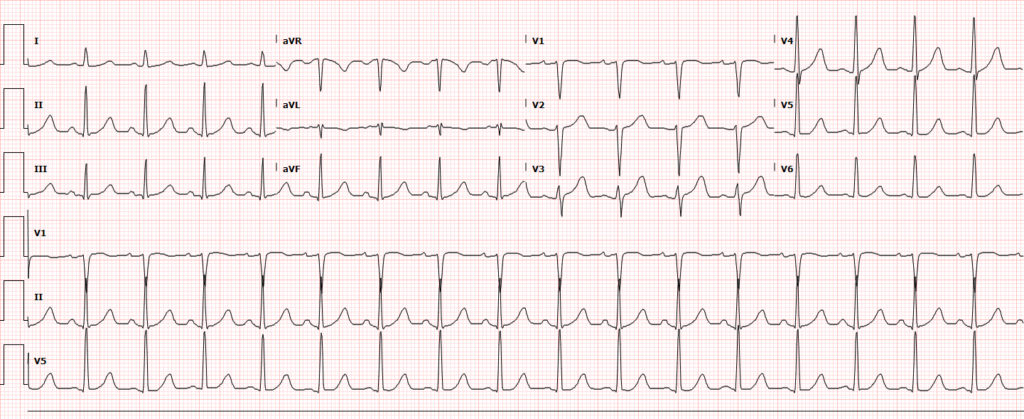
A 64-year-old male with a history of hypertension and hyperlipidemia presents with palpitations. He reports intermittent symptoms over the past 3 days, associated with dyspnea on exertion, but no chest pain, dizziness or syncope. His vital signs are notable for tachycardia (HR 129bpm) without hypotension or hypoxia. An ECG shows atrial fibrillation with rapid ventricular response.

Evaluation and Management

While the patient’s tachyarrhythmia is not yet associated with hypotension or evidence of malperfusion, preparation is key and includes continuous telemetry and vital sign monitoring, establishment of intravenous access, and application of cardioversion pads 1.

The presence of atrial fibrillation is new and the rapid ventricular response (RVR) may be symptomatic of more serious and potentially reversible pathology. A thorough history and physical examination may elucidate a precipitant and should precede attempts at rate- or rhythm-control. RVR may be provoked by any of the processes that would otherwise induce a sinus tachycardia, including bleeding, infection, toxic/metabolic etiologies and endocrinopathies 2, 3

Candidacy for Cardioversion

In hemodynamically stable patients with new-onset atrial fibrillation, candidacy for cardioversion includes:4-6

  • Stable without ischemia, hypotension or acute CHF
  • Clear onset of <48 hours
  • Non-severe symptoms
  • Few prior episodes/treatments
  • Existing anti-coagulation with warfarin and therapeutic INR (at least 3 weeks)
  • Absence of high-risk features: rheumatic/valvular disease, severe left-ventricular dysfunction, prosthetic valves, or history of thromboembolism

Cardioversion may be pharmacologic (with procainamide, or amiodarone), or electrical (synchronized at 100-200J). Electrical cardioversion for acute atrial fibrillation is both more effective and results in shorter lengths-of-stay in the emergency department – though stable patients should participate in shared decision-making7. Another important consideration when cardioversion is pursued is the prevention of systemic embolization. While atrial fibrillation of duration less than 48-hours is rarely associated with systemic embolization, certain populations are at higher risk8. One retrospective study of 3143 patients with atrial fibrillation for less than 48-hours demonstrated an overall risk of 0.7% for thromboembolic events – though the rate was significantly higher in patients older than 60 years or with other comorbidities (heart failure, diabetes)9. The risk of embolic events should be weighed against the risk of bleeding.

CHA2DS2VASc10-12

C Congestive Heart Failure 1
H Hypertension 1
A2 Age >75 2
D Diabetes Mellitus 1
S2 Stroke, TIA, Thromboembolism 2
V Vascular disease 1
A Age >65 1
Sc Sex Category Female 1


  • 0: low risk (may not require anti-coagulation)
  • 1: low-moderate risk (consider anti-platelet or anti-coagulation)
  • ≥ 2: moderate-high risk (anti-coagulation recommended)

HAS-BLED13,14

H Uncontrolled hypertension 1
A Abnormal renal/liver function
Renal (renal replacement therapy, creatinine >2.3mg/dL) 1
Liver (cirrhosis, bilirubin >2x, AST/ALT >3x) 1
S Stroke 1
B Bleeding history/anemia 1
L Labile INR 1
E Elderly (>65) 1
D Drugs
Anti-platelet agent, NSAID 1
Alcohol (>8 drinks/week) 1


  • 0: low risk (0.6-1.13% annual risk of major bleeding)
  • 1-2: intermediate risk (1.02-3.2% annual risk of major bleeding)
  • ≥ 3: high risk (4.9-19.6% annual risk of major bleeding)

Pharmacologic Management

For patients who are not candidates for cardioversion, rate-control should be pursued. Options include AV nodal blocking agents such as calcium channel blockers and beta-blockers15. The most frequently studied agents of each category are metoprolol and diltiazem. Both classes show comparable efficacy and safety profiles with trends favoring diltiazem16, 17.

Algorithm for the management of atrial fibrillation with rapid ventricular response:

Algorithm for the management of atrial fibrillation with rapid ventricular response

References

  1. Atzema, C.L. and T.W. Barrett, Managing atrial fibrillation. Ann Emerg Med, 2015. 65(5): p. 532-9.
  2. January, C.T., et al., 2014 AHA/ACC/HRS Guideline for the Management of Patients With Atrial Fibrillation: Executive Summary. A Report of the American College of Cardiology/American Heart Association Task Force on Practice Guidelines and the Heart Rhythm Society, 2014. 64(21): p. 2246-2280.
  3. Scheuermeyer, F.X., et al., Emergency Department Patients With Atrial Fibrillation or Flutter and an Acute Underlying Medical Illness May Not Benefit From Attempts to Control Rate or Rhythm. Ann Emerg Med, 2015. 65(5): p. 511-522 e2.
  4. Stiell, I.G., et al., Association of the Ottawa Aggressive Protocol with rapid discharge of emergency department patients with recent-onset atrial fibrillation or flutter. CJEM, 2010. 12(3): p. 181-91.
  5. von Besser, K. and A.M. Mills, Is discharge to home after emergency department cardioversion safe for the treatment of recent-onset atrial fibrillation? Ann Emerg Med, 2011. 58(6): p. 517-20.
  6. Cohn, B.G., S.M. Keim, and D.M. Yealy, Is emergency department cardioversion of recent-onset atrial fibrillation safe and effective? J Emerg Med, 2013. 45(1): p. 117-27.
  7. Bellone, A., et al., Cardioversion of acute atrial fibrillation in the emergency department: a prospective randomised trial. Emerg Med J, 2012. 29(3): p. 188-91.
  8. Weigner, M.J., et al., Risk for clinical thromboembolism associated with conversion to sinus rhythm in patients with atrial fibrillation lasting less than 48 hours. Ann Intern Med, 1997. 126(8): p. 615-20.
  9. Airaksinen, K.E., et al., Thromboembolic complications after cardioversion of acute atrial fibrillation: the FinCV (Finnish CardioVersion) study. J Am Coll Cardiol, 2013. 62(13): p. 1187-92.
  10. Friberg, L., M. Rosenqvist, and G.Y. Lip, Evaluation of risk stratification schemes for ischaemic stroke and bleeding in 182 678 patients with atrial fibrillation: the Swedish Atrial Fibrillation cohort study. Eur Heart J, 2012. 33(12): p. 1500-10.
  11. Lip, G.Y., et al., Refining clinical risk stratification for predicting stroke and thromboembolism in atrial fibrillation using a novel risk factor-based approach: the euro heart survey on atrial fibrillation. Chest, 2010. 137(2): p. 263-72.
  12. Ntaios, G., et al., CHADS(2), CHA(2)S(2)DS(2)-VASc, and long-term stroke outcome in patients without atrial fibrillation. Neurology, 2013. 80(11): p. 1009-17.
  13. Lip, G.Y., et al., Comparative validation of a novel risk score for predicting bleeding risk in anticoagulated patients with atrial fibrillation: the HAS-BLED (Hypertension, Abnormal Renal/Liver Function, Stroke, Bleeding History or Predisposition, Labile INR, Elderly, Drugs/Alcohol Concomitantly) score. J Am Coll Cardiol, 2011. 57(2): p. 173-80.
  14. Pisters, R., et al., A novel user-friendly score (HAS-BLED) to assess 1-year risk of major bleeding in patients with atrial fibrillation: the Euro Heart Survey. Chest, 2010. 138(5): p. 1093-100.
  15. Goralnick, E. and L.J. Bontempo, Atrial Fibrillation. Emerg Med Clin North Am, 2015. 33(3): p. 597-612.
  16. Demircan, C., et al., Comparison of the effectiveness of intravenous diltiazem and metoprolol in the management of rapid ventricular rate in atrial fibrillation. Emerg Med J, 2005. 22(6): p. 411-4.
  17. Fromm, C., et al., Diltiazem vs. Metoprolol in the Management of Atrial Fibrillation or Flutter with Rapid Ventricular Rate in the Emergency Department. J Emerg Med, 2015. 49(2): p. 175-82.
  18. DiMarco, J.P., Atrial fibrillation and acute decompensated heart failure. Circ Heart Fail, 2009. 2(1): p. 72-3.

Blunt Cardiac Injury

Case Presentation

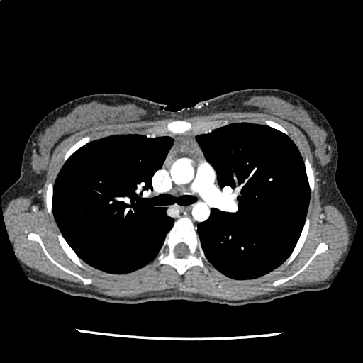
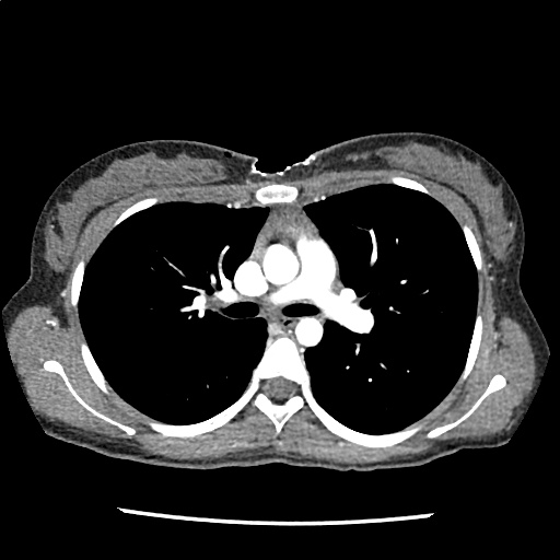
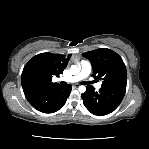
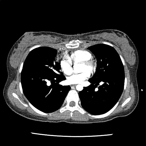
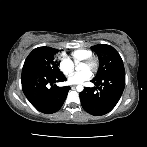
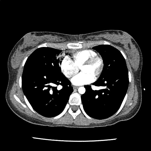
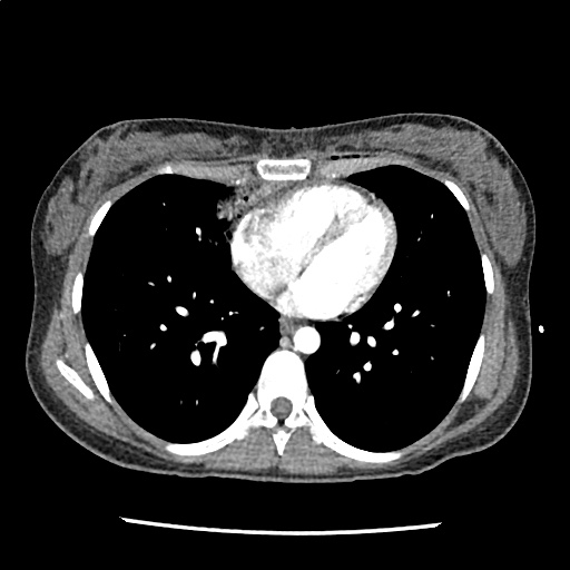
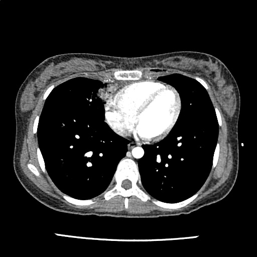
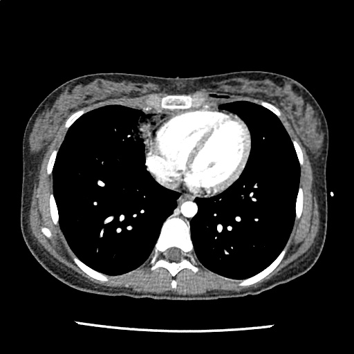
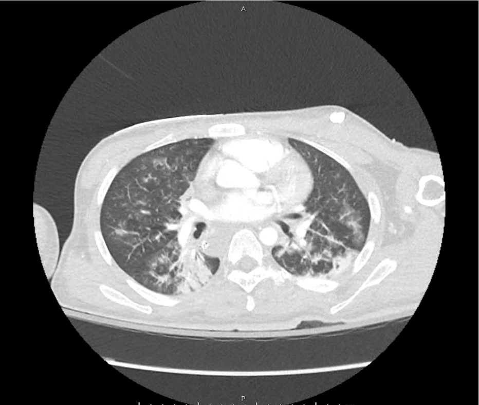
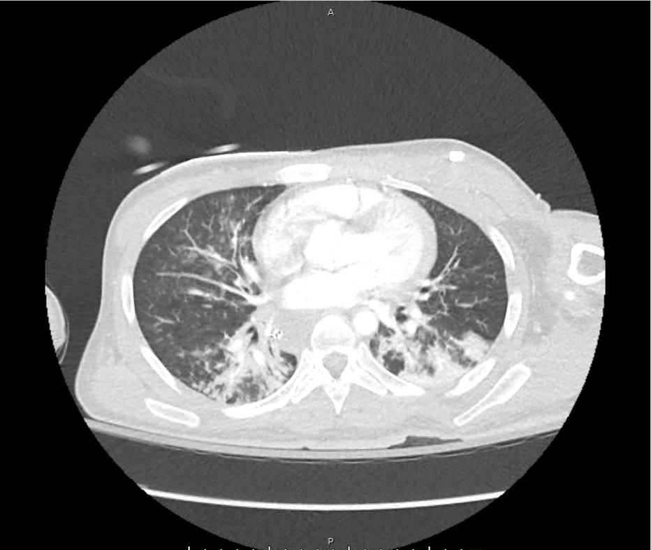
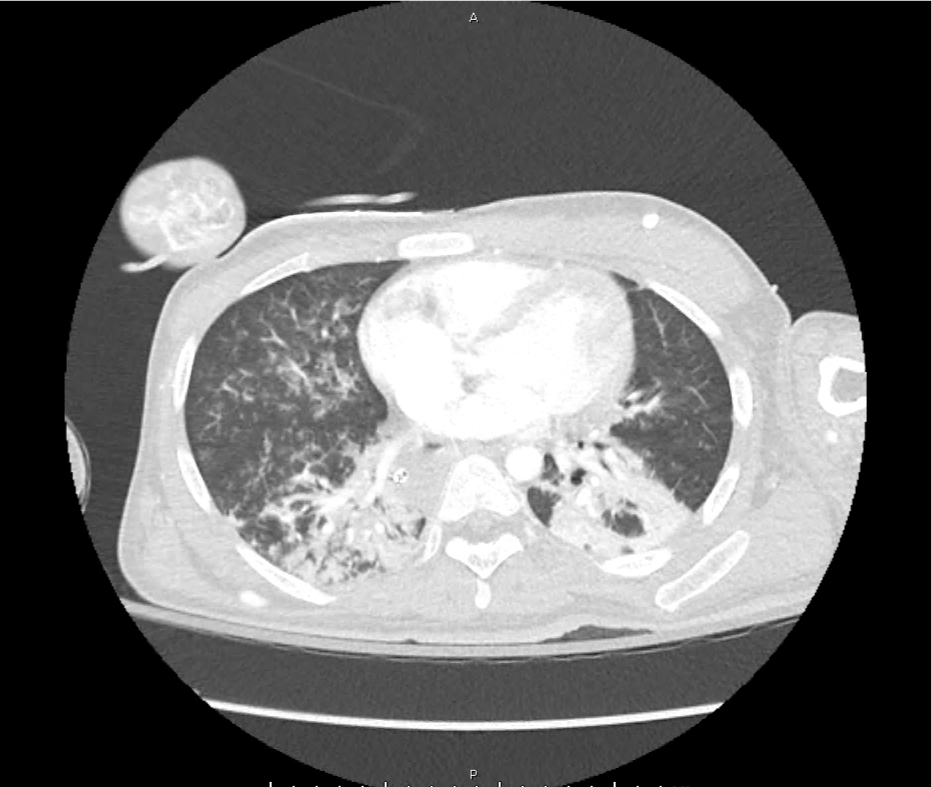
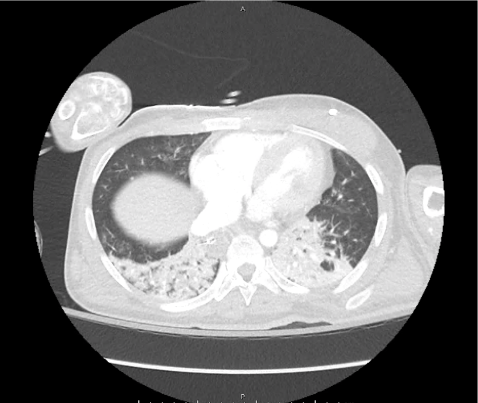
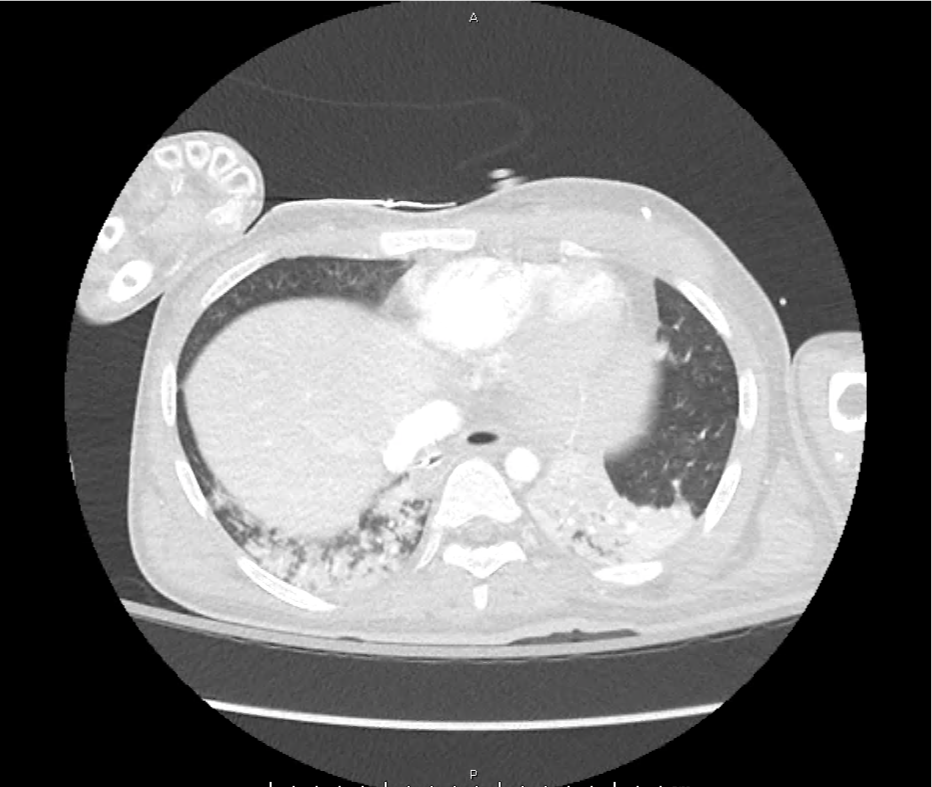
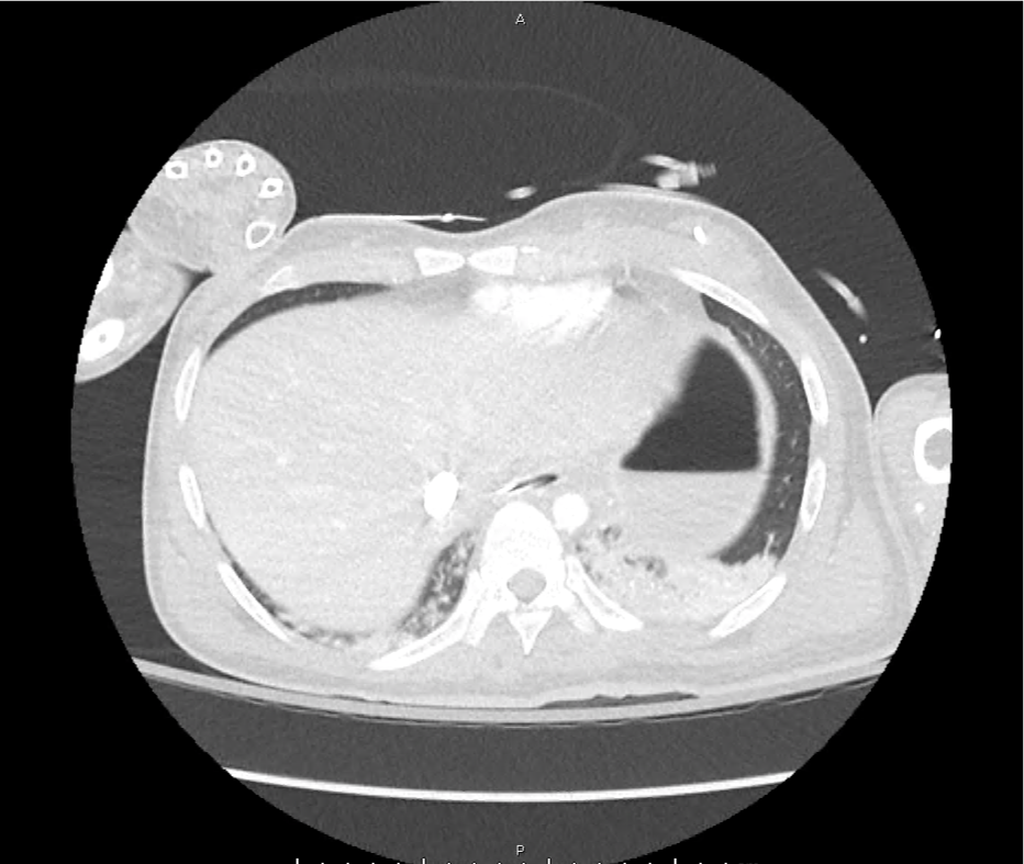
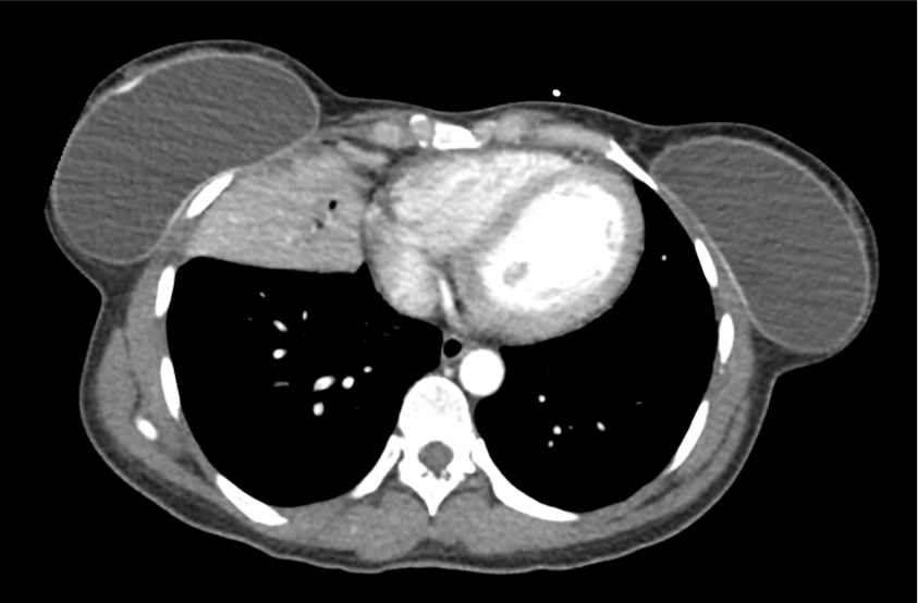
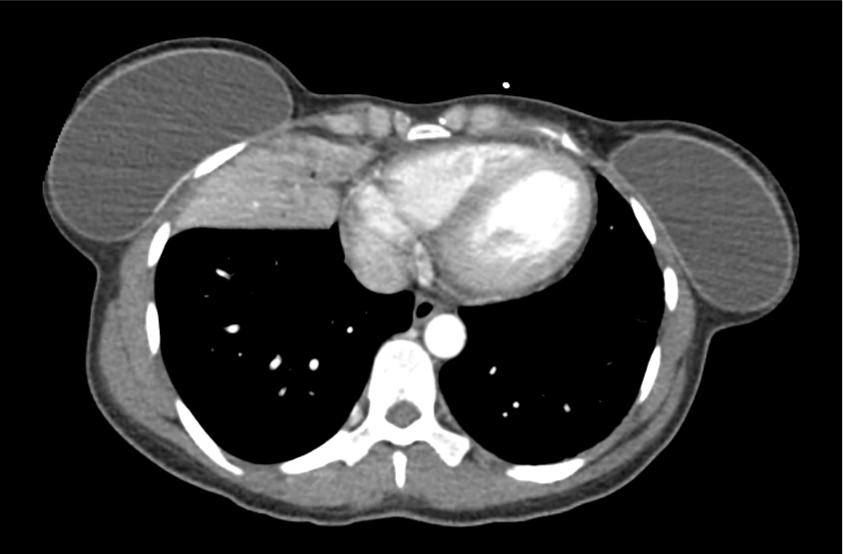
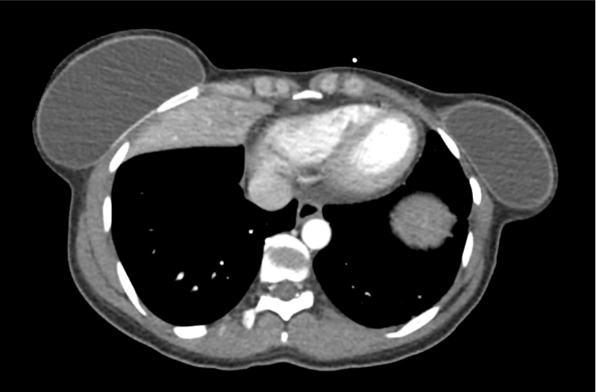
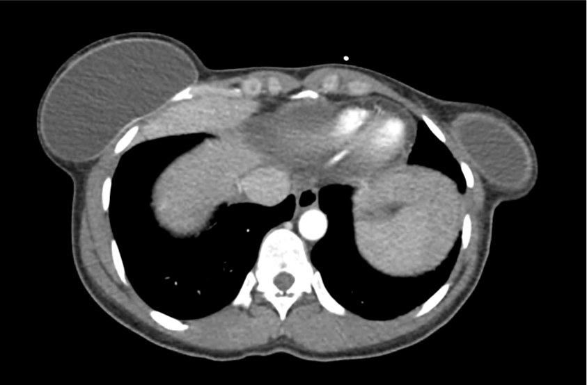
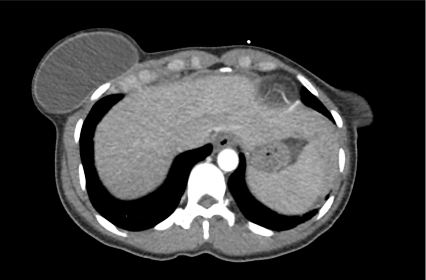
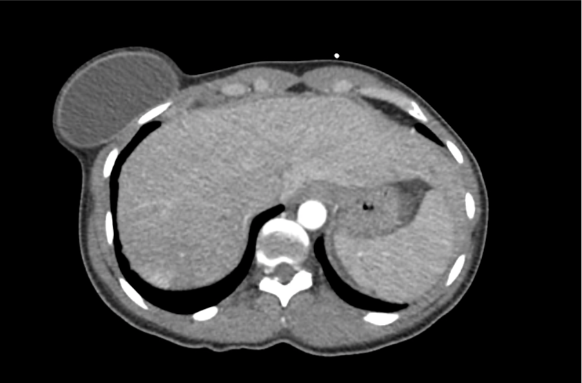
A 35-year-old female with no past medical history is brought in by ambulance to the emergency department. She was struck by a firework (“Roman Candle”) which lodged in her mid-chest until the propellant was consumed. She transiently lost consciousness but was awake upon EMS arrival. She complains of pleuritic chest pain. Examination reveals a circular 4x4cm full-thickness burn to the mid-chest with surrounding deep and superficial partial-thickness burns. Her ECG shows normal sinus rhythm, the initial serum troponin I is 32.9 (normal <0.012). CT angiography of the thorax is obtained.

bci_01
bci_02
bci_03
bci_04
bci_05
bci_06
bci_07
bci_08
bci_09
bci_10
bci_11
bci_12
bci_13

Mechanisms

Blunt cardiac injury (BCI) may be induced by multiple forces including direct thoracic trauma, crush injury of mediastinal contents between the sternum and thoracic spine, rapid deceleration causing tears at venous-atrial confluences, abrupt pressure changes from rapid compression of abdominal contents, blast injury, or laceration from bone fracture fragments1. The most common mechanisms of injury are motor vehicle collisions (50%), auto versus pedestrian (35%), motorcycle accidents (9%) and falls from significant height (>6m)2.

BCI represents a spectrum of conditions. Diagnosis is both challenging and critical as clinical manifestations can be absent or rapidly fatal.

At one end of the spectrum is myocardial contusion. The lack of a gold-standard for the diagnosis of this clinical entity has led to a preference for describing associated abnormalities if present3,4, including cardiac dysfunction (identified on echocardiography) or the next entity along the spectrum – arrhythmia.

The most common arrhythmia identified in blunt cardiac injury is sinus tachycardia, followed by premature atrial or ventricular contractions, T-wave changes, and atrial fibrillation or flutter5. Commotio cordis is a unique arrhythmia induced by untimely precordial impact (often in sports) during a vulnerable phase of ventricular excitability, resulting in ventricular fibrillation2.

ST-segment elevations after blunt cardiac injury should raise concern for myocardial infarction due to coronary artery dissection, laceration or thrombosis (often in already-diseased vessels) 5,6.

The remaining disease entities are increasingly rare, require careful examination or imaging for diagnosis, and are more likely to be non-survivable. Septal injury can range from small tears to rupture. Valvular injury most commonly affects the aortic valve (followed by mitral and tricuspid valves) and involves damage to leaflets, or rupture of papillary muscles or chordae tendineae. The clinical presentation is of acute valvular insufficiency, including acute heart failure and murmur2,7,8. A widened pulse pressure may be noted with aortic valve injury, and the manifestations of valvular injury may be delayed9. Finally, myocardial wall rupture is unlikely to be survivable, though patients may present with cardiac tamponade if rupture is small or contained2.

Evaluation

The primary diagnostic modalities for the assessment of BCI in the emergency department include assessment for pericardial fluid during the Focused Assessment with Sonography for Trauma (FAST), electrocardiography and cardiac enzymes.

While specific for identifying patients at risk of complications of BCI, electrocardiography alone is not sufficient to exclude BCI. In one study, only 59% of patient with echocardiographic evidence of BCI (wall-motion abnormalities, other chamber abnormalities) had initially abnormal ECG’s10. In another study, 41% of patients with initially normal ECG’s developed clinically significant abnormalities11. The use of specialized electrocardiography including right-sided ECG (proposed to better detect right-ventricular abnormalities which are more commonly associated with BCI) and signal-averaged ECG is not supported11,12.

Several studies have supported the use of serum troponin for the detection of clinically significant BCI – particularly in combination with electrocardiography. A prospective study in 2001 evaluating patients with blunt thoracic trauma using ECG at admission and 8-hours, as well as troponin I at admission, 4- and 8-hours had a negative predictive value of 100% for significant BCI (arrhythmia requiring treatment, shock, or structural cardiac abnormalities) in patients with initially normal ECG and troponin13.

Another prospective study adding to the population evaluated by Salim et al. included 41 patients with normal ECG’s and troponin levels at admission and 8-hours who were admitted for significant mechanisms, none developed significant BCI (again described as arrhythmia requiring treatment, shock, or structural cardiac abnormalities) after 1 to 3 days of observation14. The precise timing of serum troponin analysis remains unclear.

While FAST may detect hemopericardium warranting immediate intervention, formal echocardiography is indicated for patients with unexplained hypotension (to evaluate for valvular injury or regional wall-motion abnormalities) or persistent arrhythmias (to evaluate for arrhythmogenic intramural hematomas)15. The presence of sternal fractures was previously thought to increase risk of BCI and mandate echocardiography, however this notion is no longer supported16-18. The role of advanced imaging including helical CT (cardiac-gated), and MRI remains unclear19.

Algorithm for the Evaluation of Blunt Thoracic Trauma

Notes:
† Arrest in ED, immediate chest tube output >20ml/kg (>1.5L) or >200mL/hr for 2-4hr.

Management

Management of BCI depends on the pathologic process localized along the spectrum defined above. Persistent hypotension after appropriate evaluation for alternative etiologies may represent myocardial contusion with cardiac dysfunction and should be evaluated with echocardiography. Similarly, echocardiography and observation with continuous telemetry monitoring is indicated for any new arrhythmia or persistent and unexplained tachycardia. Patients with only elevation of the serum troponin without electrocardiographic abnormalities or obvious cardiac dysfunction should also be admitted for observation and serial cardiac enzymes. Traumatic myocardial infarction, valvular injury, or post-traumatic structural myocardial defects should be managed in consultation with cardiothoracic surgery5,19-21.

Case Conclusion

The CT interpretation noted the soft-tissue defect identified on examination as well as associated pulmonary contusions and a non-displaced sternal fracture. The patient went to the operating room for washout and debridement. A transthoracic echocardiogram demonstrated trace mitral regurgitation and a small pericardial effusion. She remained hemodynamically stable and serial troponin measures downtrended – no dysrhythmias were noted on telemetry monitoring. She was discharged on hospital day four with a negative-pressure wound dressing.

References

  1. Schultz JM, Trunkey DD. Blunt cardiac injury. Crit Care Clin. 2004;20(1):57-70.
  2. Yousef R, Carr JA. Blunt cardiac trauma: a review of the current knowledge and management. Ann Thorac Surg. 2014;98(3):1134-1140. doi:10.1016/j.athoracsur.2014.04.043.
  3. Mattox KL, Flint LM, Carrico CJ, et al. Blunt cardiac injury. The Journal of Trauma: Injury, Infection, and Critical Care. 1992;33(5):649-650.
  4. Sybrandy KC, Cramer MJM, Burgersdijk C. Diagnosing cardiac contusion: old wisdom and new insights. Heart. 2003;89(5):485-489.
  5. Elie M-C. Blunt cardiac injury. Mt Sinai J Med. 2006;73(2):542-552.
  6. Edouard AR, Felten M-L, Hebert J-L, Cosson C, Martin L, Benhamou D. Incidence and significance of cardiac troponin I release in severe trauma patients. Anesthesiology. 2004;101(6):1262-1268.
  7. Cordovil A, Fischer CH, Rodrigues ACT, et al. Papillary Muscle Rupture After Blunt Chest Trauma. Journal of the American Society of Echocardiography. 2006;19(4):469.e1-469.e3. doi:10.1016/j.echo.2005.12.005.
  8. Pasquier M, Sierro C, Yersin B, Delay D, Carron P-N. Traumatic Mitral Valve Injury After Blunt Chest Trauma: A Case Report and Review of the Literature. The Journal of Trauma: Injury, Infection, and Critical Care. 2010;68(1):243-246. doi:10.1097/TA.0b013e3181bb881e.
  9. Ismailov RM, Weiss HB, Ness RB, Lawrence BA, Miller TR. Blunt cardiac injury associated with cardiac valve insufficiency: trauma links to chronic disease? Injury. 2005;36(9):1022-1028. doi:10.1016/j.injury.2005.05.028.
  10. García-Fernández MA, López-Pérez JM, Pérez-Castellano N, et al. Role of transesophageal echocardiography in the assessment of patients with blunt chest trauma: correlation of echocardiographic findings with the electrocardiogram and creatine kinase monoclonal antibody measurements. Am Heart J. 1998;135(3):476-481.
  11. Fulda GJ, Giberson F, Hailstone D, Law A, Stillabower M. An evaluation of serum troponin T and signal-averaged electrocardiography in predicting electrocardiographic abnormalities after blunt chest trauma. The Journal of Trauma: Injury, Infection, and Critical Care. 1997;43(2):304–10–discussion310–2.
  12. Walsh P, Marks G, Aranguri C, et al. Use of V4R in patients who sustain blunt chest trauma. The Journal of Trauma: Injury, Infection, and Critical Care. 2001;51(1):60-63.
  13. Salim A, Velmahos GC, Jindal A, et al. Clinically significant blunt cardiac trauma: role of serum troponin levels combined with electrocardiographic findings. The Journal of Trauma: Injury, Infection, and Critical Care. 2001;50(2):237-243.
  14. Velmahos GC, Karaiskakis M, Salim A, et al. Normal electrocardiography and serum troponin I levels preclude the presence of clinically significant blunt cardiac injury. The Journal of Trauma: Injury, Infection, and Critical Care. 2003;54(1):45–50–discussion50–1. doi:10.1097/01.TA.0000046315.73441.D8.
  15. Nagy KK, Krosner SM, Roberts RR, Joseph KT, Smith RF, Barrett J. Determining which patients require evaluation for blunt cardiac injury following blunt chest trauma. World J Surg. 2001;25(1):108-111.
  16. Roy-Shapira A, Levi I, Khoda J. Sternal fractures: a red flag or a red herring? The Journal of Trauma: Injury, Infection, and Critical Care. 1994;37(1):59-61.
  17. Hills MW, Delprado AM, Deane SA. Sternal fractures: associated injuries and management. The Journal of Trauma: Injury, Infection, and Critical Care. 1993;35(1):55-60.
  18. Rashid MA, Ortenwall P, Wikström T. Cardiovascular injuries associated with sternal fractures. Eur J Surg. 2001;167(4):243-248. doi:10.1080/110241501300091345.
  19. Clancy K, Velopulos C, Bilaniuk JW, et al. Screening for blunt cardiac injury: an Eastern Association for the Surgery of Trauma practice management guideline. J Trauma Acute Care Surg. 2012;73(5 Suppl 4):S301-S306. doi:10.1097/TA.0b013e318270193a.
  20. El-Menyar A, Thani Al H, Zarour A, Latifi R. Understanding traumatic blunt cardiac injury. Ann Card Anaesth. 2012;15(4):287-295. doi:10.4103/0971-9784.101875.
  21. Hockberger RS, Walls RM. Rosen’s Emergency Medicine. Mosby Incorporated; 2002.

Arterial Pressure Indices

Indications

  • Assess for peripheral arterial disease (PAD)
  • Assess for risk of arterial injury in trauma

Test characteristics

  • PAD: ABI <0.90 sensitivity 95%, specificity 100% for >50% stenosis on arteriography1
  • Trauma: API <0.90 sensitivity 95%, specificity 97% for major arterial injury2

Technique for obtaining arterial pressure indices3

  1. Patient lies supine with extremities at level of the heart for 10 minutes
  2. Ankle: cuff positioned just above malleolus
  3. Brachial: cuff positioned just above antecubital fossa
  4. Doppler SBP
  5. Sequence: first arm, first PT, first DP, other PT, other DP, other arm. If SBP of first arm >10mmHg compared to second arm, repeat first arm and disregard first measure
  6. Calculation: ABI = higher of DP or PT / higher arm

Interpretation of ABI for PAD3

Value Interpretation
0-0.40 Severe PAD, rest pain, gangrene
0.41-0.90 PAD, claudication
0.91-1.30 Normal
>1.30 Non-compressible, severely calcified

Algorithm for the Evaluation of Arterial Injury4, 5

Algorithm for the Evaluation of Arterial Injury

Notes:

  • † If unable to palpate pulses due to patient habitus or shock/hypothermia, reattempt with oversized cuff and after appropriate rewarming/resuscitation. If remains challenging, proceed with imaging.
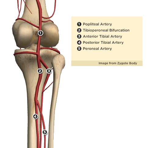
  • ‡ Proximal LE arterial injuries refers to the major named arteries of the thigh (excluding the profunda femoris) and proximal to the anterior tibial artery and tibioperoneal bifurcation.

Arteries of the Lower Leg

References:

  1. Mohler ER. Peripheral arterial disease: identification and implications. Arch Intern Med. 2003;163(19):2306-2314. doi:10.1001/archinte.163.19.2306.
  2. Johansen K, Lynch K, Paun M, Copass M. Non-invasive vascular tests reliably exclude occult arterial trauma in injured extremities. The Journal of Trauma: Injury, Infection, and Critical Care. 1991;31(4):515–9–discussion519–22.
  3. Aboyans V, Criqui MH, Abraham P, et al. Measurement and interpretation of the ankle-brachial index: a scientific statement from the American Heart Association. Circulation. 2012;126(24):2890-2909. doi:10.1161/CIR.0b013e318276fbcb.
  4. Feliciano DV, Moore FA, Moore EE, et al. Evaluation and management of peripheral vascular injury. Part 1. Western Trauma Association/critical decisions in trauma. J Trauma. 2011;70(6):1551-1556. doi:10.1097/TA.0b013e31821b5bdd.
  5. Fox N, Rajani RR, Bokhari F, et al. Evaluation and management of penetrating lower extremity arterial trauma: an Eastern Association for the Surgery of Trauma practice management guideline. J Trauma Acute Care Surg. 2012;73(5 Suppl 4):S315-S320. doi:10.1097/TA.0b013e31827018e4.
  6. Inaba K, Branco BC, Reddy S, et al. Prospective evaluation of multidetector computed tomography for extremity vascular trauma. J Trauma. 2011;70(4):808-815. doi:10.1097/TA.0b013e3182118384.

Weakness

A systematic approach to motor weakness progresses along an anatomic tract from the cerebral cortex to individual sarcomeres. Impulses are generated in the primary motor cortex mapped to the homunculus, then aggregate as they descend through the internal capsule. Fibers decussate in the medulla and descend in the contralateral lateral corticospinal tract. These upper motor neurons (UMN) synapse with the lower motor neuron (LMN) in the anterior horn of the spinal cord. The lower motor neuron is bundled with neighboring fibers into a peripheral nerve and activates the target muscle fibers at the neuromuscular junction.

Algorithm for the Evaluation of Weakness

Algorithm for the Evaluation of Weakness

Upper Versus Lower Motor Neuron Findings

Finding Upper Lower
Reflexes
Atrophy
Weakness
Fasciculation
Tone
Summary
Recalling these findings can be simplified by understanding the underlying process. Denervation near the target muscle fibers (lower motor neuron disease) results in dampening of the efferent limb of spinal reflexes, resulting in hyporeflexia. The absence of nourishing stimulation leads to muscle atrophy and disorganized interpretation of proximal activity produces fasciculation.

Comparison Between Myopathy, Neuropathy and Neuromuscular Junction Processes

Finding Myopathy Neuropathy Neuromuscular Junction
Example Polymyositis Guillain-Barre Syndrome Myasthenia Gravis
Distribution Proximal > Distal Distal > Proximal Diffuse, Bulbar
Reflexes Normal
Sensory
Fatigue
CK Normal Normal

Motor Strength Grading

Grade Description
5 Normal
4 Reduced, moves against resistance
3 Moves against gravity
2 Moves only with elimination of gravity
1 Fasciculation only
0 None

Reflex Grading

Grade Description
4 Increased amplitude, spread to adjacent, clonus
3 Increased
2 Normal
1 Decreased
0 None

References

  1. Ganti L, Rastogi V. Acute Generalized Weakness. Emerg Med Clin North Am. 2016;34(4):795-809. doi:10.1016/j.emc.2016.06.006.
  2. Asimos AW. Weakness: A Systematic Approach To Acute, Non-traumatic, Neurologic And Neuromuscular Causes. Emergency Medicine Practice. 2002;4(12):1-28.
  3. Morchi R. Weakness. In: Rosen’s Emergency Medicine. Elsevier Inc.; 2014:2521.

Epistaxis

Brief HPI:

A 63 year-old female with a history of hypertension, diabetes, and deep venous thrombosis on warfarin presents with epistaxis. She noted the spontaneous onset of nose bleeding 15 minutes prior to presentation. She had attempted compression but symptoms persisted so she was brought to the emergency department. On initial evaluation, she was in no acute distress and vital signs were normal. She was compressing her distal nares and was spitting up blood.

Oxymetolazone was administered and the patient was instructed regarding the appropriate position for compression, however bleeding continued when reassessed at 10- and then 30-minutes of compression. A bleeding focus could not be visualized on rhinoscopy so a nasal tampon was inserted with resolution of bleeding. Bleeding did not recur after two hours of observation in the emergency department. The patient’s INR was therapeutic two days prior to presentation and she was instructed to continue her usual regimen. At primary care follow-up two days later, the compression device was successfully removed.

Algorithm for the Management of Epistaxis1,2

Algorithm for the Management of Epistaxis

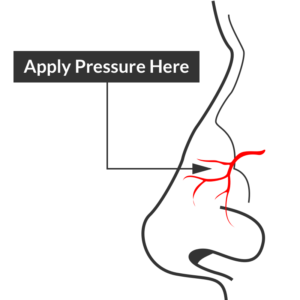
Epistaxis site of compression

Site of compression

External Compression

Begin with simple measures while preparing the necessary equipment and medications. Request that the patient gently blow their nose to clear clots, administer oxymetolazone 0.05% two sprays into the affected side. Apply firm pressure below the nasal bridge continuously for at least 10 minutes before reassessment. Commercial compression devices are available, or can be fashioned with tongue depressors3. Alternatively, the patient can apply pressure themselves.

Cautery

Again ask the patient to blow their nose to remove clots. Apply topical anesthetic for patient comfort prior to inspection with a nasal speculum. Additional suction (small tip, Frazier) may be required to improve visualization. If the bleeding site is identified, apply silver nitrate circumferentially around the source, then directly over the site. Avoid prolonged exposure or exposure to opposing sides of the nasal septum. If hemorrhage control is successful, patients may be discharged with a topical antimicrobial ointment such as polymixin-bacitracin-neomycin.

Packing 4,5

Multiple commercial anterior packing devices are available. Placement technique is similar for most, generally involving lubrication of the device with antimicrobial ointment or sterile water, sliding the device along the floor of the nasal cavity, followed by injection or inflation of the device to support tamponade. The incorporation of tranexamic acid (500mg in 5mL) into any phase of anterior packing may be beneficial 6,7. Packing the contralateral side to further support tamponade may be required.

Commonly used commercial devices are:

  • Merocel: lubricate with antimicrobial ointment, once deployed can rehydrate with saline or topical vasoconstrictor
  • Rapid Rhino
  • Rhino Rocket

Packing material should remain for 48-72 hours, during which patients should be re-evaluated. Prophylactic systemic antibiotics for the prevention of sinusitis or toxic shock are likely not required8.

Thrombogenic materials such as Floseal or Surgicel can also be used and may be better tolerated than packing materials9.

Posterior Control

If bleeding persists despite the above measures, a posterior site should be considered. Dual-balloon commercial devices are available for the control of posterior epistaxis and are deployed in a similar fashion to anterior devices. Once inserted, the posterior balloon should be inflated with air – with the volume guided by tension of the pilot cuff. The anterior balloon can then be inflated in a similar fashion. The posterior balloon cuff should be reinspected after 5 minutes as additional inflation may be required.

Commonly used commercial devices are:

If a commercial device is unavailable, a Foley catheter may be used. The catheter is introduced into the affected side. Once the tip is visualized in the posterior oropharynx, the balloon is inflated with approximately 10mL of sterile water. The catheter is then withdrawn gently to seat the balloon posteriorly. The catheter is secured in position against the nares with a clamp (taking care to pad the nares with gauze to prevent trauma) 10,11.

Patients with posterior epistaxis should be admitted with otolaryngology consultation. If bleeding continues despite these measures, emergent otolaryngology consultation for operative management is warranted.

Causes of Epistaxis12

Causes of Epistaxis

References

  1. Leong SCL, Roe RJ, Karkanevatos A. No frills management of epistaxis. Emerg Med J. 2005;22(7):470-472. doi:10.1136/emj.2004.020602.
  2. Barnes ML, Spielmann PM, White PS. Epistaxis: a contemporary evidence based approach. Otolaryngol Clin North Am. 2012;45(5):1005-1017. doi:10.1016/j.otc.2012.06.018.
  3. Moxham V, Reid C. Controlling epistaxis with an improvised device. Emergency Medicine Journal. 2001;18(6):518. doi:10.1136/emj.18.6.518.
  4. Singer AJ, Blanda M, Cronin K, et al. Comparison of nasal tampons for the treatment of epistaxis in the emergency department: A randomized controlled trial. Ann Emerg Med. 2005;45(2):134-139. doi:10.1016/j.annemergmed.2004.10.002.
  5. Iqbal IZ, Jones GH, Dawe N, et al. Intranasal packs and haemostatic agents for the management of adult epistaxis: systematic review. J Laryngol Otol. 2017;131(12):1065-1092. doi:10.1017/S0022215117002055.
  6. MD RZ, MD PM, MD SA, PhD AG, MD MS. A new and rapid method for epistaxis treatment using injectable form of tranexamic acid topically: a randomized controlled trial. American Journal of Emergency Medicine. 2013;31(9):1389-1392. doi:10.1016/j.ajem.2013.06.043.
  7. Kamhieh Y, Fox H. Tranexamic acid in epistaxis: a systematic review. Clin Otolaryngol. 2016;41(6):771-776. doi:10.1111/coa.12645.
  8. MD BC. Are Prophylactic Antibiotics Necessary for Anterior Nasal Packing in Epistaxis? YMEM. 2015;65(1):109-111. doi:10.1016/j.annemergmed.2014.08.011.
  9. Mathiasen RA, Cruz RM. Prospective, Randomized, Controlled Clinical Trial of a Novel Matrix Hemostatic Sealant in Patients with Acute Anterior Epistaxis. The Laryngoscope. 2005;115(5):899-902. doi:10.1097/01.MLG.0000160528.50017.3C.
  10. Holland NJ, Sandhu GS, Ghufoor K, Frosh A. The Foley catheter in the management of epistaxis. Int J Clin Pract. 2001;55(1):14-15.
  11. Hartley C, Axon PR. The Foley catheter in epistaxis management–a scientific appraisal. J Laryngol Otol. 1994;108(5):399-402.
  12. Kucik CJ, Clenney T. Management of epistaxis. Am Fam Physician. 2005;71(2):305-311.

Ocular Ultrasound

Brief HPI:

Intraocular foreign body

Intraocular foreign body

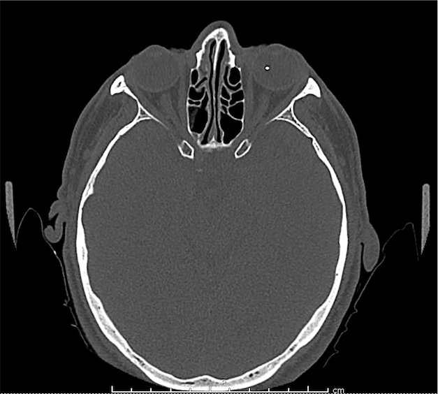
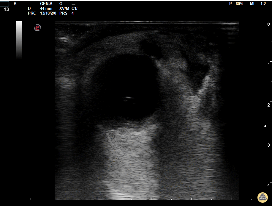
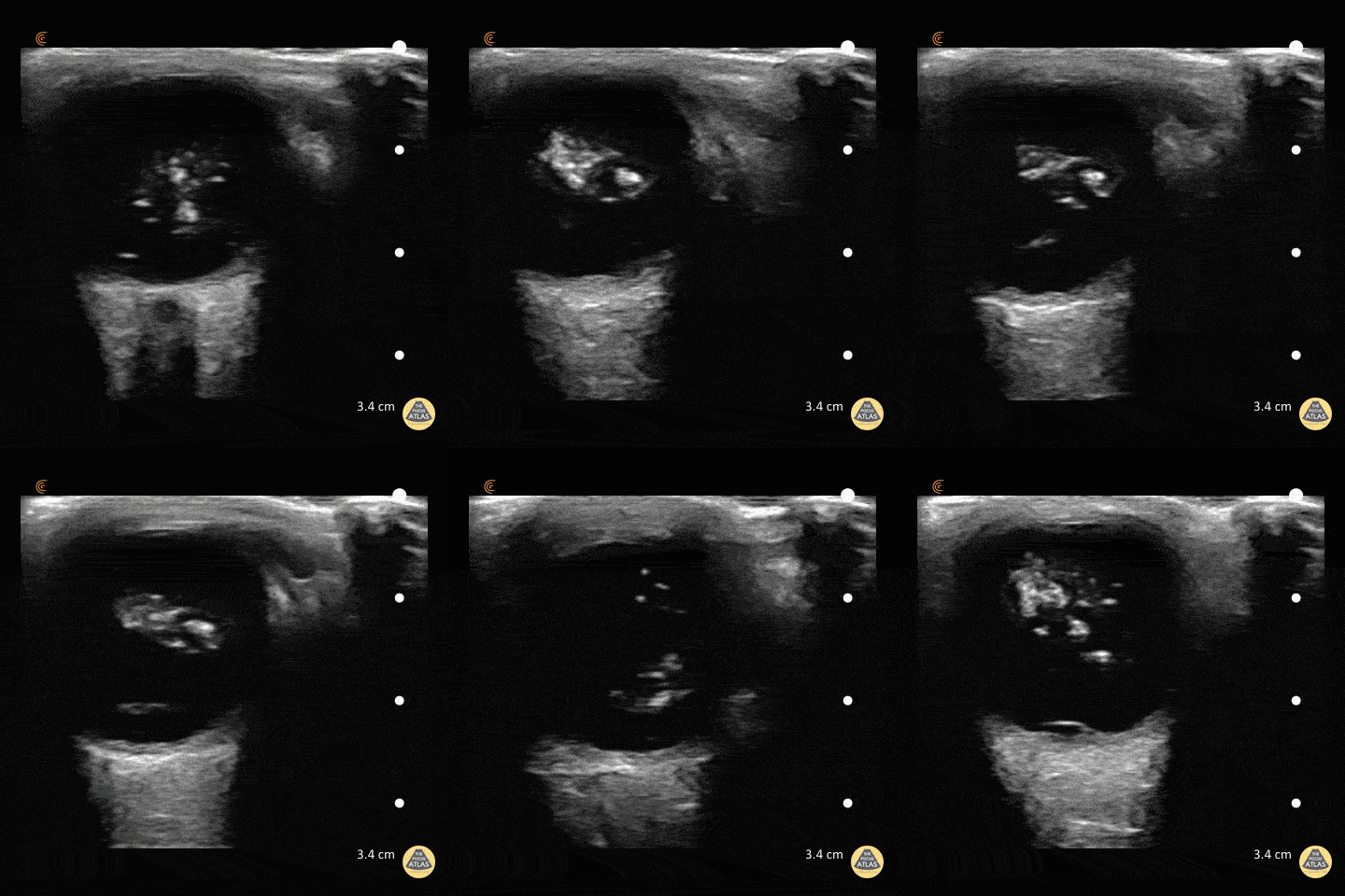
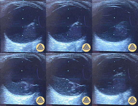
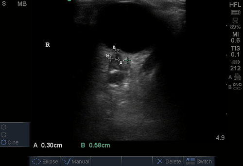
A middle-aged male with no past medical history presents with blurred vision. He reported that he was hammering while at work approximately 3 days prior to presentation and felt something enter his left eye. He denies eye pain, has noted some eye redness and increased tearing. Denies prior eye surgery or procedures. Physical examination demonstrates normal visual acuity, minimal left nasal conjunctival injection sparing the limbus, and an irregular left pupil that is minimally reactive. A no-pressure ocular ultrasound was performed and demonstrated a hyperechoic structure in the globe suggestive of foreign body which was confirmed on computed tomography of the orbit. The patient was taken to the operating room for removal.

Imaging

iofb_14
iofb_13
iofb_12
iofb_11
iofb_10
iofb_09
iofb_08
iofb_07
iofb_06
iofb_05
iofb_04
iofb_03
iofb_02
iofb_01

CT Orbit without contrast

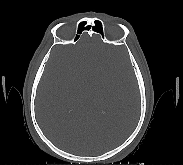
Punctate density within the left globe compatible with foreign body.

Algorithm for the Evaluation of Visual Complaints with Ocular Ultrasonography

Algorithm for the Evaluation of Visual Complaints with Ocular Ultrasonography

Gallery

The POCUS Atlas
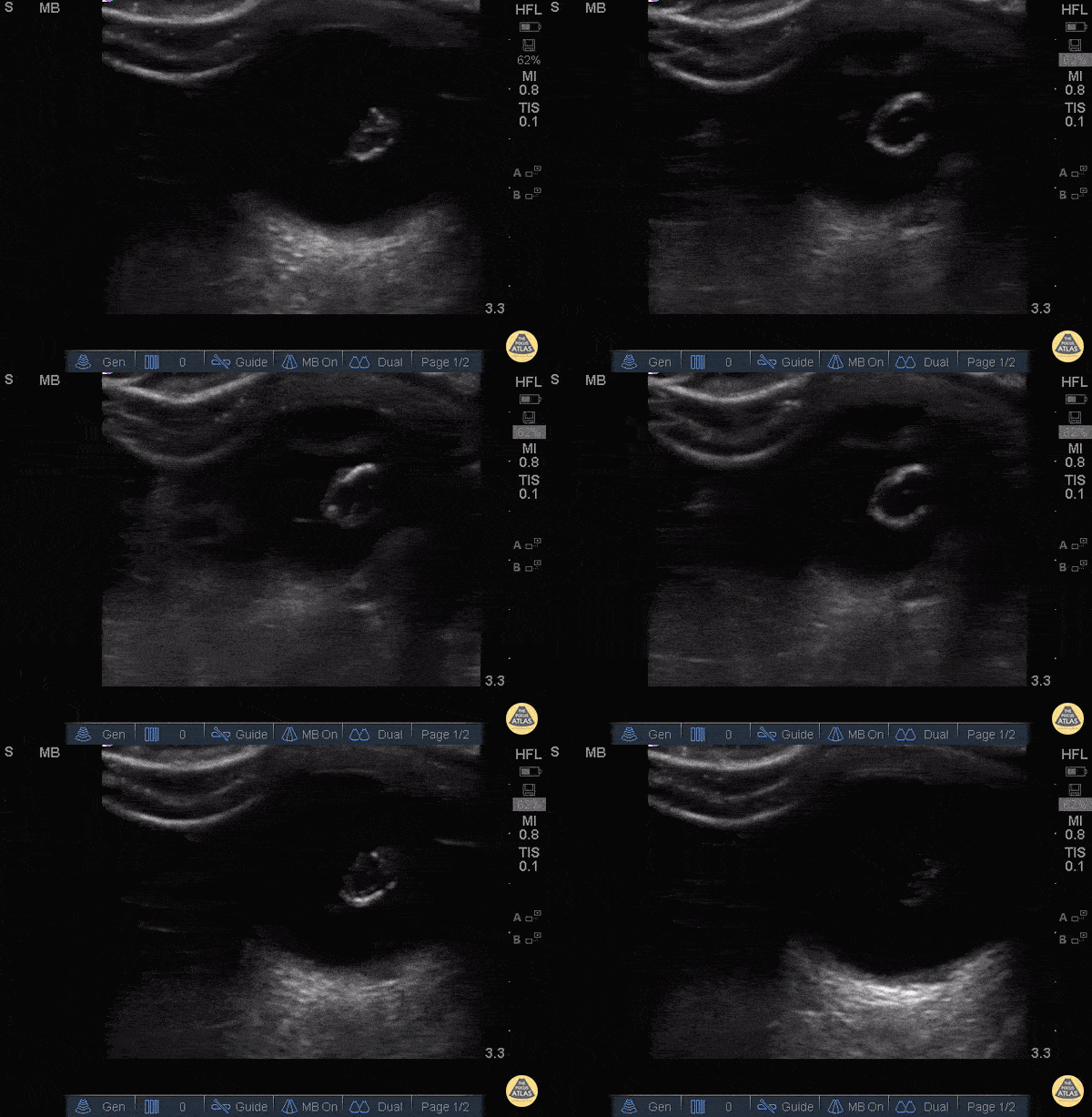
The ultrasound images and videos used in this post come from The POCUS Atlas, a collaborative collection focusing on rare, exotic and perfectly captured ultrasound images.
The POCUS Atlas

Orbital Abscess

Lens dislocation

Retinal detachment

Vitreous hemorrhage

Posterior vitreous detachment

ONSD (increased)

Central retinal artery flow (normal)

Central retinal artery occlusion


Applications1,2

Useful for the evaluation of intraocular processes:

  • Retinal detachment
  • Vitreous hemorrhage/detachment
  • Intraocular foreign body
  • Lens dislocation
  • Retroorbital hemorrhage/abscess
  • Retinal vascular processes (CRAO)

Augmentation of physical examination when limited due to facial swelling or trauma:

  • Pupil size and reactivity
  • Extraocular movements

ONSD

  • Normal <5mm adults (>15yo)
  • Normal <4.5mm children (1-15yo)
  • Normal <4mm infants (<1yo)
  • By convention, measurements of the optic nerve sheath diameter are made 3-mm posterior to the globe

Technique

  • Apply Tegaderm, ensuring no air bubbles trapped
  • Apply copious ultrasound gel
  • Use no-pressure technique, anchoring hand against the patient’s forehead, nasal bridge or maxilla
  • Probe indicator to the patient’s right for transverse views
  • Probe indicator to the patient’s head for longitudinal views
  • Start with medium gain then increase to identify subtle findings

Test Characteristics

Prospective observational study evaluating patients presenting with ocular trauma or acute vision complaints underwent ocular ultrasound. Ultrasound findings agreed with the confirmatory test: ophthalmology consultation or advanced imaging (usually computed tomography) in 60 of 61 cases3.

Specific Findings 4,5

Retinal Detachment6-9

Appears as a highly reflective membrane floating in the substance of the vitreous body, moves within vitreous body with eye movement. Remains anchored at the optic nerve and ora serrata.

Posterior Vitreous Detachment10

Both retinal detachments and posterior vitreous detachments show a linear hyperechoic line in the posterior chamber. However, posterior vitreous detachments are not tethered to the optic nerve and will appear to cross midline.

Vitreous Hemorrhage

Seen more easily with high-gain, enhanced by eye movements which demonstrate hyperechoic particles swirling around in the vitreous body.

Retrobulbar Hematoma

Identified by the presence of a hypoechoic structure posterior to the globe. Should prompt a measurement of intra-ocular pressure if simultaneous globe rupture is not suspected.

Lens Dislocation

Usually secondary to blunt trauma, lens displaced from normal position and appears as an echogenic ovoid structure floating freely in the vitreous or over the retina.

Globe Rupture

If the diagnosis of globe rupture is obvious, ultrasound should be avoided. However, the “no-pressure” technique described above likely does not significantly impact intra-ocular pressure and should be safe11,12. Globe rupture can be identified by scleral buckling, anterior chamber collapse, or globe collapse/irregularities.

Optic Nerve Evaluation13-18

Though not a direct assessment of ocular pathology, evaluation of the optic nerve sheath diameter (ONSD) serves as a reliable surrogate for elevated intracranial pressure – emulating fundoscopy for papilledema. See normal measurements and image acquisition above.

Intraocular Foreign Body

The preferred imaging modality for evaluation of intraocular foreign body is orbital computed tomography. Ultrasonographically, foreign bodies are typically hyperechoic.

Central Retinal Artery Occlusion19,20

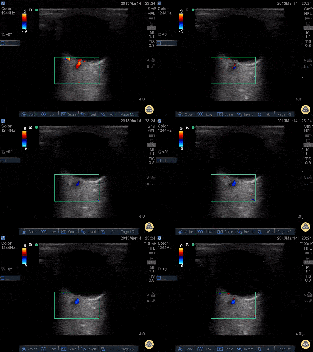
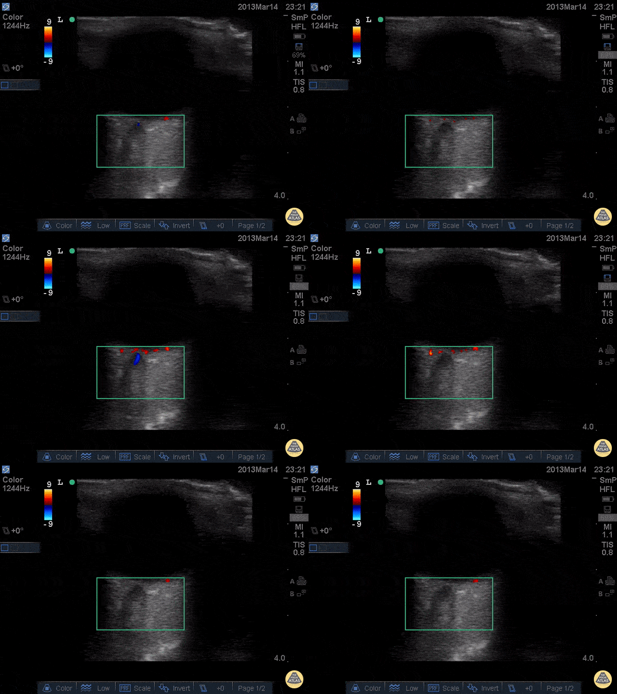
A more advanced technique, the addition of color Doppler over the central retinal artery may reveal decreased systolic amplitude and diastolic flow in embolic or thrombotic occlusion.

All illustrations are available for free, licensed (along with all content on this site) under Creative Commons Attribution-ShareAlike 4.0 International Public License.

Downloads Page License

References

  1. Kimberly HH, Stone MB. Chapter E5 – Emergency Ultrasound. Ninth Edition. Elsevier Inc.; 2018:e49-e66. doi:10.1016/B978-0-323-35479-0.00204-X.
  2. Knoop KJ, Dennis WR. Ophthalmologic, Otolaryngologic, and Dental Procedures. Seventh Edition. Elsevier Inc.; 2019:1295–1337.e2. doi:10.1016/B978-0-323-35478-3.00062-2.
  3. Blaivas M, Theodoro D, Sierzenski PR. A study of bedside ocular ultrasonography in the emergency department. Academic Emergency Medicine. 2002;9(8):791-799.
  4. Roque PJ, Hatch N, Barr L, Wu TS. Bedside ocular ultrasound. Crit Care Clin. 2014;30(2):227–41–v. doi:10.1016/j.ccc.2013.10.007.
  5. Kilker BA, Holst JM, Hoffmann B. Bedside ocular ultrasound in the emergency department. European Journal of Emergency Medicine. 2014;21(4):246-253. doi:10.1097/MEJ.0000000000000070.
  6. Yoonessi R, Hussain A, Jang TB. Bedside ocular ultrasound for the detection of retinal detachment in the emergency department. Acad Emerg Med. 2010;17(9):913-917. doi:10.1111/j.1553-2712.2010.00809.x.
  7. Shinar Z, Chan L, Orlinsky M. Use of ocular ultrasound for the evaluation of retinal detachment. J Emerg Med. 2011;40(1):53-57. doi:10.1016/j.jemermed.2009.06.001.
  8. Chu HC, Chan MY, Chau CWJ, Wong CP, Chan HH, Wong TW. The use of ocular ultrasound for the diagnosis of retinal detachment in a local accident and emergency department. Hong Kong Journal of Emergency Medicine. 2017;24(6):263-267. doi:10.1177/1024907917735085.
  9. Vrablik ME, Snead GR, Minnigan HJ, Kirschner JM, Emmett TW, Seupaul RA. The diagnostic accuracy of bedside ocular ultrasonography for the diagnosis of retinal detachment: a systematic review and meta-analysis. Ann Emerg Med. 2015;65(2):199–203.e1. doi:10.1016/j.annemergmed.2014.02.020.
  10. Schott ML, Pierog JE, Williams SR. Pitfalls in the Use of Ocular Ultrasound for Evaluation of Acute Vision Loss. J Emerg Med. 2013;44(6):1136-1139. doi:10.1016/j.jemermed.2012.11.079.
  11. Chandra A, Mastrovitch T, Ladner H, Ting V, Radeos MS, Samudre S. The utility of bedside ultrasound in the detection of a ruptured globe in a porcine model. West J Emerg Med. 2009;10(4):263-266.
  12. Berg C, Doniger SJ, Zaia B, Williams SR. Change in intraocular pressure during point-of-care ultrasound. West J Emerg Med. 2015;16(2):263-268. doi:10.5811/westjem.2015.1.24150.
  13. Tsung JW, Blaivas M, Cooper A, Levick NR. A rapid noninvasive method of detecting elevated intracranial pressure using bedside ocular ultrasound: application to 3 cases of head trauma in the pediatric emergency department. Pediatr Emerg Care. 2005;21(2):94-98.
  14. Kimberly HH, Shah S, Marill K, Noble V. Correlation of optic nerve sheath diameter with direct measurement of intracranial pressure. Acad Emerg Med. 2008;15(2):201-204. doi:10.1111/j.1553-2712.2007.00031.x.
  15. Blaivas M, Theodoro D, Sierzenski PR. Elevated intracranial pressure detected by bedside emergency ultrasonography of the optic nerve sheath. Academic Emergency Medicine. 2003;10(4):376-381.
  16. Moretti R, Pizzi B. Optic Nerve Ultrasound for Detection of Intracranial Hypertension in Intracranial Hemorrhage Patients. Journal of Neurosurgical Anesthesiology. 2009;21(1):16-20. doi:10.1097/ANA.0b013e318185996a.
  17. Rajajee V, Vanaman M, Fletcher JJ, Jacobs TL. Optic Nerve Ultrasound for the Detection of Raised Intracranial Pressure. Neurocrit Care. 2011;15(3):506-515. doi:10.1007/s12028-011-9606-8.
  18. Major R, al-Salim W. Towards evidence based emergency medicine: best BETs from the Manchester Royal Infirmary. BET 3. Ultrasound of optic nerve sheath to evaluate intracranial pressure. Emerg Med J. 2008;25(11):766-767. doi:10.1136/emj.2008.066845.
  19. Riccardi A, Siniscalchi C, Lerza R. Embolic Central Retinal Artery Occlusion Detected with Point-of-care Ultrasonography in the Emergency Department. J Emerg Med. 2016;50(4):e183-e185. doi:10.1016/j.jemermed.2015.12.022.
  20. Catalin J Dragos, Jianu S Nina, Munteanu M, Vlad D, Rosca C, Petrica L. Color Doppler imaging features in patients presenting central retinal artery occlusion with and without giant cell arteritis. VSP. 2016;73(4):397-401. doi:10.2298/VSP140814087C.

Thromboelastography

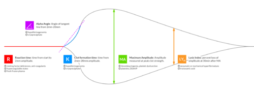
Thromboelastography (TEG) is an assessment of hemostatic function intended to evaluate in vivo coagulation parameters, guiding the targeted correction of coagulopathy1. TEG has predominantly been studied in cardiac surgery, though research has extended to other peri-operative and peri-procedural transfusion management2-5.

Recently, a randomized trial explored the use of TEG to guide transfusion in trauma patients requiring massive transfusion6. 111 patients meeting requirements for massive transfusion protocol activation were randomized to a conventional coagulation assay (CCA) or TEG-guided transfusion algorithm. Patients in the TEG group demonstrated significantly decreased mortality at 28 days and reductions in plasma and platelet transfusion requirements.

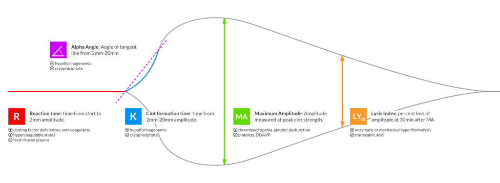
More research is needed before TEG can be recommended for use in trauma resuscitation or other common emergency department applications7,8, however it may be useful to prepare by becoming familiar with the most basic aspects of thromboelastography.

Thromboelastography Summary

Thromboelastography Summary

Examples

Normal

Normal

Anti-coagulants

Anti-coagulants

R,K: Increased
Angle: Decreased

Anti-Platelet

Anti-Platelet

R: Normal
K: Increased
MA: Decreased

Hypercoagulable

Hypercoagulable

R,K: Decreased
MA: Increased

FIbrinolysis

FIbrinolysis

MA: Decreasing
LY30: Increased

DIC (Phase 1)

DIC (Phase 1)

R,K: Decreased
MA: Increased
LY30: Increased

DIC (Phase 2)

DIC (Phase 2)

R,K: Increased
MA: Decreased

References

  1. Bolliger D, Seeberger MD, Tanaka KA. Principles and Practice of Thromboelastography in Clinical Coagulation Management and Transfusion Practice. Transfusion Medicine Reviews. 2012;26(1):1-13. doi:10.1016/j.tmrv.2011.07.005.
  2. Porte RJ, Bontempo FA, Knot EA, Lewis JH, Kang YG, Starzl TE. Systemic effects of tissue plasminogen activator-associated fibrinolysis and its relation to thrombin generation in orthotopic liver transplantation. Transplantation. 1989;47(6):978-984.
  3. Rahe-Meyer N, Solomon C, Hanke A, et al. Effects of fibrinogen concentrate as first-line therapy during major aortic replacement surgery: a randomized, placebo-controlled trial. Anesthesiology. 2013;118(1):40-50. doi:10.1097/ALN.0b013e3182715d4d.
  4. Weber CF, Klages M, Zacharowski K. Perioperative coagulation management during cardiac surgery. Current Opinion in Anaesthesiology. 2013;26(1):60-64. doi:10.1097/ACO.0b013e32835afd28.
  5. De Pietri L, Bianchini M, Montalti R, et al. Thrombelastography-guided blood product use before invasive procedures in cirrhosis with severe coagulopathy: A randomized, controlled trial. Hepatology. 2016;63(2):566-573. doi:10.1002/hep.28148.
  6. Gonzalez E, Moore EE, Moore HB, et al. Goal-directed Hemostatic Resuscitation of Trauma-induced Coagulopathy. Ann Surg. 2016;263(6):1051-1059. doi:10.1097/SLA.0000000000001608.
  7. Afshari A, Wikkelsø A, Brok J, Møller AM, Wetterslev J. Thrombelastography (TEG) or Thromboelastometry (ROTEM) to Monitor Haemotherapy Versus Usual Care in Patients with Massive Transfusion. Vol 24. (Afshari A, ed.). Chichester, UK: John Wiley & Sons, Ltd; 1996:404–3. doi:10.1002/14651858.CD007871.pub2.
  8. da Luz LT, Nascimento B, Rizoli S. Thrombelastography (TEG®): practical considerations on its clinical use in trauma resuscitation. Scand J Trauma Resusc Emerg Med. 2013;21(1):29. doi:10.1186/1757-7241-21-29.

Simplified Airway Management Algorithm

An airway management algorithm, developed with Dr. Diane Birnbaumer, has been previously developed on ddxof: airway management algorithm. The algorithm provides detailed, step-by-step recommendations for specific airway classifications – divided into normal, anticipated difficult, crash, and failed airways.

While helpful as an educational tool, the algorithm is likely too complex for rapid review or bedside application. Admittedly sacrificing some detail, this simplified airway management algorithm highlights the critical steps, incorporates only the most commonly-used airway adjuncts, assumes imminent respiratory decompensation and failure of progressive intubation attempts.

Simplified Airway Management Algorithm

Simplified Airway Management Algorithm

Febrile Seizure

Brief HPI:

An 8-month old female, fully-immunized, otherwise healthy is brought in by paramedics after 1 minute of witnessed generalized tonic-clonic shaking. The patient had otherwise been well, eating and behaving normally earlier that day. On EMS arrival, the patient was post-ictal but grew increasingly responsive en-route and upon presentation to the pediatric emergency department she was crying and appeared normal to her parents. Capillary glucose was 118g/dL. On examination the patient was noted to be febrile with a rectal temperature of 39.4°C. The remainder of the physical examination was normal.

ED Course:

The patient received anti-pyretics and a urinalysis was obtained which was not suggestive of urinary tract infection. During the 3-hour period of observation in the emergency department the patient remained at her normal baseline, had no further seizure activity, and tolerated oral intake with difficulty. The patient was suspected to have a simple febrile seizure and was discharged home.

Algorithm for the Diagnosis of Febrile Seizure

Algorithm for the Evaluation of Febrile Seizure

References

  1. Syndi Seinfeld DO, Pellock JM. Recent Research on Febrile Seizures: A Review. J Neurol Neurophysiol. 2013;4(165). doi:10.4172/2155-9562.1000165.
  2. Whelan H, Harmelink M, Chou E, et al. Complex febrile seizures-A systematic review. Dis Mon. 2017;63(1):5-23. doi:10.1016/j.disamonth.2016.12.001.
  3. Millichap JJ, Gordon Millichap J. Methods of investigation and management of infections causing febrile seizures. Pediatr Neurol. 2008;39(6):381-386. doi:10.1016/j.pediatrneurol.2008.07.017.
  4. Subcommittee on Febrile Seizures, American Academy of Pediatrics. Neurodiagnostic evaluation of the child with a simple febrile seizure. Pediatrics. 2011;127(2):389-394. doi:10.1542/peds.2010-3318.

Leukemoid Reaction

Brief HPI:

An approximately 80-year-old male with unknown medical history is brought to the emergency department from a skilled nursing facility after unwitnessed arrest – EMS providers established return of spontaneous circulation after chest compressions and epinephrine. On arrival, the patient was hypotensive (MAP 40mmHg) and hypoxic (SpO2 85%) with mask ventilation. The patient was intubated, resuscitated with intravenous fluids and started on vasopressors. Imaging demonstrated lung consolidation consistent with multifocal pneumonia versus aspiration. Laboratory studies were obtained:

  • CBC: WBC: 49.2 (N: 64%, Bands: 20%)
  • ABG: pH: 7.07, pCO2: 73mmHg
  • Lactate: 9.1mmol/L
leukemoid_ct_01
leukemoid_ct_02
leukemoid_ct_03
leukemoid_ct_04
leukemoid_ct_05
leukemoid_ct_06
leukemoid_ct_07
leukemoid_ct_08
leukemoid_ct_09
leukemoid_ct_10
leukemoid_ct_11
leukemoid_ct_12

CT Pulmonary Angiography

Peribronchial opacities and patchy consolidation in the lungs which may represent multifocal pneumonia and/or aspiration in the appropriate clinical setting.
Mildly dilated main pulmonary artery suggestive of pulmonary arterial hypertension.

ED Course:

The patient was admitted to the medical intensive care unit for cardiopulmonary arrest presumed secondary to hypoxia and septic shock from healthcare-associated pneumonia or aspiration. The markedly elevated white blood cell count was attributed to a combination of infection and tissue ischemia from transient global hypoperfusion.


Definition: 1

  • Markedly elevated leukocyte (particularly neutrophil) count without hematologic malignancy
  • Cutoff is variable, 25-50k

Review of Available Literature

Retrospective review of 135 patients with WBC >25k 2
48% infection
15% malignancy
9% hemorrhage
12% glucocorticoid or granulocyte colony stimulating therapy
Retrospective review of 173 patients with WBC >30k 3
48% infection (7% C. difficile)
28% tissue ischemia
7% obstetric process (vaginal or cesarean delivery)
5% malignancy
Observational study of 54 patients with WBC >25k 4
Consecutive patients presenting to the emergency department
Compared to age-matched controls with moderate leukocytosis (12-24k)
Patients with leukemoid reaction were more likely to have an infection, be hospitalized and die.

Differential Diagnosis of Leukemoid Reaction 1,5-8

Differential Diagnosis of Leukemoid Reaction

References

  1. Sakka V, Tsiodras S, Giamarellos-Bourboulis EJ, Giamarellou H. An update on the etiology and diagnostic evaluation of a leukemoid reaction. Eur J Intern Med. 2006;17(6):394-398. doi:10.1016/j.ejim.2006.04.004.
  2. Reding MT, Hibbs JR, Morrison VA, Swaim WR, Filice GA. Diagnosis and outcome of 100 consecutive patients with extreme granulocytic leukocytosis. Am J Med. 1998;104(1):12-16.
  3. Potasman I, Grupper M. Leukemoid reaction: spectrum and prognosis of 173 adult patients. Clin Infect Dis. 2013;57(11):e177-e181. doi:10.1093/cid/cit562.
  4. Lawrence YR, Raveh D, Rudensky B, Munter G. Extreme leukocytosis in the emergency department. QJM. 2007;100(4):217-223. doi:10.1093/qjmed/hcm006.
  5. Marinella MA, Burdette SD, Bedimo R, Markert RJ. Leukemoid reactions complicating colitis due to Clostridium difficile. South Med J. 2004;97(10):959-963. doi:10.1097/01.SMJ.0000054537.20978.D4.
  6. Okun DB, Tanaka KR. Profound leukemoid reaction in cytomegalovirus mononucleosis. JAMA. 1978;240(17):1888-1889.
  7. Halkes CJM, Dijstelbloem HM, Eelkman Rooda SJ, Kramer MHH. Extreme leucocytosis: not always leukaemia. Neth J Med. 2007;65(7):248-251.
  8. Granger JM, Kontoyiannis DP. Etiology and outcome of extreme leukocytosis in 758 nonhematologic cancer patients: a retrospective, single-institution study. Cancer. 2009;115(17):3919-3923. doi:10.1002/cncr.24480.

Thrombocytopenia

Brief HPI:

A middle-aged female with no known medical history is brought to the emergency department with altered mental status. Her family notes worsening confusion over the past 2-3 days associated with vomiting and yellow discoloration of skin and eyes.

Initial vital signs were normal, though with borderline hypotension (99/64mmHg). Examination demonstrated an alert, but lethargic patient with jaundice and scleral icterus, no skin lesions were appreciated. Laboratory studies were obtained:

CBC

  • WBC: 21.3 (N: 83%, Bands: 11%)
  • Hb: 5.5
  • Plt: 6k
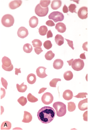
  • Marked schistocytes

Coagulation Panel

  • INR: 1.26
  • PTT: Normal
  • Fibrinogen: Normal
  • FDP: Normal
  • D-dimer: >9,000 (normal 250)
  • Haptoglobin: Undetectable
  • LDH: 1493

CMP

  • Creatinine: 1.1
  • AST/ALT: Normal
  • TB: 4.3, DB: 0.8

Imaging:

CT Head: No acute intracranial process.

ttp_ct_01
ttp_ct_02
ttp_ct_03
ttp_ct_04
ttp_ct_05
ttp_ct_06
ttp_ct_07
ttp_ct_08
ttp_ct_09
ttp_ct_10
ttp_ct_11
ttp_ct_12
ttp_ct_13
ttp_ct_14
ttp_ct_15
ttp_ct_16
ttp_ct_17
ttp_ct_18
ttp_ct_19

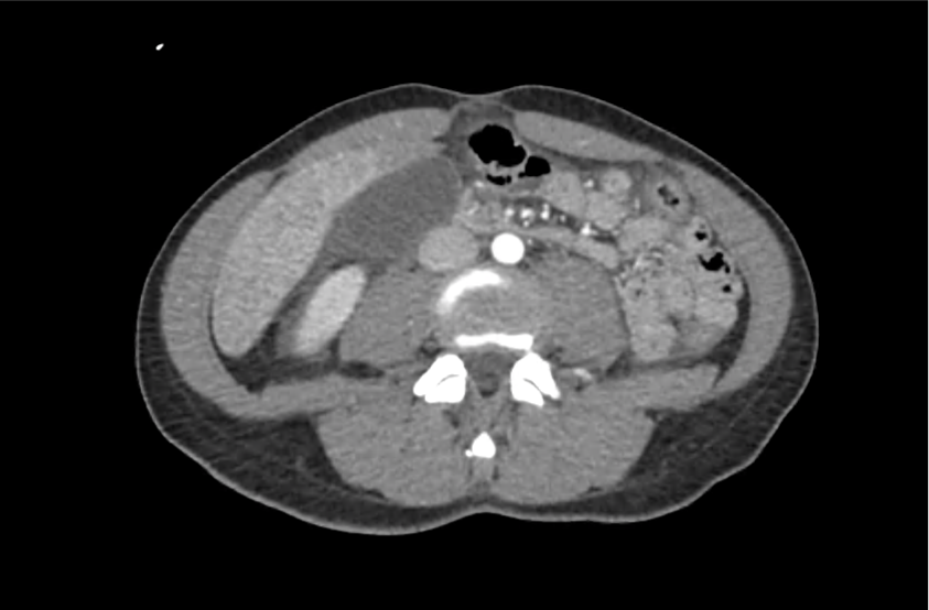
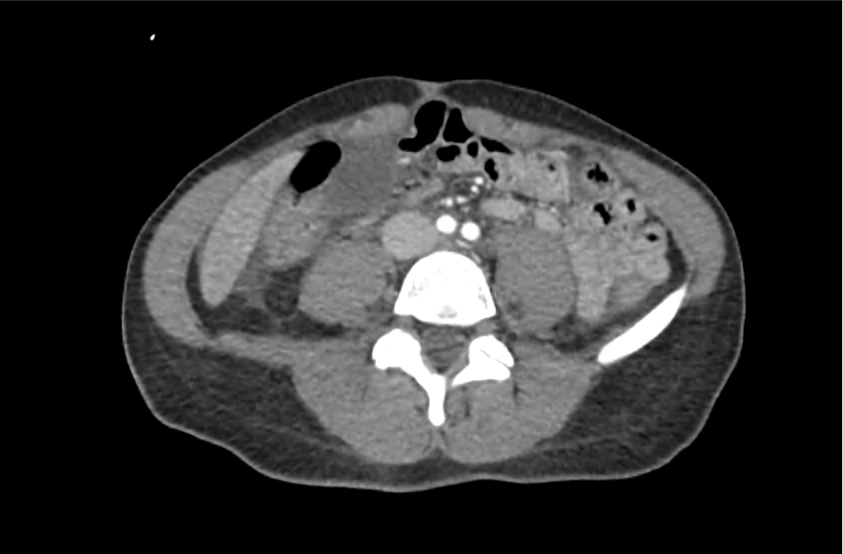
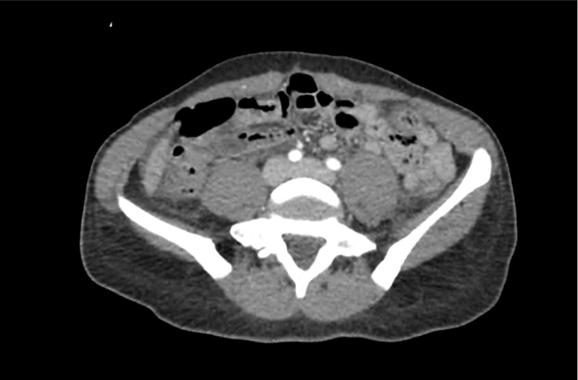
CT Abdomen/Pelvis with Contrast

Moderate free intra-abdominal fluid, heterogeneous liver with periportal edema, dense right middle lobe consolidation.

ED Course:

The patient developed worsening respiratory failure with hypoxia and tachypnea requiring endotracheal intubation. Thrombotic thrombocytopenic purpura was suspected and while awaiting emergent plasma exchange transfusion, the patient arrested and resuscitation efforts were unsuccessful.

The patient’s ADAMTS13 activity level was <3%. Autopsy demonstrated consolidation of the right middle lobe with possible lymphoproliferative mass, and lung petechial hemorrhages from microvascular thrombi.

Differential Diagnosis of Thrombocytopenia 1-7

Differential Diagnosis of Thrombocytopenia

Algorithm for the Evaluation of Thrombocytopenia 8

Algorithm for the Evaluation of Thrombocytopenia

Definition 9

  • Mild: <150k
  • Moderate: 100-150k
  • Severe: <50k
    • 10-30k: bleeding with minimal trauma
    • <10k: increased risk spontaneous bleeding

History 9,10

  • Prior platelet count
  • Family history bleeding disorders
  • Medications
    • Heparin
    • Quinine, quinidine
    • Rifampin
    • Trimethoprim-sulfamethoxazole
    • Vancomycin
  • Alcohol use
  • Travel-related infections

Physical Examination 9,10

  • Splenomegaly (liver disease)
  • Lymphadenopathy (infection, malignancy)

Workup 10,11

Schistocytes

Red blood cell fragments (schistocytes) 11

  • hCG
  • Repeat CBC
    • Detect spurious measure
    • Neutrophil-predominant leukocytosis: bacterial infection
    • Immature leukocytes (blasts): leukemia, myelodysplasia
  • Peripheral smear
    • Schistocytes: microangiopathic process (DIC, TTP, HUS)
    • Atypical lymphocytes: viral infection
    • Intracellular parasites: malaria
    • Hypersegmented neutrophils: nutritional deficiency
  • Infectious features: HIV, HCV, EBV, H.pylori, blood cultures
  • Autoimmune features: ANA, APL-Ab
  • Suspected occult liver disease: LFT, PT/PTT/INR
  • Suspected thrombotic microangiopathy: PT/PTT/INR, haptoglobin, LDH, fibrinogen, FDP, d-dimer

Specific Conditions 2-6,9,12-20

Disease Cause Presentation Laboratory Findings Treatment
DIC Sepsis
Trauma
Burn
Malignancy
Bleeding
Multi-organ failure
Shock
INR
Fibrinogen
FDP
D-dimer
Directed at underlying cause
Transfusion thresholds for hemorrhage:
FFP for INR >1.5
Platelets if <50k
Cryoprecipitate of fibrinogen <100mg/dL
TTP Insufficient ADAMTS-13 activity Non-specific constitutional symptoms (ex. weakness)
Neuro: headache, AMS, focal neuro deficit
GI: abdominal pain, nausea/vomiting
LDH
Reticulocyte
Unconjugated bilirubin
Haptoglobin
Plasma exchange
HUS Shiga-toxin-producing bacteria, E. coli O157:H7 Bloody diarrhea, anuria, oliguria, and hypertension Aggressive supportive care
HELLP Spectrum of eclampsia Hypertension
Visual symptoms
Headache
RUQ abdominal pain
AST/ALT
Uric acid
Unconjugated bilirubin
LDH
Reticulocyte
Haptoglobin
Delivery, MgSO4
ITP Primary ITP

Secondary ITP
– Drug
– Autoimmune
– Infection
– Malignancy

Usually asymptomatic, may have petechiae or easy bruising Isolated thrombocytopenia Steroids
HIT Exposure to heparin or LMWH Thrombocytopenia or a 50 percent reduction in platelet count between 5-10d exposure
New thrombosis or skin necrosis
4 T’s score
Platelet factor 4 antibodies Withdraw heparin

References

  1. Greinacher A, Selleng S. How I evaluate and treat thrombocytopenia in the intensive care unit patient. Blood. 2016;128(26):3032-3042. doi:10.1182/blood-2016-09-693655.
  2. Joly BS, Coppo P, Veyradier A. Thrombotic thrombocytopenic purpura. Blood. 2017;129(21):2836-2846. doi:10.1182/blood-2016-10-709857.
  3. Leslie SD, Toy PT. Laboratory hemostatic abnormalities in massively transfused patients given red blood cells and crystalloid. Am J Clin Pathol. 1991;96(6):770-773.
  4. Neunert C, Lim W, Crowther M, et al. The American Society of Hematology 2011 evidence-based practice guideline for immune thrombocytopenia. Blood. 2011;117(16):4190-4207. doi:10.1182/blood-2010-08-302984.
  5. Kappler S, Ronan-Bentle S, Graham A. Thrombotic microangiopathies (TTP, HUS, HELLP). Emerg Med Clin North Am. 2014;32(3):649-671. doi:10.1016/j.emc.2014.04.008.
  6. Greinacher A. Heparin-Induced Thrombocytopenia. Solomon CG, ed. N Engl J Med. 2015;373(3):252-261. doi:10.1056/NEJMcp1411910.
  7. Reardon JE Jr., Marques MB. Evaluation of Thrombocytopenia. Lab Med. 2006;37(4):248-250. doi:10.1309/R7P79KERAJHPRHLT.
  8. Stasi R. How to approach thrombocytopenia. Hematology Am Soc Hematol Educ Program. 2012;2012:191-197. doi:10.1182/asheducation-2012.1.191.
  9. Gauer RL, Braun MM. Thrombocytopenia. Am Fam Physician. 2012;85(6):612-622.
  10. Abrams CS. 172 – Thrombocytopenia. Twenty Fifth Edition. Elsevier Inc.; 2016:1159–1167.e2. doi:10.1016/B978-1-4557-5017-7.00172-0.
  11. Wilson CS, Vergara-Lluri ME, Brynes RK. Chapter 11 – Evaluation of Anemia, Leukopenia, and Thrombocytopenia. Second Edition. Elsevier Inc.; 2017:195-234.e195. doi:10.1016/B978-0-323-29613-7.00011-9.
  12. Hui P, Cook DJ, Lim W, Fraser GA, Arnold DM. The frequency and clinical significance of thrombocytopenia complicating critical illness: a systematic review. Chest. 2011;139(2):271-278. doi:10.1378/chest.10-2243.
  13. Jokiranta TS. HUS and atypical HUS. Blood. 2017;129(21):2847-2856. doi:10.1182/blood-2016-11-709865.
  14. Neunert CE. Management of newly diagnosed immune thrombocytopenia: can we change outcomes? Hematology Am Soc Hematol Educ Program. 2017;2017(1):400-405. doi:10.1182/asheducation-2017.1.400.
  15. Lambert MP, Gernsheimer TB. Clinical updates in adult immune thrombocytopenia. Blood. 2017;129(21):2829-2835. doi:10.1182/blood-2017-03-754119.
  16. Arepally GM. Heparin-induced thrombocytopenia. Blood. 2017;129(21):2864-2872. doi:10.1182/blood-2016-11-709873.
  17. Aster RH, Bougie DW. Drug-induced immune thrombocytopenia. N Engl J Med. 2007;357(6):580-587. doi:10.1056/NEJMra066469.
  18. Boral BM, Williams DJ, Boral LI. Disseminated Intravascular Coagulation. Am J Clin Pathol. 2016;146(6):670-680. doi:10.1093/ajcp/aqw195.
  19. Scully M, Hunt BJ, Benjamin S, et al. Guidelines on the diagnosis and management of thrombotic thrombocytopenic purpura and other thrombotic microangiopathies. Br J Haematol. 2012;158(3):323-335. doi:10.1111/j.1365-2141.2012.09167.x.
  20. Levine RL, Hursting MJ, Drexler A, Lewis BE, Francis JL. Heparin-induced thrombocytopenia in the emergency department. Ann Emerg Med. 2004;44(5):511-515. doi:10.1016/j.annemergmed.2004.06.004.

Hepatobiliary Ultrasound

Brief H&P:

A 43-year-old female with a history of hypertension, diabetes and obesity presents with right-upper quadrant abdominal pain for the past 1 week. The pain is characterized as burning, non-radiating, intermittent (with episodes lasting 10-30 minutes), resolving spontaneously and without apparent provoking features. She notes nausea but no vomiting, no changes in bowel or urinary habits. She similarly denies fevers, chest pain or shortness of breath. Vital signs were normal, and physical examination was notable only for right upper quadrant tenderness to palpation without rigidity or guarding.

An ECG demonstrates normal sinus rhythm, laboratory tests including liver function tests and lipase were normal and a bedside ultrasound of the right upper quadrant was performed demonstrating gallstones and a positive sonographic Murphy sign. The patient was diagnosed with acute cholecystitis, antibiotics were initiated, the patient was maintained NPO while general surgery was consulted.

Evaluation of Right-Upper Quadrant Abdominal Pain

The initial evaluation of a patient presenting with right-upper quadrant (or adjacent) abdominal pain typically includes laboratory tests such as a complete blood count, chemistry panel, liver function tests and serum lipase. In patients at risk for atypical presentations for an acute coronary syndrome or with other concerning symptoms, electrocardiography and cardiac enzymes may be indicated.

The differential diagnosis is broad. A systematic approach proceeds anatomically from superficial to deeper structures centered around the site of maximal pain.

Skin

Skin

Herpes zoster, erysipelas, or cellulitis

Connective Tissue

Connective Tissue

Intercostal muscle strain, myositis, fasciitis

Bone

Bone

Rib contusion or fracture

Hepatobiliary

Hepatobiliary

Hepatitis (infectious, toxin-mediated), perihepatitis (Fitz-Hugh-Curtis), hepatic abscess, symptomatic cholelithiasis, acute cholecystitis, ascending cholangitis, pancreatitis

Gastric

Gastric

Peptic ulcer disease, gastroesophageal reflux, gastritis, gastroparesis

Small Bowel

Small Bowel

Duodenal ulcer, small bowel obstruction

Large Bowel

Large Bowel

Retrocecal appendicitis, inflammatory bowel disease

Genitourinary

Genitourinary

Pyelonephritis, ureterolithiasis

Referred

Referred

Acute coronary syndrome, lower-lobe pneumonia, pulmonary embolus

Ultrasound in the Evaluation of Right Upper Quadrant Abdominal Pain

The diagnosis is unlikely to be made based on laboratory tests alone 1. However, the addition of bedside ultrasound, particularly for the evaluation of gallbladder pathology, is both rapid and reliable 2-8. The algorithm below provides a pathway for the incorporation of bedside ultrasound of the right upper quadrant in the evaluation of suspected gallbladder disease.

Algorithm for the Use of Ultrasound in the Evaluation of Right Upper Quadrant Abdominal Pain

A normal-appearing gallbladder absent gallstones should prompt a traversal of the anatomic approach to the differential diagnosis detailed above. If gallstones are identified, the association with a positive sonographic Murphy sign is highly predictive of acute cholecystitis 2,5,6,9. Acute cholecystitis may be associated with inflammatory gallbladder changes such as wall-thickening (>3mm) or pericholecystic fluid 3,5,6,10-13. However, in the absence of cholelithiasis or a positive sonographic Murphy sign, these features are non-specific and may be the result of generalized edematous states such as congestive heart failure, renal failure, or hepatic failure and critically-ill patients may develop acalculous cholecystitis 7,11,14. Finally, common bile duct dilation may be due to intra-luminal obstruction as in choledocholithiasis, luminal abnormalities such as strictures, or extra-luminal compression from masses or malignancy.  Dilation is generally described as a diameter >6mm – allowing an additional 1mm for every decade over 60 years-old as well as more vague accommodations for patients with prior cholecystectomy 3,5,7,15.

Gallery

The POCUS Atlas
The ultrasound images and videos used in this post come from The POCUS Atlas, a collaborative collection focusing on rare, exotic and perfectly captured ultrasound images.
The POCUS Atlas

Gallstones

Many gallstones

Gallbladder wall thickening

Pericholecystic fluid

Choledocholithiasis

Common bile duct dilation

All illustrations are available for free, licensed (along with all content on this site) under Creative Commons Attribution-ShareAlike 4.0 International Public License.

Downloads Page License

References

  1. Trowbridge RL, Rutkowski NK, Shojania KG. Does this patient have acute cholecystitis? JAMA. 2003;289(1):80-86.
  2. Scruggs W, Fox JC, Potts B, et al. Accuracy of ED Bedside Ultrasound for Identification of gallstones: retrospective analysis of 575 studies. West J Emerg Med. 2008;9(1):1-5.
  3. Ross M, Brown M, McLaughlin K, et al. Emergency physician-performed ultrasound to diagnose cholelithiasis: a systematic review. Acad Emerg Med. 2011;18(3):227-235. doi:10.1111/j.1553-2712.2011.01012.x.
  4. Jang T, Chauhan V, Cundiff C, Kaji AH. Assessment of emergency physician-performed ultrasound in evaluating nonspecific abdominal pain. Am J Emerg Med. 2014;32(5):457-460. doi:10.1016/j.ajem.2014.01.004.
  5. Kendall JL, Shimp RJ. Performance and interpretation of focused right upper quadrant ultrasound by emergency physicians. J Emerg Med. 2001;21(1):7-13.
  6. Summers SM, Scruggs W, Menchine MD, et al. A prospective evaluation of emergency department bedside ultrasonography for the detection of acute cholecystitis. Ann Emerg Med. 2010;56(2):114-122. doi:10.1016/j.annemergmed.2010.01.014.
  7. Rubens DJ. Ultrasound Imaging of the Biliary Tract. Ultrasound Clinics. 2007;2(3):391-413. doi:10.1016/j.cult.2007.08.007.
  8. Rosen CL, Brown DF, Chang Y, et al. Ultrasonography by emergency physicians in patients with suspected cholecystitis. American Journal of Emergency Medicine. 2001;19(1):32-36. doi:10.1053/ajem.2001.20028.
  9. Shea JA. Revised Estimates of Diagnostic Test Sensitivity and Specificity in Suspected Biliary Tract Disease. Arch Intern Med. 1994;154(22):2573-2581. doi:10.1001/archinte.1994.00420220069008.
  10. Miller AH, Pepe PE, Brockman CR, Delaney KA. ED ultrasound in hepatobiliary disease. J Emerg Med. 2006;30(1):69-74. doi:10.1016/j.jemermed.2005.03.017.
  11. Shah K, Wolfe RE. Hepatobiliary ultrasound. Emergency Medicine Clinics of NA. 2004;22(3):661–73–viii. doi:10.1016/j.emc.2004.04.015.
  12. Matcuk GR, Grant EG, Ralls PW. Ultrasound measurements of the bile ducts and gallbladder: normal ranges and effects of age, sex, cholecystectomy, and pathologic states. Ultrasound Q. 2014;30(1):41-48. doi:10.1097/RUQ.0b013e3182a80c98.
  13. Engel JM, Deitch EA, Sikkema W. Gallbladder wall thickness: sonographic accuracy and relation to disease. American Journal of Roentgenology. 1980;134(5):907-909. doi:10.2214/ajr.134.5.907.
  14. Gerstenmaier JF, Hoang KN, Gibson RN. Contrast-enhanced ultrasound in gallbladder disease: a pictorial review. Abdom Radiol (NY). 2016;41(8):1640-1652. doi:10.1007/s00261-016-0729-4.
  15. Becker BA, Chin E, Mervis E, Anderson CL, Oshita MH, Fox JC. Emergency biliary sonography: utility of common bile duct measurement in the diagnosis of cholecystitis and choledocholithiasis. J Emerg Med. 2014;46(1):54-60. doi:10.1016/j.jemermed.2013.03.024.

Neonatal Congenital Heart Disease

Brief H&P

An 8-day old male infant, ex-full term, born by normal spontaneous vaginal delivery and discharged home 2 days after birth without identified complications or maternal infections presents with parents to the emergency department due to decreased activity. Starting on day-of-life six, the family noted that feeding appeared to be taking longer and the mother felt her infant was breathing faster.

On presentation, the patient was pale, dusky, lethargic and with mottled skin. Temperature 36.3°C (rectal), HR 170, RR 60, BP 62/35, SpO2 70%. Physical examination demonstrated flat fontanelle, coarse breath sounds, regular rate and rhythm without additional heart sounds or murmurs, and hepatomegaly with liver edge 3cm below costal margin. Capillary refill was delayed at 5-6 seconds. Supplemental oxygen was applied without effect.

Algorithm for the Evaluation and Management of Suspected Congenital Heart Disease in Neonates

Algorithm for the Evaluation of Neonatal Congenital Heart Disease

Neonates with undiagnosed congenital heart disease may present to the emergency department with nonspecific symptoms, and may be considerably unstable requiring immediate life-saving interventions.

Key Historical Features

  • Respiratory difficulty
  • Feeding difficulty (small quantities, diaphoresis during feeding)
  • Poor weight gain
  • Chromosomal abnormalities, syndromes
  • Maternal risk factors: diabetes, teratogen exposure, substance use
  • Sibling of affected child

Key Examination Findings

  • Vital signs: tachycardia, tachypnea, hypotension
  • Blood pressure differential (RUE vs. LE >8mmHg difference)
  • Pulse oximetry differential (RUE vs. LE >4% difference, <95%)
  • Cardiac examination: murmur, thrill, pulse differential, capillary refill, hepatomegaly

Workup

  • CXR: Evaluate for cardiomegaly, pulmonary vascular congestion
  • ECG: Evaluate for axis deviation (right axis deviation is normal for neonate)
  • ABG with co-oximetry

References

  1. Special thanks to Dr. Kelly Young, MD, MS, FAAP. Director, Pediatric Emergency Medicine Fellowship. Harbor-UCLA Medical Center Department of Emergency Medicine.
  2. Association AAOPAAH. Textbook of Neonatal Resuscitation. 2016.
  3. Lissauer T, Fanaroff AA, Miall L, Fanaroff J. Neonatology at a Glance. John Wiley & Sons; 2015.
  4. Steinhorn RH. Evaluation and Management of the Cyanotic Neonate. Clinical Pediatric Emergency Medicine. 2008;9(3):169-175. doi:10.1016/j.cpem.2008.06.006.
  5. MD MR. Chapter 7 – Cardiology. Twenty First Edition. Elsevier Inc.; 2018:156-202. doi:10.1016/B978-0-323-39955-5.00007-7.
  6. Gomella T, Cunningham M. Neonatology 7/E. McGraw-Hill Prof Med/Tech; 2013.
  7. Yee L. Cardiac emergencies in the first year of life. Emergency Medicine Clinics of NA. 2007;25(4):981–1008–vi. doi:10.1016/j.emc.2007.08.001.
  8. Yates MC, Rao PS. Pediatric cardiac emergencies. Emerg Med. 2013. doi:10.4172/2165-7548.1000164.
  9. Silberbach M, Hannon D. Presentation of congenital heart disease in the neonate and young infant. Pediatr Rev. 2007;28(4):123-131.
  10. Mastropietro CW, Tourner SP, Sarnaik AP. Emergency presentation of congenital heart disease in children. Pediatric Emergency …. 2008.
  11. Brousseau T, Sharieff GQ. Newborn Emergencies: The First 30 Days of Life. Pediatric Clinics of North America. 2006;53(1):69-84. doi:10.1016/j.pcl.2005.09.011.

Hyperthermia

Brief H&P

A young male with unknown medical history is brought in by ambulance with altered mental status. EMS reports that the patient was agitated, requiring restraints for transportation. On arrival, the patient is agitated, uncooperative and unable to provide history. Vital signs are notable for tachycardia, tachypnea and hypertension. Physical examination demonstrates diaphoresis and mydriasis, as well as increased muscle tone – particularly in the lower extremities with ankle clonus. A core temperature is obtained and noted to be elevated at 41.5°C. Point-of-care glucose is normal.

Rapid external cooling measures were instituted and several doses of intravenous benzodiazepines were administered with improvement in agitation. Laboratory studies were notable for a modest leukocytosis (WBC 18.4 without immature forms), serum sodium was 135 without osmolar gap, creatine kinase was slightly elevated without renal dysfunction, and thyroid function tests were normal. Toxicology screen was negative. ECG revealed sinus tachycardia but was otherwise normal and non-contrast computed tomography of the head was normal.

After a brief admission in the intensive care unit, the patient’s mental status improved and he reported MDMA use on the evening of presentation, he also described a history of major depression and was taking paroxetine.

Evaluation of Elevated Temperature

The designation of 38°C as “suspicious” for fever dates to 1868 and the analysis of over one million (axillary) temperature measurements by Carl Wunderlich1. Any cutoff is arbitrary and requires recognition of the clinical context and normal daily variations (with nadir in the morning and peak in evening) 2,3. What is clear is that peripheral thermometry (unless demonstrating fever) is unreliable and a core temperature should be sought4.

Thermoregulation

Temperature homeostasis is a balance between heat production and dissipation maintained by the anterior hypothalamus. Heat production is a byproduct of normal metabolic processes and skeletal muscle activity. Conservation, maintenance or dissipation of heat is aided by cutaneous vasodilation, sweating, or behavioral responses.

Fever is caused by endogenous or exogenous pyrogens which alter the homeostatic set-point, inducing thermogenesis and elevating the body temperature. Precipitants of fever are usually infectious, however non-infectious processes (ex. malignancy, tissue ischemia/infarction, auto-immune disease) resulting in inflammation can provoke a similar response 5-7.

There is no explicit temperature distinction to diagnose hyperthermia, instead the physiologic mechanism is different. In hyperthermia, the body’s homeostatic mechanisms are dysfunctional or overwhelmed due to heat exposure, excess production, ineffective dissipation or hypothalamic malfunction 8.

Algorithm for the Evaluation of Hyperthermia 8-15

Algorithm for the Evaluation of Hyperthermia

Implicated Agents in Drug-Induced Hyperthermic Syndromes 9,10

Serotonin Syndrome

Class Examples
SSRI sertraline, fluoxetine, paroxetine
Other anti-depressants trazodone, venlafaxine, lithium
MAOI phenelzine, isocarboxazid
Anti-epileptic drugs valproate
Analgesics meperidine, fentanyl, tramadol
Anti-emetic ondansetron, metoclopramide
Anti-migraine sumatriptan
Antimicrobial linezolid, ritonavir
Illicit substances MDMA, LSD

Neuroleptic Malignant Syndrome (NMS)

Class Examples
Typical anti-psychotic haloperidol, prochlorperazine
Atypical anti-psychotic risperidone, olanzapine, quetiapine, aripiprazole
Anti-dopaminergic metoclopramide, droperidol

References:

  1. Wunderlich CA. Das Verhalten Der Eigenwärme in Krankheiten. 1870.
  2. Mackowiak PA, Wasserman SS, Levine MM. A critical appraisal of 98.6 degrees F, the upper limit of the normal body temperature, and other legacies of Carl Reinhold August Wunderlich. JAMA. 1992;268(12):1578-1580.
  3. Lee-Chiong TL, Stitt JT. Disorders of temperature regulation. Compr Ther. 1995;21(12):697-704.
  4. Niven DJ, Gaudet JE, Laupland KB, Mrklas KJ, Roberts DJ, Stelfox HT. Accuracy of peripheral thermometers for estimating temperature: a systematic review and meta-analysis. Ann Intern Med. 2015;163(10):768-777. doi:10.7326/M15-1150.
  5. Dinarello CA. Infection, fever, and exogenous and endogenous pyrogens: some concepts have changed. J Endotoxin Res. 2004;10(4):201-222. doi:10.1179/096805104225006129.
  6. Greisman LA, Mackowiak PA. Fever: beneficial and detrimental effects of antipyretics. Curr Opin Infect Dis. 2002;15(3):241-245.
  7. Dinarello CA. Thermoregulation and the pathogenesis of fever. Infect Dis Clin North Am. 1996;10(2):433-449.
  8. Simon HB. Hyperthermia. N Engl J Med. 1993;329(7):483-487. doi:10.1056/NEJM199308123290708.
  9. Boyer EW, Shannon M. The serotonin syndrome. N Engl J Med. 2005;352(11):1112-1120. doi:10.1056/NEJMra041867.
  10. Berman BD. Neuroleptic malignant syndrome: a review for neurohospitalists. Neurohospitalist. 2011;1(1):41-47. doi:10.1177/1941875210386491.
  11. Hayes BD, Martinez JP, Barrueto F. Drug-induced hyperthermic syndromes: part I. Hyperthermia in overdose. Emerg Med Clin North Am. 2013;31(4):1019-1033. doi:10.1016/j.emc.2013.07.004.
  12. Oruch R, Pryme IF, Engelsen BA, Lund A. Neuroleptic malignant syndrome: an easily overlooked neurologic emergency. Neuropsychiatr Dis Treat. 2017;13:161-175. doi:10.2147/NDT.S118438.
  13. Musselman ME, Saely S. Diagnosis and treatment of drug-induced hyperthermia. Am J Health Syst Pharm. 2013;70(1):34-42. doi:10.2146/ajhp110543.
  14. Ahuja N, Cole AJ. Hyperthermia syndromes in psychiatry. Adv psychiatr treat (Print). 2018;15(03):181-191. doi:10.1192/apt.bp.107.005090.
  15. Tomarken JL, Britt BA. Malignant hyperthermia. Ann Emerg Med. 1987;16(11):1253-1265. doi:10.1016/S0196-0644(87)80235-4.

Wide-complex Tachycardia

Several algorithms exist for the electrocardigraphic evaluation of regular, wide-complex tachycardias with the objective of distinguishing ventricular tachycardia (VT) from a supraventricular tachycardia (SVT) with aberrant conduction. The algorithm detailed below, developed by Dr. James Niemann, presents an ED-centric approach favoring the diagnosis of the more life-threatening dysrhythmia. This approach recognizes that SVT with aberrancy is rare, particularly in patients with a history of cardiac disease where the likelihood of ventricular tachycardia exceeds 90%. The algorithm requires the use of only the most simple and easily-recalled criteria, and any point of failure along the algorithm lends to the universally-appropriate management as ventricular tachycardia.

Algorithm for the Evaluation of Regular, Wide-Complex Tachycardia

Algorithm for the Evaluation of Wide-Complex Tachycardia

  1. aVR: Is the initial deflection in aVR positive? If yes, then VT.
  2. Concordance: Is there concordance (monophasic with same polarity) in all of the precordial leads? If yes, then VT.
  3. AV Dissociation: Is there evidence of AV dissociation (fusion or capture beats)? If yes, then VT.
  4. Bundle-branch morphology: Is the QRS morphology in V1 and V6 consistent with either LBBB or RBBB? If no, then VT.

References

  1. Neimann J. Wide QRS Complex Tachycardias. Lecture. Harbor-UCLA Department of Emergency Medicine. 2014:1-19.
  2. Vereckei A, Duray G, Szénási G, Altemose GT, Miller JM. New algorithm using only lead aVR for differential diagnosis of wide QRS complex tachycardia. Heart Rhythm. 2008;5(1):89-98. doi:10.1016/j.hrthm.2007.09.020.
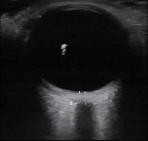
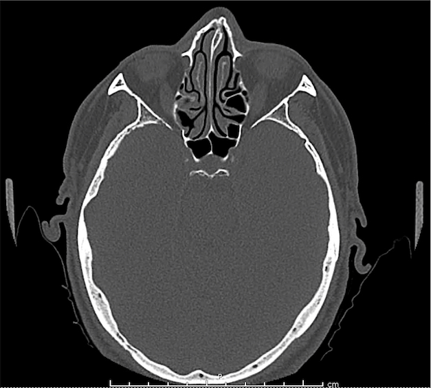
  3. Szelényi Z, Duray G, Katona G, et al. Comparison of the “real-life” diagnostic value of two recently published electrocardiogram methods for the differential diagnosis of wide QRS complex tachycardias. Acad Emerg Med. 2013;20(11):1121-1130. doi:10.1111/acem.12247.
  4. Brugada P, Brugada J, Mont L, Smeets J, Andries EW. A new approach to the differential diagnosis of a regular tachycardia with a wide QRS complex. Circulation. 1991;83(5):1649-1659.
  5. Lau EW, Pathamanathan RK, Ng GA, Cooper J, Skehan JD, Griffith MJ. The Bayesian approach improves the electrocardiographic diagnosis of broad complex tachycardia. Pacing Clin Electrophysiol. 2000;23(10 Pt 1):1519-1526.
  6. B Garner J, M Miller J. Wide Complex Tachycardia – Ventricular Tachycardia or Not Ventricular Tachycardia, That Remains the Question. Arrhythm Electrophysiol Rev. 2013;2(1):23-29. doi:10.15420/aer.2013.2.1.23.
  7. Vereckei A. Current algorithms for the diagnosis of wide QRS complex tachycardias. Curr Cardiol Rev. 2014;10(3):262-276.
  8. Garmel GM. Wide Complex Tachycardias: Understanding this Complex Condition: Part 1 – Epidemiology and Electrophysiology. West J Emerg Med. 2008;9(1):28-39.
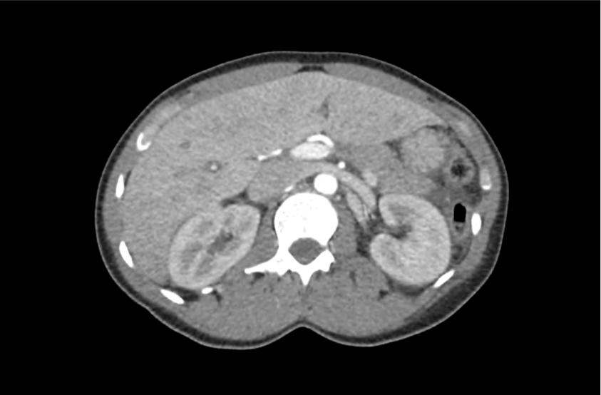
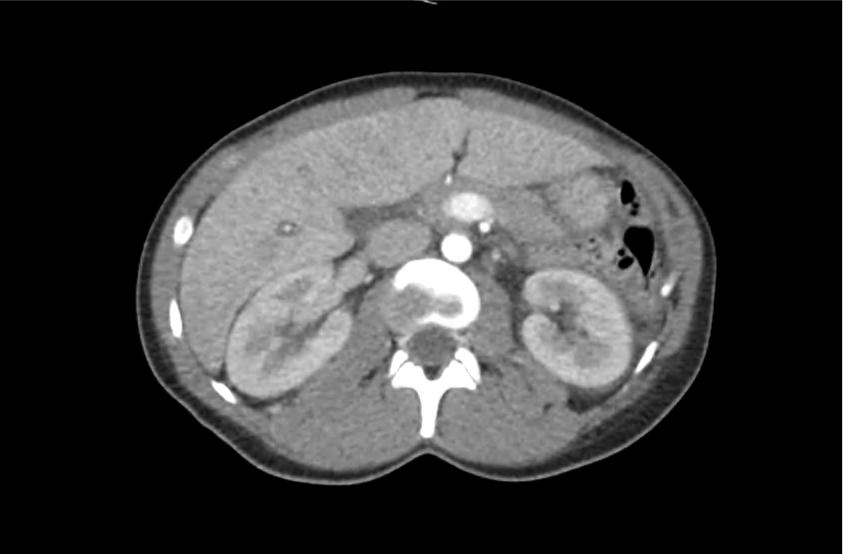
  9. Garmel GM. Wide Complex Tachycardias: Understanding this Complex Condition Part 2 – Management, Miscellaneous Causes, and Pitfalls. West J Emerg Med. 2008;9(2):97-103.
  10. Griffith MJ, Garratt CJ, Mounsey P, Camm AJ. Ventricular tachycardia as default diagnosis in broad complex tachycardia. The Lancet. 1994;343(8894):386-388.